HAL Id: tel-02145218
https://tel.archives-ouvertes.fr/tel-02145218
Submitted on 2 Jun 2019HAL is a multi-disciplinary open access archive for the deposit and dissemination of sci-entific research documents, whether they are pub-lished or not. The documents may come from teaching and research institutions in France or abroad, or from public or private research centers.
L’archive ouverte pluridisciplinaire HAL, est destinée au dépôt et à la diffusion de documents scientifiques de niveau recherche, publiés ou non, émanant des établissements d’enseignement et de recherche français ou étrangers, des laboratoires publics ou privés.
Role of caveolin-3 and caveolae mechanics in muscle
pathophysiology
Melissa Dewulf
To cite this version:
Melissa Dewulf. Role of caveolin-3 and caveolae mechanics in muscle pathophysiology. Cellular Biol-ogy. Université Paris-Saclay, 2018. English. �NNT : 2018SACLS067�. �tel-02145218�
Thèse de doctorat de l'Université Paris-Saclay
préparée à l’Université Paris-Saclay
École doctorale n°568
Signalisations et réseaux intégratifs en biologie (Biosigne) Spécialité de doctorat: Biologie cellulaire et moléculaire
Thèse présentée et soutenue à Paris, le 29 mars 2018, par
Melissa Dewulf
Role of caveolin-3 and caveolae mechanics
in the muscle pathophysiology
Composition du Jury :
Pr. Karim Benihoud Président du jury
Dr. Soazig Le lay Rapporteur
Dr. Edgar Gomes Rapporteur
Dr. Julie Dam Examinateur
Dr. Stéphane Vassilopoulos Examinateur Dr. Christophe Lamaze Directeur de thèse
! ! ! ! ! ! !
« Et voilà, on a les droits ! »
François Pignon
Acknowledgements
The last four years were probably the most challenging but also the most exciting of my life, and it is time for me to thank many people who were involved, in so many different ways, in this adventure. I will take this opportunity to address my first words to my supervisor. Christophe, thank you so much for trusting me with this project, which was not always easy, but you were always very optimistic and in the end, I believe we made it work! You have always been very supportive concerning the choices I made for my project, but also concerning my “extra-research” projects, which I particularly appreciated. At the personal level, you are a man of all the extremes, extremely scandalous, but also extremely interesting and funny! You are a “personnage haut en couleurs” as we would say in French, and I am very happy Graça put me on your way. I would like now to mention the other person “qui tire les ficelles”. Cédric, I am so grateful that you are such a nice and patient person! I think we can say that we have different standards of organization, clumsiness and the list could go on for a while, but it worked anyway! It probably has to do with the fact that we also share many different interests, food being the most important one, but we cannot exclude that science was also part of the game… You have been a great teacher and thanks to you, I have now quite a list of skills, both useful for the experiments and for trying to do nice illustrations, that I can brag about to a very limited amount of people, but still…Finally, thank you for all the time you invested in making sure my manuscript would be as perfect as possible while you were preparing yourself for the famous concours. I would also like to thank Ludger who would always bring precious feedbacks whenever I would be on stage during our weekly labmeetings. I am also very grateful for your trust with the organization of the unit retreat and different team building events, I enjoyed it a lot!
I would like now to warmly thank Dr Soazig Le lay and Dr Edgar Gomes for the time they dedicated to review my manuscript and the nice and pertinent feedback they gave me. I would also like to address a few words to Dr Stéphane Vassilopoulos who I have met a few years ago and who I always enjoyed having animated scientific discussions with. I am thus very happy that he accepted to evaluate my work. I would also like to thank Pr Karim Benihoud who was my PhD tutor and will preside over my thesis jury, and Dr Julie Dam for kindly accepting my invitation. This work wouldn’t have been possible without our collaborators from the Institut de myologie. In addition to Stéphane that I mentioned before, I would like to thank Gillian Butler-Browne for the scientific discussion and support on this project, Anne Bigot who generated all the muscle cell lines we used for our work and who has always been available for advices. Finally, I thank Catherine Coirault who trained and advised me on the use of the Flexcell device, and who also was one of my thesis tutors. Part of the work has also been done through collaborations within our unit. Valérie and Christine, thank you for the precious time you dedicated to reveal the dirty secrets of my cells, our paper looks much more fancy now thanks to you!
To me, a good thesis requires a good scientific and human environment, and regarding this, I have been very lucky. The Lamaze and Johannes teams are a melting pot of people, each of them with its own little hint of craziness, and I will definitely remember having a great time and laughing a lot! First, Nicolas, who actually asked me to mention him first, has been my perfect sidekick during this thesis. Although I have never been supportive of his sense of humor, I really hope I have actually been as supportive for everything else as he has been with me during these last four years. PhD sidekicks actually come in different flavors: Carlos, thank you for being you, so annoying and so great at the same time! And Thomas, thank you for always being present whenever I need to tell a story, complain, or just drink a coffee on the terrace, no matter the weather… For great discussions, scientific or others, I could also always count on Cédric, Christine, Estelle, Valérie or Massi! I will always remember Cédric’s enthusiasm for the construction of a new stretcher device with a 3D printer or for the discovery of the saucisson paradise, our games of “which emoji movie am I?”, the long series of quotes from Le diner de con, and we should not forget about our 80’s aerobic moments with Christine, our endless talks on life with Valérie, our mutual teasing with Estelle and our daily rendez-vous with Massi at his bench to do a summary of our day. I will of course remember all of our big laughs, sushi a volonté parties, and many coffee breaks and lunches that are perfect times to talk about anything that crosses our minds. More recently, we got to see many new faces arriving, which fortunately allowed me to expand the number of great people I met during my time in the lab. Satish with his wisdom and his great repartee that makes me laugh so much, Manon la force tranquille with her calm and patience, although as we say in French “il faut se méfier de l’eau qui dort” and our very own tornado Alison who, together with Ewan, brought us a very nice British (and neapolitan) ambiance to our daily life. This is without forgetting about past members of the lab who left us with nice memories. Ma ptite Steph, who has always been a great support and who I shared so much with. Natacha, Weiwei, Henri, Daniela, whose names we can still come across when using old pipetboys or tube racks, as if they never left. I would also like to thank all of the members of the Lamaze and Johannes team, as scientific and non-scientific support was something I could find very easily between the walls that we share.
Beyond these walls, I got to meet many other different people, who for some of them became very good friends. Fortunately, I started this adventure in Curie with Léa, with the two other members of the bras cassés, Arthur and Maxence who were never too far away from us. Since university, we were always there to support each other through every single step of the process and I am so happy we got to share all of this together! Léa and Arthur were leading the dance, showing us the way on the road of excellency, while Mikou would always be there to share with us his latest discovery while drinking and spilling beer everywhere. Léa, knowing that your were there, just 5 floors away, made everything easier for me and as we were both trying to make our way into the life of the institute by attending a young scientist retreat, we got to meet the people who would become our good friends. Suddenly, it made me feel like I was going back to school again and I would get to see my friends every day. Camilla is this very calm, precise, and loving person that anyone should have in his life. Luckily for me, since then, she never left me and I am so grateful that she allowed me to integrate her fully in my professional and especially
personal life, as I would be completely lost if she ever decided to leave it. She is the definition of a good friend and I am so glad I can count on her for anything. Imène is in the category of person that might seem a little bit inaccessible at first, but it is so worth it to scratch the surface a little, and I am already missing so much our long brainstormings about life and science. Long and intense discussions were always one of our favorite activities with Alex as well who will also always be there if you need a shoulder to cry on. If you want kindness and comfort, as well as a good laugh, just go to Guillaume, he has way too much kindness for himself and will not hesitate to share some with you, he will always have a funny story “malgré lui” to tell you, and also, he does a great Shakira dance that will stick in our minds forever. Hugo and Bruno are never the last ones to entertain us with their stories either! Hugo has this capacity to put on smile on my face simply by talking with his lovely southern accent, that sure we all laugh a lot about but to tell the truth I think we all love it! And as for Bruno, he may look very calm with his “tête d’ange” but beware! Bruno can be a real tsunami…Thanks to all of you, I have now countless beautiful memories: all these great times together at ADIC parties, retreats, conferences, bars, diners and so many apéros in my old apartment! I cannot mention my old apartment without talking about Mélanie. Living with someone is always particular, especially if this someone is Mel. You were the witness of my first steps into emancipation, not without mocking me a little of course, and I could not imagine anyone better to do that with than you. The Saturday mornings watching tons of documentaries about plastic and environment, the long exchanges on our daily life as PhD students when we would come home at night, the cooking attempts, and all the laughs we had will be beautiful memories I will keep forever.
Finally, I would like to dedicate this last chapter to the people who might not be involved in my scientific life directly, but who are still of huge importance. First, my parents and moutch, who have always been extremely supportive. I guess the infinite curiosity and language skills of my father, the hard working and networking skills of my mother, and the extreme commitment of my brother to his objectives were my source of inspiration. Inspiration also comes from the family I chose, my lovely Lea and Juliette, but also my second mother Patricia, who I can count on, always! The whole gang of crazy people I call my friends and who make my life more colorful every day. And finally, Félix, I cannot imagine a better life partner for me, and I would like to thank you for being there every step of the way, and for giving me all of your kindness, loyalty and support.
! ! ! "!
TABLE OF CONTENT
!"#$%&'(&)'*!%*!&++++++++++++++++++++++++++++++++++++++++++++++++++++++++++++++++++++++++++++++++++++++++++++++++++++++++++&,! $-.!&'(&(-/01%.&"*2&!"#$%&++++++++++++++++++++++++++++++++++++++++++++++++++++++++++++++++++++++++++++++++++++++++&3! "##1%4-"!-'*.&++++++++++++++++++++++++++++++++++++++++++++++++++++++++++++++++++++++++++++++++++++++++++++++++++++++++++++++++&5! .066"17&+++++++++++++++++++++++++++++++++++++++++++++++++++++++++++++++++++++++++++++++++++++++++++++++++++++++++++++++++++++++++++++&8! 1%.06%&+++++++++++++++++++++++++++++++++++++++++++++++++++++++++++++++++++++++++++++++++++++++++++++++++++++++++++++++++++++++++++++++++&9! ,&!:;&<=;>;?@>&AB<C>;D&@E&;<<;E?F@>&@EG&:FH:>I&JKH@EFL;G&?F<<B;&+++++++++++++++++++&,,! ,+M&(KJA&?:;&AB<C>;&?F<<B;&?J&?:;&<FEH>;&AIJNFO;K&++++++++++++++++++++++++++++++++++++++++++++++++++++++&,,! ,+3&!:;&AIJNFO;KD&@&:FH:>I&<P;CF@>FL;G&@KC:F?;C?BK;&+++++++++++++++++++++++++++++++++++++++++++++++++++&,M! "#$#"!%&'!()*+,-'*'!)./!+,.0*)+012103!#########################################################################################!"4! "#$#4!%&'!+,(0)-'*'5!-3,617'*!1.0'8*103!)./!-'+&).,0*).(/9+01,.!#############################!"$! "#$#$!:3(6'*21.!)./!-'-7*).'!*';)1*!##########################################################################################!"<! "#$#<!=9(+2'!)((,+1)0'/!(18.)21.8!;)0&>)3(!#############################################################################!"?! "#$#<#"!%&'!829+,('!;)0&>)3!1.!0&'!-9(+2'!###########################################################################################!"?! "#$#<#4!%&'!@ABCD%E%$!;)0&>)3!1.!0&'!-9(+2'!###################################################################################!"B! ,+5&!:;&<P;CFNFC&JKH@E;>>;&JKH@EFL@?FJE&@EG&A;AOK@E;&?JPJ>JHI&JN&AIJNFO;K<&,9! "#<#"!F9+2'1!##############################################################################################################################################!"G! "#<#4!%&'!0*1)/H!0&'!()*+,;2)(-1+!*'01+929-!)./!0&'!%I09792'!(3(0'-!##########################!"J! M&)@Q;J>@;&+++++++++++++++++++++++++++++++++++++++++++++++++++++++++++++++++++++++++++++++++++++++++++++++++++++++++++++++++++++++++&M,! M+,&)@Q;J>@;D&<P;CF@>FL;G&P>@<A@&A;AOK@E;&FEQ@HFE@?FJE<&++++++++++++++++++++++++++++++++++++&M,! M+M&)@Q;J>@;&OFJH;E;<F<&++++++++++++++++++++++++++++++++++++++++++++++++++++++++++++++++++++++++++++++++++++++++++++++++++++++++&M,! 4#4#"!K)L',2)'!;*,0'1.!+,-;,(101,.!#############################################################################################!4"! 4#4#"#"!K)L',21.(!###############################################################################################################################################!4"! 4#4#"#4!K)L1.(!#####################################################################################################################################################!4<! 4#4#"#$!E++'((,*3!;*,0'1.(!###########################################################################################################################!4?! 4#4#4!K)L',2)'5!)!(;'+161+!21;1/!+,-;,(101,.!##############################################################################!4?! 4#4#$!K)L',2)'!6,*-)01,.5!)!(0';!73!(0';!;*,+'((!###################################################################!4B! 4#4#$#"!=1.1-9-!*'M91*'-'.0(!#################################################################################################################!4B! 4#4#$#4!K)L',21.!,218,-'*1N)01,.!)./!0*)661+O1.8!0,!0&'!;2)(-)!-'-7*).'!###########################!4P! 4#4#$#$!K)L1.(!*'+*910-'.0!)./!+)L',2)'!1.L)81.)01,.!####################################################################!4G! 4#4#$#<!Q'+*910-'.0!,6!)++'((,*3!;*,0'1.(!############################################################################################!4G! M+3&)@Q;J>@;&NBEC?FJE<&+++++++++++++++++++++++++++++++++++++++++++++++++++++++++++++++++++++++++++++++++++++++++++++++++++++++++++&MR! 4#$#"!K)L',2)'!)./!+'22!0*)661+O1.8!#################################################################################################!4J! 4#$#4!K)L',2)'!)./!21;1/!&,-',(0)(1(!##########################################################################################!$"! 4#$#$!K)L',2)'!)./!(18.)21.8!############################################################################################################!$4! 4#$#<!K)L',2)'5!-'-7*).'!0'.(1,.!7966'*1.8!)./!-'+&).,;*,0'+01,.!########################!$<! M+5&)@Q;J>@;&K;>@?;G&GF<;@<;<&+++++++++++++++++++++++++++++++++++++++++++++++++++++++++++++++++++++++++++++++++++++++++++++&3S! 4#<#"!A1;,/3(0*,;&3!############################################################################################################################!$B! 4#<#4!R)(+92)*!)./!;92-,.)*3!/3(69.+01,.!###############################################################################!$P! 4#<#$!K).+'*!#############################################################################################################################################!$G! 4#<#<!=9(+2'!/1(')('(!##########################################################################################################################!$J! 3&)@Q3&@EG&C@Q;J>@;&FE&AB<C>;&P:I<FJ>JHI&@EG&P@?:JP:I<FJ>JHI&++++++++++++++++++&5M! 3+,&)@Q3&AB?@?FJE<&@EG&AB<C>;&GF<;@<;<&++++++++++++++++++++++++++++++++++++++++++++++++++++++++++++++++++++++++&5M! 3+M&)@Q3&@EG&C@Q;J>@;&FE&AB<C>;<&++++++++++++++++++++++++++++++++++++++++++++++++++++++++++++++++++++++++++++++++++++++&53! $#4#"!=3,617'*!1.0'8*103!#####################################################################################################################!<$! $#4#"#"!='+&).,;*,0'+01,.!##########################################################################################################################!<$! $#4#"#4!='-7*).'!*';)1*!###############################################################################################################################!<<! $#4#"#$!=3,72)(0!69(1,.5!-9(+2'!)0*,;&3!)./!-9(+2'!&3;'*0*,;&3!###########################################!<?!! 4! $#4#4!=3,617'*!69.+01,.!######################################################################################################################!<B! $#4#4#"!%*1)/!6,*-)01,.!)./!69.+01,.!######################################################################################################!<B! $#4#4#4!K,(0)-'*'!6,*-)01,.!#######################################################################################################################!<P! $#4#4#<!K)2+19-5!FS!)./!-9(+2'!+,.0*)+01,.!########################################################################################!<G! $#4#4#<!T29+,('!-'0)7,21(-!#########################################################################################################################!<J! 5&$@C=&JN&NBEC?FJE@>&C@Q;J>@;&FE&)@Q3&TM9$&@EG&)@Q3&1MUV&:BA@E& GI<?KJP:FC&AIJ?BO;<&K;<B>?<&FE&G;NFCF;E?&A;C:@EJPKJ?;C?FJE&@EG& -$UW.!"!3&A;C:@EJ<FHE@>FEH+&+++++++++++++++++++++++++++++++++++++++++++++++++++++++++++++++++++++++++++++++++++&S3! 5+,&'OX;C?FQ;<&@EG&<BAA@KI&+++++++++++++++++++++++++++++++++++++++++++++++++++++++++++++++++++++++++++++++++++++++++++++++&S3! 5+M&6;?:JG&+++++++++++++++++++++++++++++++++++++++++++++++++++++++++++++++++++++++++++++++++++++++++++++++++++++++++++++++++++++++++++++++++&SU! 5+3&"K?FC>;&+++++++++++++++++++++++++++++++++++++++++++++++++++++++++++++++++++++++++++++++++++++++++++++++++++++++++++++++++++++++++++++++++++&U5! S&2F<CB<<FJE&@EG&P;K<P;C?FQ;<&+++++++++++++++++++++++++++++++++++++++++++++++++++++++++++++++++++++++++++++++++&,YM! S+,&)"43&AB?@?FJE<D&K;?;E?FJE&FE&?:;&/J>HF&CJAP>;Z&@EG&>J<<&JN&C@Q;J>@;&@?&?:;& P>@<A@&A;AOK@E;&++++++++++++++++++++++++++++++++++++++++++++++++++++++++++++++++++++++++++++++++++++++++++++++++++++++++++++++++&,YM! S+M&)@Q;J>@;&F<&K;[BFK;G&NJK&P>@<A@&A;AOK@E;&FE?;HKF?I&FE&AB<C>;&C;>><& BEG;KHJFEH&A;C:@EFC@>&<?K;<<&++++++++++++++++++++++++++++++++++++++++++++++++++++++++++++++++++++++++++++++++++++++++++&,Y5! S+3&)@Q;J>FE\3&@EG&A;C:@EFC@>&<?K;<<&@K;&E;H@?FQ;&K;HB>@?JK<&JN&?:;&-$UW.!"!3& P@?:]@I&++++++++++++++++++++++++++++++++++++++++++++++++++++++++++++++++++++++++++++++++++++++++++++++++++++++++++++++++++++++++++++++++++++&,YS! S+5&)@Q;J>FE\3&AB?@?FJE<&@K;&@<<JCF@?;G&]F?:&GFNN;K;E?&C>FEFC@>&P:;EJ?IP;<&+++&,Y9! 1%(%1%*)%.&+++++++++++++++++++++++++++++++++++++++++++++++++++++++++++++++++++++++++++++++++++++++++++++++++++++++++++++++++++&,,M! "**%^&+++++++++++++++++++++++++++++++++++++++++++++++++++++++++++++++++++++++++++++++++++++++++++++++++++++++++++++++++++++++++++++++&,SM! &!
! $!
LIST OF FIGURES AND TABLE
Figure 1. The structure of the skeletal muscle Figure 2. The sarcomere
Figure 3. Actin filament sliding allows muscle contraction Figure 4. The costamere
Figure 5. Model for the mechanism of membrane patching in muscle fibers.
Figure 6. Insulin and exercise mediated GLUT4 translocation at the plasma membrane. Figure 7. The IL6/STAT3 pathway.
Figure 8. IL6 autocrine functions in skeletal muscle.
Figure 9. Nuclear positioning during myogenic differentiation.
Figure 10. Calcium release and uptake during excitation-contraction coupling controlled by the Triad.
Figure 11. Caveolae observation by electron microscopy.
Figure 12. Caveolin protein family domains and insertion in the plasma membrane. Figure 13. Cavin structure.
Figure 14. Putative model for caveolar coat assembly and organization. Figure 15. Fate of Cav1 following caveolar endocytosis.
Figure 16. Caveolae components behavior after caveolae flattening. Figure 17. Potential role of caveolae in tumor progression.
Figure 18. Muscle disorders associated caveolin-3 mutations. Figure 19. Model for caveolin dependent dysferlin trafficking. Figure 20. Caveolin-3 promotes muscle growth.
Figure 21. Modulation of myoblast fusion by caveolin-3. Figure 22. Synchonization of Cav3 trafficking.
Figure 23. Membrane repair defect in Cav3 P28L and R26Q myotubes. Figure 24. Cav3 interacts with JAK2.
! <!
ABBREVIATIONS
aa : amino acid
AMPK: AMP-activated protein kinase ATP: adenosine triphosphate
BAR: bin, amphiphysin and rvs
Ca2+: Calcium ion
CAF: cancer associated fibroblasts Cav1, 2, 3: Caveolin-1, -2, -3 CBM: caveolin binding motif CCP: clathrin coated pits
cdc42: Cell division control protein 42 C/EBPδ: CCAAT-enhancer-binding proteins CHC: clathrin heavy chain
CK: creatine kinase
CLIC/GEEC: clathrin-independent carriers/GPI-AP enriched early endosomal compartment
COP: coat protein
CRAC: cholesterol recognition/interaction amino acid consensus CSD: caveolin scaffolding domain
CTx: cholera Toxin Cys: cystein
DGC: dystrophin glycoprotein complex DHPR: dihydropyridine receptor
DM: distal myopathy ECM: extra cellular matrix EGF: epidermal growth factor
EGFR: epidermal growth factor receptor EHD2: EH-domain containing 2
EM: electron microscopy
eNOS: endothelial nitric oxide synthase ER: endoplasmic reticulum
ERK: extracellular signal–regulated kinase EV: extracellular vesicles
FRET: Förster resonance energy transfer GD3, GD1a: disialoganglioside 3, 1a GLUT: glucose transporter
GSL: glycosphingolipids
GM1, GM3: monosialotetrahexosylganglioside 1, 3 gp130: glycoprotein 130
! ! ! ?! GTP: guanosine triphosphate HCK: hyperCKemia HR1/2: helical region 1/2
ICAM-1: intercellular adhesion molecule 1 IF: intermediate filament
IL-6: interleukin-6 IL-6R: IL-6 receptor
IRS-1: insulin receptor substrate 1 JAK1, JAK2: Janus Kinases 1, 2 kDa: kilo Dalton
KO: knock-out
LDL: low density lipoprotein
LGMD 1C: limb girdle muscular dystrophy 1C LINC: linker of nucleoskeleton and cytoskeleton MAPK: mitogen-activated protein kinase
MG53: mitsugumin 53
MHC: major histocompatibility complex MJ: myotendinous junction
mRNA: messenger ribonucleic acid MT: microtubules
MTOC: microtubule organizing center MURC: muscle-restricted coiled- coil protein MVB: multivesicular bodies
NE: nuclear envelop
NFAT: nuclear factor of activated T-cell NLS: nuclear localization signaling NMJ: neuromuscular junction NO: nitrite oxide
OE: overexpression
Pacsin2: protein kinase C and casein kinase substrate in neurons protein 2 PCM-1: pericentriolar material protein 1
PEST: sequences enriched in proline, glutamic acid, serine and threonine Phe: phenylalanine
PI3K: phosphatidylinositol-4,5-bisphosphate 3-kinase
PIP2, or PI(4,5)P2: phosphatidylinositol (4,5)-bisphosphate PKB/akt: protein kinase B
PKC: protein kinase C
PLA: proximity ligation assay PM: plasma membrane PS: phosphatydileserine
PTRF: polymerase I and transcript release factor rac1: ras-related C3 botulinum toxin substrate 1
! B! RMD: rippling muscle disease
RUSH: Retention Using Selective Hook RyR1: ryanodine receptor 1
SDPR: serum deprivation-response protein Ser: serine
SERCA: sarco-endoplasmic reticulum calcium ATPase pumps
SHP2/PTPN11: Tyrosine-protein phosphatase non-receptor type 11 SOCS: suppressor of cytokine signaling 3
SR: sarcoplasmic reticulum
SRBC: sdr-related gene product that binds to c-kinase STAT3: signal transducer and activator of transcription 3 TG: triglyceride
TGF- β: transforming growth factor β Trp: tryptophan
TRPC1: transient receptor potential channel 1 T-tubule: transverse tubule
Tyr: tyrosine
VCAM: vascular cell adhesion molecule VEGF: vascular endothelial growth factor VIP-21: vesicular integral protein 1 WT: wild type
! P!
SUMMARY
Caveolae are plasma membrane invaginations that require caveolin proteins for their biogenesis. Recently, our laboratory reported a new role for caveolae in the cell response to mechanical stress (Sinha et al, Cell, 2011). Mutations in the CAV3 gene (muscle isoform), which lead to Cav3 retention in the Golgi complex, are associated with muscle disorders. My project consists in identifying the functional link between CAV3 mutations and muscle disorders, which exhibit defects in membrane integrity and repair, and in muscle homeostasis. Giving the rising interest on caveolae in the cell mechanical response, we focused on their specific role in human CAV3 related muscle diseases. To do this, we studies human myotubes from healthy patients, or bearing the CAV3 P28L or R26Q mutation, allowing the study of endogenously expressed CAV3 mutations. First, we showed a drastic decrease of caveolae structures at the plasma membrane of CAV3 mutant myotubes due to Cav3 mutant retention in the Golgi complex. As a consequence, we could show that mutant myotubes were not capable of buffering the increase of membrane tension upon mechanical stress, resulting in failed mechanoprotection. Considering the possible role of caveolae mechanics in signaling (Nassoy & Lamaze, 2012), we next wanted to know whether the interleukin-6 (IL6) signaling pathway could be differently regulated in mutant and wild type myotubes under rest and upon mechanical stress, as it is one of the major signaling pathway in muscle cells, and is tightly linked to muscle contraction. We found that Cav3 could negatively regulate the IL6 signaling pathway. Furthermore, IL6 signaling pathway is also negatively regulated by mechanical stress in a Cav3-dependent manner. As a result, hyperactivation of the IL6 pathway, and failed mechano-regulation of the pathway were observed in mutant myotubes. Interestingly, mutated myotubes phenocopy Cav3 depletion, and the phenotype is reversible with caveolae reformation upon re-expression of the WT form of Cav3. This confirms the direct link between CAV3 mutations, the absence of caveolae and the observed phenotype.
! ! !
G!
RESUME
Les cavéoles sont des invaginations de la membrane plasmique dont la biogénèse nécessite la présence d’une famille de protéines appelées cavéolines. Récemment, mon laboratoire d’accueil a décrit un nouveau rôle pour les cavéoles dans la réponse des cellules à des stress mécaniques (Sinha et al., 2011). Des mutations dans le gène CAV3 (isoforme spécifique du muscle), qui ont pour conséquence la rétention de la protéine Caveoline-3 (Cav3) dans l’appareil de Golgi, ont été décrites comme associées à des pathologies musculaires. Mon projet consiste en l’identification du lien fonctionnel entre ces mutations de CAV3 et les pathologies musculaires associées qui ont pour caractéristiques un défaut d’intégrité et de réparation membranaire, ainsi que des dérégulations dans l’homéostasie du muscle. Le rôle des cavéoles dans la réponse mécanique de la cellule suscitant un intérêt grandissant, nous nous sommes plus particulièrement intéressé a cet aspect dans le contexte des mutations de CAV3 associées a des pathologies musculaires. Pour ce faire, nous avons étudié des myotubes humains immortalisés à partir de biopsies de patients sains, ou portant les mutations CAV3 P28L ou CAV3 R26Q, ce qui nous a permis d’étudier ces mutations à un niveau d’expression endogène.
Dans un premier temps, nous avons montré une diminution importante de structures caveolaires à la membrane plasmique des myotubes mutés pour Cav3, ce qui résulte de la rétention du mutant de Cav3 dans l’appareil de Golgi. En conséquence, nous avons montré que les myotubes mutants n’étaient plus capable de tamponner l’augmentation de la tension de membrane lorsqu’ils sont soumis à un stress mécanique, ce qui conduit à un défaut de mécanoprotection de ces myotubes.
En considérant le potentiel rôle de la mécanique des cavéoles dans la signalisation cellulaire (Nassoy & Lamaze, 2012), nous avons ensuite voulu savoir si la voie de l’interleukine-6 (IL6) pouvait être régulée différemment dans les myotubes sains et les myotubes mutants lorsqu’ils sont au repos ou soumis à un stress mécanique. En effet, la
! J!
voie IL6 est une des voies de signalisation majeures dans le muscle et est étroitement associée avec la contraction musculaire. Nous avons pu montré que Cav3 régule négativement la voie IL6, et celle ci est également inhibée lorsque les myotubes sont soumis à un stress mécanique et ce, dépendamment de Cav3. Cela a pour conséquence une hyperactivation de la voie IL6 au repos, ainsi qu’un défaut de mécanorégulation de la voie IL6 dans les myotubes mutés soumis à un stress mécanique. De manière intéressante, nous avons pu montré que les myotubes mutants avaient le même phénotype que des myotubes dans lesquelles Cav3 a été déplétée. De plus, le phénotype observé chez les mutants peut être inversé lorsque l’on reforme des cavéoles à la membrane après réexpression de la forme sauvage de Cav3. Ceci confirme donc un lien direct entre les mutations de CAV3, l’absence de cavéoles à la membrane plasmique et le phénotype observé dans les mutants. !
! "U!
INTRODUCTION
-! ""!
1 The skeletal muscle, an essential and highly
organized tissue
Several types of muscle tissues exist within the organism: cardiac muscle, smooth muscle and skeletal muscle. In the context of my PhD work, I will focus on the most represented one in our body, the skeletal muscle.
1.1 The skeletal muscle, a central role in the organism
In human, skeletal muscles represent approximately 40% of total body weight and concentrates 50-70% of total proteins. It is composed of 20% proteins, 70% of water, and the rest consists in carbohydrates, mineral fat and inorganic salts (Frontera & Ochala, 2014). Skeletal muscles, as the name indicates, are linked to the skeleton, through tendons attached to the bones, and have two main functions in the body. One is to regulate glucose and lipid homeostasis, and the second is to provide support and movements through force generation initiated by muscle contractility.
In this chapter, the particular organization and composition of the skeletal muscle will be detailed, highlighting how the fine structure of this organ enables muscle functions.
1.2 From the muscle tissue to the single myofiber
The muscle tissue is composed of satellite cells, myofibers, connective tissue, adipocytes and motoneurons fibers. Satellite cells, also called myoblasts, differentiate and fuse into myotubes, which undergo further maturation process. The fully differentiated muscle cells are myofibers, which are multinucleated cells that can reach several cm long with an average diameter of 100µm (Bentzinger et al., 2012). Myofibers are surrounded by connective tissue called the epimysium and assemble in bundles surrounded by the
! !
! !
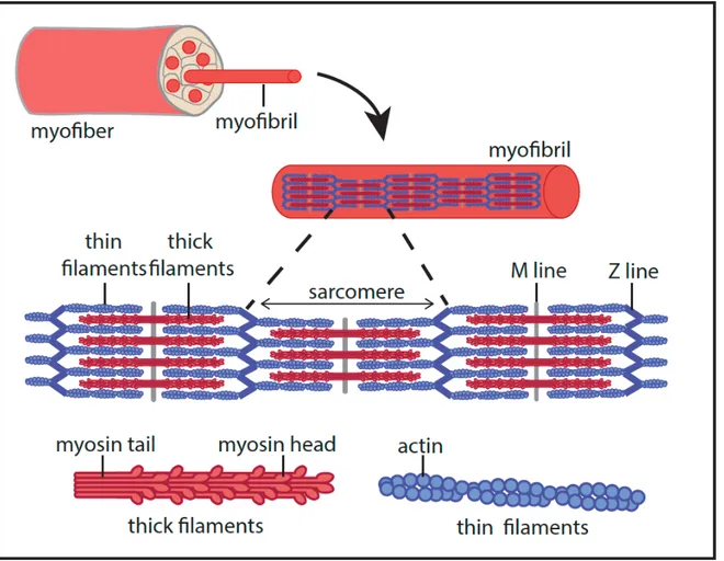
Figure 1. The structure of the skeletal muscle
In the skeletal muscle, satellite cells differentiate and fuse together to form the single unit of the muscle tissue: the myofiber. Myofiber contains a large number of myofibrils, which play a key role in contraction. Myofibers are surrounded by connective tissues called endomysium, assembled in bundles, themselves surrounded by the perimysium. Based on (Frontera & Ochala, 2014).
Figure 2. The sarcomere
The sarcomere is the structural unit of a myofibril, allowing contraction. Sarcomere formation relies on the association between thick filaments made of myosin and thin filaments made of actin. Thick filaments are docked on both sides of the M line at the center of the sarcomere. They are interdigitated with thin filaments, which are docked on the Z line at each extremity of the sarcomere. Based on (Frontera & Ochala, 2014).
!
! !
Figure 3. Actin filament sliding allows muscle contraction
1- ATP binds to myosin head. 2- Myosin ATPase activity triggers ATP hydrolysis. ADP + PI remain bound to myosin and induce conformational change of its head. 3- In this conformation, the myosin head binds to actin and forms a cross bridge. 4- The release of PI leads to the rotation of myosin head which pushes the actin filament. Finally, the release of ADP allows new ATP molecule binding and makes the cycle start again. Based on (Frontera & Ochala, 2014). !
! "4!
perimysium (Frontera & Ochala, 2014). Satellite cells are located between the myofiber’s plasma membrane (PM), called sarcolemma, and the basal lamina (Figure 1). They can self-renew and are key for muscle regeneration (Collins et al., 2005).
The myofiber, the single unit of the muscle, is highly specialized and has a particular protein and organelle content, as well as a specific sarcolemma topology further detailed in the next chapters.
1.3 The myofiber, a highly specialized architecture
1.3.1 The sarcomere and contractility
Each myofiber contains multiple myofibrils, themselves composed of an assembly of sarcomeres. A sarcomere is the muscle contractile unit consisting of a specific arrangement of myofilaments. Myofilaments can be of two types: they can be thick filaments composed of myosin and thin filaments made of actin and troponin. Thin actin filaments are docked on one side to a Z line, mainly composed of α-actinin. One Z line can be found at each extremity of a sarcomere. These actin filaments, emanating symmetrically from each Z line are connected and interdigitate with thick myosin filaments that compose the H zone. Myosin filaments are docked on both sides of a central M line (Figure 2). The contraction of skeletal muscle relies on myosin and actin interaction, that form cross-bridge through myosin heads binding to actin (Huxley, 1969), and is dependent on calcium release (Szent-Györgyi, 1975). This is followed by the sliding of actin filaments, provoked by myosin heads movement in an ATP (adenosine triphosphate) dependent manner (Holmes & Geeves, 2000; Huxley, 1969; Huxley, 1957). ATP is provided by the mitochondria network or by glycogenolysis. This mechanism is detailed in Figure 3.
!
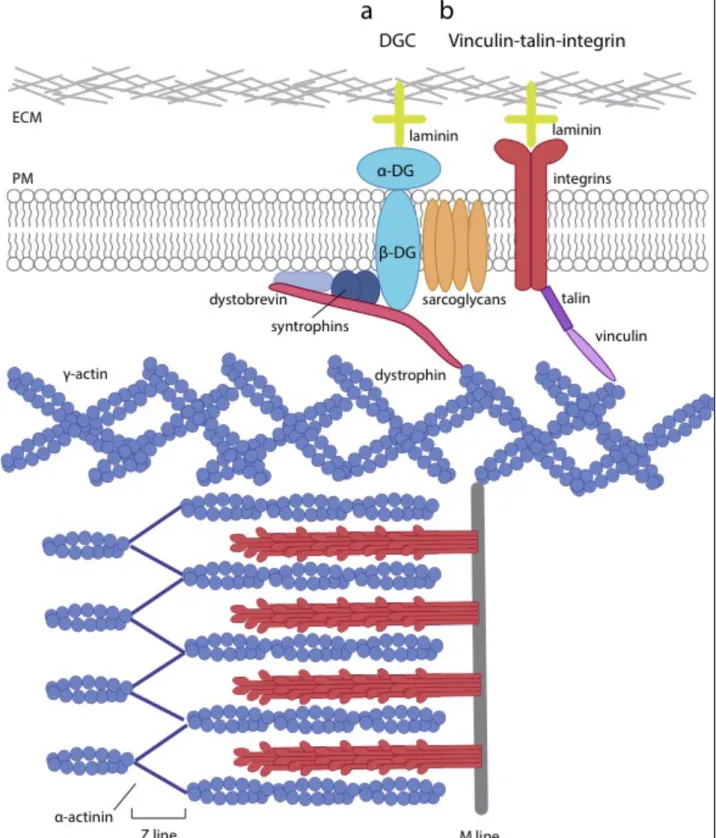
Figure 4. The costamere
Costameres are composed of two protein complexes, the dystrophin glycoprotein complex (DGC) and the vinculin-talin-integrin complex. They are located at the plasma membrane of myofibers and aligned with Z and M lines of sarcomeres.
a) The DGC contains dystrophin, the scaffold protein linking the actin cytoskeleton to the plasma membrane and the extracellular matrix (ECM). Dystrophin bridges γ–actin through its N-terminal domain, to β-dystroglycan through its C-terminal domain. β-dystroglycan is a transmembrane protein that forms a complex with the α-dystroglycan binding directly to laminin in the ECM. In addition, dystrophin also interacts with dystobrevin and syntrophins, both cytosolic proteins associated with the plasma membrane. The sarcoglycans, another set of proteins, stabilizes this complex. Based on (Cohn & Campbell, 2000). b) Vinculin interacts with the extremity of actin bundles linked to sarcomeres, on one side, and with talin on the other. Talin is the scaffold protein linking vinculin to integrins, transmembrane proteins that bind laminin in the ECM. Based on (Sabatelli et al., 2012).
! "$!
Other actors are involved in the filament stabilization, such as troponin, tropomyosin, titin, nebulin or desmin (the role of desmin is further detailed in the next chapter) (Au, 2004).
1.3.2 The costamere, myofiber integrity and mechanotransduction
Costameres are protein complexes located at the sarcolemma, which are arranged perpendicularly to myofilaments, at a specific distance and are mainly aligned with M lines, but also with Z lines (Pardo et al., 1983). They allow sarcomeres to anchor to the sarcolemma, but also to link the sarcolemma, the sarcomeres, and the cortical actin to the extracellular matrix. The costameres are mainly composed of two protein complexes, the dystrophin-glycoprotein complex (DGC) and the vinculin-talin-integrin complex. The DGC is the protein complex that allows to link the actin-based cortical cytoskeleton to laminin in the extracellular matrix (ECM), and is crucial for sarcolemma integrity (Yoshida & Ozawa, 1990). It contains both cytoplasmic and transmembrane proteins such as dystrophin, sarcoglycans, dystroglycan, dystrobrevins and syntrophins (Figure 4a). Dystrophin is the scaffold protein, binding γ-actin through its N-terminal domain (Ervasti & Campbell, 1993), a transmembrane protein, the β-dystroglycan its carboxyl-terminus, and cytoplasmic proteins such as dystobrevins and syntrophins. Dystroglycans consist in a heterodimer of two dystroglycan isoforms. β-dystroglycan is a transmembrane protein that binds dystrophin on its C-terminal part, and its extracellular partner α-dystroglycan that binds laminin in the extracellular matrix (Ervasti & Campbell, 1993). The precise role of sarcoglycans is not yet eluded, but they definitely contribute to the stabilization of the DGC complex, as deficient mice show loss of sarcolemmal integrity (Duclos et al., 1998). Finally, syntrophin and dystrobrevin complete this complex to supply an additional layer of dystrophin anchorage to the sarcolemma (Lapidos et al., 2004).
The vinculin-talin-integrin complex allows a link between the sarcomeres, the sarcolemma and the ECM (Figure 4b). Vinculin is a membrane associated cytoplasmic protein that
! "<!
was initially found to bind the extremity of actin filament bundles (Geiger et al.,1980; Hüttelmaiere et al., 1997). Vinculin interacts with talin, a scaffold protein that can also bind β-integrin and actin (Burridge & Mangeat, 1984; Calderwood et al., 2002; Lee et al., 2004). Furthermore, interaction between talin and β-integrin induces β-integrin activation and further binding to collagen or laminin in the ECM (Calderwood et al., 2002), which allows mechanotransduction (De Palma et al., 2013).
A few more proteins are involved in the organization and stability of sarcomeres, in particular desmin and the clathrin heavy chain (CHC). Desmin is an intermediate filament (IF) that is essential for muscle architecture since it connects sarcomeres with the sarcolemma, nuclei and other organelles (Paulin & Li, 2004). Desmin filaments are cross-linked by plectins, create a network and bind to integrins (Steinböck & Wiche, 1999). Recently, an unconventional role for CHC usually associated with endocytosis was described in muscle. At the sarcolemma, CHC was shown to be organized as large plaques that recruit α-actinin for proper anchorage and organization of sarcomere (Vassilopoulos et al., 2014).
Costameres are thus essential for sarcomere anchorage and stability, myofiber integrity and mechanotransduction. No surprisingly, mutations in most of costamere proteins are associated with different types of myopathies (reviewed in Jaka et al., 2015).
1.3.3 Dysferlin and membrane repair
Dysferlin is a transmembrane protein that is key for membrane repair in muscle (Bansal et al., 2003). It localizes at the sarcolemma or near T-tubules (transverse-tubule) (further detailed in chapter 1.4.2) and can be recruited on site of membrane injury. Upon
membrane damage, dysferlin is recruited in a Ca2+-dependent manner and, together
with MG53 (mitsugumin 53) and caveolin-3 (Cav3), will induce fusion of intracellular vesicles, and injured sarcolemma in order to reseal the wound (Cai et al., 2009; Lennon et al., 2003). The role of Caveolin-3 in this process will be further detailed in chapter 3.1.2.4.
! !
! !
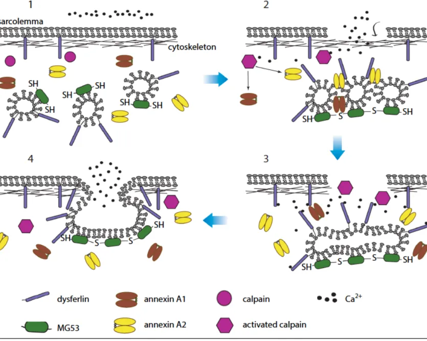
Figure 5. Model for the mechanism of membrane patching in muscle fibers.
1) In the intact muscle fiber, Mitsugumin 53 (MG53) is on sarcoplasmic vesicles in a reduced form, and the concentration of calcium in sarcoplasm is lower than in the extracellular matrix.
2) Upon membrane damage, exposure of the intracellular environment to oxidative agents leads to oxidation and oligomerization of MG53 proteins on sarcoplasmic vesicles. Increased calcium concentration around the injury site also activates calpain. Activated calpain may cleave annexin proteins, which subsequently mediate accumulation of vesicles near the site of damage.
3) Dysferlin senses the calcium that floods into the fiber and triggers fusion of accumulated vesicles. Activated calpain also degrades the cytoskeleton near the damaged area, making easier the fusion of vesicles with the plasma membrane.
4) Vesicles fuse with the sarcolemma and form a patch that seals the membrane. Abbreviation: SH, reduced sulfur moieties in MG53. Adapted from (Tabebordbar, Wang, & Wagers, 2013).
! "?!
Dysferlin can also be associated with intracellular vesicles and upon membrane damage, it is proposed that these vesicles could fuse with lysosomes to form larger vesicles ready to be recruited for membrane resealing (McDade & Michele, 2014). Dysferlin-dependent membrane repair in muscle is summarized in Figure 5.
1.3.4 Muscle associated signaling pathways
Many signaling pathways have been shown to be essential for muscle integrity and functions. In this chapter, I chose to focus on the glucose pathway that is tightly regulated in muscle tissue to maintain glucose and lipid homeostasis, and the IL6 pathway, which is important for muscle tissue integrity and homeostasis (reviewed in Muñoz-Cánoves et al., 2013).
1.3.4.1 The glucose pathway in the muscle
The glucose pathway in skeletal muscle is very important for homeostasis of the muscle and the whole organism. It has been observed very early that glucose was uptaken in skeletal muscle during exercise. Glucose can be transported in cells through specific transporters of the GLUT (glucose transporter) family, and more specifically GLUT1 and GLUT4 in skeletal muscle (Devaskar & Mueckler, 1992). GLUT1 being expressed at very low levels, GLUT4 is the major glucose transporter in muscle cells (Gaster et al., 2000; Zisman et al., 2000). In resting condition, GLUT4 is stored in large endosomal structures, which formation requires the specific clathrin isoform CHC22 (clathrin heavy chain 22), and that can be recruited upon insulin stimulation or muscle contraction (Karlsson et al., 2009; Lauritzen et al., 2010; Ploug et al., 1998; Vassilopoulos et al., 2009). These two stimuli have been proposed to recruit different pools of GLUT4 containing vesicles, as an additive effect was observed for their recruitment when muscle fibers are stimulated with both insulin and muscle contraction (Ploug et al., 1998). Once in the cell, glucose can
!
Figure 6. Insulin and exercise mediated GLUT4 translocation at the plasma membrane.
Upon insulin binding, the insulin receptor gets phosphorylated, triggering the phosphorylation of the insulin receptor substrate (IRS) that recruits and activates the Phosphoinositide 3-Kinase (PI3K).
This kinase converts PIP2 to PIP3, a docking platform for the recruitment of PDK1 and Akt. Once
activated, Akt promotes the recruitment of GLUT4 containing vesicles at the plasma membrane, leading to glucose entry in the cell. Based on (Leto & Saltiel, 2012).
Muscle contraction can have many cellular consequences. Among them, Ca2+ release increases NO,
AMPK and TBC1D1 activities, all promoting the recruitment of GLUT4 vesicles at the plasma membrane. Based on (Richter & Hargreaves, 2013).
! "B!
undergo glycolysis to provide the ATP needed for muscle contraction or glycogenesis for muscle glycogen storage (Hansen et al., 1995). Then, the stored glycogen will be used for rapid ATP production during the next exercise, or will follow glycogenolysis pathway to provide glucose in fasting diet for example. Glucose uptake mechanism is summarized in Figure 6.
1.3.4.2 The IL6/STAT3 pathway in the muscle
Interleukin-6 (IL6) is one of the major cytokine produced by the muscle and plays autocrine, paracrine and endocrine roles (Muñoz-Cánoves et al., 2013; Pedersen & Febbraio, 2008). IL6 is produced and secreted during exercise, and thus during muscle contraction (Steensberg et al., 2002). Several pathways can trigger IL6 production, all dependent on contraction. It was proposed that nitric oxide (NO), produced during exercise, could induce IL6 mRNA expression (Steensberg et al., 2007). NO also increases IL6 protein expression through the MAPK (Mitogen activated kinase) pathway (Makris et al., 2010). Increased calcium levels observed during contraction might also regulate IL6 mRNA (messenger ribonucleic acid) expression. Indeed calcium binding to calcineurin leads to the dephosphorylation of nuclear factor of activated T-cell (NFAT). Then, NFAT translocates to the nucleus where it controls, among others, the IL6 mRNA transcription (Crabtree, 2001; Keller et al., 2006). The decrease of glycogen concentration, induced by glycogenolysis, are linked to the phosphorylation of P38 MAPK (mitogen-activated protein kinase) that promotes IL6 mRNA expression (Chan et al., 2004). Finally, IL6 itself can trigger its own expression probably via activation of AMP-activated protein kinase (AMPK) although this mechanism is not fully elucidated (Keller et al., 2003; Weigert et al., 2007).
IL6 signal transduction depends on its binding to the glycoprotein 130 (gp130), a ubiquitously expressed transmembrane receptor, and to the IL6 receptor (IL6R). IL6 binds first to the IL6R, that can be either at the plasma membrane or soluble in the extracellular
!
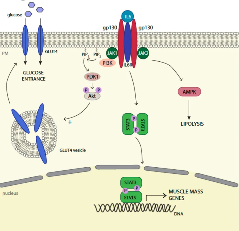
Figure 7. The IL6/STAT3 pathway.
IL6 first binds to the IL6 receptor (IL6R). Then, the complex associates with two gp130 sub-units. JAK1 and JAK2 are then recruited and activated by phosphorylation, leading to the recruitment of
the PI3K. PI3K converts PIP2 to PIP3, a docking platform for the recruitment of PDK1 that
phosphorylates Akt. Finally, Akt activation promotes cell survival. The activation of JAKs also leads to STAT3 phosphorylation, followed by its dimerization and nuclear translocation. In the nucleus, it acts as a transcription factor and regulates the expression of many genes, including STAT3 itself but also SOCS3. SOCS3, together with SHP2, are negative inhibitors of the pathway and therefore key are for the termination of the signal. Based on (Heinrich et al., 2003).
!
! !
! !
Figure 8. IL6 autocrine functions in skeletal muscle.
In skeletal muscle, the IL6 pathway is associated with glucose and lipid homestasis. IL6 signaling activation can participate to glucose entry through Akt-dependent GLUT4 vesicles translocation at the plasma membrane. It is also involved in lipolysis through AMPK activation. IL6 activated-STAT3 controls the transcription of several genes such as FBXO32 or C/EBPδ, essential for the control of muscle mass.
! "P!
environment, and then to gp130, leading to initiation of the signal (Heinrich et al., 2003). Janus Kinases 1 and 2 (JAK1 and JAK2) are recruited to the plasma membrane, and associate to the intracellular tail of gp130 to be activated (Babon et al., 2014; Haan et al., 2001; Haan et al., 2000). Cytosolic STAT3 (signal transducer and activator of transcription 3) is then recruited to the complex and undergoes phosphorylation and dimerization, both required for its subsequent nuclear translocation and activation of STAT related gene transcription (Milocco et al., 1999). JAK activation can also activate AMPK and the subsequent PI3K/Akt (PI3K: phosphatidylinositol-4,5-bisphosphate 3-kinase PKB/Akt: protein kinase B) pathway (Heinrich et al., 2003). Two different ways to terminate the signal have been described, and rely on SOCS3 (suppressor of cytokine signaling 3) and SHP2/PTPN11 (Tyrosine-protein phosphatase non-receptor type 11) proteins (Heinrich et al., 2003). The IL6/JAK/STAT pathway in muscle is summarized in Figure 7.
The IL6/STAT3 pathway in muscle tissue has been shown to be important for satellite cell activation during muscle regeneration, and its overactivation can cause satellite cell exhaustion (Price et al., 2014; Tierney et al., 2014; Toth et al., 2011). Interestingly, it seems that the main IL6 source involved in muscle regeneration is secreted from infiltrated macrophages and neutrophils (Zhang et al., 2013).
In myotubes, the IL6 pathway contributes to homeostasis, as it enhances lipolysis and glucose uptake by triggering GLUT4 translocation through the activation of the AMPK and PI3K pathways (see chapter 1.3.3.1) (Carey et al., 2006; Wolsk et al., 2010). STAT3 activity is mostly associated with muscle mass. Indeed, in extreme conditions such as high dose and long term IL6 exposure or IL6 overexpression, increased muscle proteolysis with decreased muscle mass have been observed (Ebisui et al., 1995; Franckhauser et al., 2008). This explains the description of IL6/STAT3 induced muscle wasting phenotype in the context of cancer cachexia (Bonetto et al., 2011). Autocrine functions of the IL6 pathway in skeletal muscle are summarized in Figure 8.
! "G!
1.4 The specific organelle organization and membrane topology
of myofibers
In myofibers, the organelle organization and distribution is adapted to the very particular elongated shape of these multinucleated cells, highly enriched with myofibrils. I will focus here on two main organelles that are highly adapted to muscle function: the nuclei and the sarcoplasmic reticulum. The sarcoplasmic reticulum (SR) will be described together with the T-tubule system, as they form a particular structure called the triad.
1.4.1 Nuclei
Nuclei are located at the periphery of the myofibers, right below the sarcolemma, in a regular spaced manner (Bruusgaard, 2006). This specific localization requires nuclear movements occurring through sequential steps (Roman & Gomes, 2017). The first step, nuclear centration is dependent on microtubules (MT) that allow the gathering in the center of the cell of newly integrated nuclei when myoblasts fuse with a myotube. In myotubes, the microtubule organizing center (MTOC) has the particularity to be located on the nuclear envelop (NE) instead of the centrosomes as classically observed in other cell types, leading to MT nucleation from the nuclei (Tassin et al., 1985). In addition to MT, other important players are involved in this process, such as the dynein/dynactin motor and cdc42, Par3 and Par6 proteins (Cadot et al., 2012). Nuclei then align and spread along the longitudinal axis. This process is dependent on the LINC (linker of nucleoskeleton and cytoskeleton) complex, and more particularly on nesprin-1α, that recruits centrosomal proteins such as PCM-1 (pericentriolar material protein 1) and Akap450. PCM-1 allows the recruitment of motors such as dynein/dynactin and kinesins, although kinesin-1 can be recruited by nesprin-1 itself, leading to nuclear movement (Espigat-Georger et al., 2016; Wilson & Holzbaur, 2015). Akap450 regulates MT nucleation and is crucial for nuclear positioning (Gimpel et al., 2017). Finally, nuclei move towards the periphery of the cell where they are anchored. Once myofibrils are formed,
!
Figure 9. Nuclear positioning during myogenic differentiation.
a) Nuclear movements during myogenesis. Blue rectangles represent zoomed illustrations of B–F. b) Centration. Nucleus from newly fused myoblast is pulled toward the center of the myotube by dynein and microtubules (MT).
c) Alignment. Nesprin-1α and PCM-1 recruit centrosomal and motor proteins to the NE.
d) Spreading. Kinesin-1 and Map7 attached to microtubules allow use the anti-parallel microtubular network to spread nuclei along the length of the myotube. Kinesin-1 and kinesin light chain (KLC) at the nuclear envelop walk towards the (+) end of surrounding microtubules to induce nuclear rotation. e) Peripheral migration. Myofibril crosslinking, contraction and nuclear stiffness variations drive nuclear movement to the periphery. Myofibril crosslinking is mediated by desmin organization at the z-lines. f) Anchoring. Nesprin organizes an astral microtubular network to anchor nuclei juste below the plasma membrane. Anchoring is reinforced by a desmin network. (Roman & Gomes, 2017)
! "J!
they are cross-linked at the level of the Z line with the help of desmin, which proper organization for cross-linking depends on the Arp2/3 complex and γ-actin. All together, these proteins will act like closing zippers that push nuclei towards the periphery. This process is further enhanced by myofibril contraction and lower nuclear stiffness tuned by local lamin A/C level decrease (Roman et al., 2017). Nuclei can either be aggregated to the myotendinous junction (MJ), involved in stretching, or to the neuromuscular junction (NMJ), involved in contraction, for localized expression of specific proteins. They can also be isolated and evenly distributed at the periphery of myofibers. The two different anchorage mechanisms involved may depend on nucleus fate, but are so far poorly understood (Roman & Gomes, 2017). The mechanisms of nuclear positioning are summarized in Figure 9.
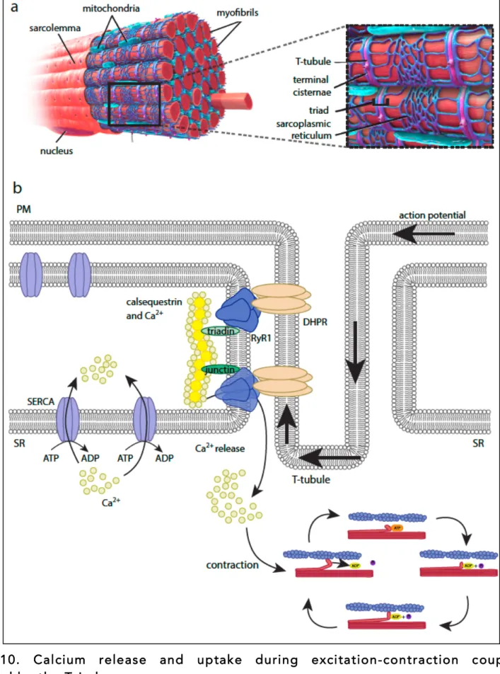
1.4.2 The triad: the sarcoplasmic reticulum and the T-tubule system
The triad, a specific structure only found in skeletal muscle, is key for muscle contraction. It is composed of the association of one T-tubule and two terminal cisternae of the sarcoplasmic reticulum (Al-Qusairi & Laporte, 2011).
T-tubules are deep membrane invaginations that insert between myofibrils. Their main
role is to induce Ca2+ release from the SR, needed for muscle contraction (see chapter
1.3.1). Prior to release, calcium is stored in the SR through a calsequestrin-dependent
process (Costello et al., 1986). Calsequestrin is localized at terminal cisternae where its anchorage depends on other proteins such as junctins, triadin and RyR1 (ryanodine
receptor 1) (Zhang et al., 1997). It can bind Ca2+ with low affinity but in a high capacity
manner, allowing high calcium concentration close to release sites (MacLennan & Wong, 1971). Calcium release depends on the tight association between the L-type calcium channel DHPR (dihydropyridine receptor), located at the T-tubule, and RyR1, located at the level of the terminal cisternae of SR (Marty et al., 1994; Rios & Brum, 1987). Action potential spreading into T-tubules triggers DHPR conformational changes, leading to
!
Figure 10. Calcium release and uptake during excitation-contraction coupling controlled by the Triad.
a) Localization of the triad in myofibers, composed of one T-tubule and two terminal cisternae of the sarcoplasmic reticulum (SR) are surrounding. (Blausen.com staff (2014). "Medical gallery of Blausen Medical 2014". WikiJournal of Medicine)
b) At rest, Ca2+ is concentrated and associated with calsequestrin, located right under Ryanodin
Receptor 1 (RyR1) at SR terminal cisternae through its association with triadins and junctins. Upon excitation, the action potential propagates through the plasma membrane and the T-tubules, where it induces dihydropyridine receptor (DHPR) activation. DHPR is tightly associated with RyR1 and its
activation induces RyR1 conformational change, leading to Ca2+ release and muscle contraction. After
contraction, the excess of Ca2+ is re-uptaken in the SR through SERCA (sarco-endoplasmic reticulum
calcium ATPase pumps) pumps, in an ATP-dependent process. !
! 4U!
RyR1 opening, allowing the massive Ca2+ release in the cytoplasm, and subsequent
contraction (Marty et al., 1994). Finally, during muscle relaxation, the excess of calcium needs to be re-captured by the SR. This process is mainly handled by sarco-endoplasmic reticulum calcium ATPase pumps (SERCA), located both at terminal cisternae and longitudinal SR (Yu et al., 1993). The mechanism of calcium release and uptake by the Triad is summarized in Figure 10.
In addition to T-tubules, the plasma membrane of muscle cells is also highly enriched in a particular membrane invagination called caveolae. The next chapter will describe their constituents, biogenesis and functions. I will then discuss the particular functions of caveolae in the skeletal muscle, as well as their implication in muscle diseases.
! ! ! ! ! ! ! !
Figure 11. Caveolae observation by electron microscopy. a, b) Caveolae can adopt different organizations. It can be single caveolae individually connected to the plasma membrane (indicated with arrowheads), or interconnected caveolae forming bigger structures, called rosettes that can still be connected to the plasma membrane (indicated with asterisks).
c) Visualization of the caveolar coat at the plasma membrane of myotubes. Survey view of the cytoplasmic surface of an unroofed mouse myotube presenting caveolae at the plasma membrane. Different types of caveolae structures are apparent, ranging from fully budded (1), circular (2), to flat (3). Scale bar: 500 nm. Scale bar in insets: 50 nm. Adapted from (Lamaze et al., 2017).
! 4"! !
!
2 Caveolae
2.1 Caveolae, specialized plasma membrane invaginations
Caveolae were observed for the first time by electron microscopy in the ’50s by analyzing the structure of blood capillaries and the gall bladder epithelium (Palade, 1953; Yamada, 1955). They are defined as 50-100 nm diameter plasma membrane invaginations, and can be distinguished from clathrin coated pits when observed by rapid-freeze deep-etch electron microscopy (Rothberg et al., 1992). In mammals, caveolae are present in all type of cells, with the notable exception of neurons and lymphocytes, although they express caveolin-1, and are particularly enriched in adipocytes, endothelial and muscle cells. Caveolae can be found as single caveolae, associated to specialized structures such as T-tubule in differentiating muscle cells (Parton et al., 1997), or interconnected as a multi-caveolar structure called “rosette” (Pelkmans & Zerial, 2005) (Figure 11). After their discovery, it took almost 40 years to identify caveolin-1 (Cav1) as their main protein component (Glenney, 1992; Kurzchalia et al., 1992; Rothberg et al., 1992). This identification was followed by the characterization of two other members of the caveolin family, caveolin-2 (Cav2) and caveolin-3 (Cav3), and was the starting point of a better understanding of caveolae biogenesis and function in cells.
2.2 Caveolae biogenesis
2.2.1 Caveolae protein composition 2.2.1.1 Caveolins
Caveolin-1, the first identified member of the caveolin family, was discovered by two distinct teams at the same time but was thought to be two different proteins, VIP-21
! 44!
(vesicular integral protein 1) and caveolin-1 (Kurzchalia et al., 1992; Rothberg et al., 1992). It was then established that VIP-21 and caveolin-1 were the same protein, and the name caveolin-1 was kept (Glenney, 1992). Cav1 is a small protein of 21-24 kDa with two isoforms: α-Cav1 and β-Cav1. The β-Cav1 isoform lacks the first 31 amino acids (aa) compared to α-Cav1. Nevertheless, they both have a 33 aa hydrophobic domain, suggested to be a β-sheet inserted in the plasma membrane (Glenney & Soppet, 1992), with the N- and C-terminus facing the cytosol (Dupree et al., 1993; Monier et al., 1995).
α-Cav1 (not β-) can be phosphorylated on its Tyrosine 14 (Tyr14) residue upon Rous
sarcoma virus, or after insulin or Src activation (Glenney, 1989; Shengwen et al., 1996; Mastick, et al., 1995). The consequences of this modification will be further discussed in chapter 2.2.3. Cav1 can also be phosphorylated on its serine 80 (ser80) residue, which allows Cav1 binding to ER (endoplasmic reticulum) proteins and its secretion in the particular case of regulated secretion by pancreatic cells (Schlegel et al., 2001). Controlled Ser80 phosphorylation is also crucial for proper caveolae formation and shape (Ariotti et al., 2015). Cav1 presents also three palmitoylation sites (Cys133, Cys153 and Cys156) which are not necessary for membrane anchorage but may influence caveolin oligomerization (Dietzen et al., 1995; Monier et al., 1996). Using velocity sucrose gradient centrifugation, Cav1 was shown to form 300 kDa high molecular complexes. Caveolin proteins can form homo- or hetero- oligomers of α-/β-Cav1 and Cav2 (Root et al., 2015; Sargiacomo et al., 1995). This phenomenon occurs through the Cav1 oligomerization domain (residues 61-101). The oligomerization process and its purpose will be further detailed in chapter 2.2.3.4.
Few years after Cav1 discovery, a potential homologue of Cav1 was found by nucleotide alignment. It allowed the identification of the muscle specific isoform that was first called M-caveolin, to be later renamed caveolin-3 (Cav3) (Way & Parton, 1995). Compared to α-Cav1, Cav3 has 65% of identity and 85% of similarity, and lacks the first 27 aa. Its expression is restricted to cardiac, smooth and skeletal muscle, and is only expressed upon differentiation in skeletal muscle. Because it lacks the Tyr14 present in Cav1, it is
! !
Figure 12. Caveolin protein family domains and insertion in the plasma membrane.
a) Caveolin protein family exhibit several very conserved domains: the oligomerization domain containing the 8aa stretch signature sequence (FEDVIAEP) and the caveolin scaffolding domain (CSD), and the intramembrane domain. Size variability within the caveolin family is due to length difference of the N-terminal part of caveolins.
b) Caveolin 1 (Cav1) is inserted in the plasma membrane through its intramembrane domain, with the N- and C-terminal parts facing the cytosol, conferring a hairpin shape. In addition, three palmitoylation sites allow anchoring and stability of the protein at the plasma membrane. Based on (Parton & del Pozo, 2013).
! 4$!
not phosphorylated, but it still shares a lot of common features with Cav1. Indeed, Cav3 is palmitoylated, has the same conserved hydrophobic domain and is able to oligomerize (Tang et al., 1996; Way & Parton, 1995). Furthermore, in cardiac myocytes, one of the few cells expressing both Cav1 and Cav3, it has been shown that these caveolins could form hetero-oligomers (Volonte et al., 2008).
The last member of the family, and the least studied, is caveolin-2. Based on the purification of membrane fractions enriched in caveolae, it was first defined as a protein with 38% identity and 58% similarity with Cav1. Furthermore, a specific stretch of 8 aa (FEDVIAEP) in the N-terminal part, extremely conserved with Cav1, and found later to be also present in Cav3, was found (Scherer et al., 1996; Tang et al., 1996). It was thus suggested to be the caveolin family signature. Cav2 exists under two isoforms, α-Cav2 and β-Cav2, but not much is known about their potential differences in expression or function. Compared to α-Cav1, α-Cav2 and β-Cav2 lack the first 16 aa and 29 aa respectively. Cav2 localizes at caveolae structures, and colocalize with Cav1. As it is expressed in skeletal muscle cells, contrary to Cav1, it also colocalizes with Cav3. It has the capability to dimerize but not oligomerize further, and can form hetero-oligomers with other caveolin isoforms (Scherer et al., 1996, 1997). As Cav1 and Cav3, α-Cav2 is also triple palmitoylated and although α-Cav2 lacks the Tyr14, it can still be phosphorylated on its Tyr19 and Tyr27 residues upon insulin stimulation (Kwon et al., 2015). Interestingly, there is no evidence that these residues can also be phosphorylated in the other caveolins.
In summary, all three members of the caveolin family have a very similar structure, composed of an N-terminal part containing a very conserved stretch of 8 aa, a very conserved 33 aa hydrophobic region (residues 102-134 of Cav1) with N- and C-terminus facing the cytosol. This allows a particular “hairpin” conformation of the protein, inserted in the plasma membrane (Figure 12).
!!
! !
! !
Figure 13. Cavin structure.
Cavins are heterogeneous in size but they all contain PEST and Leucine rich domains for potential interaction with other proteins. Cavin-1 is the only member of the cavin family with a NLS sequence, favoring nuclear translocation. Based on (Bastiani et al., 2009).