EUMELANIN FILMS FOR ORGANIC BIOELECTRONICS:
GROWTH, CHARGE TRANSPORT, AND INTERACTION WITH METAL ELECTRODES
JULIA WUENSCHE
D´EPARTEMENT DE G´ENIE PHYSIQUE ´
ECOLE POLYTECHNIQUE DE MONTR´EAL
TH`ESE PR´ESENT´EE EN VUE DE L’OBTENTION DU DIPL ˆOME DE PHILOSOPHIÆ DOCTOR
(G ´ENIE PHYSIQUE) JUIN 2014
´
ECOLE POLYTECHNIQUE DE MONTR´EAL
Cette th`ese intitul´ee :
EUMELANIN FILMS FOR ORGANIC BIOELECTRONICS:
GROWTH, CHARGE TRANSPORT, AND INTERACTION WITH METAL ELECTRODES
pr´esent´ee par : WUENSCHE Julia
en vue de l’obtention du diplˆome de : Philosophiæ Doctor a ´et´e dˆument accept´ee par le jury d’examen constitu´e de :
M. YELON Arthur, Ph.D., pr´esident
Mme SANTATO Clara, Doct., membre et directrice de recherche M. ROSEI Federico, Ph.D., membre et codirecteur de recherche M. BUSCHMANN Michael, Ph.D., membre
ACKNOWLEDGEMENTS
First, I would like to express my deepest thanks to my research director, Clara Santato, for guiding, supporting, and encouraging me during these last four years. Your passion for science, your personal involvement in each student’s project, and your persistence are extraordinary and helped me to overcome the more difficult moments. You always have a sympathetic ear for the concerns of your students, professional and personal, which is remarkable. Thank you for putting so much confidence in me and for entrusting me with such a challenging project. I also very much appreciate that you gave me the chance to meet and discuss with so many great scientists.
I thank my codirector, Federico Rosei, for providing me with a “quick start” in Montreal by means of an internship at INRS, for encouraging me to apply for scholarship, and for his advice during my PhD. Furthermore, I am grateful to the jury members, Arthur Yelon, Michael D. Buschmann, and Siegfried Bauer, for their time and interest in my work.
Financial support from NSERC through a Vanier Canada Graduate Scholarship is grate-fully acknowledged.
Many thanks to all the collaborators that have contributed to my research projects over the last years and who made my work more enjoyable and interesting. I would like to thank Alessandro Pezzella for sharing his invaluable knowledge on melanin chemistry. I am very grateful to Francesca Soavi and Marco Rolandi for the long and insightful discussions on the electrical and electrochemical properties of eumelanin. Special thanks to Fabio Cicoira for his continuous, pleasant support, and in particular for his advice on microfabrication. I very much thank Luis Cardenas for his support in the characterization of Au-eumelanin dendrites. I really enjoyed working with you. Many thanks to Yingxin Deng for the PdH measurements and her kind collaboration. I am very grateful to Suzie Poulin who took and analyzed many ToF-SIMS and XPS measurements for our project. I would like to thank Carlos F. O. Graeff for providing melanin samples and for his advice. I am also grateful to Reynald Gauvin for the SEM images taken in his lab. Many thanks to Alberto Salleo for GIXRD measurements by his group.
I have not yet thanked the collaborators who were also my colleagues at Polytechnique. Special thanks to Prajwal Kumar for the many CV measurements, which required perseve-rance and patience. A big thank you to Jonathan Sayago for the many times he helped me out in the lab and for being such a nice and generous colleague over all these years. Many thanks to Eduardo di Mauro for his enthusiastic start into this project and for proof-reading the first part of the thesis.
Of course, I would also like to thank my other colleagues, former and present, for their unlimited support in the lab, their advice, for making work pleasant, and for the interesting discussions about different cultures. Special thanks to Fr´ed´eric Venne for revising the French abstract.
I am very grateful to P. Moraille for her king support with AFM, to Alireza H.Mesgar for support in microfabrication to Josianne Lefebvre (XPS and ToF-SIMS), N. Brodusch (SEM), C. Chilian (NAA), and Samir Elouatik (Raman) for measurements.
Merci beaucoup `a Jo¨el Bouchard, Christophe Cl´ement, Yves Drolet, Jean-Paul L`evesque et Lucas Majeau pour leur support technique et les discussions dans le corridor. Je remercie aussi tous les ´etudiants qui ont contribu´e aux diff´erents projets r´eli´es `a la m´elanine dans le cadre des cours ou des stages d’´et´e.
I am grateful to Patrick Desjardins, Antonella Bad´ıa, Richard Martel, Mihai Irimia-Vladu, Alon Gorodetsky, David Ordinario, Paul Meredith and the many other scientists I met for fruitful discussions.
I would also like to thank former collegues and collaborators that worked with me during the last years on research projects other than those presented in this thesis, in particular Simone Bertolazzi, Giuseppe Tarabella, Ivan A. Velasco Davalos, and Andreas R¨udiger.
Merci beaucoup `a mes amis `a la Polytechnique pour rendre les pauses de midi plus que agr´eables. Merci `a toute mon ´equipe de volleyball qui m’a donn´e plein de distractions apr`es le travail. Je remercie tous mes amis `a Montr´eal pour les nombreux moments de plaisir, leur soutien et pour me recevoir si bien quand je suis arriv´ee. Ich danke auch meinen Freunden in Deutschland, die mich nach so vielen Jahren Abwesenheit immer noch gerne empfangen und die mich aus der Ferne unterst¨utzt haben.
Ich m¨ochte mich ganz herzlich bei meiner Familie, insbesondere meinen Eltern und meiner Schwester, bedanken. Vielen Dank f¨ur eure uneingeschr¨ankte Unterst¨utzung, auch als ich nach Montreal zog, und f¨ur eure Ermutigungen.
Finalement, je remercie de tout cœur Jean-Francois Desjardins pour son soutien, pour me faire venir `a Montr´eal et pour la traduction du r´esum´e. Merci beaucoup pour me faire profiter de la vie `a tous les jours !
ABSTRACT
Organic semiconductors are mechanically soft materials and generally exhibit mixed ionic and electronic conduction. These properties are exploited in organic bioelectronics, a new field of research at the interface of electronics and biology that aims at providing knowledge and technology required for applications such as biosensing, drug delivery, and neuronal stim-ulation. In addition to well-characterized organic electronic materials already used in organic light-emitting diodes and solar cells, researchers in the field of organic bioelectronics are ex-ploring the use of biomolecular electronic materials. The search for suitable biomolecular materials is motivated by potential benefits such as biocompatibility and biodegradability, for applications at the interface with living systems. On a more general level, biomolecular electronic materials are relevant because they could lead to more environmentally friendly electronics.
The biomolecular material eumelanin has been considered to be an amorphous semicon-ductor since the 1970’s. Eumelanin is a pigment ubiquitous in flora and fauna and has many functions in the human body including UV-protection, free radical scavenging, metal ion chelation, and thermoregulation. Eumelanin has a π-conjugated backbone and is character-ized by a high degree of energetic and structural disorder. Different types of eumelanin can be distinguished according to their (bio-)synthetic origin, resulting in different molecular and supramolecular structures. Pressed powder pellets of eumelanin show thermally activated conductivity and photoconductivity. However, in contrast to typical organic semiconductors, the conductivity of eumelanin pellets strongly increases with hydration. Although there are reports about mobile ions in eumelanin, the amorphous semiconductor model prevailed and was used to interpret the electrical properties of eumelanin during the last four decades. Dur-ing the work for this thesis, a new study on eumelanin pellets was published showDur-ing that this model cannot correctly describe the hydration-dependent conductivity of eumelanin. In-stead, the work suggests that eumelanin is a mixed ionic-electronic conductor. This report underlines the need to reinvestigate the electrical properties of eumelanin and reinforces the recent interest in eumelanin as a material for organic bioelectronics. The integration of eu-melanin in device structures has been facilitated by the recent progress in the preparation of eumelanin thin films. Indeed, first attempts to employ eumelanin in chemical sensors and batteries have been reported. However, the properties of eumelanin thin films are far from being fully characterized. In particular, the electrical properties of hydrated films are largely unexplored.
inves-tigated in view of possible applications in bioelectronics. The first focus of this work is to identify a suitable procedure for the solution-processing of eumelanin films to achieve homogeneous and smooth films, a non-trivial task due to the insolubility of eumelanin in common solvents. Several combinations of different synthesis routes and processing solvents are compared in terms of the morphology and the chemical composition of the films pre-pared, using atomic force microscopy (AFM) and x-ray photoelectron spectroscopy (XPS). It is shown that the use of ammonia solution, suggested in the literature, severely affects the chemical composition of eumelanin. The use of dimethyl sulfoxide (DMSO) as process-ing solvent in combination with commercial synthetic eumelanin yields homogeneous films with sub-nanometer surface roughness, good adhesion on hydrophilic substrates, and without significantly changing the elemental composition of the films. Based on the comparison of eumelanins of different origins and a more detailed XPS study, it is argued that there is a trade-off between the solution-processibility of a eumelanin-derived material and its chemical resemblance to natural eumelanin. Furthermore, the growth of eumelanin films deposited from DMSO is investigated by AFM. This investigation reveals that the films are composed of nanoaggregates and assemble in a quasi layer-by-layer fashion. The observation of nanoag-gregates supports the stacked-oligomer model for the eumelanin samples investigated in this work, as opposed to the heteropolymer model.
The core of this thesis is devoted to a better understanding of the electrical response of hydrated eumelanin films and the charge carrier transport properties of eumelanin. Eu-melanin is characterized in thin film form with coplanar metal electrodes under controlled humidity, using voltages ≤ 1 V. A surprisingly strong interaction of hydrated eumelanin thin films with Au electrodes under electrical bias is revealed. This interaction leads to the growth of Au-eumelanin dendrites between the electrodes paralleled by a sudden resistive change of the sample. The growth of the dendrites is investigated by AFM, scanning elec-tron microscopy, and time-of-flight secondary ion mass spectroscopy. Samples of eumelanin of various sources are compared to gain insight into the role of molecular structure and im-purities. Based on this study, a mechanism for the formation of the dendrites is proposed, involving the electrochemical oxidation of Au enabled by the presence of chloride traces and enhanced by the metal chelation properties of eumelanin. The phenomenon described herein is interpreted considering electrochemical metallization cells and suggests the possibility to conceive biocompatible eumelanin-Au based memory devices.
To investigate the intrinsic electrical properties of eumelanin, further experiments are con-ducted with Pt electrodes. Transient current measurements, current-voltage measurements, and electrochemical impedance spectroscopy (EIS) are employed to distinguish different con-tributions to the electrical current by their voltage and time/frequency-dependence. The
effect of sample hydration, film and electrode dimensions on the electrical current is investi-gated. The strong hydration dependence of the current, previously investigated for pellets, is confirmed for eumelanin thin films. It is shown that the electrical current is capacitive at voltages below 0.2 V (over 10 µm) and that electrochemical reactions dominate the non-capacitive currents at higher voltages. Current measurements with proton-transparent PdHx electrodes reveal the strong contribution of protons to the current in hydrated eumelanin films. EIS provides a first estimate (10−4 S cm−1) for the proton conductivity at 90% rela-tive humidity. In contrast, the experimental results do not show clear evidence of electronic conduction. Based on the recent suggestion that the density of mobile electrons is governed by a pH-dependent comproportionation equilibrium, a qualitative model is developed for the electrical response of hydrated eumelanin films. The model suggests how the interplay of pro-ton migration, the redox properties of eumelanin, and the comproportionation equilibrium could limit electron transport over micrometric distances. Subsequently, results of this thesis and from the literature are discussed in terms of protonic conduction and the possibility of electronic conduction in eumelanin.
In view of bioelectronic applications, the main contribution of this thesis is the first de-tailed investigation of the electrical properties of strongly hydrated eumelanin films. This investigation revealed the importance of electrochemical processes in the electrical response of hydrated eumelanin films. Such processes can yield interesting new phenomena such as the growth of Au-eumelanin dendrites. For the study of charge transport in eumelanin, it is important to consider the contribution of electrochemical processes to the electrical current. This thesis provides new insights into the charge carrier properties of eumelanin films. It demonstrates and quantifies protonic conduction in hydrated eumelanin films. In contrast, it places a question mark over the paradigm of electronic conduction in eumelanin. Further-more, this work emphasizes the necessity to consider the specific chemical composition of the eumelanin under investigation and the effect of the processing solvent on this composition. In a wider context, this thesis promotes the awareness of the effects of water, impurities, and electrochemical processes on the electrical performance of organic bioelectronic devices.
R´ESUM´E
Les semi-conducteurs organiques sont des mat´eriaux souples `a conduction ionique et ´ elec-tronique mixte. Ces propri´et´es sont exploit´ees dans la bio´electronique organique, un nouveau champ de recherche `a l’interface de l’´electronique et de la biologie qui vise `a fournir les connaissances et la technologie pour des applications telles que les biocapteurs, la livraison de m´edicaments et la stimulation neuronale. Outre les mat´eriaux ´electroniques organiques qui ont ´et´e bien caract´eris´es `a des fins d’applications de diodes ´electroluminescentes organiques et de cellules solaires, les chercheurs dans le domaine de la bio´electronique organiques explorent l’utilisation des mat´eriaux ´electroniques biomol´eculaires. La recherche de mat´eriaux biomo-l´eculaires est motiv´ee par ses b´en´efices potentiels en biocompatibilit´e et en biod´egradabilit´e, essentiels pour des applications `a l’interface avec des syst`emes vivants. Sur un plan plus g´en´ e-ral, les mat´eriaux ´electroniques biomol´eculaires pourraient ´eventuellement ˆetre utilis´es pour la conception de syst`emes ´electroniques plus ´ecologiques.
L’eum´elanine, un mat´eriau biomol´eculaire, a ´et´e consid´er´e comme un semi-conducteur amorphe depuis les ann´ees 1970. L’eum´elanine est un pigment omnipr´esent dans la flore et la faune. Il poss`ede de nombreuses fonctions dans le corps humain: la protection UV, le pi´egeage des radicaux libres, la ch´elation des ions m´etalliques, et la thermor´egulation. L’eu-m´elanine a une structure ´electronique mol´eculaire de type pi-conjugu´e qui se caract´erise par son haut degr´e de d´esordre ´energ´etique et structurel. Il y a diff´erents types d’eum´elanine et ils se distinguent par leurs structures mol´eculaires et supramol´eculaires, lesquelles d´ependent des conditions de (bio-)synth`ese. Par exemple, les pastilles de poudre press´ee d’eum´elanine poss`edent une conductivit´e et photoconductivit´e activ´ee thermiquement. Cependant, contrai-rement aux semi-conducteurs organiques typiques, la conductivit´e des pastilles d’eum´elanine augmente drastiquement avec l’hydratation. Bien que la mobilit´e des ions dans l’eum´elanine ait ´et´e rapport´ee, le mod`ele de semi-conducteur amorphe est le mod`ele pr´evalent dans la communaut´e scientifique depuis quatre d´ecennies et il est encore utilis´e pour interpr´eter les propri´et´es ´electriques de l’eum´elanine. Pendant le travail qui a men´e `a cette th`ese, une nou-velle ´etude sur les pastilles d’eum´elanine a ´et´e publi´ee d´emontrant que ce mod`ele ne peut pas d´ecrire correctement la conductivit´e de l’eum´elanine en fonction de l’hydratation. Cette ´etude sugg`ere plutˆot que l’eum´elanine est un conducteur ionique-´electronique mixte. Cela souligne la n´ecessit´e de r´eexaminer les propri´et´es ´electriques de l’eum´elanine et renforce l’int´erˆet d’uti-liser l’eum´elanine comme mat´eriau de base dans la bio´electronique organique. L’int´egration d’eum´elanine dans des dispositifs est facilit´ee par les r´ecents progr`es dans la pr´eparation de couches minces d’eum´elanine. R´ecemment, des premiers essais `a employer l’eum´elanine
dans les capteurs chimiques et les piles ont ´et´e rapport´es dans la litt´erature. Cependant, des propri´et´es des couches minces d’eum´elanine restent `a ˆetre caract´eris´ees, particuli`erement les propri´et´es ´electriques des couches hydrat´ees qui demeurent inexplor´ees.
Dans cette th`ese, l’´etude des propri´et´es structurales et ´electriques des couches d’eum´ ela-nine est pr´esent´ee dans l’optique d’une ´eventuelle application dans la bio´electronique. L’ob-jectif premier est de mettre au point une proc´edure ad´equate pour la pr´eparation des couches d’eum´elanine homog`enes et lisses `a partir de solutions. Cette tˆache est un d´efi en raison de l’insolubilit´e de l’eum´elanine dans les solvants usuels. Dans ce travail, des combinaisons de diff´erentes voies de synth`ese et de solvants de d´epˆot sont compar´ees en termes de la mor-phologie et de la composition chimique des couches obtenues. La caract´erisation s’est faite `
a l’aide de la microscopie `a force atomique (AFM) et la spectroscopie photo´electronique par rayons X (XPS). L’utilisation d’une solution d’ammoniaque comme solvant est rapport´ee dans la litt´erature. Pourtant, les r´esultats pr´esent´es dans cette th`ese montrent que ce solvant affecte grandement la composition chimique de l’eum´elanine. L’utilisation de dim´ ethylsul-foxyde comme solvant de d´epˆot combin´ee `a l’eum´elanine synth´etique commerciale permet la synth`ese de couches homog`enes dont la rugosit´e de surface est sub-nanom´etrique. Elles pr´ e-sentent une bonne adh´erence sur des substrats hydrophiles, et ce, sans faire de modification significative dans la composition ´el´ementaire des couches. S’appuyant sur une comparaison de diff´erents types d’eum´elanine et une ´etude XPS d´etaill´ee, il est sugg´er´e qu’un compro-mis doit ˆetre fait entre la possibilit´e de pr´eparer des couches lisses de l’eum´elanine `a partir des solutions et sa ressemblance `a l’eum´elanine naturelle. La caract´erisation par AFM de la croissance de couches d´epos´ees `a partir d’une solution d’eum´elanine r´ev`ele que les couches sont compos´ees de nanoagr´egats et sont assembl´ees dans une mode quasi couche-par-couche. L’observation de nanoagr´egats supporte le mod`ele d’oligom`eres empil´es pour les ´echantillons d’eum´elanine examin´es dans cette th`ese, par opposition au mod`ele d’h´et´eropolym`ere.
Le noyau de cette th`ese est consacr´e `a une meilleure compr´ehension de la r´eponse ´ elec-trique de couches d’eum´elanine hydrat´ees et des propri´et´es de transport de charges dans l’eum´elanine. L’eum´elanine est caract´eris´ee sous forme de couche mince en utilisant des ´ elec-trodes coplanaires m´etalliques dans un environnement d’humidit´e contrˆol´ee, sous des tensions ≤ 1 V. Sous tension ´electrique, une forte interaction des couches minces d’eum´elanine hydra-t´ees avec les ´electrodes Au est remarqu´ee. Cette interaction se traduit par une croissance de dendrites Au-eum´elanine entre les ´electrodes, en parall`ele avec un changement brusque de la r´esistance de l’´echantillon. La croissance des dendrites est ´etudi´ee par AFM, par microscopie ´
electronique `a balayage et par spectrom´etrie de masse `a ionisation secondaire `a temps de vol. Les ´echantillons d’eum´elanine provenant de diverses sources sont compar´es afin de mieux comprendre le rˆole de la structure mol´eculaire et des impuret´es. `A partir de cette ´etude,
un mod`ele est propos´e pour le m´ecanisme de formation des dendrites, impliquant l’oxyda-tion ´electrochimique de Au activ´ee par la pr´esence de traces de chlorure et renforc´ee par les propri´et´es de ch´elation de m´etal de l’eum´elanine. Le ph´enom`ene d´ecrit est interpr´et´e en consid´erant les cellules de m´etallisation ´electrochimiques et sugg`ere la possibilit´e de concevoir des dispositifs de m´emoire biocompatibles bas´es sur l’eum´elanine et Au.
Pour ´etudier les propri´et´es ´electriques intrins`eques de l’eum´elanine, des mesures ´ elec-triques ont ´et´e men´ees avec des ´electrodes en Pt. Des mesures de courant en r´egime transitoire, de courant-tension et de spectroscopie d’imp´edance ´electrochimique (EIS) ont ´et´e effectu´ees pour distinguer les diff´erentes contributions au courant ´electrique. L’effet de l’hydratation et des dimensions des couches et des ´electrodes sur le courant ´electrique a ´et´e ´etudi´e. Tel que d´emontr´e dans des ´etudes sur les pastilles d’eum´elanine, la forte d´ependance du courant ´
electrique `a l’hydratation de l’´echantillon est confirm´ee pour les couches minces de l’eum´ ela-nine. Il est observ´e que le courant ´electrique est capacitif `a des tensions inf´erieures `a 0.2 V (distance d’´electrodes 10 µ m) et que les r´eactions ´electrochimiques dominent les courants non-capacitifs `a des tensions plus ´elev´ees. Les mesures de transport de charges avec ´electrodes en PdHx transparent aux protons r´ev`elent que les protons contribuent fortement au courant dans les couches d’eum´elanine hydrat´ees. Les mesures d’EIS fournissent une premi`ere estima-tion (10−4 S cm−1) pour la conductivit´e protonique `a 90 % d’humidit´e relative. En contraste, les r´esultats exp´erimentaux ne montrent pas de preuve claire sur la pr´esence d’une conduction ´
electronique. En consid´erant la r´ecente suggestion que la densit´e des ´electrons mobiles est r´egie par un ´equilibre de m´ediamutation d´ependant du pH, un mod`ele qualitatif est propos´e pour d´ecrire la r´eponse ´electrique de couches d’eum´elanine hydrat´ees. Le mod`ele sugg`ere com-ment la migration de protons, les propri´et´es d’oxydor´eduction de l’eum´elanine et l’´equilibre de m´ediamutation pourraient limiter le transport d’´electrons sur des distances microm´etriques. Subs´equemment, les r´esultats de cette th`ese et de la litt´erature sont discut´es en termes de conduction protonique et de la possibilit´e de conduction ´electronique dans l’eum´elanine.
En vue d’applications bio-´electroniques, la contribution principale de cette th`ese est la pre-mi`ere ´etude d´etaill´ee des propri´et´es ´electriques des couches d’eum´elanine fortement hydrat´ees. Cette ´etude r´ev`ele l’importance des processus ´electrochimiques dans la r´eponse ´electrique des couches d’eum´elanine hydrat´ees. De tels processus peuvent mener `a de nouveaux ph´enom`enes int´eressants, tels que la croissance des dendrites Au-eum´elanine. Pour l’´etude du transport de charges dans l’eum´elanine, il est important de consid´erer la contribution des processus ´
electrochimiques pour le courant ´electrique. Cette th`ese fournit de nouvelles connaissances sur les propri´et´es des porteurs de charge dans les couches d’eum´elanine. Elle d´emontre et quantifie la conduction protonique dans les couches d’eum´elanine hydrat´ees. En revanche, elle met un point d’interrogation sur le paradigme de la conduction ´electronique dans l’eum´
e-lanine. En outre, ce travail met l’accent sur la n´ecessit´e de consid´erer la composition chimique sp´ecifique `a l’eum´elanine ´etudi´ee et de comprendre l’effet des solvants utilis´es lors des d´epˆots sur cette composition. Dans un contexte plus large, cette th`ese contribue `a sensibiliser aux effets de l’eau, des impuret´es et des processus ´electrochimiques sur la performance ´electrique des dispositifs bio´electroniques organiques.
TABLE OF CONTENTS
ACKNOWLEDGEMENTS . . . iii
ABSTRACT . . . v
R´ESUM´E . . . viii
TABLE OF CONTENTS . . . xii
LIST OF TABLES . . . xv
LIST OF FIGURES . . . xvi
LIST OF APPENDICES . . . xxii
LIST OF ABBREVIATIONS . . . .xxiii
LIST OF SYMBOLS . . . xxv
CHAPTER 1 INTRODUCTION . . . 1
1.1 Eumelanin - a natural pigment and a potential material for bioelectronics . . . 2
1.2 Objectives and organization of the work . . . 3
CHAPTER 2 LITERATURE REVIEW . . . 7
2.1 Synthesis, molecular and supramolecular structure of eumelanin . . . 7
2.2 Physical and chemical properties of eumelanin . . . 11
2.2.1 Hydration . . . 11
2.2.2 Optical properties . . . 12
2.2.3 Paramagnetic properties . . . 12
2.2.4 Redox and proton-exchange properties . . . 13
2.2.5 Metal ion chelation properties . . . 14
2.3 Thin film growth and processing . . . 15
2.3.1 Characteristics of organic thin film growth . . . 15
2.3.2 Solution processing . . . 16
2.3.3 Processing and growth of eumelanin thin films . . . 17
2.4 Charge carrier transport . . . 19
2.4.2 Fundamentals of proton transport . . . 26
2.4.3 Charge carrier transport properties of eumelanin . . . 30
CHAPTER 3 ARTICLE 1: Eumelanin thin films: solution-processing, growth, and charge transport properties . . . 38
3.1 Authors . . . 38 3.2 Abstract . . . 38 3.3 Introduction . . . 39 3.4 Experimental . . . 41 3.4.1 Sample preparation . . . 41 3.4.2 Sample characterization . . . 42
3.5 Results and discussion . . . 42
3.5.1 Thin film processing . . . 42
3.5.2 Film growth . . . 44
3.5.3 Charge transport properties . . . 46
3.6 Conclusions . . . 49
3.7 Acknowledgments . . . 49
CHAPTER 4 ARTICLE 2: In situ formation of dendrites in eumelanin thin films be-tween gold electrodes . . . 50
4.1 Authors . . . 50
4.2 Abstract . . . 50
4.3 Introduction . . . 51
4.4 Results . . . 52
4.4.1 Formation of Au-eumelanin nanoaggregates and dendrites: morpholog-ical and photoluminescent properties . . . 52
4.4.2 Resistive change in eumelanin films on Au electrodes . . . 55
4.4.3 Effect of electrical bias, sample hydration, and processing solvent on the formation of nanoaggregates and dendrites . . . 58
4.4.4 Investigation of eumelanins from different sources . . . 59
4.5 Discussion . . . 60
4.6 Conclusions . . . 62
4.7 Experimental . . . 62
4.7.1 Materials and sample preparation . . . 62
4.7.2 Sample characterization . . . 63
CHAPTER 5 ARTICLE 3: Proton conduction and transient nature of electronic
cur-rents in hydrated eumelanin thin films . . . 65
5.1 Authors . . . 65
5.2 Abstract . . . 65
5.3 Introduction . . . 66
5.4 Results and discussion . . . 68
5.4.1 Measurement of proton current using PdHx electrodes . . . 68
5.4.2 Chemical characterization by XPS: Proton donor sites . . . 69
5.4.3 Exploring electronic transport by EIS and I-V measurements . . . 69
5.4.4 Discussion on the interplay of proton migration, redox processes, and electronic transport . . . 72 5.5 Conclusion . . . 75 5.6 Experimental . . . 76 5.6.1 Sample preparation . . . 76 5.6.2 Sample characterization . . . 76 5.7 Acknowledgement . . . 77
CHAPTER 6 SUPPLEMENTARY METHODS AND RESULTS . . . 78
6.1 Thin film processing: Effect of the molecular structure of eumelanin, substrate, and post-treatments . . . 78
6.2 Patterning of eumelanin thin films . . . 81
6.3 Thin film structure . . . 84
6.4 Degree of hydration for different relative humidities . . . 86
6.5 The effect of molecular dopants on the electrical properties of eumelanin . . . 86
6.6 Electrical characterization of eumelanin thin films in thin film transistor con-figuration with ionic liquid as gating medium . . . 90
CHAPTER 7 GENERAL DISCUSSION . . . 92
7.1 Processibility and chemical identity of eumelanin . . . 92
7.2 Charge carrier transport properties . . . 93
7.3 The role of electrochemistry . . . 97
CHAPTER 8 CONCLUSION AND PERSPECTIVES . . . 99
REFERENCES . . . 103
LIST OF TABLES
Table 3.1 XPS results on the chemical composition (in atomic %) of eumelanin films prepared according to different synthesis routes and deposited from DMSO and NH3(aq) suspensions. The difference to 100% in sum is due to impurities not specified here. The atomic ratios of the three main elements most abundantly present in eumelanin apart from H (C/N, C/O, O/N) are also given. This permits to compare such ratios with those expected for eumelanin, which is composed of DHI and DHICA building blocks and their redox forms. . . 44 Table 4.1 Summary of results from electrical measurements, AFM/optical
mi-croscopy, and nuclear activation analysis of Cl− concentration on eu-melanins obtained from different sources. . . 59 Table 6.1 Thickness of eumelanin thin films deposited by spin-coating from DMSO
suspension at 1000 rpm (2 min) and 4000 rpm (30 s). . . 79 Table 6.2 FT-IR peak positions for a pristine eumelanin films and eumelanin films
LIST OF FIGURES
Figure 2.1 Raper-Mason scheme for the biosynthesis of eumelanin. . . 8 Figure 2.2 Constituent monomers of eumelanin: : 5,6-dihydroxyindole (DHI) and
5,6-dihydroxyindole-2-carboxylic acid (DHICA). The various redox forms that coexist in the eumelanin macromolecule are shown for DHI: H2Q, SQ, and Q. QI is the tautomeric form of Q. . . 9 Figure 2.3 Examples for different macromolecular structures of eumelanin
sug-gested in the literature: (a) polymer structure for eumelanin obtained from DHI, (b) and (c) tetramer structures for DHI, (d) tetramer struc-ture for DHICA. In (b) the color orange refers to C, blue to N, and red to O atoms. . . 10 Figure 2.4 Stacked oligomer model according to Clancy et al. . . 11 Figure 2.5 Comproportionation equilibrium between the different redox states of
the eumelanin building blocks. . . 13 Figure 2.6 Illustration of island growth (a), plus-island growth (b), and
layer-by-layer growth (c). . . 16 Figure 2.7 Illustration (a) and electron energy level diagram (b) of the formation
of molecular σ and π orbitals from atomic orbitals in benzene. . . 21 Figure 2.8 Energy level diagram for (a) a single molecule, (b) a solid with weak
intermolecular interaction, and (c) a solid with strong interaction. The electron affinity A and the ionization potential I are indicated for the solid (s) and gas (g) phase. P is the polarization energy, Eg the energy gap, and EF the Fermi level. VL is the vacuum level. . . 22 Figure 2.9 Energy diagram for the electron transfer according to Marcus theory.
The configurational coordinate R describes the configuration of all nu-clei of the molecule simplified to one dimension. The activation energy EA equals (λ+∆G)
2
4λ . . . 23 Figure 2.10 Illustration of proton conduction by structure diffusion in bulk water.
The plots at the top of the figure schematically represent the potential seen by the proton in the various configurations. . . 27
Figure 2.11 Temperature and hydration dependence of the proton conductivity dif-fusion coefficient, Dσ, and the water self-diffusion coefficient, DH2O, for Nafion®, representing proton conduction by structure diffusion and ve-hicle mechanism, respectively. n is the number of water molecules per sulfonic acid group. . . 29 Figure 2.12 Energy diagram for proton conduction in H+- and OH−-type chitin
derivatives according to a semiconductor model. The diagram shows valence and conduction band for protons with hopping barriers. The “doping” effect of acidic and basic groups is also illustrated. . . 30 Figure 2.13 Temperature-dependence of the conductivity of synthetic eumelanin
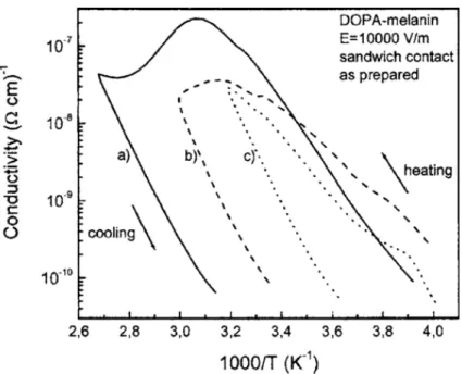
pellets under vacuum using different maximum temperatures: 373 K (a), 333 K (b), and 313 K (c). An activation energy of about 0.5 eV was deduced from the cooling curve. . . 33 Figure 2.14 (a) Hydration dependence of the conductivity of eumelanin pellets with
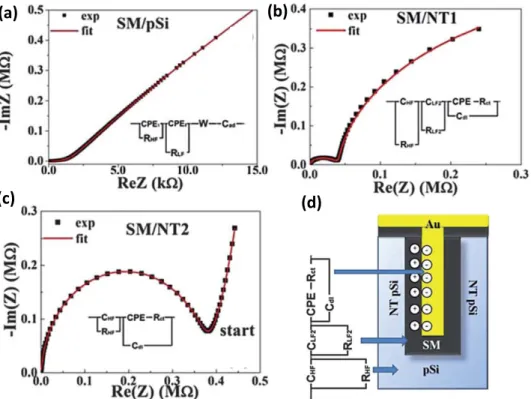
electrodes in van der Pauw configuration and best fit according the modified dielectric theory. (b) Results from the muon spin relaxation experiments. λ is the relaxation rate for paramagnetic muons (a mea-sure for the unpaired spin density), ∆ is the relaxation rate for diamag-netic muons (a measure for the local field experienced by free protons), and ν is the muon hopping rate (a measure for the proton mobility). The inset shows ∆ vs hydration on a different scale. . . 34 Figure 2.15 Nyquist plots of the impedance spectroscopy results for a planar
p-Si/eumelanin/Au device (a) and for two devices with differently nanos-tructures p-Si/eumelanin interfaces, (b) and (c). (d) schematically il-lustrates the assignments of equivalent circuit elements used to fit the Nyquist plots. . . 37 Figure 3.1 Main building blocks of eumelanins: 5,6-dihydroxyindole (DHI) and
5,6-dihydroxyindole-2-carboxylic acid (DHICA). . . 39 Figure 3.2 10µm × 10 µm AFM images of 30 nm thick films of different eumelanins
spin-coated from DMSO and NH3(aq) suspensions as indicated. The root mean square roughness Rq is indicated for each film. . . 43 Figure 3.3 1 µm × 1 µm AFM image of a 30 nm thick film of Sigma melanin
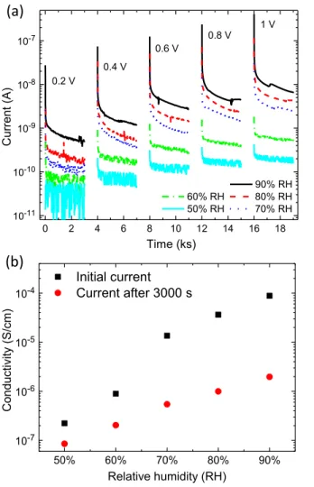
Figure 3.4 AFM images of an about 8 nm thick film of Sigma melanin spin-coated from DMSO suspension on SiO2((a) and (b), z-scale 6 nm) and of drop-cast film of DMSO melanin on glass (thickness > 100 nm), also using DMSO as a solvent ((d) and (e), z-scale 8 nm). (c) and (f) are sections along the lines indicated in the images (b) and (e), respectively. Point A corresponds to x = 0, B to x = 275 nm and 325 nm, respectively. . . 45 Figure 3.5 (a) Transient current measurement of Sigma melanin film for 3000 s
long voltage pulses from 0.2 to 1 V at relative humidity (RH) varied between 90% and 50%. Zero voltage was applied during the 1000 s between subsequent voltage pulses. The curves have been smoothened to reduce spikes caused by external noise. (b) Effective conductivity derived from the electrical current at 0.6 V at 0 s and 3000 s as a function of relative humidity. . . 48 Figure 4.1 a) Main building blocks of the eumelanin macromolecule: 5,6-dihydroxyindole
(DHI) and 5,6-dihydroxyindole-2-carboxylic acid (DHICA). b) Planar structure of Au electrodes and eumelanin film used in this work. . . 51 Figure 4.2 AFM images of eumelanin thin films between Au electrodes (L = 6µm)
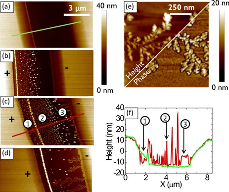
a) before electrical biasing and b–d) after biasing at 1 V for increas-ing times. Numbers in (c) mark the distinct features of the growth of the nanostructures: (1) Decomposition of the positively biased Au electrode, (2) NAs moving towards the negatively biased electrode, and (3) dendrite growth. e) AFM height and phase image of dendrite struc-tures. f) Height profile for the sections in (a) (green) and (c) (red). . . 53 Figure 4.3 AFM and corresponding conductive AFM image of the dendrite-like
structures extending from the negative to the positive electrode. The voltage applied to the AFM tip was -10 V. . . 55 Figure 4.4 a) SEM images of the dendrites growing on the eumelanin film between
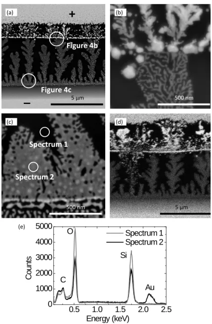
the Au electrodes using the backscattered electron signal (BSE). b,c): High-resolution SEM-BSE images taken at positions similar to those marked in (a). d) Mapping of the gold distribution based on the EDS signal. e) EDS spectra taken at positions similar to those marked in (c). 56
Figure 4.5 a) Transient current measurement for a eumelanin film between Au electrodes (L = 10µm, W = 2 mm, applied voltage 1 V) at 90% relative humidity. The sample was left hydrating for 24 h before biasing. b) Example for the resistive change of eumelanin films with Au-eumelanin dendrites between states of discrete resistance (L = 6µm, W = 4 mm, applied voltage 1 V). c) Conductance histogram corresponding to (b). d) Voltage-step measurement of an Au-eumelanin-Au structure (L = 6µm, W = 4 mm). The sample was biased at 1 V beforehand to attain the highly conductive state. The dashed lines separate the region of linear I − V characteristics from the region of electrochemical reactions. 57 Figure 5.1 Redox forms of the final monomer precursors of eumelanin (5,6-dihydroxyindole
(DHI) and 5,6-dihydroxyindole-2-carboxylic acid (DHICA)). Hydro-quinone (H2Q), semiquinone (SQ), and quinone (Q) forms. The quinone imine form (QI) is the tautomer of Q. Reversibility of the redox pro-cesses is indicated assuming that no other reactions occur after oxida-tion/reduction. . . 67 Figure 5.2 Transient current measurements of a eumelanin film (d = 50 nm) with
Pd electrodes (L = 9 µm, W = 20 µm) at 60, 70, and 80 % RH, under proton- and electron-injecting (with H2) vs electron-injecting (no H2) conditions. The applied bias is 0.5 V. . . 68 Figure 5.3 Nyquist plots of the EIS measurement on a eumelanin film at 90%
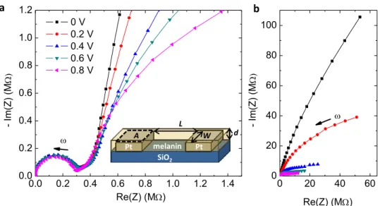
RH, applying a bias between 0 and 0.8 V (L = 10 µm, W = 24.5 mm, d = 50 nm). (a) High-frequency range. The inset illustrates the sample geometry. (b) Entire frequency range (10−2− 106 Hz). . . 70 Figure 5.4 Current-voltage characteristics of a eumelanin film (d = 50 nm) at 90%
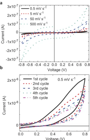
RH, measured with coplanar Pt electrodes (L = 10 µm, W = 24.5 mm). (a) Voltage sweep rate dependence of the first cycle. (b) First five cycles of the 0.5 mV s−1 measurements for positive voltages. . . 72
Figure 5.5 Illustration of the most important concepts of the model proposed for the electrical response of hydrated eumelanin films between co-planar Pt electrodes. (a) Composition of the eumelanin film before biasing in-cluding the various redox forms of DHI and DHICA (H2Q, SQ, Q, QI tautomer). (b) Under electrical bias, proton migration affects proto-nation and the comproportioproto-nation equilibrium (Eq. 5.1). (c) Possible electron transfer reactions at higher bias include the oxidation (brighter background) and reduction of eumelanin (darker background) accord-ing to Fig. 5.1. Moieties that changed their redox state are marked in green. While H2Q might form an insulating layer at the negative electrode, Q provides a transport path for the electronic charge carriers. 74 Figure 6.1 10 µm×10 µm AFM images of thin films of degraded DMSO melanin
spin-coated from 15 mg ml−1 NH3(aq) suspension (a) and DHICA melanin spin-coated from 15 mg ml−1 DMSO suspension (b). . . 79 Figure 6.2 TGA (weight) and DTG (weight loss derivative) curves of (a) pristine
eumelanin powder and eumelanin deposited from DMSO and (b) eume-lanin deposited from DMSO and treated according to the procedures given in the legend. . . 80 Figure 6.3 Process flow for the preparation of patterned eumelanin thin films
be-tween Pt electrodes using a parylene C layer as a mask for metal etching and eumelanin deposition. . . 82 Figure 6.4 Process flow for the preparation of patterned eumelanin thin films
be-tween Pt electrodes using an orthogonal resist layer as a mask for the etching of eumelanin and in the lift-off process to pattern the electrodes. 83 Figure 6.5 20 µm×20 µm AFM image (a) and corresponding height profile (b) of a
eumelanin film patterned to fit the interelectrode region between two Pt electrodes. The sample was fabricated by the lift-off process in Fig. 6.4 but using orthogonal resist as a lift-off material and a conventional negative photoresist on top. The red circles indicate interstices between the eumelanin film and the Pt electrode leading to a bad electrical contact. 84 Figure 6.6 2D plot (a) and intensity profile along the y-axis (b) of GIXRD data
taken on a drop cast film of Sigma melanin on a Si substrate. . . 85 Figure 6.7 TGA data of eumelanin powder hydrated at the RH as indicated in the
legend. The water content, also given in the legend, was determined from the weight loss up to 140 . . . 86
Figure 6.8 FT-IR measurements on pristine eumelanin, eumelanin with DDQ and TTF (molar ratio 1:30). The spectra are normalized and shifted along the absorption axis for better comparability. . . 88 Figure 6.9 (a) I-V measurement for thin films of eumelanin, eumelanin:DDQ
(30:1), and eumelanin:TTF (30:1) under vacuum. L = 10 µm, W = 24.5 mm, d = 30 nm. Currents are below noise level . (b) Measure-ment of current vs time at 90% RH for the same samples. The applied voltage is 0.8 V. . . 90 Figure 6.10 OTFT structure as used in this work, with bottom source and drain
electrodes (Pt), unpatterned channel material (eumelanin), gating medium (BMPyrr-TFSI ionic liquid in Durapore® filter), and top gate (high surface-area activated carbon). Voltages are shown for n-type opera-tion but p-type operaopera-tion was also tested. . . 91
LIST OF APPENDICES
A - Supporting Information for Article 1 . . . 125 B - Supporting Information for Article 2 . . . 129 C - Supporting Information for Article 3 . . . 140 D - List of publications at ´Ecole Polytechnique de Montr´eal . . . 150
LIST OF ABBREVIATIONS
AFM Atomic force microscopy
BMPyrr-TFSI 1-Butyl-1-methylpyrrolidinium bis(trifluoromethylsulfonyl)imide BSE Backscattered electron
CPE Constant phase element
CV Cyclic voltammetry
DDQ 2,3-Dichloro-5,6-dicyano-1,4-benzoquinone DHI 5,6-Dihydroxyindole
DHICA 5,6-Dihydroxyindole-2-carboxylic acid
DMF Dimethylformamide
DMSO Dimethyl sulfoxide DNA Deoxyribonucleic acid
DTG Differential thermogravimetric analysis EDS Energy-dispersive X-ray spectroscopy EIS Electrochemical impedance spectroscopy ESR Electron spin resonance
FET Field-effect transistor
FT-IR Fourier-transform infrared spectroscopy GIXRD Grazing incidence X-ray diffraction H2Q Ortho-hydroquinone
HOMO Highest occupied molecular orbital
IL Ionic liquid
IPES Inverse photoelectron spectroscopy
ITO Indium tin oxide
LUMO Lowest unoccupied molecular orbital MAPLE Matrix-assisted pulsed laser deposition
NA Nano-aggregate
NAA Nuclear activation analysis OLED Organic light-emitting diode OTFT Organic thin film transistor
Q (Indole)quinone
QI Quinone imine
RH Relative humidity
SCE Saturated Calomel Electrode SEM Scanning electron microscopy
SQ Semiquinone
TGA Thermogravimetric analysis
ToF-SIMS Time-of-flight secondary ion mass spectroscopy TTF Tetrathiafulvalene
UPS Ultaviolet photoelectron spectroscopy XPS X-ray photoelectron spectroscopy XRD X-ray diffraction
LIST OF SYMBOLS
A In-plane electrode area
C Capacitance
CDL Double layer capacitance d Film thickness
Dσ Proton conductivity diffusion coefficient DH2O Water self-diffusion
∆q XRD peak width EF Fermi level Eg Energy gap
EHOM O Energy of the highest occupied molecular orbital ELU M O Energy of the lowest unoccupied molecular orbital Eox◦ Standard oxidation potential
Ered◦ Standard reduction potential −
→
F Electric field
g Paracrystalline parameter ∆ G Change in Gibbs free energy G0 Quantum of conductance
H Hydration
I Current
kb Boltzmann constant kel Electron transfer rate κ Dielectric constant L Interelectrode distance λ Reorganization energy µ Charge carrier mobility µh Hole mobility
µe Electron mobility n Charge carrier density
p Pressure
pKa Acid dissociation constant
q Charge
qz XRD scattering vector
R Resistance
RCT Charge-transfer resistance
Rq Root mean square surface roughness
σ Conductivity t Time T Temperature − →v Carrier velocity V Voltage W Electrode width
CHAPTER 1
INTRODUCTION
Since the discovery of the first conductive polymers four to five decades ago [1–3], research on organic electronics has made substantial progress. Displays based on organic light-emitting diodes (OLEDs) are now commercially available on a large scale and other organic electronic devices such as white OLEDs for lighting and organic solar cells might follow soon [4]. Organic electronic devices make use of thin films of π-conjugated small molecules or polymers with semiconducting or conducting properties. The structural, optical, and electrical properties of these materials can be tuned by chemical synthesis to meet the requirements of a wide range of applications. Organic thin film devices can be produced under low energy consumption by printing or low-temperature evaporation. They are light-weight and conform to flexible substrates. The goal of organic electronics is not to exceed the performance of Si-based devices but rather to reduce the cost and the environmental impact of electronics, as well as to enable new fields of applications such as semitransparent and flexible electronics [5, 6]. Thousands of peer-reviewed publications per year indicate both that the potential of organic electronics has not yet been fully exploited and that some challenges have not yet been met. These challenges include understanding the effect of microstructure on charge transport [7], the optimization of charge separation in organic solar cells [8], printable electrode and semiconductor materials [9], stable and efficient n-type materials and blue emitters [10, 11], and large scale processing [12].
Starting about ten years ago, another property of organic electronic materials has gained much attention. While inorganic (semi-)conductors usually conduct only electronic charge carriers, their organic counterparts often support both electronic and ionic/protonic cur-rents. This property allows for ion-to-electron transduction and opens new possibilities for the interaction of electronic and biological systems, now explored in the field termed Organic Bioelectronics [13]. Potential applications include bio-sensing for health and environmental monitoring, drug delivery, and neuronal signal recording and stimulation [13–16]. Naturally, biocompatibility is an important requirement for such applications. Indeed, biocompatibility and biodegradability, in addition to low power consumption during production and opera-tion, is a desirable feature for any electronic device in the effort to reduce its environmental footprint. The vision of sustainable electronics and the potential benefits from coupling elec-tronic and biological systems has recently encouraged several research groups to search for biomolecular and bio-inspired materials suitable as components of electronic and ionic
de-vices [17–20]. While the list of biomolecular materials that have been successfully employed as substrate or dielectric layer in transistors is long [17], the identification of biomolecules that are semiconducting or conducting in thin film form is much more challenging. Remarkably, during the work for this thesis, hydrogen-bonded pigments emerged as a new class of organic semiconductors, also including well-known natural pigments such as indigo [20]. Proton con-ductors such as maleic chitosane fibers have been employed in bio-protonic transistors [21]. In addition to the properties mentioned above, biomolecules often have remarkable mechanical, chemical, or optical properties related to their biological function.
Organic bioelectronics, a multidisciplinary field of research, is still in its infancy. The systems investigated are generally characterized by a high degree of structural and energetic disorder, the simultaneous conduction of ions and electrons, and a complex environment, possibly including water and redox-active species. Understanding the effect of all these factors on device performance requires models and characterization methods beyond inorganic solid state physics [18]. There is thus an urgent need for further studies contributing to a better understanding of charge carrier transport, structure-property relationships, and the interaction of organic and biomolecular materials with their environment and other device components.
1.1 Eumelanin - a natural pigment and a potential material for bioelectronics The biomolecular pigment melanin, more precisely eumelanin, was reported to have semi-conducting properties already in the 1960’s and 70’s [22–25]. Melanins are bio-macromolecules ubiquitous in flora and fauna that hold important functions in the human body including photo-protection, coloration of skin, hair and eyes, free radical scavenging, metal ion chela-tion, thermo-regulachela-tion, and might also be involved in neuronal signal transmission [26–28]. Furthermore, melanins play a role in diseases such as melanoma skin cancer and Parkin-son’s disease [29–31]. Eumelanin is the predominant form of melanin in the human body. Properties of eumelanin that caught the attention of material scientists and physicists in-clude a persistent electron spin resonance signal, indicating stable free radicals [32], strong optical absorption with a featureless spectrum [33], and the hydration-dependent (photo-)conductivity of eumelanin pellets [34, 35]. These properties were mostly explained within the framework of the amorphous semiconductor model, building on the discovery of reversible threshold switching of eumelanin pellets by McGinness in 1974 [25]. Most recent works re-ferring to the electrical properties of eumelanin still consider eumelanin as an amorphous semiconductor [19, 36–40]. However, there have also been reports about mobile ions, in par-ticular protons, in eumelanin [41–44]. In 2012, Mostert et al. resumed the debate on the
origin of the electrical conductivity of eumelanin [42]. Their work on eumelanin pellets seems to disprove the amorphous semiconductor model and suggests that both electrons and pro-tons are mobile in eumelanin pellets. Definitive proof of electron and proton transport over device-relevant distances, insights into the relative contribution of electrons and protons to the electrical current at different sample hydration states, and a new model for the charge carrier transport mechanism in eumelanin are still missing to date. In particular, reports on the electrical properties of hydrated eumelanin films are very sparse.
The characterization of eumelanin is complicated by its high degree of chemical and structural disorder and its insolubility in common solvents [45]. Furthermore, many properties of eumelanin are strongly affected by hydration [46]. It is now accepted that eumelanin does not have a well-defined chemical structure but is rather a mixture of different oligomers and polymers of 5,6-dihydroxyindole (DHI), 5,6-dihydroxyindole-2-carboxylic acid (DHICA) and their various redox forms. The stucture of eumelanin depends on (bio-)synthetic conditions and precursors [45, 47]. Chemical and structural disorder also implies energetic disorder and makes any structure-property correlation challenging. The insoluble nature of eumelanin defies many conventional techniques for the characterization of organic molecules and it is furthermore an obstacle for the preparation of eumelanin thin films. Indeed, strategies for the solution processing of eumelanin films have been suggested only recently [48, 49]. Thin films enable the use of a wider range of characterization techniques and facilitate the integration in device architectures. The next step in this line of research is to optimize film processing, characterize film structure and functional properties, and to investigate the interaction of eumelanin thin films with other device components.
The development of organic bioelectronics revived the interest in eumelanin as a func-tional material [17, 18, 50, 51]. The electrical and chemical properties of eumelanin in com-bination with its intrinsic biocompatibility have encouraged researchers to explore the use of eumelanin in tissue engineering [39], biocompatible batteries [52], memory devices [53], and sensors [37,54]. The possibility of mixed ionic-electronic conduction makes eumelanin an interesting candidate for ion-to-electron transduction [42]. A glance at the vast literature on polydopamine, a synthetic eumelanlike polymer, reveals further potential applications, in-cluding biosensing, drug delivery, and membranes for fuel cells [40,55]. However, the electrical properties of polydopamine films are largely unexplored.
1.2 Objectives and organization of the work
The primary objective of this thesis is to investigate the structural and electrical prop-erties of eumelanin thin films for potential applications in organic bioelectronics. The rich
biofunctionality of eumelanin in combination with its hydration-dependent electrical conduc-tivity, possibly based on mixed ionic-electronic conduction, opens diverse possibilities for its use at the interface of electronics and biological systems. A better understanding of the fundamental properties of eumelanin is also of great fundamental interest considering its role in biology and medicine.
For device integration, it is advantageous to use eumelanin in the form of thin films. Fur-thermore, thin films offer a better control over sample morphology and dimensions, and enable the use of a wider range of characterization techniques such as scanning probe microscopy. Therefore, the first objective of this work is:
(1) The identification of a procedure for the solution-processing of eumelanin films that yields homogeneous, smooth films with good adhesion to the substrate and has little effect on the chemical composition of the eumelanin film.
Since the electrical properties of thin films strongly depend on film morphology and structure, the second objective is
(2) The investigation of the growth and morphology of eumelanin thin films. Finally, the main focus of this thesis is
(3) The characterization of the electrical response of hydrated eumelanin thin films and to shed light on the charge carrier transport properties of eumelanin.
The thesis starts with a literature review of the thin film processing and the physical and chemical properties of eumelanin (Chapter 2). Special attention is paid to the charge carrier transport properties of eumelanin. Chapter 2 also contains a brief introduction to the processing, the growth, and the electron and proton transport properties of organic thin films. Subsequently, Chapter 3 to 5 are reprints of three articles resulting from the work for this thesis:
– Article 1: Eumelanin thin films: solution-processing, growth, and charge transport properties
– Article 2: In situ formation of dendrites in eumelanin thin films between gold electrodes – Article 3: Proton conduction and transient nature of electronic currents in hydrated
eumelanin thin films.
thesis. In Article 1, published in the Journal of Materials Chemistry B, different synthesis routes and processing solvents for eumelanin suggested in literature are compared in terms of the morphology and the chemical composition of spin-coated thin films. The combination of the synthetic eumelanin and the processing solvent that was found to yield the best films with respect to the objective defined above is used in all subsequent studies of this thesis. Article 1 also addresses the second objective. Studying the growth of eumelanin films gave insight into the supramolecular organization of eumelanin in the form of nanoaggregates and the 3D film morphology of the films, suggesting that the processing technique employed is suitable to obtain continuous eumelanin films.
The first electrical characterizations in this project were carried out with Au electrodes, often employed in organic electronics for their chemical inertness and also in several pre-vious studies on eumelanin. In Article 2, published in Advanced Functional Materials, an unexpected interaction between hydrated eumelanin films and Au electrodes, the growth of Au-eumelanin dendrites, is reported. The dendrites completely dominate the electrical prop-erties of the sample and suggest interesting new applications of eumelanin. These results point to the importance of the electrode material and the consideration of electrochemical reactions in the electrical response of hydrated eumelanin films.
Further electrical measurements on eumelanin were conducted with Pt and Pd electrodes. In Article 1 the strong hydration dependence of the conductivity of eumelanin thin films is confirmed and the time-dependence of the current is investigated. The current transients suggest several contributions to the current. In Article 3, which has recently been submitted, these contributions are disentangled for strongly hydrated eumelanin films using a combina-tion of electrical and electrochemical characterizacombina-tion techniques. The eumelanin films are shown to be proton conductive over micrometric distances and the proton conductivity is estimated. Furthermore, the results of Article 3 indicate that electrochemical processes play an important role in the electrical response of hydrated eumelanin films. In contrast, the results of Article 3 show no clear evidence of electronic conduction. A qualitative model is developed describing how the interplay of proton migration, the redox properties of eume-lanin, and the comproportionation equilibrium, determining the density of mobile electrons, could limit electron transport in eumelanin thin films.
Following these three articles, supplementary methods and results that have been obtained during the work for this thesis are presented (6). These include details on the film processing, strategies for the patterning of eumelanin films, and attempts to enhance the conductivity of eumelanin films.
In Chapter 7, the results of this thesis are discussed as a whole, supported by the literature review in Chapter 2. Finally, conclusions are drawn and perspectives on future work are given
CHAPTER 2
LITERATURE REVIEW
Eumelanin can be found in many plants and animals and has various functions in the human body. In addition, some of its intriguing solid-state properties have already been dis-covered in the 1970’s. Nevertheless and despite considerable progress during the last years, many fundamental properties of eumelanin are still a matter of debate. This is partly due to the high chemical heterogeneity and the insolubility of eumelanin, making characterization challenging. This chapter starts with an overview of the current knowledge about the molec-ular and supramolecmolec-ular structure of eumelanin (Sec. 2.1), followed by a brief description of the physical and chemical properties of eumelanin (Sec. 2.2). Following this, the processing, growth (Sec. 2.3), and charge carrier transport properties of eumelanin (Sec. 2.4) are reviewed in more detail. The latter two sections also include a brief introduction to general concepts of organic thin film growth, electron transport in organic semiconductors, and proton transport in hydrated polymers.
2.1 Synthesis, molecular and supramolecular structure of eumelanin
The terms melanin and eumelanin are used ambiguously in literature. In 2013, leading chemists in melanin research made a new attempt in establishing a common nomenclature [47]:
“Melanins: Pigments of diverse structure and origin derived by the oxidation and polymerization of tyrosine in animals or phenolic compounds in lower or-ganisms. Eumelanins: Black-to-brown insoluble subgroup of melanin pigments derived at least in part from the oxidative polymerization of L-dopa via 5,6-dihydroxyindole (DHI) intermediates.”
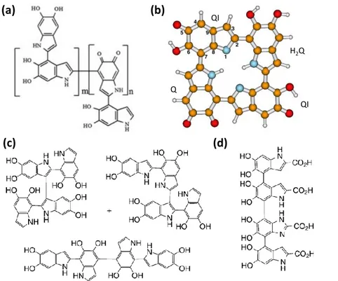
Accordingly, eumelanins are defined via their oxidative formation pathway, which can lead to different macromolecular structures depending on precursors and oxidation conditions. The biosynthetic formation of eumelanin from tyrosine is described by the Raper-Mason scheme shown in Fig. 2.1 [56]. The last step of the formation is the oxidative polymerization of DHI and 5,6-dihydroxyindole-2-carboxylic acid (DHICA). In eumelanin, different oxidation states of these monomer units coexist, the ortho-hydroquinone (H2Q), -semiquinone (SQ), and -(indole)quinone (Q) form (Fig. 2.2). Furthermore, the tautomer of Q, quinone imine (QI) is often found [46, 57, 58]. Eumelanin thus contains phenolic hydroxyl, amine, and
O H COOH NH2 N H O H O H COOH N H O H O H N H O H O H COOH N H O O COOH O H COOH NH2 O H O COOH NH2 O dopa 5,6-dihydroxyindole-2-carboxylic acid (DHICA) 5,6-dihydroxyindole (DHI) eumelanin tyrosinase dopachrome -CO2 oxidative polymerization dopaquinone leucodopachrome
Figure 2.1 Raper-Mason scheme for the biosynthesis of eumelanin. Reprinted from Ref. [50].
carboxyl groups, which can act as proton donors or acceptors depending on their degree of protonation. The ratio of oxidized and reduced forms as well as of monomers with and without carboxyl groups strongly depend on the biological origin of eumelanin or, in the case of synthetic eumelanins, on precursors and reaction conditions. Due to difficulties in the extraction and purification of natural eumelanin, synthetic forms of eumelanin are used in most chemical and physical studies. A list of the different synthetic procedures reported in literature can be found in Ref. [50]. Apart from the monomer units shown in Fig. 2.2, eumelanin can contain uncyclized units (residues of precursors or intermediates) and pyrroles (eumelanin degradation products) [47, 59]. The variety of monomers that can be present in eumelanins is a source of both significant chemical diversity between eumelanins of different origin and the chemical heterogeneity of a certain eumelanin.
Considerable variety and disorder is furthermore present on the macromolecular and supramolecular level of the eumelanin structure. Coupling between monomers can occur at the 2, 3, 4, and 7 position as indicated in Fig. 2.2 leading to the formation of mainly planar oligomers of different structure and size [45]. The presence of a carboxyl group at the 2 position in DHICA reduces the number of potential coupling sites and results in the formation of more linear structures.
(H2Q) Q
Figure 2.2 Constituent monomers of eumelanin: : dihydroxyindole (DHI) and 5,6-dihydroxyindole-2-carboxylic acid (DHICA). The various redox forms that coexist in the eumelanin macromolecule are shown for DHI: H2Q, SQ, and Q. QI is the tautomeric form of Q. Reprinted with permission from Ref. [46] (Copyright © 2006, John Wiley and Sons).
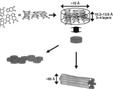
For a long time, it was commonly believed that eumelanin is a high-molecular-weight het-eropolymer [23]. Evidence for polymeric structures of DHI of more than 30 monomer units was also recently collected by mass spectroscopy [60]. A possible heteropolymeric structure for eumelanin formed by polymerization of DHI is shown in Fig. 2.3a. However, most ex-periments, including AFM, scanning tunneling microscopy, transmission electron microscopy, and X-ray diffraction (XRD), point to a model commonly referred to as the “stacked-oligomer model” (Fig. 2.4) [61–65]. This model predicts the formation of planar sheets composed of 4-8 monomers, which stack in graphite-like manner via π−π interaction with a spacing of 3.3-4.0 ˚
A. The so-called protomolecule is about 1.5-2 nm large and 1-2 nm high and self-assembles via non-covalent binding to larger structures. The structure of eumelanin oligomers has been ex-tensively studied [45, 50, 66] and examples for suggested structures are reported in Fig. 2.3b, c and d. The discussion above reveals that eumelanin has a conjugated backbone, whose extension depends on the (bio-)synthetic origin of eumelanin.
A comment should be made on the use of the term polydopamine at this point. Poly-dopamine is the “black oxidation product of Poly-dopamine” [67] and is also commonly referred to as “the dopamine-derived synthetic eumelanin polymer” [40]. Since the discovery of poly-dopamine as a versatile platform for biofunctional coatings in 2007 [55], publications on its application in biomedicine and other domains have been numerous [67]. It is commonly be-lieved that polydopamine is a polymer of DHI or uncyclized catecholamines. However, it has recently been demonstrated that polydopamine can also contain other units and should be regarded as a mixture of various macromolecules [67], in analogy to eumelanin. It is gener-ally accepted that the polydopamine structure strongly depends on the synthetic conditions. A notable difference to eumelanin derived from L-dopa or tyrosine lies in the apparent ab-sence of DHICA and its redox forms. However, carboxyl groups can be formed by oxidative
QI H2Q Q QI (a) (b) (c) (d)
Figure 2.3 Examples for different macromolecular structures of eumelanin suggested in the literature: (a) polymer structure for eumelanin obtained from DHI, (b) and (c) tetramer structures for DHI, (d) tetramer structure for DHICA. In (b) the color orange refers to C, blue to N, and red to O atoms. Adapted with permission from (a) Ref. [60] (Copyright © 2012 John Wiley & Sons, Ltd), (b) Ref. [66] (copyright © 2006 by the American Physical Society) and (c) and (d) Ref. [45] (copyright © 2009 WILEY-VCH Verlag GmbH & Co. KGaA, Weinheim).
Figure 2.4 Stacked oligomer model according to Clancy et al. Reprinted with permission from Ref. [63] (copyright © 2001, American Chemical Society).
degradation of polydopamine [67]. To date, it is still unclear under which conditions the oxidation product of dopamine is similar in structure and properties to eumelanins derived from other precursors. Thus polydopamine and eumelanin should not be used as synonyms and, in general, the origin of eumelanin has to be considered when referring to literature. In this work, the precursor of the eumelanin investigated will often be included by referring to dopa melanin, DHI melanin etc. An exception is DMSO melanin, which is eumelanin derived from L-dopa by oxidation in dimethyl sulfoxide (DMSO) instead of aqueous suspen-sion. Commercial synthetic eumelanin prepared by the oxidation of tyrosine with hydrogen peroxide (Sigma Aldrich) will be indicated as Sigma melanin and natural eumelanin from sepia officinalis (Sigma Aldrich) as Sepia melanin.
2.2 Physical and chemical properties of eumelanin
This section describes the physical and chemical properties of eumelanin that are of particular relevance for this work. The charge carrier transport properties of eumelanin are discussed separately and in more detail in Sec. 2.4.3.
2.2.1 Hydration
Eumelanin is hygroscopic and its properties are strongly dependent on hydration. Mostert et al. measured the absorption isotherms for water in eumelanin pellets [68]. The pellets contained about 11.5 wt% water at 50% RH and 16 wt% at 80% RH. Eumelanin is known
to contain at least two types of water, often referred to as weakly and strongly bound water [69, 70]. Weakly bound water is easily removed by reducing the relative humidity in air or by gentle heating, while the sample has to be placed in vacuum or heated well above 100 to, at least partially, remove strongly bound water [36,71]. Recently, Bridelli et al. investigated the interaction of eumelanin and water by infrared spectroscopy [72]. The -OH stretching band revealed the presence of three different water species in synthetic dopa melanin and natural Sepia melanin drop cast from aqueous suspension: strongly bound water in the vapor-phase and two more weakly bound water species in the liquid phase, characterized by H-bonding. The presence of the different water species depend upon the pore size distribution of the sample investigated. For dopa melanin, only the liquid-phase water, which exist in larger pores, was found and hydration/dehydration was reversible. In contrast, in Sepia melanin vapor-phase water was also detected and dehydration was partly irreversible, i.e., the sample could reabsorb only a smaller amount of water after dehydration.
2.2.2 Optical properties
One of the characteristics of eumelanin is its strong broad-band absorption in the UV-visible, exponentially decreasing with wavelength. This was often considered an indication that eumelanin shows extended π-conjugation and can be described as an amorphous semi-conductor (Section 2.4.3). However, it has been shown that the absorption spectrum can result from the superposition of the absorption of eumelanin oligomers of different size and structure [73]. Pezzella et al. furthermore reported that the coexistence of oxidized and reduced monomers contributes to the broad-band absorption [57].
In accordance with the role of eumelanin in photoprotection, the pigment efficiently trans-forms absorbed photon energy into heat via nonradiative relaxation pathways, which are still under investigation today [74–76]. It has been suggested that intramolecular proton transfer plays an important role in the dissipation process [74–76], as well as chemical and structural disorder [77].
Consequently, the fluorescence quantum yield of eumelanin is very low (about 10−4) [78]. The fluorescence spectrum of eumelanin extends between 250 and 500 nm with the peak position and intensity depending on the excitation wavelength. The multi-exponential decay of the fluorescence is in agreement with the chemical disorder model [79].
2.2.3 Paramagnetic properties
Eumelanin shows a persistent electron spin resonance (ESR) signal, reflecting the presence of stable free radicals [32, 80]. The ESR is affected by pH, various metal ions, light, and
H2Q Q SQ-
Figure 2.5 Comproportionation equilibrium between the different redox states of the eume-lanin building blocks. Reprinted with permission from Ref. [42] (copyright © 2012 National Academy of Sciences).
hydration [41, 42, 80–84]. Two different types of unpaired spins can be distinguished in eumelanins, carbon-centered “intrinsic” spins and the “extrinsic” spins of the semiquinone radicals [42]. In aqueous suspension, the ESR signal at neutral and basic pH has been assigned to SQ radicals [85]. Numerous experiments have shown that the SQ radical population is controlled by the comproportionation equilibrium (Fig. 2.5) [80, 85]. The equilibrium strongly favors the reactants over the products resulting in only one SQ radical per 1000-1500 monomers in aqueous suspension at pH 7 [83, 86]. The SQ population can be increased by increasing the pH [42, 83, 86] or by complexation of SQ with suitable metal ions [80, 82].
The ESR signal of hydrated eumelanin pellets is more complex and was investigated only very recently [85]. The signal is dominated by carbon-centered radicals, resulting from the incomplete polymerization of the monomers, but also shows the response of SQ radicals as a minor component. An increase of hydration resulted in a decrease of the total signal intensity. The separation of the two components was difficult and no clear trend of the SQ population with hydration was observed. However, the relative contribution of SQ radicals to the ESR signal was stronger in basic pellets than in acidic or neutral pellets, in agreement with the comproportionation reaction [85]. A muon spin relaxation study by the same authors furthermore suggested the increase of the radical density with hydration [42].
2.2.4 Redox and proton-exchange properties
Eumelanin acts as a redox buffer in biological systems [87] and its redox ability is pH-dependent [88]. Eumelanin contains oxidized as well as reduced moieties [57]. Due to the insolubility and the aggregated structure of eumelanin, characterization of the redox activity of eumelanin in solution is difficult [58]. Serpentini et al. reported cyclic voltammetry (CV) measurements on DOPA melanin incorporated into a carbon paste electrode (0.1 M KCl aqueous solution, pH 5.6) [89]. Two irreversible oxidation peaks were observed at 460 and 550 mV (vs SCE), attributable to the oxidation of either a SQ or the amino group of an indole unit (QI) of eumelanin, and two reduction peaks at 20 and -355 mV. The absence of a
![Figure 2.1 Raper-Mason scheme for the biosynthesis of eumelanin. Reprinted from Ref. [50].](https://thumb-eu.123doks.com/thumbv2/123doknet/2352066.36504/34.918.302.601.106.508/figure-raper-mason-scheme-biosynthesis-eumelanin-reprinted-ref.webp)