by Gry Houeland
Département de physiologie Faculté de médicine
Thesis submitted at Faculté des études supérieures
in accordance with the requirement for the degree of Philosophiae Doctor (Ph.D.)
in Sciences Neurologiques
Janvier 2007
C
(-*
L)
-q
>
AVIS
L’auteur a autorisé l’Université de Montréal à reproduire et diffuser, en totalité ou en partie, par quelque moyen que ce soit et sur quelque support que ce soit, et exclusivement à des fins non lucratives d’enseignement et de recherche, des copies de ce mémoire ou de cette thèse.
L’auteur et les coauteurs le cas échéant conservent la propriété du droit d’auteur et des droits moraux qui protègent ce document. Ni la thèse ou le mémoire, ni des extraits substantiels de ce document, ne doivent être imprimés ou autrement reproduits sans l’autorisation de l’auteur.
Afin de se conformer à la Loi canadienne sur la protection des renseignements personnels, quelques formulaires secondaires, coordonnées ou signatures intégrées au texte ont pu être enlevés de ce document. Bien que cela ait pu affecter la pagination, il n’y a aucun contenu manquant.
N 011 CE
The author of this thesis or dissertation has granted a nonexciusive license allowing Université de Montréal to reproduce and publish the document, in part or in whole, and in any format, solely for noncommercial educational and research purposes.
The author and co-authors if applicable retain copyright ownership and moral rights in this document. Neither the whole thesis or dissertation, nor substantial extracts from it, may be printed or otherwîse reproduced without the author’s permission.
In compliance with the Canadian Privacy Act some supporting forms, contact
information or signatures may have been removed from the document. While this may affect the document page count, it does not represent any Ioss of content from the document.
This thesis is entitled:
Kinase C substrates and snaptic plasticity inAplysia.
Presented by: Gry Houeland
Has been evaluated by a jury composed of the following people:
Richard Robitaille, reporting president Vincent Castelucci, research director
Wayne Sossin, co-director Lue Desgroseillers, jury member John H. Byme, extemal examinor
étudié en particulier le rôle de la kinase C (PKC) dans la transmission synaptique. J’ai tenté d’élucider les modifications par la phosphorylation des protéines impliquées dans la relâche de neurotransmetteurs par la PKC. J’ai étudié ces questions dans le mollusque ApÏysia cailfornica, un modèle expérimental propice à 1 ‘étude des relations qui existent entre l’apprentissage et la plasticité du système nerveux. Synaptotagmine-I ($yt-I) est une protéine vésiculaire qui fonctionne comme un détecteur de calcium et joue un rôle essentiel dans la relâche des neurotransmetteurs. Dans le processus de cloner Syt-I, nous avons trouvé une nouvelle isoforme avec deux acides aminés +VQ insérés dans le domaine N-terminal de C2-A (Syt-IvQ). La surexpression de $yt-IvQ bloque la facilitation par la sérotonine. Ceci indique que la région VQ de la synaptotagmine joue un rôle important dans la facilitation des synapses déprimées. J’ai ensuite testé si EGFP-Syt-IVQ pouvait bloquer l’effet de PDBu, un phorbol ester qui active le PKC indépendamment de 5-HT. Le EGFP-Syt-IvQ n’était plus capable de bloquer le reversement de la facilitation, suggérant que le déficit est lié à la capacité de 5-HT d’activer PKC. Cet effet spécifique de Syt-IvQ sur la transmission s’explique si cette isoforme est la seule qui se trouve dans les vésicules synaptiques. Nous avons observé une différence d’expression dans les cônes de croissance et dans les axones entre les 2 isoformes. Les résultats indiquent que le site VQ est impliqué dans la régulation de la localisation des $yt-I. Nous avons aussi cloné une autre isoforme de synaptotagmine, $yt-I C2B-f3 avec 39 changements dans la région C2B. Une mesure de protéolyse avec la chymotrypsine démontre que cette isoforme peut, en absence de calcium supporter un changement de conformation normalement déclenché par celui-ci. Nous avons également montré que Syt-I C2B-f3 avait une plus grande affinité pour $NAP-25 que Syt-I C2B-ct. En conclusion, ces résultats suggèrent que l’épissage change la structure du domaine C2B et pourrait ainsi affecter la fonction de la protéine.
$NAP-25 est une protéine impliquée dans la relâche de neurotransmetteur et membre du complexe SNARE. Cette protéine est aussi un substrat de PKC. J’ai cloné
de cette protéine par PKC joue un rôle dans la transmission synaptique. La surexpression de SNAP-25 n’avait aucun effet sur les cinétiques de la dépression et de la facilitation synaptique. Cependant, nous suggérons que la phosphorylation de $NAP-25 par PKC joue un rôle dans la modulation synaptique. Notamment, nous avons trouvé qu’en présence de PDBu ou de protéines phosphomimétiques les cinétiques de la dépression ralentissaient. Cet effet a été bloqué dans les cellules exprimant un site de phosphorylation PKC non phosphorylable ainsi qu’en présence de bisinolylmalemide-I (Bis), un bloqueur de PKC.
Mots-clés: Plasticité, Aplysia, synapse, synaptotagmine, épissage, SNAP-25, PKC, phosphorylation.
The principal aim of my research was to study the synaptic plasticity at the sensori-motor synapse of Aptysia, and how it contributes to behavior, leaming and memory. In my studies I focused on the role of protein kinase C (PKC) in synaptic transmission. I sought to define the step or steps in the transmitter release process that invoive PKC phosphorylation and that contribute to synaptic plasticity, with the ultimate goal of identifying the PKC posttransiationai (phosphorylation) modifications of specific proteins in the exocytotic pathway that regulate the number of vesicles available for release. I studied these questions in cuitured Aplysia sensory-motor neurons using moiecular biologicai and electrophysiologicai techniques. We discovered two novel splice forms in Aplysia synaptotagmin. We found a novel spiice form with two amino acids VQ (SytIvQ), inserted in the juxtamembrane domain of Syt I. The VQ and -VQ forms are expressed at equai levels in the Aplysia nervous system. Overexpression of the two forms have distinct effects, suggesting that the splice is of physiological importance. Overexpression of SytIVQ biocks
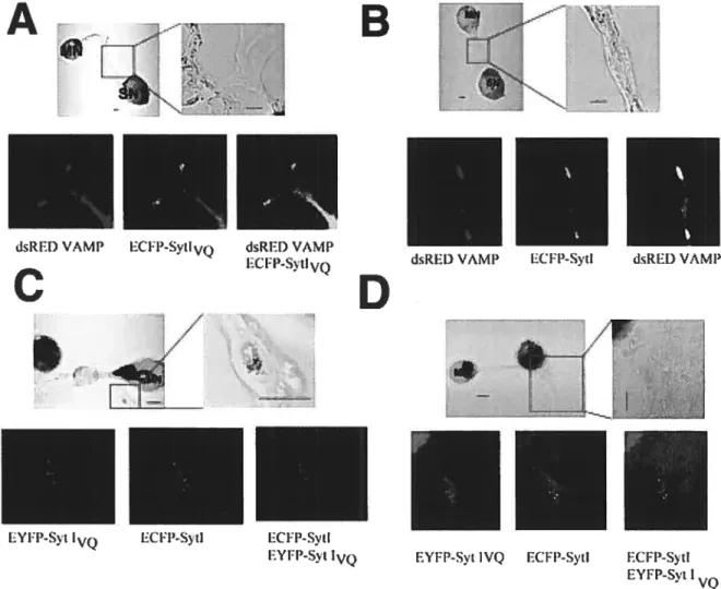
the ability of serotonin to reverse synaptic depression, whiie overexpression of VQ does flot. This effect is specific to the reversai of depression as there is no effect on the ability of serotonin to facilitate non-depressed synapses. The reversai of depression is mediated by PKC and synaptotagmin is a putative PKC substrate. However, despite the phosphorylation of Aplysia synaptotagmin at a conserved site adjoining the VQ, there is no change in phosphoryiation between the two forms. Moreover, overexpression of synaptotagmin does not block the effect of phorbol esters, suggesting that the effect is due to a biock of the abiiity of 5-HT to activate PKC in the cells overexpressing synaptotagmin. We discovered that while both VQ and -VQ forms are found in synaptic vesicies, they appear to be found in distinct vesicies in axons and growth cones, suggesting that we uncovered an important sorting signai for synaptotagmin.
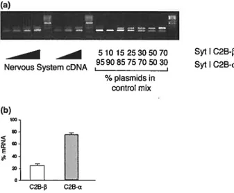
A second splice in Aplysia synaptotagmin is an alternative exon used in the C2B domain. This spiice form (C2B spiice) is aiso expressed in the Aplysia nervous system. Whiie ail of
stability in the absence of calcium than the previously identified form.
$NAP-25 is a member of the SNARE protein complex implicated in synaptic vesicle docking and fusion and a substrate of PKC. We cloned Aplysia SNAP-25 and generated SNAP-25 constructs mutated in the PKC phosphoiylation site Ser98, simulating either a phosphorylated or a non-phosphorylated form of SNAP-25. From our results, we suggest a role for SNAP-25 and its phosphorylation in some forms of synaptic modulation. Notably, we have found that phosphorylation of SNAP-25 by PKC in Aplysia neurons is required for PDBu mediated facilitation of depressed synapses.
Keywords: Plasticity, Aplysia, synapse, synaptotagmin, splicing, SNAP-25, PKC, phosphorylation.
ABSTRACT y
INDEX ii
LIST 0F TABLES xi
LIST 0F FIGURES xii
LIST 0F ABREVIATIONS xv
ACKN0WLEDGEMENTS xvii
I.
1.1 The dichotomy of memory functions 3
1.1.2 The brain must undergo changes that are maintained as long as the memory is stored.
What are these changes’ 5
1.1.3 What is the neuroanatomical support of our extTaordinary capacity to store memory
functions 6
1.2 The “engram” 7
1.2.1 Synaptic Plasticity 9
1.3 Vertebrates versus invertebrates 10
1.3.1 The ideal model 11
1.3.2ApÏysia 11
1.3.2.1 habituation 19
1.3.2.2 sensitization 20
1.3.4Mouse 25
1.4 The synapse 26
1.4.1 $ignaling within fleurons: the action potential 27
1.4.2 Synaptic transmission 27
1.4.3 The “calcium hypothesis” 29
1.4.4 The “quantal hypothesis” 29
1.4.4.1 What morphological feature might account for the quantum of transmitter7 30
1.5 The molecular biology ofvesicle fusion and release 32 1.5.1 The initial trigger for neurotransmitter release is the activation of voltage-gated
calcium channels and the influx of calcium 32
1.5.2 Calcium triggered exocytosis 33
1.6 The SNARE complex: structure assembly and disassembly 35
1.6.1 Key stages involved in synaptic vesicle fusion 39
1.6.2 The functional expression ofengrams 39
1.6.3 SNAP-25 40
1.6.4 What is the Ca2 sensor and how does it work to trigger release9 40
1.7 Protein phosphorylation 43
1.7.1 Protein Kinases 44
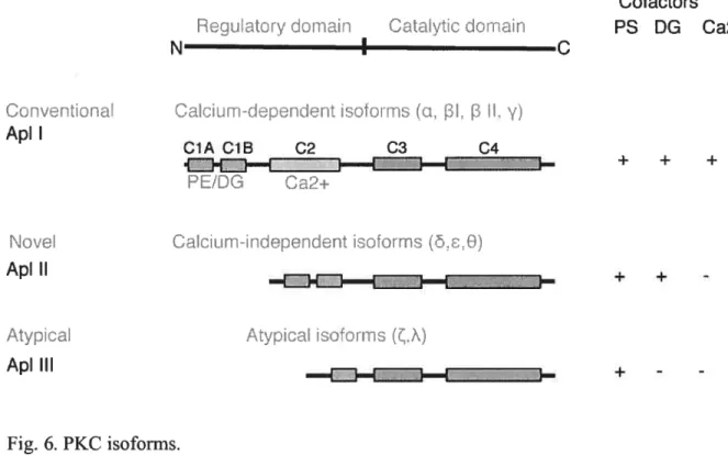
1.7.2PKC 47
1.7.3 PKA 49
forms of synaptotagmin I. Nakhost A, Houeland G, Castellucci Vf, Sossïn
WS (2003).
JNettrosci23:6238-44.
ABSTRACT 52 INTRODUCTION 52 METHODS 52 RESULTS 53 DISCUSSION 56 REFERENCES 57III.
41LIIJE
2...59
Identification and characterization of a novel C2B splice variant of
synaptotagmin I. Nakhost A, Houeland G, Blandford VE, Castellucci
VF,Sossin WS (2004).
JNetirochern89:354-63.
ABSTRACT 60 INTRODUCTION 60 METHODS 61 RESULTS 62 DISCUSSION 65 REFERENCES 69synapses in Aplysia. Houeland G, Nakhost A, Sossin WS, Castellucci VF
(2007).
JNeurophysiot
97:134-143.
ABSTRACT 71 INTRODUCTION 71 METHODS 71 RESULTS 73 DISCUSSION 76 REFERENCES 79\.
. . . ..81
5.1 Contribution of this thesis for the comprehension PKC modulation of transmitters
release by PKC 81
5.1.1 Mutation of Syt I at phosphoiylation sites does not alter transmitter release 81 5.1.2 Phorbol esters increase transmission at naïve synapses through phosphorylation of
SNAP-25 $2
5.1.3 Pharmacological tools that interfere with PKC function 85 5.1.4 PKC and short-term plasticity: rested versus depressed synapses $7 5.2 The role of calcium in neurotransmitter release in Aplysia 8$ 5.2.1 Does SNAP-25 modulation of calcium dynamics play a role in depression9 91
5.3 Localization of Syt IsinAplysia 92
5.3.1 How does our work compare with studies ofSNAP-25 in chromaffin cells9 93
5.4 Overexpression studies 96
5.4.1 The use ofEGFP-fusion proteins in Aplysia 99
5.5 Pretransiational modifications 101
5.5.1 Alternative spiicing during development 103
5.5.2 Possible roles of an alternative splice form in the $yt I juxtamembrane domain....105
5.6 Possible roles ofanalternative splice form in the Syt Ijuxtamembrane domain 106
7I. Conclusion...
...108
‘7!!. !efercnces...11O
VIII. 4nnex. . . .
....161
Table 1. Comparison of intrinsic and synaptic properties of sensory neurons expressing
Figure 1: Aplysia CNS 13
Figure 2: A dorsal view of ApÏysia showing the gui-siphon withdrawal reflex and its modifications by experience through simple forms ofleaming 15
figure. 3: A highly simpiified scheme of a fragment of the circuit that subserves the GSW
reflex 22
Figure. 4: The current models for synaptic vesicie exocytosis propose that fusion requires
assembly and activation of the $NARE compiex 36
Figure 5: the effect of the facilitating neurotransmitter serotonin (5-HT) on sensorimotor
(SM) synapses inAptysia 46
f igure 6: PKC isoforms 49
II ARTICLE 1
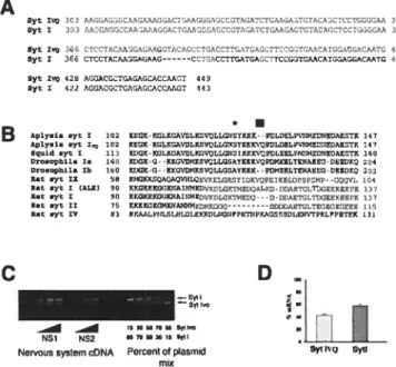
Figure 1: Cloning of a novel spiiced isoform of Syt I 54
f igure 2: Localization of Syt I and SytIVQ in sensory neurons 55
Figure 3: Syt I and
Syt
IVQ are phosphorylated at serine 123 by PKC in vitro 55Figure 4: Syt IvQbiocks the reversai of synaptic depression at a step upstream of PKC
Figure 2: RT-PCR of Syt I C2B-a and Syt I C2B-b 64
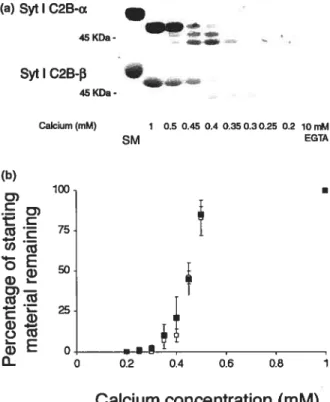
Figure 3: Chymotrypsin proteolysis of the cytoplasmic domain of Syt I
C2B-a and Syt I C2B-b 64
Figure 4: Calcium dose—response experiments 65
Figure 5: Effect of spiicing in C2B on binding to SNAP 25 66
Figure 6: Chymotrypsin proteolysis ofthe C23 domains of Syt I C2B-a
and Syt I C2B-B 66
Figure 7: SDS—PAGE analysis ofvarious soluble Syt I proteins 67
Figure 8: Localization of CFP-tagged Syt I C2B-13 in sensory neurons 67
Figure 9: Similar levels ofbacterial contamination in the splice isoforms 6$
II ARTICLE 3
Figure 1: Cloning ofAplysia synaptosomal-associated protein of 25 KDa (SNAP-25) 73
Figure 2: Localization of SNAp-25 constructs in sensory neurons 74
Figure 5: Scatterpiot of initial EP$Pfor ail constructs 77
Figure 6: Short-term facilitation of depressed synapses is partly blocked by overexpression of EGFP-SNAP-25 and EGFP-SNAP-25 S-D but not by any of the other mutants 77
AC - adenylyl cyclase
ADfP - activity dependent presynaptic facilitation
5-HT- 5-hydroxytryptamine
ANOVA - Analysis of Variance
cAMP- cyclic adenosine monophosphate ATP - Adenosine 5’-triphosphate
Bis- bisindolylmaleimide
CAl - Cornu Ammonis 1
CDR- cysteine-rich domain
CREB - cAMP-response element-binding protein
CS - conditioned stimulus
DAG - diacylglycerol EM - Electron Microscopy
EPSP - excitatory postsynaptic potentials
GSW- gill-and siphon-withdrawal
GWSR- gui-siphon withdrawal reflex
HRP - horseradish peroxidase
Hz - Hertz
1P3 - inisitol 1,4,5 triphosphate
IPSP- inhibitory postsynaptic potentials
KO - Knock out
LTP - Long-term-potentiation
mEPP - miniature end plate potentials
NMJ - neuromuscular junction
PC12 - pheochromocytoma
PK- Protein Kinase
PKA- Protein Kinase A
PKC- Protein Kinase C
PLC- phospholipase C
RRP - Readily Releasable Pool
RT-PCR- reverse transcription-polymerase chain reaction
S-A - Serine-Alanine S-C - Serine-Cysteine S-D - Serine-Aspartate S-E - Serine-Glutamate SCP - small cardiopeptides 5cr - Serine SM - Sensory-motor
SNARE- soluble NSF attachment receptor
SNAP - 25 -synaptosome-associated protein of 25 kD
SRP - Slow Releasable Pool
Syt - Synaptotagmin
US - unconditioned stimulus
To my thesis director Dr. Vincent Castellucci first of ail for liaving accepted me in his laboratory, for aiways being available, even when he hundred administrative task to complete. For his etemal optimism, his endless patience, and for numerous stimulating discussions about science and everything else.
To my codirector, Dr. Wayne
$055m,
for having integrated me in his lab and for his infinite knowledge in molecular biologyb Dr. Marc Klein for numerous stimulating discussions.
To ail the former labmembers, especially Frederic Manseau, for his help with experimental setup and general advice. For his friendship and support.
To Annie Campbell for her technical assistance
Finally, I am forever indebted to my parents for their understanding, endless patience and encouragement.
I. Introduction
You have to begin to lose your memoîy, fonly in bits andpieces, to realize that memoiy is what makes our lives. Lfe without memory is no life at ail, just as an intelligence without the possibitity of expression is flot really an intelligence. Our memoiy is our coherence, our reason, ourfeeling, even our action. Without it, we are nothing.
Luis 3uiuet (Sacks, 1985)
The question of how we leam, remember, and forget has long been the subject of much speculation. Considering how success and failures of our memory abilities affect our lives, this interest seems highly justified. Diseases that affect memories, like Alzheimer’s clearly show that our personality does not survive the destruction of our capacity to form memories.
The brain is an astonishingly complex computational device that constructs our perception of the extemal world, fixes our attention, and controls our actions. What we perceive in the extemal world transform itself in our brain in sensations and impressions that constitute our memories, and modifies and reorganizes the ones we already have. They are the foundation of our personality, of our imagination and our creative spirit. Memory is the function in our brain that constitutes a link between what we have been and what we are. We depend on our memories for so much of what we do, such as whenever we are engaged in identifying, appreciating, and responding appropriately to the objects and persons we encounter in our environment and to the events in which we participate; in speaking, reading, writing, or communicating; in thinking, reasoning, and problem solving; and in reminiscing about our experiences. So much do we depend on our memory that we have become mostly unaware of its constant contributions, other than appreciate the pleasant memories that are sometimes triggered by an idle thought or to curse the occasional inability to retrieve some particular name or word on demand.
One key question that has been asked ever since the Antiquity is that of the localization of memories. In the ancient world, the function of the brain was flot clear. Among the Greeks
controversy centred on whether the brain or the heart was the home of perception, intellect, emotion, imagination, memory and other functions of the mmd. Homer was the first to compare the “heart of the soul” to a block of wax —“We assume that it exist in our soul an “impregnable piece of wax”, a gift from Mnemosyne, the mother of all the mythological muses the goddess of memory. Descartes described it as a block of wax that can be engraved and that keeps this impression, which can be retrieved whenever needed for subsequent use (Tadié and Tadié, 1999).
Prior to the 19th century, when Freud and his followers wrestled with the mmd, others devised methods to penetrate the brain’s substance, the popular concept of memory as a private storehouse of images was acceptable. In fact it was not before 1885, when Herman Ebbinhaus, dissatisfied with treating memory as a philosophical problem described objective techniques for measuring memory (Ebbinhaus, 1913). He leamed lists of non sense syllables and at later times tested his recail by measuring the time necessary for releaming. A few years later, Edward Thomdike described similarly objective methods for measuring memory in cats and other laboratory animals. He placed an animal in a box fitted with an intemal release and measured the effect of repeated exposures on the time elapsed before the animal triggered the release and escaped from the box (Thomdike, 1923). In fact, he discovered that memories were flot stored in a specific centre where they were stockpiled, but that the entire brain participated in retrieving them. The development of these simple experimental methods for studying leaming and memory started a rigorous and empirical school of psychology called behaviorism. James Watson and Burrhus Skinner argued that behavior could be studied with the precision achieved in the physical sciences, but only if students of behavior abandoned speculation about what goes on in the mmd and focuses instead on observable aspects ofbehavior (Milner et al., 1998).
Before the introduction of functional neuroimaging, the study of amnesia was the only practical approach to the investigation of brain substrates of memory in humans. Whereas the application of functional neuroimaging could identify correlations between the activity
of distinct brain regions and the performance on memory tasks, the study of anmesiacs could potentially identify those structures that are obligatory for normal memory. The real hero in this research is the anmesic patient H.M., unfortunately unaware of his profound continuai role in modem neuroscience. H.M developed severe memory deficits following an experimental operation (Scoviile and Mimer, 1957) to alleviate uncontrollable epiiepsy. The operation surgicaiiy removed his medial temporal iobes, inciuding the hippocampus (Mayford and Kandel, 1999).
1.1 The dichotomy ofmemo,yJunctions
Our brain is able to store different kinds of information and form different kinds of memories that fali intotwo different categories: impiicit and declarative.
Declarative or expiicit memory is the conscious recali of knowledge about people, places and things and is particular well developed in the vertebrate brain. This memory involves ail our sensory perceptions, our feelings and motivations. When we remember an experience we recali what we have seen, heard, smelled, tasted, touched and felt. Implicit or non-declarative memory on the other hand is the non-conscious recali of motor skills and other tasks and includes simple associative forms, such as classical conditioning, and non associative forms, such as sensitization and habituation. Whereas explicit memory is mostly studied in mammals, implicit forms of memory can be effectively studied in both non mammalian vertebrates and invertebrates. To what degree do these two different forms of memory share common molecular components? Specifically, what parts of the nervous system are critical for leaming? How is information about a leamed event acquired and encoded in neural ternis? How is the information stored, and once stored how is it retrieved?
Most neuroscientists believe that the answers to these questions lie in understanding how the properties of individual nerve celis in general, and how synapses in particular are changed when learning occur.
First I will consider the major scientific insights that have helped to identify and delineate signaling and communication in nerve celis and then I will place signaling in the broader context of modem cell and molecular biology.
Finding the underlying cellular basis for memory is like the Holy Grau for many neurobiologists. 0f the various higher-cognitive abilities a human being possesses, such as reasoning and language, memory is the only one that can be studied effectively in simple experimental organisms. An average neuron forms and receives about 1000 to 20000 synaptic connections and the human brain contains about 1011 neurons. Thus at least 1014
synaptic connections are formed in the brain, making it the most complex structure, natural and artificial on earth (Kandel, 2000n). Although nerve celis can be classified into different types that share many common features, a key discovery in the organization of the brain is that nerve cells with basically similar properties are able to produce very different actions because of precise connection with each other. The complexity and relative inaccessibility of the nervous system, particularly in humans and other vertebrates limits the design and execution of effective memory research.
Most nineteenth century anatomists failed to appreciate that the cell body of the neuron, which housed the nucleus almost invariably gave rise to two types of extensions: to dendrites, that serves as input elements for neurons and that receive information from other ceils, and to an axon which serves as the output element of the neuron and conveys information to other cells. Appreciation of the full extent of the neuron and its processes came ultimately with the histological studies of Ramôn y Cajal. Cajal used silver salts to stain fleurons, a specialized method developed by Camillo Golgi that revealed their entire stucture under the microscope. Cajal observed that neurons are in fact discrete cells, bounded by membranes, and suggested that nerve celis communicate with one another only at specialized points of appositions, contacts points that Charles Sherrington later
called synapses (Sherrington, 1897) (see section 1.4) As Cajal continued to examine neurons in almost every region of the nervous system, he showed an uncanny ability to conclude from static images remarkable functional insight into the dynamic properties of neurons. One of his most remarkable insights was the principie of dynamic polarization. According to this principle electrical signaling within neurons is unidirectional i.e., the signal propagate from the receiving pole of the neuron, the dendrites and the ceil body, and then along the axon to the output of the pole of the fleuron- the presynaptic axon terminal (Albright et al., 2000; Kandel, 2000)
1.1.2 The brain must undergo changes that are maintained as long as the memory is stored What are these changes?
Because ail transformation of neural information in the brain involve only fleurons, glia, and their interconnections, neurobiologists since Ramén y Cajal (1911) believed that elementary aspects of leaming and memory storage were likely to be resolved on a cellular level. In his lecture to the Royal Society in 1894, Cajal proposed that memory is stored in the growth of new connections. This prescient idea was neglected for almost half a century as students of leaming fought over newer competing ideas. First, Lashley and a number of other gestalt psychologists proposed that leaming leads to changes in the electric fields or chemical gradients, which they postulated surround neural populations and are produced by aggregate activity of cells recruited by the leaming process. At about the same time Lorente de Né (1938), proposed that memory is stored dynamically by a self-re-exciting chain of neurons (Kandel, 2000n). f inally Holger Hyden demonstrated that learning changes the base composition of nuclear RNA, and thus induces an alteration in gene expression. From these findings, Hyden concluded that establishment of memory is correlated with protein synthesis, and he demonstrated de novo synthesis of several high-molecular protein species after leaming (Hertz et al., 2001).
The development of cellular techniques in the late 1940, early 1950s for the study of synaptic connections redefined Cajal’s ideas. Intracellular microelectrode recording
methods were introduced to study the synaptic actions of individual neurons and electron microscopy was applied to visualize the fine structure of synapses. These methodological advances allowed central synapses to be studied directly and revived Cajal’s idea that memory is stored in the growth of new connections. Prior to the last fourty years, evidence for this type of neuronal growth was restricted to embryonic development and regeneration of peripheral tissues.
1.1.3 What is the neuroanatomical support of our extraordinaiy capacity to store memories?
The question of where memory is stored emerged at the begiiming of the 19th century as part of the larger question- to what degree can any mental processes be localized within the brain? The first person to address this question was Franz Joseph Gail, a German neuroanatomist who argued that the cerebral cortex is not homogenous but contains distinctive centers that control specific mental functions. Gali proposed that particular mental functions are discretely localized and believed that one can determine a mental profile from the shape of the skull. He asserted that the brain does not act as a unitary organ but is divided into at least 27 faculties, each corresponding to a specific mental faculty. He thought that even the most abstract and complex human traits, such as generosity and spirituality were assigned their spot in the brain. This anatomically oriented approach to personality became known as phrenology. Despite Gall’s failure of his mental diagnostic methods, neuropsychological analysis of brain damaged patients with highly circumscribed behavioral deficits lias been used as evidence for the existence of highly specific cognitive modules (Damasio, 1990; Baynes et al., 1998).
In the middle of the nineteenth century, Pierre Paul Broca, a French clinical neurobiologist, analyzed the behavioral consequences following selective lesions of the brain, much influenced by Gall’s idea that functions could be localized. Using this approach he discovered that different regions of the cerebral cortex of the human brain are not functionally equivalent. Lesions to different brain regions produced defects in distinctively
different aspects of cognitive function. $ome lesions interfered with the comprehension of language, others with its expression (Schiller, 1992). However, the relevance of the breakdown unveiled by pathology, did flot reveal, Gall’s clear-cut mental faculties.
1.2 The “engrain“.
100 years ago a German scholar, Richard Semon, termed the material recorded engraved by a stimulus in living tissues as the “engram” (Semon, 1904). The etymological roots of the term are Greek, and it means, “something converted into writing”. Most of the popularity of the term “engram” stems from a paper by Karl Lashley (1950) entitled “In search of the engram”. Lashley aimed at identifying “the habits of the conditioned reflex type” in the brain. His methodology was interference of fiinction from dysfunction. He inflicted anatomical lesions on various parts of the rat or monkey brain, and tested the effect of the intervention on brightness discrimination and maze leaming (Lashley, 1929; 1950). In spite bis failure to locate the site of leamed discriminations, bis groundwork was influential for future models of memory research. His seminal contribution was flot however in his ad hoc experimental conclusions: He reached the conclusion that the engram is widely distributed throughout the brain (Lashley, 1929). Rather, his contribution was in the methodology and the concepts. On the methodological side, he was a pioncer in combining careful lesion techniques with behavioral analysis. On the conceptual side, he found that the severity of the leaming defect seemed to depend on the size of the lesion, not on their precise anatomical position, and he concluded that leaming and other mental functions have no special locus in the brain and consequently cannot be pinned down to specific collections of fleurons. However, the main objections were that his behavior tasks and the behavioral strategies required for successful performance were flot well defined in terms of the sensory inputs. A subject with partial brain damage could stiil succeed in the task by using the undamaged areas, and furthermore, the lesions were flot delicate enough to differentiate the contribution of distinct functional division in the brain.
At almost the same time that Lashley’s research indicated that there was no memory engram, a Canadian neurosurgeon, Wilder Penfield produced evidence to the contrary. Penfield electrically stimulated the exposed brains of epileptic patients to determine the location of abnormal brain tissue and the functions its removal might endanger. When Penfield probed a region of the temporal lobe, the patients’ responses took him by surprise. Stimulation anywhere on the cerebral cortex could bring responses of one kind or another, but lie found that when he stimulated the temporal lobes, he could elicit meaningful, integrated responses such as memory, including sound, movement, and color. These memories were mucli more distinct than usual memory, and were often about things unremembered under ordinary circumstances. Yet, if Penfield stimulated the same area again, the exact same memory popped up. The temporal region lie theorized, must be “part of an automatic mechanism which scans the record of the past,” linldng the cortex to deeper brain structures tliought to be involved in memory. “In the vast circuitry of the liuman brain,” Penfield confidently wrote, “the evidence of an engram is clear” (Penfield, 1951).
Later, Lashley’s student, Donald Hebb developed lis famous theoretical principles of memory formation at a cel!ular level (Hebb, 1949). At the synaptic level, Hebb coined a postulate of use-dependent synaptic plasticity. In its original version it states the following: “When an axon of ce!! A is near enough to excite a ceil B and repeatedly and persistently takes place in firing it, some growtli processes or metabolic changes takes place in one or both cells such that A’s efficiency, as one of the cells firing B, is increased”. In a Hebbian synapse, the increase in synaptic weight is thus a function of correlation of pre- and postsynaptic activity. Hebb proposed that the strength of the connection between the two neurons is increased for a long period of time when the firing of the presynaptic and postsynaptic neurons are closely correlated in time. Subsequently, this synaptic strengthening has been termed associative, because it associates the firing of a postsynaptic neuron with that of a presynaptic neuron. After such an event, when the first of the two neurons is activated, the chance for the postsynaptic neuron firing is increased. Hebb
implied that the synaptic strengthening is input-specific: when two neurons fire coincidentally the synapse between them is strengthen, but other synapses on either neuron remain unchanged (Bailey et al., 2000). Kandel and Tauc showed that synapses could be strengthened or weakened without requirement for an activity of either the pre- or the postsynaptic neurons as a resuit of the firing of a third, modulatory intemeuron. They also suggested that this heterosynaptic modulation could have two forms, associative or non associative. The associative form, combines features of both homosynaptic and heterosynaptic mechanisms, whereas the non-associative form is purely homosynaptic (Kandel and Tauc, 1965a; Kandel and Tauc, 1965b).
1.2.1 Synaptic Flasticity
Understanding how memories are stored and retrieved has been the goal of work in several scientific disciplines, including neuroscience, neuropsychology, psychology, and cognitive sciences. In neuroscience, research on leaming and memory has been conducted largely within the context of plasticity i.e., the malleability of the synapses. Chemical synapses can be modified functionally and anatomically during development and regeneration, and most importantly, through experience and leaming. Use dependent neuronal plasticity embodies stimulus induced regulation of intracellular signal transduction cascades, which culminates in either: 1) Functional alterations, typically short term, and invoïve changes in the effectiveness of existing synaptic connections and requires only covalent modification of pre-existing proteins, or 2) Anatomical alterations, typically long-term that comprise the modulation of gene expression, new protein synthesis, and the establishment of new synaptic connections (Goelet et al., 1986; Bailey et al., 1996). In homosynaptic plasticity, it is the intrinsic activity of the synapse itself that subsequently alters its own functional state. These functional changes can be triggered by biochemical processes localized within either the presynaptic terminal or the postsynaptic ce!!. In heterosynaptic plasticity, in contrast, a synapse between two neurons is modified by extrinsic action of a third neuron. This modu!atory neuron can alter synaptic transmission either by direct synaptic action or
through diffuse release of a transmitter or hormone, to alter the function of the first synapse. An important question has been whether these different types of plasticity involve fundamentally different mechanism, or whether they share an alphabet of basic mechanisms that are combined in different ways.
Synaptic plasticity contributes to a variety of physiological and pathological processes in the aduit brain- from leaming and memory to amnesia and dementia, from drug dependence to epilepsy (Bliss and Collingridge, 1993; Martin and Kandel, 1996).
Different mechanisms underlie synaptic plasticity at different types of synapses. In theory, the efficacy of synaptic transmission could be modulated at any of the steps that lead to vesicle fusion, release of transmitters and activation of postsynaptic receptors. It is therefore important to thoroughly elucidate the basic mechanisms of synaptic transmission to ultimately understand the meanders of synaptic transmission.
1.3 Vertebrates versus invertebrates
As learning and memory have become accessible to study with the techniques of cellular and molecular biology, a variety of cellular mechanisms of neuronal plasticity have been identified that are thought to contribute to different forms of leaming in invertebrates and vertebrates. Now, researchers can aftempt to define the basic principles of leaming by comparing these mechanisms and asking: what do they have in common, and how are they different?
Although the ultimate goal is to delineate the mechanisms by which memory in our own mmd is formed, stored and retrieved, the favorite models are for obvious reasons species that are amenable for experimentation in laboratories. Unlike other mental process such as thought, language, and consciousness, learning is relatively accessible to cellular and molecular analysis.
Interest in the intelligence of simple organisms and their use to investigate probiems of leaming and memory, draws mainly from two conceptual frameworks: Darwinism (Boakes,
1984) and reductionism. The notion that evolution applies to the mmd as well, combined with an interest in zooiogy and behavior, directed some investigators already a century ago to study the mental powers of organisms such as protozoa and insects (Peckham and Peckham, 1887; Jennings 1906; Day and Bentley, 1911). There are, for example few functional or biochemical differences between the nerve celis and synapses of humans and those of ApÏysia. Since behavior and learning is an expression of nerve celi activity, it would be surprising if the leaming capability of people did flot have some elementary features in common with learning in simple organisms. And, if elementary leaming are
common to ail animais with an evolved nervous system, there must be conserved features in the mechanisms of leaming at a cellular —and molecular level that can be studied effectively even in simple invertebrates (Krasne, 1978).
1.3.1 The idealmodeÏ
The ideal model should have the following qualities: 1) The animal should be able to perform a simple task that can be modified by experience 2) the neuronal network responsible for the task should be characterized in detail. 3) the neurons and synapses in the network where the physiological changes associated with the leaming or the modification of behavior take place should be identified, and finally 4) knowing ail the important modifications we should be able to reveal the cellular and molecular processes that are involved (Castellucci and Trudeau, 1997).
1.3.2 Aplysia
The possibility of acquisition by repetition of a stimulus is found in ah animal species, even the simplest ones: Aplysia californica has proven to be an ideal model for this task. The main asset of this marine mollusk is:
1) Aplysia has a relatively simple and accessible nervous system. Whereas the mammalian brain has a 1011 nerve celis, the central nervous system of Aplysia bas only 20,000 arranged
in widely spaced ganglia (Kandel, 2000n).
2) In Aplysia, certain elementary behaviors that can be modified by leaming may use fewer than 100 celis. This neural simplicity makes it possible to delineate in detail the wiring diagram of the behavior and thus pinpoint the contribution of each individual nerve ccli to the behavior in which they participate (Kandel, 1976).
3) In addition to being few in numbers these celis are the largest nerve ceils in the animal kingdom, reaching up to 1000 tmin diameter, as big as the entire brain of Drosophila and big enough to be seen with the naked eye. Because of their large size, distinctive pigmentation, location, shape, and firing pattem, it is possible to identify many of the celis as unique individuals. One can record from these cells for hours, and the same ccli can be returned to and rerecorded over a period of days. The celis can easily be dissected out for biochemical studies, so that from a single cdl one can obtain sufficient mRNA to make a cDNA library.
Finally these ceils can readily be injected with labeled compounds or genetic constructs (Kandel, 2000n), and since both sides of the synapses are accessible to substances injected into the ccli bodies, it is possible to manipulate presynaptic as well as postsynaptic mechanisms (lin and Hawkins, 2003).
The Aplysia have attracted several cellular physiologists (Arvanitaki and Chalazonitis, 1958; Tauc and Gershenfeld, 1961; Kandel and Tauc, 1965). It is however, the research on plasticity and leaming that is Aptysia, especialiy Aplysia calfornica ‘s daim to fame. It gained immortai fame from studies in the laboratory of Eric Kandel. He was a pioneer and bas continued to be a major driving force in Aplysia research. In 2000 he shared the Nobel
Prize in medicine for 40 years of research in the cellular processes underlying learning and memory, first in Aplysia, and recently in mice.
Aplysia calfornica, a hermaphroditic marine snail with mottled purpie skin, is found in shallow ocean waters, have small repertoire of stereotyped behaviors that includes feeding, egg-laying, and a variety of protective mechanisms. These responses include withdrawal of the tau, siphon and gui, and the release of defensive secretions such as ink and opaline (Walters et al., 1983)
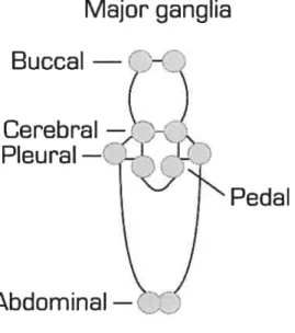
The central nervous system consists of eight paired ganglia arranged around the eosophages, and a large fused ganglion in the abdomen.
Major ganglia
Pedal
Fig. 1. Aplysia nerve celis are clustered together in five major bilateral pairs of ganglia, each containing about 2000 nerve celis.
Following a series of reductive and simplifying steps, the cellular and molecular mechanisms of leaming in Aplysia has been pursued from the behaving animal via
Buccal
—Pleural
preparations of isoiated ganglia, to identified nerve celis and synapses in culture (Carew et ai, 1971; Castellucci et ai, 1970; Rayport and $chacher, 1986).
Not ail synapses are equaliy adaptable. The strength of some synapses in Aplysia rarely changes, even with repeated activation. However, with synapses specially involved in learning and memory storage, such as the synaptic connections between the sensory neurons and their follower neurons in the gui-siphon -and tau withdrawal reflexes, a relatively small amount of training, especially if it is appropriately spaced, can produce large and long-lasting changes in synaptic strength. The study of these systems in ApÏysia has provided an exceptionally favorable opportunity for relating synaptic changes in specific celis to behavior, because the monosynaptic pathway in the giil-and siphon withdrawal (GSW) reflexes and the related pathway activated by tau stimuli belong to a reflex system that can be studied on the cellular level (Byrne et al., 1993; Hawkins et al.,
1993).
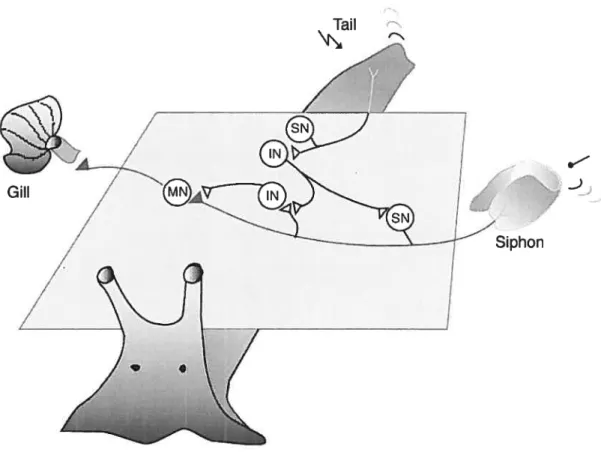
The analysis of ieaming in Aplysia has focused mainiy on the defensive reflexes, illustrated primarily by the gili-and siphon-withdrawal (GSW) reflex. The GSW is a very simple defensive reflex: the withdrawal of the gili upon stimulation of the siphon, an action that is like the quick withdrawal of a hand from a hot object (Pinsker et ai., 1970). This reflex is advantageous because the neuronal circuit is partly monosynaptic, and it has been shown to undergo several simple forms ofleaming (Murphy and Glanzman, 1997).
Fig. 2. A dorsal view of Aplysia, showing the gui-siphon withdrawal (GSW) reflex. The gui lies in the mantle cavity and is normally covered partially by the mantie shelf. A light touch to the siphon causes the siphon to contract and the gui to withdraw into the mantie cavity under the protection of the mantle shelf. Repetitive monotonous tactile stimuli resuit in habituation. A shock to the tau resuits in sensitization of the reflex.
In ApÏysia, the gill is the extemal respiratory organ that is housed in the mantie cavity. The cavity is a respiratory chamber covered by the mantle shelf. When the animal is in a normal
Simple forms of reflexive learning leaU te
changes in effectiveness of synaptic transmission
Habituation S e n
s itizat ion
Snmuli Sî=2Oec TauI
Stimu{irelaxed state, the gui is partially covered by a sheet of skin (the mantle sheif), which ends in a fleshy spout, called the siphon. If a tactile stimulus is applied to the siphon, both the siphon and the gui are drawn into the mantle cavity. The G$W reflex can be habituated by repetitive monotonous tactile stimuli to the skin; sensitized by noxious or strong tactile stimuli to the tail or head; and it can also undergo classical conditioning.
The circuitry underlying the Aplysia siphon-elicited siphon-withdrawal reflex bas been widely used to study the cellular substrates of simple forms of leaming and memory. Although the basic form of the behavior is quite simple, the underlying circuitry is rather complex, consisting of monosynaptic and polysynaptic pathways, excitatory and inhibitory intemeurons (frost and Kandel, 1995). A groundbreaking finding that allowed biochemical and molecular investigation of this memory at the single cdl level was provided by the work of Montarolo et al. (1986). These investigators showed that it is possible to reconstitute in vitro the main components of the neural circuit underlying sensitization and to reproduce synaptic responses in vivo by learning. The parallel use of both in vitro and in vivo models and the work of several groups in the last 20 years led to an understanding of the biological processes underlying sensitization of the G$W-reflex (Bailey et al., 1996; Byme and Kandel, 1996, Carew, 1996). For the studies described in this thesis, we used the pleural ventrocaudal (VC) sensory neuron cluster of the pleural ganglion and the LFS siphon motor neurons of the abdominal ganglion reconstituted in vitro (Frost et ai,, 1997; Walters and Cohen, 1997). These cells in particular, together with the gill motorneuron L7 have been widely used to study the cellular substrates of simple forms of leaming and memory. The bilateral VC clusters are notable for their uniform appearance and the tight packing of their ceils (Walters et ai,, 1983) The VC cluster us comprised of mechanoafferent neurons that innervate a large part of the body surface of the animal. In situ, the LFS motor neurons receive monosynaptic afferents from the LE sensory neurons in the abdominal ganglion. The Aptysia siphon-elicited siphon-withdrawal reflex circuit consists of the LFS motor neurons and 10 intemeurons (5 L29s, 3 L30s, 2 L34s) known to
convey excitatory input to them (frost et ai,, 1997). The full circuit contains tbree other centrai siphon motor neuron groups (e.g., LBS, LD$, RDS) (Frost and Kandei, 1995). Aplysia sensory-motor neuron synapses in isoiated ce!! culture exhibit both short and long lasting forms of piasticity characteristic of the intact animai (Rayport and Schacher, 1986; Montarolo et al., 1986; Eliot et al., 1994a,b; Lin and Glanzman, 1997; Bao et al., 1997; 1998). In addition, they have a number of different advantages for examining the mechanisms of plasticity. 1) The neurons are identified as individuals with known behaviora! functions. 2) There are no other neurons in the dish and they do not form autapses. 3) One can unambiguously distinguish between homosynaptic and heterosynaptic effects and also know the source of spontaneous miniature EPSP with certainty (Jin and Hawkins, 2003). The neuronal networks causally related to these reflexes inciude two main components, a monosynaptic one, the sensory-to-motor neurone synaptic junctions, and a polysynaptic one made up of various excitatory and inhibitory interneuronal synapses (Frost et al., 1988; Trudeau and Castellucci, 1992; Wbite et al., 1993). There are many sites that can be modified when the reflex is facilitated or depressed (Fischer and Carew, 1995; Trudeau and Castellucci, 1993a; Xu et ai., 1995). Like many forms of leaming-reiated synaptic plasticity, facilitation at Aplysia sensory-motor neuron synapses can involve different molecular mechanisms depending on experimental variables such as the history of activity and the duration of 5HT exposure (Byme and Kandel, 1996).
The transmitter at the sensory to motor junction is most likely glutamate (Trudeau and Castellucci, 1993; Lechner and Byme, 1998; Storozhuk and Castellucci, 1999; Levenson et ai, 2000; Cohen et al., 2003; Antzoulatos and Byrne, 2004). The sensitizing or facilitating stimulation activates severa! types of facilitator intemeurons, some of which have been identified (Hawkins et al., 1981; Goelet et al., 1986; Hawkins and Schacher, 1989; Trudeau and Castellucci, 1993; Liu et al., 2004;
).
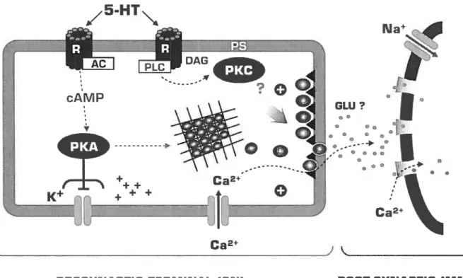
These neurons use at least four different transmitters: 5-hydroxy-tryptamine (serotonin or 5-HT), the small cardiopeptides SCPA andMost of the work on synaptic facilitation has focused on the action of 5-HT. Serotonin induces changes in ionic conductances leading to spike broadening and enhancement of excitability in the sensory neurons and in some motor neurons. Serotonin leads to an increase in synaptic release in two ways, one that is dependent on spike broadening and the other that is independent of spike broadening. These two facilitatory actions of 5-HT are mediated by at least two second messenger activated protein kinase systems, protein kinase A (PKA) and protein kinase C (PKC). The two biochemical cascades overlap in their contributions to synaptic facilitation; their contributions are not simply synergistic but are state- and time-dependent (Burreli and Sahley 2001; Kandel, 2001; Leal et al., 2005). Recent observations by Glanzman and colleagues (Li et al., 2005) have indicated that facilitation of the sensori-motor synapses during and after 5-HT exposure depends also on a rise in postsynaptic intracellular calcium and release of calcium from postsynaptic stores, which then signals back to the presynaptic terminal by some unknown mechanism. In ccli culture, application of 5-HT can be substituted for a harmful modulatory stimulus. A single application of 5-HT produces short-terni facilitation, same as does single tail shock, whereas four more repeated applications of 5-HT produce long-term synaptic facilitation (Montarolo et al., 1986 from Castellucci et al., 1988). Blocking the action of these serotonergic celis blocks the effect of sensitizing stimuli (Glanzman et al., 1989).
The sensory to motor connection can undergo homosynaptic depression (Armitage and Siegelbaum, 1998; Byme and Kandel, 1996), homosynaptic facilitation (Bao et al., 1997; Li et al., 2005; Lin and Glanzman, 1994b; Muller and Carew 1998) and heterosynaptic depression involving FMRFamide and dopamine (Abrams et al., 1984; Montarolo et al., 1986). There are also unidentified sensory neurons, probably at the periphery that can contribute to the reflex; they have lower threshold of activation than those of the other groups but their modulation seems to be similar (Frost et al., 1988). Anatomical studies conducted by Bailey and Chen (Bailey and Chen, 1988) indicated that long-term memory (lasting several weeks) is accompanied by a plethora of alterations at identified sensory neuron synapses. Their resuits indicate that the size of the active zones and the total number
of varicosities are larger in sensitized animais compared to controls and smaller in habituated animais. Castellucci and Kandel attempted to iocalize the change produced by habituation and sensitization to either the presynaptic or postsynaptic component of the synapse. They applied a quantal analysis to the synaptic connections between the sensory and motor celis, and found that the short-term homosynaptic depression that accompanies habituation involves a reduction in the amount of transmitter released from the presynaptic neuron (Castellucci and Kandel, 1974).
The Aplysia G$W reflex can be modified by four simple forms of leaming paradigms: 1) habituation, 2) sensitization, 3) dishabituation, and 4) ciassical conditioning
1.3.2.1 Habituation is the simplest form of implicit leaming, where the animal show a progressive decrease in reflex strength with repeated iimocuous stimulation. If the stimulus is neither beneficial nor harmful, the animal leams after repeated exposure, to ignore it. Habituation prevents recurrent non-threatening environmental stimuli from endlessly distracting the animal from potentially meaningful stimuli of behavioral significance. Habituation is caused by a homosynaptic depression in the activity of a sensory-motor connection (Castellucci et al., 1970, 1974) and is associated with depression of the synapses from sensory neurons onto gui and siphon or tau motor neurons produced by a reduction in transmitter release from the presynaptic terminais following each action potential. Habituation can last from a few minutes to up to severai weeks if repeated training sessions are administrated over several days (Carew et al., 1973). Release of neurotransmitter typically occurs at axonai swellings, termed varicosities, in contact with the processes of a postsynaptic neuron. Anatomical studies show that the number of varicosities (Bailey and Chen, 198$) as well as the number and size of the vesicle pool in the active zone are significantly decreased in long-term habituated synapses (Bailey and Chen, 1983), indicating that morphological changes accompany the changes in behavior. At Aplysia synapses, short-term depression does not appear to involve a depletion of releasable
vesicles or a decrease in presynaptic Ca2 influx, but may involve an inactivation of the release process itseif (Eliot et al., 1994b; Armitage and Siegelbaum, 199$). However, Baiiey and Chen (19$$) and study by the group of Marc Klein (2002) suggest depletion as a possible mechanism. Although considered the simplest of ail types of leaming, the cellular and molecular analysis of habituation in Aplysia has so far yielded less elaborate mechanistic models than the analysis of sensitization or classical conditioning.
1.3.2.2 Sensitization is the erihancement of reflex responses. When a harmful stimulus is applied to the neck or tau of Aplysia, facilitating neurons are activated that in tum act on the sensory neurons to enhance transmitter release (Castellucci and Kandel, 1976). Defensive reflexes for withdrawal and escape becomes amplified, the animal leams to respond more vigorously, flot only to that stimulus but also to other stimuli, even harmless ones. The phylogenic value of sensitization is rather straightforward: An Aptysia that has just avoided being eaten by a lobster that has pinched its tau, is indeed well advised to respond with a lower threshold to a similar stimulus, because it is almost certain that the lobster will attack again. During short-term sensitization or dishabituation of the withdrawal reflex, the monosynaptic connections between sensory neurons and motor neurons are enhanced by heterosynaptic facilitation (Byme and Kandel, 1996). Facilitation or dishabituation is the enhancement of a habituated reflex response by a noxious stimulus, whereas sensitization is an enhancement of a non-habituated reflex response by that stimulus (Carew et al., 1971).
1.3.2.3 Dishabituation has been thought to be due either to reversai of the process of habituation or to a second process equivalent to sensitization superimposed on habituation (Hawkins et al., 2006). Hoclmer and colleagues (1986 a, b) suggested that dishabituation and sensitization in adult Aplysia are produced, at least in part, by different cellular mechanisms. This reversai of synaptic depression parallels the behavioral process of dishabituation. They also proposed that this facilitatory mechanism might represent a direct
modulation of transmitter release, either an increase in the availability of transmitters for release, or a modulation of the release mechanism itself. In contrast to a unitary process view, dishabituation and sensitization emerge as separate behavioral processes according to very different developmental timetables in Aplysia. The magnitude of dishabituation appears to be determined by the interaction of 3 underlying processes: (1) the dishabituation process itself, (2) an inhibitory process that competes with dishabituation and, (3) a facilitatory process (sensitization) that augments dishabituation (Rankin and Carew, 1988). At depressed Aplysia sensoiy to motor synapses, 5-HT facilitates transmitter release by dishabituating the synapse primarily through activation of a Ca2tindependent form ofPKC, PKC Apl II (Ghirardi et al., 1992; Manseau et al., 2001). While activation of PKC by 5-HT increases transmitter release only at depressed synapses, activation of PKC by phorbol esters (PE) increases transmitter release at both naïve and depressed synapses (Braha et al., 1990; Ghirardi et al., 1992; Nakhost et al., 2003). At naïve synapses 5-HT facilitates transmitter release primarily through a protein kinase A (PKA) pathway. In the prescence of a PE, the Ca2-dependent PKC, Apl I is also recmited. It is also possible that the initial increase in synaptic strength caused by activation of PKA may make further modifications induced by 5-HT irrelevant. However, recently Jin et al. (2005) showed that PKC-mediated increases in synaptic strength at naïve synapses could be activated through prolonged applications of 5-HT.
1.3.2.4 Classical conditioning is a more complex form of leaming than sensitization. Rather than learning only about one stimulus, the organism leams to associate one type of stimulus with another. This form of leaming, originally described in dogs by the Russian physiologist and psychologist Ivan Pavlov (Pavlov, 1927), occurs when a behaviorally neutral stimulus, usually a light tone or a tactile stimulus (the conditioned stimulus or CS) is presented to an animal together with a reinforcing stimulus (the unconditioned stimulus
aiI
Ciii
Siphon
Fig. 3. A highly simplified scheme of a fragment of the circuit that subserves the GSW reflex. The neuronal networks causally related to these reflexes include two main components, a monosynaptic one (marked in red), the sensory-to-motor neurone synaptic junctions, and a polysynaptic one made up of various excitatory and inhibitory
or US). Classical conditioning has been portrayed as sharing cellular mechanisms with sensitization, however, in contrast to sensitization, which enhances the responses of subsequent stimulations of the skin at any location; the facilitation in classical conditioning is specific to the pathway that has mediated the conditioned input. The GSW-reflex of Aplysia, can be enhanced by classical conditioning, however, the timing of the CS and US is critical. It depends on close forward pairing, (about 0.5 s) of sensory neuron activity, the CS (siphon touch) and the excitatory intemeuron activity, the US, (a strong shock to the tau). The sequential activation of the sensory neuron during a critical interval by the CS and the US leads to greater presynaptic facilitation than when the two stimuli are flot accurateÏy paired. Classical conditioning in Aplysia was first described by Kandel and colleagues in 1981 (Carew et al., 1981). This form ofinvertebrate learning was originally hypothesized to be due to an exclusively presynaptic mechanism, known as activity dependent presynaptic facilitation (ADFP). A 1984 study (Carew et al. 1984) where postsynaptic hyperpolarization during the US did not block the associative enhancement of EPSP, concluded that ADfP does not involve a Hebbian mechanism. A decade later, the discovery that sensory-motor synapses of Aplysia possessed the capacity for NMDA receptor dependent LTP (Lin and Glanzman, 1997; Lin and Glanzman, 1994a; Lin and Glanzman, 1994b) indicated that classical conditioning also depends in part, on Hebbian LTP (Glanzman, 1995).
1.3.3 Drosophila
The fruitfly, Drosophila melanogaster has long been an organism of choice for molecular geneticists. A tiny creature, 3mm in length, equipped with complex body structures, including a brain of 250,000 neurons. Drosophilas are conveniently small, and remarkably inexpensive. Their generation time is about 10 days at room temperature, and their life cycle includes easily identifiable phases (Ashbumer, 1989). The small number of chromosomes, the convenient chromosomal cytology, the availability of spontaneous mutants, the short generation time, and ease of breeding, initiated a meticulous, systematic
analysis of Drosophila genetics. The fact that flues can leam to remember a variety of associative tasks makes Drosoph lia an excellent system to characterize genes involved in learning and memory (Davis, 1993). Studies of leaming in flues frequentiy use a classical conditioning paradigm that involves the temporal coupling of an electric shock with a particular odor; flues leam to avoid the shock-associated odor after training. Behaviorai screening of mutagenized flues using such learning paradigms have led to the isolation of genes invoived in leaming and memory. Over the years, “leaming mutants” of Drosophila have contributed significantly to our current knowiedge about the molecular mechanisms of acquisition and consolidation of simple memory. It has created remarkable evidence in support of the cAMP signal transduction cascade and of the cAMP-response element binding protein (CREB), a type of protein that regulates the expression of genes, and plays a key role in neuronal plasticity.
In Drosoph lia synaptic transmission is usually anaiyzed at the neuromuscular junction (NMJ) during the embryonic, third instar larvai, and adult stages of development. Although this methodology does not involve intemeuronal connections, the basic release mechanisms us unlikely to be substantiaiiy different, as the moiecular component of these two glutamatergic synapses are highiy conserved (Littieton and Bellen, 1995). In the Drosoph lia NMJ identified motor neurons ilmervate specific muscle targets in a highiy stereotypic manner, ailowing consistent and reproducible analysis of synaptic parameters between animais (Yoshihara and Montana, 2004). Yet Drosophila, in spite of offering unique advantages to the geneticist, is flot the dream machine of the neurophysioiogist, centrai neurons in the fruit fly are flot easiiy avaiiable to electrode recordings in the same way as central mammaiian neurons. Furthermore, being invertebrate, Drosophila is incapable ofproviding dues ofthe mammaiian brain at the circuit and system ievei.
In recent years, some of the enthusiasm for using simple organisms to anaiyze the neuroiogical bases of behavior has declined, because state of art molecular bioiogy can now be used to approach probiems in higher organisms that previously were oniy approachabie
in lower ones. Powerful neurogenetics, for exampie, is already practiced in mice, depriving Drosophila of its monopoiy in the neurogeneticai analysis of memory.
1.3.4 Mouse
Mus muscuÏus, the common mouse is a pest for househoiders, a pet for animal loyers, and a biessing for molecular bioiogists. Mice are the mammalian counterparts of Drosophila for leaming and behavioral studies. Aithough they lack Drosophila advantages for genetics studies, the ability to generate mice with specific gene “knock outs”, and possibility of assessing the effect of the targeted mutations in classical behavioral tests, made mice attractive animais for memory studies. Unlike Drosophila, where specific genes required for memory can be identified in specific screens, studies in mice are somewhat lirnited to guessing the players. With mice, it is now possible to add engineered genes to the mouse genome, or remove other genes at will, and to generate mouse unes that will express the mutation and propagate it to their progeny. Knock out (KO) mice, where a gene is ablated in situ, have been used to identify the roles of a variety of protein kinases, of subtypes of glutamate receptors and of a variety of transcription factors invoÏved in long-term potentiation (LTP). They have also proved useful in particular for probing the relations between LTP and leaming, and the role of hippocampus in learning and memory (Grant et al., 1992; Mayford et al., 1996; Isien et al., 1996a). Furthermore, novel techniques now permit the generation of tissue, -celi-type, -and temporarily restricted gene knockouts (Tsien et al., 1996b; Shimizu et al., 2000). These conditional mice techniques offer considerable advantages for the study of leaming and memory, because they could be used to dissociate the effect of a mutation on development from those on behavioral plasticity, and furthermore, localize the defect to specific brain regions and circuits. In parallel with the aforementioned trend, the use of brain suces (e.g. Dobrunz and Stevens, 1999) and of neuronal cell cultures (e.g. Tardin et al., 2003; Jaskolski and Mulle, 2004; Abel et al., 1997) from complex nervous system has gained much popularity, because such simplffied preparations permit exploitation of highly advanced molecular and computational
techniques e.g. the investigation of LTP in the hippocampus and fear-conditioning in the amygdala. Among mammais the mouse is stiil unique in this respect; appropriate neurogenetic techniques are flot yet available, for example in rats. In addition, they are small (20-35g), the size oftheir brain is manageabie, and the generation time is 3-4 months.
1.4 The synapse
The neurons in our brain communicate with one another through specialized structures caiied synapses. The term synapse (Greek for syn-haptein “to make contact”) is commonly attributed to Sherrington. Sherrington was a neurophysiologist who beiieved that nerves terminate in free endings and that the transfer of information from these endings to their targets differs markedly from the propagation of information along neuronal branches. When requested to revise his contribution to a textbook of physiology (foster and Sherrington, 1897), he reasoned that since the research on this functional junction between nerve celis had already matured to become an important topic in physiology, this type of junction deserved a speciai term. Hence, the synapse was bom (Sherrington, 1941).
Synapses come in many flavors. They can be classified by their morphology, location, function (e.g. inhibitory vs. excitatory), types of neurotransmitters and their receptors. A major taxonomy distinguishes chemical from electrical synapses. John Eccles, (Sherington’s student), believed ail synaptic transmission was electrical, that the action potential in the presynaptic neuron generates a current that flows passively into the postsynaptic cell. He resisted initiaily the idea of a chemicai transmission proposed by Henry Dale and his followers, but later he became a major proponent of it (Kandel, 2000). It is now accepted that although most synapses use a chemical transmitter, some operate purely by electricai means.
Neurobioiogists now accept the existence of two major modes of synaptic transmission: eiectrical, which depends on current, through gap-junctions that bridge the cytoplasm of pre- and postsynaptic ceiis. Eiectrical transmission piays a roie in synchronizing neural
activity. In chemical transmission, pre- and postsynaptic celis have no structural continuity they are separated by a discrete extracellular space, the synaptic clefi (Bennett, 2000). The most common transmitters of the CNS are glutamate, GABA, dopamine and serotonin. Eccles and his collaborators (Eccles, 1963) showed that synaptic communication in the CNS, as in the periphery, is mediated by ionic currents that flow across the postsynaptic membrane, generating excitatory or inhibitory postsynaptic potentials (EP$Ps and IPSPs). Depending on which ions carry the postsynaptic currents, the respective transmitters are classified as excitatory or inhibitory.
1.4.1 Signaling within fleurons: the action potential
Nerve cells are able to carry signals over long distances because of their ability to generate an action potential. An action potential generates a local flow of current that is sufficient to depolarize the adjacent region of the axonal membrane and is propagated without failure along the axon to the nerve terminal. In 1939 while recording from the giant axon of the squid Kenneth Cole and Howard Curtis found that the ionic conductance across the membrane increases dramatically during the action potential, suggesting that the action potential reflects the flow of the ionic current. Hodgkin, Huxley and Katz extended these observations in a series of papers in the early 1950s. They found that the amplitude of the action potential is reduced when external Na concentration is lowered, indicating that Na influx is responsible for the rising phase of the action potential (Hodgkin and Katz, 1949). Their data also suggested that the falling phase of the action potential was caused by a later increase in K permeability (Hodgkin et al., 1952).
1.4.2 Synaptic transmission
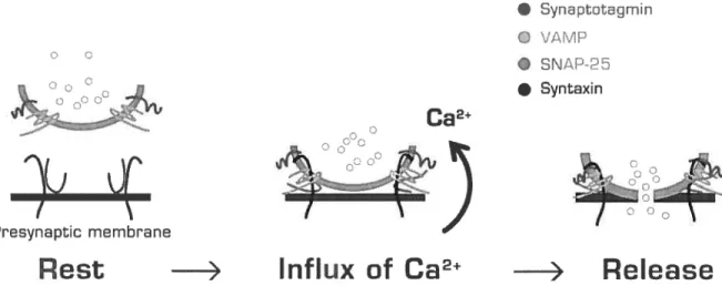
Synaptic transmission is initiated when an action potential triggers neurotransmitter release from a presynaptic nerve terminal (Katz, 1969) and is determined by the amount of transmitter release from the presynaptic neurons and by the transduction of the chemical signal into an electrical response by the target cell. An action potential induces the opening
of Ca2-channe1s and the resulting Ca2 transient stimulates synaptic vesicle exocytosis. Transmitter release depends on the size of the readily releasable pool (RRP) of transmitter (Rosemund and $tevens, 1996; Gillis et al., 1996), thought to represent the release-ready synaptic vesicles docked at the active zone (Schikorski and Stevens, 2001), and the efficacy of the release process.
Synaptic transmission includes a chemical step, where the signaling substance, called a transmitter, is released very locally from the sending, presynaptic ceil and then acts transientïy on receptors of the receiving, postsynaptic ceil. The receptor is part of an ion channel and mediates, upon occupation by the transmitter, a brief flux of ions across the postsynaptic membrane generating a change in the postsynaptic membrane potential (Sakmann, 1991).
The signal that actually initiates the cellular response of the postsynaptic ceil is the flux of ions across the postsynaptic membrane. The size, duration and direction of this ion flux, as well as the nature of the ions traversing the postsynaptic membrane, determines whether this response will either activate voltage sensitive membrane conductance and initiate action potentials, or instead reduce the celis electrical activity. The cellular response may also be determined by a change in intracellular ion concentrations, in particular the concentration of calcium ions, which act as a second messenger for many cellular responses. Receptors can also gate ion channels indirectly. These receptors often referred to as metabotropic receptors, produce slow synaptic responses, which persist for seconds or minutes. They are coupled via a detachable transducer, called G-protein and act by altering intracellular metabolic reactions. Activation of these receptors stimulates the production of second messengers, small freely diffusible intracellular metabolites such as cAMP and diacylglycerol (DAG). Many such second messengers activate protein kinases (PKs), an ubiquitous type of enzyme that modifies proteins and regulates their function by catalyzing the addition of a phosphate group. Some of the modified proteins are other enzymes, others are signaling and regulatory molecules, stili others transiocate to the nucleus and modify transcriptional regulatory proteins, in this way controlling gene expression. Second