6646
An appraisal of the potency of roots of Anogeissus
leiocarpus (DC.) Guill. & Perr. and Terminalia
glaucescens Benth. in the management of E. coli related
infections
Gbadamosi IT and AO Ogunsuyi
Department of Botany, University of Ibadan, Nigeria
*Correspondence to: Idayat T. Gbadamosi, 08035505173, gita4me2004@yahoo.com
Original submitted in on
14
th March 2014. Published online at www.m.elewa.org on 30th June 2014.http://dx.doi.org/10.4314/jab.v78i1.10
ABSTRACT
Objective: This study analysed the roots of
Anogeissus leiocarpus and Terminalia glaucescens for their
chemical constituents and investigated their therapeutic potential in Escherichia coli related infections with a
view to combating resistant strains and providing basis for future pharmacological research on the two plants.
Methodology and results: The phytochemical analysis of the powdered plant samples was done using
qualitative technique. The water and ethanol extracts of the two plants were prepared using cold extraction
method. An extract concentration of 10 mg/ml was employed for the antibacterial screening using agar-well
diffusion method. The test organisms were clinical isolates of E. coli obtained from the University College
Hospital (UCH), Ibadan, Nigeria. All data were statistically analysed. The plants contained alkaloids, saponins,
tannins, phenols and glycosides. Seventy (70) % of the test organisms were susceptible to the water extracts of
both plants at 10
-6cfu/ml inoculum concentration of isolates. The ethanol extracts of the plants were active
against 100 % of the organisms at 10
-6cfu/ml.
Conclusion and application of results: The plants have significant therapeutic potential in the management of E.
coli associated infections. The isolation of active compounds from the two plants and the study of their mode of
actions in infections could lead to the discovery of novel phytodrugs that could be useful in combating
multi-drug resistant strains of E. coli. The roots of the two plants are sold as chewing sticks (for the prevention of oral
infections and mouth odour) in Nigeria. This study indicates the possible antibacterial activity of the plants
against oral microbes hence they could be useful in the prevention of tooth decay, gum and throat infections. In
addition, the antioxidant screening of the plants could form basis for the assessment of their therapeutic
potential in the management of metabolic diseases such as diabetes. The roots of the two plants are commonly
used in ethnomedicine in Nigeria; therefore efforts should be directed at their sustainable use via conservation.
Keywords: Anogeissus leiocarpus, Terminalia glaucescens, herbal recipe, Escherichia coli, antibacterial
activity, phytochemical analysis.
INTRODUCTION
Anogeissus leiocarpus (African birch) and Terminalia
glaucescens (Bedda nut tree) belong to family
Combretaceae. The root of the two plants in
combination (1:1) are sold as a recipe in herbal
Journal of Applied Biosciences 78:6646 – 6653
6647
markets in Ibadan, Nigeria and is commonly used in
the management of gastrointestinal diseases and
lower back pain. Both plants are also sold as
chewing sticks for the prevention or treatment of oral
infections in southwest Nigeria. In folk medicine, A.
leiocarpus is used for the treatment of skin
infections, wounds, mouth infections, parasitic
infection such as malaria, as well as jaundice
(Andary et al., 2005). The decoction of young leaves
and the bark of T. glaucescens are used for the
treatment of stomach ache and abdominal pains. It is
a valuable plant in the management of malaria;
diarrhoea and tooth decay (Ojo et al., 2006). The
stem bark and root are used as laxatives. The fruits
are used as vermifuge i.e. it expels intestinal worms.
The leaf and bark find application in the treatment of
naso-pharyngeal infections. The roots are used as
medicines for the management of diarrhoea,
dysentery; genital stimulants, depressants; leprosy
and liver disorder. Also, the roots are used as
pain-killers, and for the treatment of skin eruptions and
venereal diseases (Burkill, 1985). The potential of T.
glaucescens in the management of dental and oral
infections has been reported (Okunade et al., 2007;
Ogundiya et al., 2008; Adebayo & Ishola, 2009). The
plant forms part of traditional recipe for the treatment
of diabetes in Nigeria (Sonibare & Gbile, 2008).
Escherichia coli is a Gram negative bacterium found
in the intestinal tract of animals. It is the causative
organism of neonatal meningitis; an inflammation of
meninges of babies characterised by respiratory
problems, convulsion, nausea and jaundice. It is the
major organism implicated in Urinary Tract Infections
(UTIs), the symptoms being diarrhoea, fever,
recurrent urge to urinate, pains and kidney
infections. Some strains of E. coli are responsible for
pneumonia, gastroenteritis, septicaemia, and
haemolytic-uremic syndrome (Neugebauer, 1983).
Globally, the resistance of bacteria to conventional
drugs is high, particularly in developing countries. E.
coli has antibiotic resistance genes (Bailey et al.,
2010) partly caused by uncontrolled use of
antibiotics (Yagupsky, 2006; WHO, 2011). Its
resistance to fluoroquinolones and cephalosporins is
fast increasing in the community setting (Mesa et al.,
2006; Laupland et al., 2008). Bacterial
fluoroquinolone resistance has been reported in both
humans and animals (Hordijk et al., 2011). In view of
increasing resistance of pathogenic bacteria to
antibiotics, there has been a renewed interest in
plant extracts, oils and compounds as antiseptics
and antimicrobial agents in medicine (Ahmad & Beg,
2001; Khan et al, 2009; Sekeroglu et al., 2007; Raj et
al., 2008). This study examined the chemical
components and antibacterial activity of roots of A.
leiocarpus and T. glaucescens against ten clinical
isolates of E. coli with a view to providing information
on the therapeutic potentials of the two plants in the
management of E. coli related infections, as well as
providing basis for future pharmacological research
on the two plants.
MATERIALS AND METHODS
Ethnobotanical information: The ethnobotanical
investigation revealed that the roots of A. leiocarpus and
T. glaucescens are combined in equal proportions and
sold as a recipe in herbal markets in Ibadan, Nigeria. The herbal recipe is used for the management and treatment of diarrhoea, dysentery, stomach ache, lower abdominal pain, pile and lower back pain.
Collection and identification of plants: Fresh and
healthy plant-parts of A. leiocarpus and T. glaucescens were collected from Idi-ayunre in Ibadan, Nigeria. The plants were identified at species level in the University of Ibadan Herbarium (UIH). The plant specimens were prepared and deposited at UIH.
Preparation of powdered plant materials: The test
plants were washed, cut into small pieces and completely
dried at room temperature (27oC) for two weeks. The dry
plant materials were ground into powder and stored in air-tight glass bottles at room temperature prior to experiments.
Phytochemical analysis of plant samples: The
powdered plant materials were screened for the presence of phytochemicals using qualitative method (AOAC, 2005) in the laboratory of the Department of Pharmacognosy, University of Ibadan.
Preparation of extracts: Water extract: Powdered plant
material (400g) of each plant was shaken in sterile distilled (1000 ml) water for 24 h. The extract was filtered (Whatman No 1 filter paper) and freeze dried to complete dryness. Hundred (100) mg of the extract was dissolved in 10 ml of sterile distilled water to give an extract
6648
concentration of 10 mg/ml which was used for the antibacterial bioassay.
Ethanol extract: Powdered plant material (500g) of each
plant was extracted in 80% ethanol (1000 ml) for 48 h. The extract was filtered (Whatman No 1 filter paper) and
evaporated to dryness at 40oC using a rotary evaporator.
Ten (10) mg/ml of the extract was used for the antibacterial bioassay.
Source and maintenance of test organisms: The test
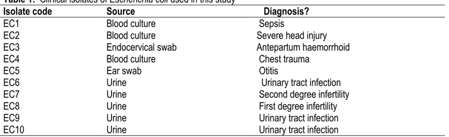
organisms (Table 1) were 10 clinical isolates of E. coli obtained through due process from the Medical Microbiology Laboratory, University College Hospital (UCH), Ibadan, Nigeria. The isolates were maintained on nutrient broth (Difco Laboratories, USA).
Table 1: Clinical isolates of Escherichia coli used in this study
Isolate code Source Diagnosis?
EC1 Blood culture Sepsis
EC2 Blood culture Severe head injury
EC3 Endocervical swab Antepartum haemorrhoid
EC4 Blood culture Chest trauma
EC5 Ear swab Otitis
EC6 Urine Urinary tract infection
EC7 Urine Second degree infertility
EC8 Urine First degree infertility
EC9 Urine Urinary tract infection
EC10 Urine Urinary tract infection
Antibacterial assay of plant extracts: The agar-well
diffusion method was used for the antibacterial screening. The isolates were grown in nutrient broth for 18 h at 37oC.
Six concentrations of each isolate of E. coli were prepared from the broth in sterile distilled water to give a range of concentrations at 1 × 10⁻¹ to 1 × 10⁻⁶ cfu/ ml via
serial dilution method prior to use. 1ml of the inoculum was thoroughly mixed with 19 ml of sterile nutrient agar and poured into sterile Petri dish. The agar was left to solidify. Two wells of 6 mm in diameter were punctured in each agar plate and 60 μl of each extract was filled into the wells with the aid of a sterile micropipette. Sterile distilled water was used instead of extract in the control
experiment. Also plates containing the test organisms in agar without extract were used as control. All experiments were done aseptically and each experiment was replicated three times. The plates were incubated at 37oC
for 36 - 48 h. Readings were taken after 36 h and 48 h. The zone of inhibition was measured in millimetres (mm).
Statistical analysis of data: All data were statistically
analysed using One-way Analysis of Variance (ANOVA) and expressed as mean ± SD. The Duncan Multiple Range Test (DMRT) was used to test means for significance (P ˂ 0.05).
RESULTS AND DISCUSSION
The two plants contained alkaloids, saponins, tannins, phenols and glycosides (Table 2). The phytochemical constituents of T. glaucescens have been reported by previous authors (Adebayo & Ishola, 2009; Oshomoh &
Idu, 2011). T. glaucescens contains tannins and astringents (Burkill, 1985). The therapeutic value of the two plants in folk medicine might be attributed to their tannin content.
Table 2: Phytochemical constituents of Anogeissus leiocarpus and Terminalia glaucescens
Plant sample Alkaloids Saponins Tannins Phenols Glycosides
A. leiocarpus + + + + +
T. glaucescens + + + + +
+ = present
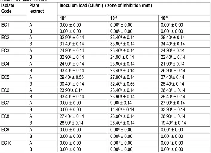
The antibacterial activity of water extracts of the plant samples is presented in Table 3. The E. coli isolates EC1, EC9 and EC10 were not susceptible to the water extracts
of the two plants. Also not susceptible was isolate EC7 at 10-1 cfu/ml inoculum concentration. The highest inhibitory
6649
(33.40 mm) against isolates EC4 and EC6, and the least
(23.90 mm) was for A. leiocarpus also against EC6 at 10-1
cfu/ml. T. glaucescens had the highest (33.90 mm) activity against EC2 at 10-3 cfu/ml and A. leiocarpus (9.90
mm) the least against EC7. The highest (34.40 mm)
activity at 10-5 cfu/ml was observed for T. glaucescens
against EC2 and the least (19.40 mm) against EC8 was for the same plant. Overall, EC2 was the most
susceptible to water extract of T. glaucescens (10-3 and
10-5 cfu/ml).
Table 3: Inhibitory activity of water extracts of Anogeissus leiocarpus and Terminalia glaucescens against ten clinical
isolates of Escherichia coli
Isolate
Code Plant extract Inoculum load (cfu/ml) / zone of inhibition (mm)
10-1 10-3 10-5 EC1 A 0.00j ± 0.00 0.00k ± 0.00 0.00n ± 0.00 B 0.00j ± 0.00 0.00k ± 0.00 0.00n ± 0.00 EC2 A 32.90b ± 0.14 23.40d ± 0.14 28.40d ± 0.14 B 31.40c ± 0.14 33.90a ± 0.14 34.40a ± 0.14 EC3 A 24.90h ± 0.14 23.40h ± 0.14 24.90j ± 0.14 B 32.90b ± 0.14 24.90f ± 0.14 22.40k ± 0.14 EC4 A 24.90h ± 0.14 23.90g ± 0.14 21.90l ± 0.14 B 33.40a ± 0.14 28.40c ± 0.14 26.90g ± 0.14 EC5 A 29.40e ± 0.56 27.90d ± 0.14 27.40f ± 0.14 B 30.40d ± 0.14 32.40b ± 0.56 25.40i ± 0.14 EC6 A 23.90i ± 0.14 23.40h ± 0.14 26.40h ± 0.14 B 33.40a ± 0.14 23.90g ± 0.14 29.40c ± 0.14 EC7 A 0.00j ± 0.00 9.90j ± 0.14 27.90e ± 0.14 B 0.00j ± 0.00 14.40e ± 0.14 33.90b ± 0.14 EC8 A 27.40g ± 0.14 23.90g ± 0.14 26.90g ± 0.14 B 28.90f ± 0.14 26.40e ± 0.14 19.40m ± 0.14 EC9 A 0.00j ± 0.00 0.00k ± 0.00 0.00n ± 0.00 B 0.00j ± 0.00 0.00k ± 0.00 0.00n ± 0.00 EC10 A 0.00j ± 0.00 0.00 k± 0.00 0.00 n± 0.00 B 0.00j ± 0.00 0.00k ± 0.00 0.00n ± 0.00
*Value = Mean ± standard deviation. Values within a column followed by the same superscript are not significantly different at P < 0.05. Diameter of the cork borer = 6.0 mm. A = Anogeissus leiocarpus extract. B = Terminalia
glaucescens extract.
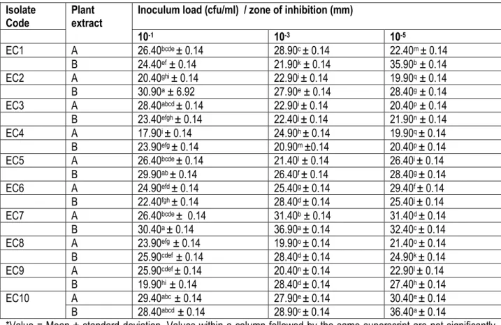
In Table 4, all isolates were susceptible to the ethanol extracts of the two plants at all inoculum concentrations. The highest antibacterial activity was observed for T.
glaucescens (30.90 mm) against EC2 and the least
(17.90 mm) was for A. leiocarpus against EC4 at 10-1
cfu/ml of all isolates. The highest (36.90 mm) antibacterial effect was observed for T. glaucescens against EC7 and
the least (19.90 mm) for A. leiocarpus against EC8 at 10-3
cfu/ml of all isolates. At 10-5 cfu/ml, T. glaucescens (36.40
mm) was most active against EC10 and the least (19.90 mm) activity was from A. leiocarpus against EC2 and EC4. Generally, isolate EC7 was the most susceptible to the ethanol extract of T. glaucescens at high inoculums concentrations (10-1 and 10-3 cfu/ml).
6650
Table 4: Inhibitory activity of ethanol extracts of Anogeissus leiocarpus and Terminalia glaucescens against ten clinical
isolates of Escherichia coli
Isolate
Code Plant extract Inoculum load (cfu/ml) / zone of inhibition (mm)
10-1 10-3 10-5 EC1 A 26.40bcde ± 0.14 28.90c ± 0.14 22.40m ± 0.14 B 24.40ef ± 0.14 21.90k ± 0.14 35.90b ± 0.14 EC2 A 20.40ghi ± 0.14 22.90i ± 0.14 19.90q ± 0.14 B 30.90a ± 6.92 27.90e ± 0.14 28.40g ± 0.14 EC3 A 28.40abcd ± 0.14 22.90i ± 0.14 20.40p ± 0.14 B 23.40efgh ± 0.14 22.40j ± 0.14 21.90n ± 0.14 EC4 A 17.90i ± 0.14 24.90h ± 0.14 19.90q ± 0.14 B 23.90efg ± 0.14 20.90m ±0.14 20.40p ± 0.14 EC5 A 26.40bcde ± 0.14 21.40l ± 0.14 26.40i ± 0.14 B 29.90ab ± 0.14 26.40f ± 0.14 28.40g ± 0.14 EC6 A 24.90efd ± 0.14 25.40g ± 0.14 29.40f ± 0.14 B 22.40fgh ± 0.14 28.40d ± 0.14 25.40j ± 0.14 EC7 A 26.40bcde ± 0.14 31.40b ± 0.14 31.40d ± 0.14 B 30.40a ± 0.14 36.90a ± 0.14 32.40c ± 0.14 EC8 A 23.90efg ± 0.14 19.90o ± 0.14 21.40o ± 0.14 B 25.90cdef ± 0.14 28.40d ± 0.14 24.90k ± 0.14 EC9 A 25.90cdef ± 0.14 20.40n ± 0.14 22.90l ± 0.14 B 19.90hi ± 0.14 28.40d ± 0.14 27.40h ± 0.14 EC10 A 29.40abc ± 0.14 27.90e ± 0.14 30.40e ± 0.14 B 28.40abcd ± 0.14 28.90c ± 0.14 36.40a ± 0.14
*Value = Mean ± standard deviation. Values within a column followed by the same superscript are not significantly different at P < 0.05. Diameter of the cork borer = 6.0 mm. A = Anogeissus leiocarpus extract. B = Terminalia
glaucescens extract.
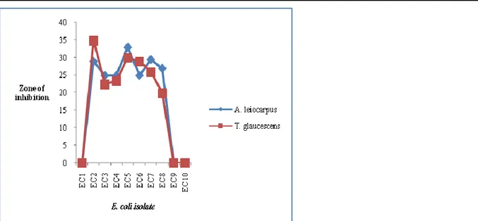
Fig. 1 shows the comparative antibacterial activity of water extracts of A. leiocarpus and T. glaucescens against all isolates at 10-6 cfu/ml. The extracts of the two
plants were inactive on EC1, EC9 and EC10. Of the remaining isolates (7), A. leiocarpus had higher activity against 5 isolates (EC3, EC4, EC5, EC7 and EC8). A.
leiocarpus is traditionally acclaimed to be effective in
treating infectious diseases in man and animals (Dweek, 1996). Overall, the water extract of A. leiocarpus was more active than that of T. glaucescens at 10-6 cfu/ml.
The antibacterial activity of the ethanol extracts of the two plants against all isolates at 10-6 cfu/ml is presented in
Fig. 2. T. glaucescens showed higher activity against all isolates than A. leiocarpus. The finding of this study on antibacterial activity of T. glaucescens is in line with the reports of previous authors. The extracts of T.
glaucescens demonstrated significant activity against
bacterial and fungal isolates; at 100mg/ml, the root extract was more active than the leaf extract against test organisms (P. aeruginosa, B. anthracis, S. aureus, C.
albicans, K. pneumoniae, E. coli, S. typhi and Proteus
6651
Figure 1: Comparative antibacterial activity of water extracts of Anogeissus leiocarpus and Terminalia glaucescens
against isolates of E. coli at 10-6 cfu/ml
Figure 2: Comparative antibacterial activity of ethanol extracts of Anogeissus leiocarpus and Terminalia glaucescens
against isolates of E. coli at 10-6 cfu/ml
The methanol extract (10 mg/ml) of T. glaucescens roots showed significant inhibitory activity against E. coli, it also inhibited the growth of K. pneumoniae, P. mirabilis and C
albicans at 60mg/ml (Adebayo & Ishola, 2009). In a study
on the antimicrobial activities of the aqueous and ethanol extracts of T. glaucescens against dental caries causing microorganisms, Oshomoh and Idu (2011) reported that the stem showed a significantly higher antimicrobial activity against S. aureus and Streptococcus mutans at a lower concentration of 3.13 mg/ml. The efficacy of the
antimicrobial activities of T. glaucescens plant has made both parts (stem and root) suitable for better dental care. There is scarcity of information on the antimicrobial activity of A. leiocarpus in the literature. However, Andary
et al., [2005] reported that the extracts of the different
parts of A. leiocarpus showed antimicrobial activity against pathogenic fungi and bacteria. Furthermore, the roots as chewing sticks have broad spectrum antibacterial properties against tooth and gum infections.
CONCLUSION
The phytochemical constituents of A. leiocarpus and T.
glaucescens could be responsible for their therapeutic
values in human pathogenic infections. The water and
ethanol extracts of the two plants showed significant antibacterial activity against pathogenic isolates of E. coli from clinical sources (blood, urine, ear and endocervical
6652
swab). The isolation of active compounds from the two
plants may lead to the discovery of novel drugs that may be useful in combating resistant strains of E. coli.
REFERENCES
Adebayo EA and Ishola OR, 2009. Phytochemical and antimicrobial screening of crude extracts from the root, stem bark, and leaves of Terminalia
glaucescens. African Journal of Pharmacy and Pharmacology 3(5): 217-221.
Ahmad I and Beg AZ, 2001. Antimicrobial and phytochemical studies on 45 Indian medicinal plants against multi-drug resistant human pathogens. Journal of Ethnopharmacology 74: 113–123.
Andary C, Doumbia B, Sauvan N, Olivier M, Garcia M, 2005. Anogeissus leiocarpa (DC.) Guill. & Perr. [Internet] Record from PROTA4U. Jansen, P.C.M. & Cardon, D. (Editors). PROTA (Plant Resources of Tropical Africa / Ressources végétales de l’Afrique tropicale), Wageningen, Netherlands.
<http://www.prota4u.org/search.asp>. Accessed 30 July 2013.
AOAC, 2005. Official Methods of Analysis. 18th edition.
Association of Official Analytical Chemists, Washington, DC., USA.
Ayepola OO, 2009. Evaluation of the antimicrobial activity of root and leaf extracts of Terminalia
glaucescens. Advances in Natural and Applied Sciences 3(2): 188-191.
Bailey JK, Pinyon JL, Anantham S, Hall RM, 2010. Commensal Escherichia coli of healthy humans:
A reservoir for antibiotic-resistance
determinants. Journal Medical Microbiology 59: 1331-1339.
Burkill HM, 1985. The useful plants of West Tropical Africa. 2nd edition. Royal Botanic Gardens, Kew,
UK.
Dweek AA, 1996. Plant for Africa. Part 2. http://www.dweek data.Co.uk/published papers. Hordijk J, Veldman K, Dierikx C, van Essen-Zandbergen A, Wagenaar JA, Mevius D, 2011. Prevalence and characteristics of quinolone resistance in
Escherichia coli in veal calves. Veterinary Microbiology 156(1-2): 136-142.
Khan R, Islam B, Akram M, Shakil S, Ahmad A, Ali SM, Mand MS, Khan AU, 2009. Antimicrobial activity of five herbal extracts against multi drug resistant (MDR) strains of bacteria and fungus of clinical Origin. Molecules 14: 586-597.
Laupland KB, Church DL, Vidakovich J, Mucenski M, Pitout JD, 2008. Community-onset extended-spectrum β-lactamase (ESBL)-producing
Escherichia coli: importance of international
travel. Journal of Infectious Diseases 57: 441-448.
Mesa RJ, Blanc V, Blanch AR, Cortés P, González JJ, Lavilla S, et al., 2006. Extended-spectrum β-lactamase-producing Enterobacteriaceae in different environments (humans, food, animal farms and sewage). Journal of Antimicrobial
Chemotherapy 58: 211–215.
Neugebauer J, 1983. Atlas of Infectious Diseases. ROCHE, Switzerland.
Ogundiya MO, A.L. Kolapo AL, Okunade MB, Adejumobi JA, 2008. Evaluation of phytochemical composition and antimicrobial activity of
Terminalia glaucescens against some oral
pathogens. Advances in Natural and Applied
Sciences 2(2): 89-93.
Ojo OO, Nadro MS, Tella IO, (2006). Protection of rats by extracts of some common Nigerian trees against acetaminophen-induced hepatotoxicity. African
Journal of Biotechnology 5(9): 755-760.
Okunade MB, Adejumobi JA, Ogundiya MO, Kolapo AL, 2007. Chemical, phytochemical compositions and antimicrobial activities of some local chewing sticks used in south western Nigeria.
Journal Phytopharmacotherapy and Natural products 1(1): 49-52.
Oshomoh EO and Idu M, 2011. Antimicrobial activities of the aqueous and ethanol extracts of the root and stem of Terminalia glaucescens against Dental caries causing microorganisms. International
Journal of Medicinal and Aromatic Plants 1(3):
287 – 293.
Raj G, George V, Pradeep NS, Sethuraman MG, 2008. Chemical composition, antimicrobial activity of the leaf oil from Syzygium gardneri Thw.
Journal of Essential Oil Research 20: 72–74.
Sekeroglu NS, Deveci M, Buruk CK, Gurbuz B, Ipek A, 2007. Chemical composition and antimicrobial activity of Anzer tea essential oil. Journal of the
Science of Food and Agriculture 87: 1424–1426.
Sonibare MA and Gbile ZO, 2008. Ethnobotanical survey of anti-asthmatic plants in South Western Nigeria. African Journal of Traditional,
6653
Complimentary and Alternative Medicines 5(4):340 – 345.
World Health Organization (WHO), 2011. Antimicrobial
Resistance. WHO: Geneva, Switzerland.
Yagupsky P, 2006. Selection of antibiotic-resistant pathogens in the community. Pediatric Infectious