i
Université de Montréal
A proteome-wide strategy reveals a novel
mechanism of control of cell cycle progression
through modulation of cyclin mRNA stability
par Vincent Messier Département de biochimie Programme de Biochimie Faculté de médecine Université de Montréal
Thèse présentée à la faculté des études supérieures en vue de l'obtention du grade Philosophiae Doctor (Ph.D)
en biochimie
8 February 2013
ii
Université de Montréal Faculté des études supérieures
Cette thèse s'intitule:
A proteome-wide strategy reveals a novel
mechanism of control of cell cycle progression
through modulation of cyclin mRNA stability
Présentée par:
Vincent Messier
a été évaluée par un juré composé des personnes suivantes:
Pascal Chartrand Président-rapporteur Stephen Michnick Directeur des recherches
Damien D'Amours Membre du juré
Jan Skotheim Examinateur externe
iii
Sommaire
La quantité de données générée dans le cadre d'étude à grande échelle du réseau d'interaction protéine-protéine dépasse notre capacité à les analyser et à comprendre leur sens; d'une part, par leur complexité et leur volume, et d'un autre part, par la qualité du jeu de donnée produit qui semble bondé de faux positifs et de faux négatifs. Cette dissertation décrit une nouvelle méthode de criblage des interactions physique entre protéines à haut débit chez Saccharomyces cerevisiae, la complémentation de fragments protéiques (PCA). Cette approche est accomplie dans des cellules intactes dans les conditions natives des protéines; sous leur promoteur endogène et dans le respect des contextes de modifications post-traductionnelles et de localisations subcellulaires. Une application biologique de cette méthode a permis de démontrer la capacité de ce système rapporteur à répondre aux questions d'adaptation cellulaire à des stress, comme la famine en nutriments et un traitement à une drogue.
Dans le premier chapitre de cette dissertation, nous avons présenté un criblage des paires d'interactions entre les protéines résultant des quelques 6000 cadres de lecture de Saccharomyces cerevisiae. Nous avons identifié 2770 interactions entre 1124 protéines. Nous avons estimé la qualité de notre criblage en le comparant à d'autres banques d'interaction. Nous avons réalisé que la majorité de nos interactions sont nouvelles, alors que le chevauchement avec les données des autres méthodes est large. Nous avons pris cette opportunité pour caractériser les facteurs déterminants dans la détection d'une interaction
iv
par PCA. Nous avons remarqué que notre approche est sous une contrainte stérique provenant de la nécessité des fragments rapporteurs à pouvoir se rejoindre dans l'espace cellulaire afin de récupérer l'activité observable de la sonde d'interaction. L'intégration de nos résultats aux connaissances des dynamiques de régulations génétiques et des modifications protéiques nous dirigera vers une meilleure compréhension des processus cellulaires complexes orchestrés aux niveaux moléculaires et structuraux dans les cellules vivantes.
Nous avons appliqué notre méthode aux réarrangements dynamiques opérant durant l'adaptation de la cellule à des stress, comme la famine en nutriments et le traitement à une drogue. Cette investigation fait le détail de notre second chapitre. Nous avons déterminé de cette manière que l'équilibre entre les formes phosphorylées et déphosphorylées de l'arginine méthyltransférase de Saccharomyces cerevisiae, Hmt1, régulait du même coup sont assemblage en hexamère et son activité enzymatique. L'activité d'Hmt1 a directement un impact dans la progression du cycle cellulaire durant un stress, stabilisant les transcrits de CLB2 et permettant la synthèse de Cln3p. Nous avons utilisé notre criblage afin de déterminer les régulateurs de la phosphorylation d'Hmt1 dans un contexte de traitement à la rapamycin, un inhibiteur de la kinase cible de la rapamycin (TOR). Nous avons identifié la sous-unité catalytique de la phosphatase PP2a, Pph22, activé par l'inhibition de la kinase TOR et la kinase Dbf2, activé durant l'entrée en mitose de la cellule, comme la phosphatase et la kinase responsable de la modification d'Hmt1 et de ses fonctions de régulations dans le cycle cellulaire. Cette approche peut être
v
généralisée afin d'identifier et de lier mécanistiquement les gènes, incluant ceux n'ayant aucune fonction connue, à tout processus cellulaire, comme les mécanismes régulant l'ARNm.
vi
Abstract
The quantity of data generated within the framework of protein-protein interaction network large-scale studies exceeds our capacity to analyze them and to understand their meaning; on one hand, by their complexity and their number, and on the other hand, by the quality of the produced data, which are populated with spurious interactions. This dissertation describes new applications of a protein-fragments complementation assay (PCA) to screen for interactions among all proteins in the budding yeast Saccharomyces cerevisiae. This approach is carried out in intact cells, with proteins expressed in their native contexts and under their endogenous promoter, thus assuring correct post-translational modifications and subcellular localization. A further novel application of PCA is described for investigating proteome wide changes in response to cellular adaptation to stresses, such as nutrient starvations and drug treatments. Finally, as a result of the latter strategy applied to characterizing proteome-wide response to the immunosuppressant drug, rapamycin, I describe the discovery of an unforeseen mechanism of modulating cell cycle progression through control of cyclin mRNA stability.
In the first chapter of this dissertation, I present a pairwise screen of interactions among proteins resulting from the ~6000 open reading frames in Saccharomyces cerevisiae. We identified 2770 interactions among 1124 proteins. We estimated the quality of our screen by comparing our results to curated gold standard data and coverage of known interactions to all previous
vii
studies. The majority of our interactions were novel, but overlap with data from previous studies was as high as 40%. PCA is based on refolding of the reporter protein from complementary N- and C- terminal fragments following interaction of the two proteins to which they are fused. Thus, reporter activity is sterrically limited to interactions in which the termini of the proteins to which the complementary reporter fragments are fused are sufficiently close in space. In the case of our reporter, this limit was 8 nm. Thus PCA is a molecular ruler, providing information on both direct protein-protein interactions and sterrically restricted distances between proteins in complexes. We benchmarked and demonstrated correct topological relationships for a number of known complexes, including the proteasome, RNA polymerase II and the nuclear pore complex. Thus our study provided, for the first time, a topological map of complex organization in a living cell. The integration of the results from such efforts with those of gene regulation dynamics and protein modifications will lead to a fuller understanding of how complex cellular processes are orchestrated at a molecular and structural level in the living cell.
In chapter 2, I describe the results of an application of PCA to study the dynamic rearrangement of the proteome under a specific stress; treatment of cells with rapamycin. The results of these efforts were the identification of a novel mechanism of cell cycle control at the level of cyclin mRNA. Specifically, we discovered that the balance between the phosphorylated and dephosphorylated forms of the Saccharomyces cerevisiae arginine methyltransferase, Hmt1, regulates both its assembly into a hexamer and its
viii
enzymatic activity. The Hmt1 activity modulates cell cycle progression through stabilizing the B cyclin CLB2 mRNA. We then used PCA to identify the Hmt1 regulators under rapamycin treatment. We identified the catalytic subunit of the PP2a phosphatase, Pph22, activated by the inhibition of TOR, and the kinase Dbf2, activated during entry into mitosis, as the phosphatase and the kinase responsible for the modification of Hmt1 and for its regulatory functions in the cell cycle.
I thus, in the end close the circle I began in this summary, going from large-scale discovery of protein-protein interactions, to mapping dynamics of proteome changes during an adaptation and finally to mechanistic insight into a primordial control mechanism in cellular dynamics. The strategies that we devised to discover this mechanism can be generalized to identify and mechanistically link genes together, including those of unknown function, to any cellular process.
ix
Table of contents
Sommaire ... iii
Abstract ... vi
Table of contents... ix
Table of figures ... xiv
Abbreviations ... xvii
Amino acids codes ... xx
Acknowledgements... xxi
Dedication ... xxv
Overview... 1
1.Chapter 1: An in Vivo Map of the Yeast Protein Interactome ... 4
1.1.Overview ... 4
Background ... 10
1.2. Abstract ... 12
1.3. Introduction and results ... 13
1.3.1.Genome-wide in vivo screen. ... 14
1.3.2.Data filtering, quality assessment, and overlap with existing PINs. . 16
1.3.3.General organization of the yeast DHFR PCA PIN. ... 29
1.3.4.Global structure and topology of the in vivo interactome. ... 32
1.3.5.Bird’s-eye view of the yeast in vivo PIN. ... 37
1.3.6.In vivo network at the bud neck... 41
x
1.4.Conclusions. ... 45
1.5. Methods ... 48
1.5.1. Adaptation of the mDHFR Protein-fragment Complementation Assay for studies in yeast. ... 48
1.5.2. Creation of universal DHFR PCA fragment templates and creation of homologous recombination cassettes. ... 52
1.5.3. Oligonucleotide design and synthesis. ... 54
1.5.4. Creation of homologous recombination cassettes ... 55
1.5.5. Creation of recombinant strains ... 56
1.5.6. Optimization of DHFR PCA screening conditions ... 58
1.5.7. Test for detection of structural and topological organization of a protein complex ... 59
1.5.8. Large scale PPI screen ... 61
1.5.9. Control experiments for spontaneous, interaction-independent DHFR protein fragment complementation. ... 64
1.5.10. Data acquisition, colony quantification and documentation, and statistical analyses. ... 66
1.5.11. Analysis of colony intensity distributions and benchmarking. ... 71
1.5.12. Analysis of high-quality PPI in comparison to protein abundance, gene ontology enrichment and three-dimensional structures. ... 78
xi
1.5.13. Comparison of DHFR PCA to previously determined PPI. ... 82
1.5.14. Overlap with previous large-scale studies. ... 83
1.5.15. Clustering of high confidence interactions. ... 85
1.5.16. GO enrichment. ... 86
2.Chapter 2: A pathway that controls early M-phase progression by regulation of CLB2 mRNA stability via methylation of hnRNPs ... 88
2.1. Overview ... 89
Background ... 99
2.2. Highlights ... 101
2.3. Introduction ... 102
2.4.Results ... 106
2.4.1. CLB2 mRNA fail to accumulate in early M-phase in cells treated with rapamycin ... 106
2.4.2.Rapamycin causes sequestering of hnRNPs in the nucleus ... 108
2.4.3.Rapamycin does not affect Hmt1 activity through changes in expression or localization ... 112
2.4.4.Rapamycin prevents homo-oligomerization and activation of Hmt1 ... 115
2.4.5.Hmt1 phosphorylation is essential for its homo-oligomerization ... 116
2.4.6.Phosphorylation of Hmt1 is required to regulate CLB2 mRNA stability ... 121
xii
2.4.7.hnRNP nuclear sequestering is dependent on Hmt1 phosphorylation
... 124
2.4.8.Methyltransferase activity of Hmt1 is regulated by its phosphorylation ... 125
2.4.9.Rapamycin-dependant Hmt1 protein-protein interactions include potential kinases and phosphatases ... 126
2.4.10.Hmt1 phosphorylation and oligomerization are regulated by Dbf2 kinase and Pph22 phosphatase ... 131
2.4.11.Rapamycin induces cooperative disassembly of Dbf2-Hmt1 and Hmt1-Hmt1 complexes but graded assembly of Hmt1-Pph22 ... 136
2.4.12.Dbf2 activity increases and Pph22 activity decreases CLB2 mRNA stability ... 137
2.4.13.Failure to accumulate CLB2 mRNA correlates with early M-phase progression delay ... 138
2.5.Discussion ... 139
2.6.Acknowledgement ... 150
2.7.Experimental Procedures ... 151
2.7.1.Yeast strains, plasmids, growth conditions and buffers ... 151
Growth conditions ... 151
Cell using α-factor ... 152
xiii
2.7.2.FISH probes and procedures ... 155
2.7.3.Strains, plasmids, growth conditions, buffers and primers ... 156
2.7.4.Single cell, single molecule Fluorescence in situ Hybridization (FISH) ... 156
2.7.5.Fluorescence-Activated Cell Sorter (FACS) analysis of strains expressing GFP-tagged proteins ... 156
2.7.6.Fluorescence-Activated Cell Sorter (FACS) analysis of cell DNA content ... 157
2.7.7.Fluorescent protein localization ... 158
2.7.8.Microscopy ... 159
2.7.9.Hmt1 DHFR PCA large-scale screen ... 159
2.7.10.TAP-tagged protein immunoprecipitation... 160
2.7.11.Hmt1 methyltransferase assay ... 161
2.7.12.Rluc PCA luminescence detection ... 162
2.7.13.Purification of Dbf2-Mob1 kinase complex ... 163
2.7.14.Protein kinase assay... 163
3.Chapter 3: Discussion ... 164
3.1.Implications and future directions... 164
xiv
Table of figures
FIGURE 1IN VIVO PCA SCREEN OF THE YEAST PIN. ... 15
FIGURE 2:IN VIVO PCA SCREEN OF THE YEAST PROTEIN INTERACTION NETWORK. ... 17
FIGURE 3:AUTOMATED EXTRACTION OF COLONY INTENSITIES ON PLATES. ... 18
FIGURE 4:DISTRIBUTION OF COLONY INTENSITIES ON PLATES... 19
FIGURE 5:REPRODUCIBILITY OF THE SCREENING PROCESS. ... 20
FIGURE 6:THE DHFRPCA IS REVERSIBLE. ... 21
FIGURE 7.DISTRIBUTION OF PROTEIN ABUNDANCE. ... 23
FIGURE 8:QUALITY ASSESSMENT FOR THE PCA NETWORKS AND OTHER PPI NETWORKS. ... 24
FIGURE 9:OVERLAP OF THE DHFRPCA NETWORK WITH OTHER LARGE-SCALE EXPERIMENTS. ... 26
FIGURE 10:OVERLAP OF THE DHFRPCA NETWORK WITH OTHER LARGE-SCALE EXPERIMENTS. ... 27
FIGURE 11.INTERACTIONS ARE ENRICHED WITHIN GO CATEGORIES. ... 30
FIGURE 12:INTERACTIONS ARE ENRICHED WITHIN GENE ONTOLOGY CATEGORIES. ... 31
FIGURE 13.RNA POLYMERASE II COMPLEX RECONSTITUTION THROUGH DHFR PCA. ... 34
xv
FIGURE 14.THE DHFRPCA RESULTS PROVIDE STRUCTURAL AND TOPOLOGICAL INSIGHTS. ... 35
FIGURE 15.THE DHFRPCA NETWORK IS MODULAR AND INTERCONNECTED. ... 38
FIGURE 16.THE YEAST DHFRPCA NETWORK PROVIDES INSIGHTS INTO BOTH CELL POLARITY AND AUTOPHAGY. ... 40
FIGURE 17:NETWORK REPRESENTATION OF A PROTEIN INTERACTION
REGULATORY CASCADE. ... 92
FIGURE 18:THE EVENTS OF THE EUKARYOTIC CELL CYCLE ... 93
FIGURE 19:CLB2 MRNA FAIL TO ACCUMULATE IN EARLY M-PHASE IN CELLS TREATED WITH RAPAMYCIN. ... 107
FIGURE 20: HNRNPS ARE SEQUESTERED IN THE NUCLEUS IN RAPAMYCIN
TREATED CELLS. ... 110
FIGURE 21:OTHER KARYOPHERINS AND MRNA NUCLEAR EXPORT ELEMENTS ARE HOMOGENOUSLY AFFECTED IN RAPAMYCIN TREATED CELLS. ... 111
FIGURE 22:BOTH MODIFICATIONS AND HOMO-OLIGOMERIC AFFINITIES ARE LOST ON HMT1 IN CELL TREATED WITH RAPAMYCIN. ... 113
FIGURE 23:HMT1 PHOSPHORYLATION OF SERINE 9 IS ESSENTIAL AND REQUIRED FOR HMT1 OLIGOMERIZATION, METHYLTRANSFERASE ACTIVITY AND RESULTING CLB2 MRNA ACCUMULATION IN EARLY M-PHASE AND HNRNP NUCLEAR EXPORTS. ... 118
FIGURE 24:HMT1 PHOSPHORYLATION OF SERINE 9 IS ESSENTIAL AND REQUIRED FOR HMT1OLIGOMERISATION, METHYLTRANSFERASE ACTIVITY CLB2
xvi
MRNA ACCUMULATION IN EARLY M-PHASE AND HNRNP NUCLEAR
EXPORTS. ... 123
FIGURE 25:DBF2 KINASE AND PPH22 PHOSPHATASE REGULATE HMT1 PHOSPHORYLATION STATE AND RELATED ACTIVITY ON CLB2
ACCUMULATION DURING EARLY M-PHASE DIFFERENTIALLY IN RAPAMYCIN TREATED CELLS. ... 129
FIGURE 26:DBF2 KINASE AND PPH22 PHOSPHATASE REGULATE HMT1 PHOSPHORYLATION STATE AND RELATED ACTIVITY ON CLB2
ACCUMULATION DURING EARLY M-PHASE DIFFERENTIALLY IN RAPAMYCIN TREATED CELLS. ... 134
FIGURE 27:HMT1 REGULATIONS ARE NECESSARY FOR M PHASE PROGRESSION AND INDUCED EARLY M PHASE DELAYED IN RAPAMYCIN TREATED CELLS.
... 140 FIGURE 28:PROPOSED MODELS ... 142
xvii
Abbreviations
RBD Ras binding domain on Raf
mRNA messenger Ribonucleic acid
DNA Deoxyribonucleic acid
NMR Nuclear Magnetic Resonance
S phase Synthesis phase
M phase Mitosis phase
G1 Gap 1
G2 Gap 2
G0 Gap 0
Cdk Cyclin dependant kinase
APC Anaphase Promoting Complex
Y2H Yeast two-Hybrid
PCA Protein-fragment Complementation Assay
PIN Protein Interaction Network
hnRNP heterogeneous nuclear RiboNucleoProtein
xviii
DHFR DiHydroFolate Reductase
Rluc Renilla LUCiferase
PKA Protein Kinase A
cAMP cyclic Adenosine MonoPhosphate
PPI Protein-Protein Interaction
ORF Open Reading Frame
PPV Positive Predicted Value
SC Synthetic Completed media
YFP Yellow Fluorescent Protein
MIPS Munich Information Center for Protein Sequences
CC Cell Compartment
BP Biological Process
MF Molecular Function
TAP-MS Tandem Affinity Purification coupled Mass Spectrometry
ROC curve Receiver Operating Characteristic curve
xix
ESCRT Endosomal Sorting Complex Required for Transport
MVB MultiVesicular Bodies
xx
Amino acids codes
Amino acid Three letter code One letter code
Alanine ala A
Arginine arg R
Asparagine asn N
Aspartic acid asp D
Cystein cys C
Glutamic acid glu E
Glutamine gln Q Glycine gly G Histidine his H Isoleucine ile I Leucine leu L Lysine lys K Methionine met M Phenylalanine phe F Proline pro P Serine ser S Threonine thr T Tryptophan try W Tyrosine tyr Y Valine val V
xxi
Acknowledgements
During my time in graduate school, I have been in contact with many people who have transformed my views. I thought that graduation would be a simple trip, where I would acquire important knowledge and useful techniques that could help me in my future career. The truth is more complicated and elaborate. As I think of every person that I met, had discussions with, and challenges that I encountered and, more importantly, overcame were by far more important and useful than what university claims to be.
I would like to first thank Dr. Stephen Michnick for his contribution on fostering both my professional and personal maturation. Steve was the best guide for a graduate school trip. While he did the road several times already with other past students, he chooses to do it again with me. The paths were not always easy to get through and, being in Montreal, some were probably under construction. He foresaw obstacles in the way, leaving me to choose between getting through them and finding other ways. Sometimes I stopped following his advice, perhaps thinking that he was lost even more than I was. Luckily for me, he was waiting on the side of the road so that I did something more productive and efficient to get to the end of the journey. Now, being at the end of the trip, I feel that he not only cares that his students present time in his laboratory to be productive, but also he worries even more for their future, as he leaves them with the map for a successful career.
xxii
At the beginning of my doctoral studies, I became acquainted with a young scientist in Mcgill University. She is the 'queen of the scope' and had no troubles to tell me that my first paragraphs in the science draft were just worse than a pile of 's#&t'. Dr Jackie Vogel is not only a talented researcher but also an inspiration to me. I saw a strong woman starting her lab with a sea of ideas, little help, but all the ambition of the world to succeed in it. It was probably not an easy period, but she was ready to share with me and her lab members, advice, cups of coffee, chocolate and conversations that I still value. She taught me microscopy and transmitted to me some of her passion to see these small glowing dots on a black screen. Her generosity in the time she gives to others and the confidence she shares to those who have worked with her contribute significantly to the attitude and motivation of people around, for which I am thankful to have been part of.
I described Dr Daniel Zenklusen as the 'God of FISH' several times, probably because he is. He gave me time and advices to develop FISH proficiency. More importantly, we shared our enthusiasm for the latest project, which would be a dim reflection of what it is now without his input. I admire his capacity to see details, the good and the bad ones. He shapes ideas to push them to their purest form, while optimistically looking at flaws in these diamonds as a continuous chance for improvements. I believe that the future will continue to make our path overlap in many fruitful collaborative efforts.
xxiii
It is inconceivable to me that I could be writing this section without several thoughts for my comrades, present and past members of the Michnick group. I wish to thank you one by one for your support, your enthusiasm and the bonds of friendship that make the Michnick group a special boat to get in to. Po Hien and Mohan for the ride that we shared to get our PhD, the laughs and the talks that came with it. To the old timers of the group, François-Xavier, Alexis and Ingrid, who did it before us and left us with a strong foundation. Louis-Philippe, Sinan and madame Didi, the new ones, for making me feel old and see that I progress through advice that I received and give back to you guys. To Jean-François and Edi for the shared beers, the ski trips and the unique friendship. To Jacqueline for the conversation on ecolo-, grano- and shaggying up life. To Valérie, “les Nathalies”, Jean-Marc, Livia, Émilie and Émily, the past post-doc friends, who shared their time and talents with me. To Luz, Durga, Emmanuelle, Bram, Abdellali and Punam, the new post-doc crowd, who were there during these past tough months. To Stevo, Mercedes, Kirill and Yelena (the yeast group) for your assurance in me while being part of a heroic accomplishment. To Michael and Beat for the shared diners, the conversation and that we feel all at home. To Carole, Ibrahima, Emmanuel and Christian to have been influential during my time in the lab. There will always be a place for you all in my thoughts.
For the time, that I invested in my studies, is a time that I could not always be with you, my family. I wish to thank my parents and my family members. Even if you did not know what I could be doing in the lab, you were
xxiv
understanding of my joy, my pain and all other feelings that could come and go in these years in school. There are no words for my love and admiration that I bear for each of you and only hope that you will always be as proud of me.
To my friends that were there during this time., especially, Karine Grimard, Amélie Forget and Francis Bélanger for their constant genuine friendship, using my ear to share their passion and their own senses to support my taught times. Your company has helped me to preserve my sanity and enjoy life.
I also thank my thesis committee for their advice. Sometimes for the kick that made me think better and work faster. They were supportive in both my academic progression and in the ideas that were discussed in the frame of proposed projects.
Finally, for the evaluation of this dissertation and the interesting discussion that will issue from the upcoming defense, I wish to acknowledge my jury members.
xxv
Dedication
"For many smiles, many tears and the times that we shared laughter, they were always the best."
1
Overview
Proteins are operators in all cellular processes including the cell cycle, the central subject of this thesis. Further, matter, energy and information transfer through biochemical pathways and their organization, are determined by specific protein-protein interactions at every phase of the cell cycle. For example, protein interactions during cell cycle progression such as mitosis are indispensable for the structure of sub-cellular organelles [1], the transport machinery across the various biological membranes [2], the packaging of chromatin [3], the network of microtubule filaments [4], the periodic decay of mitotically regulated molecules [5], and the cyclin-dependent kinase signal transduction [6], the regulation of DNA region by transcription factors [7], to name a few. Protein interactions during cell cycle progression have been the object of intense research for many years due to their importance in development and disease. Aberrant protein bindings are implicated in a number of neurological disorders such as Creutzfeld-Jacob, Alzheimer's disease [8] and cancer, during all developmental stages and metastasis [9]. Although disregulated protein interactions are of interest clinically, the main focus of this study is in the finely tuned recruitment of proteins and molecules involved in the regulation of cell division.
Within cells, proteins interact with other proteins, metabolites and nucleic acids. Protein interactions are measured using a variety of assays, described in method sections of chapters 1 and 2. These assays are based on
2
new innovations in various fields of biochemistry and biophysics, increasing the sensitivity and accuracy in molecular interaction cartography. Recently, many techniques were designed with the aim of scaling up to measure interactions on a proteome-wide level [10-14]. The positive and false-negative rates for networks generated from such high-throughput methods are high, stressing the need for discovering new experimental techniques and new methods that will allow to study protein interactions in intact cell, and to perform studies in various growth conditions and cellular states with endogenously expressed full-length proteins in their native post-translationally modified states and cellular locations. This knowledge could be generated to understand how genetic or environmental perturbations could be linked to these networks in disease development or to uncover how perturbations, such as drugs, of individual network components could reduce or eliminate cellular dysfunctions.
I present here the application in Saccharomyces cerevisiae of a protein-fragments complementation assay (PCA) reporter of protein-protein interactions, that is unique because: (I) it is performed in intact cells and (II) it can be used to address dynamic behaviors of protein complexes under non-toxic conditions. The goal of this dissertation is to demonstrate the limitations, the determining factors in detecting interactions and the strength of this approach in yeast and to show how it can be used to make a substantial and unique contribution to biology. In the first chapter of this dissertation, I focus on describing the PCA in the context of a systematic screen of all pairwise
3
interactions among ~6,000 yeast proteins. In the second chapter, I applied the PCA strategy to understand the mechanism regulating an arginine methyltransferase assembly and activity under stressful conditions, such as starvation and rapamycin-induced TOR kinase inhibition. These enzyme regulations have a direct impact on RNA-binding protein capacities to stabilized cyclin transcripts and overall cell cycle progression. As a result, this strategy can be generalized to identify and mechanistically link genes, including those of unknown function, to any cellular process.
4
1.Chapter 1: An in Vivo Map of the Yeast Protein Interactome
Kirill Tarassov,1* Vincent Messier,1* Christian R. Landry,1,2* Stevo Radinovic,1* Mercedes M.
Serna Molina,1 Igor Shames,1 Yelena Malitskaya,1 Jackie Vogel,3 Howard Bussey,3 Stephen W.
Michnick1,2†. Science, 2008 June 13; 320(5882): 1465-70. Epub 2008 May 8.
1.1.Overview
Communication at the level of the organism requires the transduction of physical cues or chemical information, such as turgor pressure, toxin presence or available nutrient sources, into biochemical signals. Chemical and physical signals are processed by specific interactions among heterologous proteins through non-covalent or covalent modifications. Some of these molecular transducers arose from co-evolution of complementary binding interfaces that specifically interact when simultaneously localized in space and time with corresponding molecular partners, while accumulating mutations that disfavor spurious contacts [15-17]. For example, the Haemophilus influenza SspB protein homodimer evolved a symmetrical binding surface consisting of an α-helix and β-strand forming a hydrophobic cluster between two copies of the molecule [18]. The SspB homomeric interface is essential to bind and target for degradation truncated peptide resulting from stalled ribosome activity upon severe starvation [19]. Computationally design SspB mutations at position 12, 15, 16 and 101 that optimized the stability between the heterodimer mutants, SspBFAFI and SspBLALI, would actually assemble with high stability both into
5
same position that optimize the difference in energy between the heterodimer and both homodimer mutants, SspBLSLA and SspBYGMF, show a 99% specificity
for the heteromeric assembly while bearing lower stability [17]. It is reasonable to think that, as in the design of SspB mutants, protein complex specificity in intact cell is achieved at the cost of stability, rendering in vitro purification conditions a constraint to their detection.
Proteins interact through their interfaces into stable macromolecular structures for which a panel of high-throughput screen strategy were design to capture. The quality of an unbiased PPI screen is composed of two distinct elements: (I) its completeness and (II) its fidelity to reproduce the real PPI network. Protein complexes have been extensively studied by many small-scale techniques and have been curated to create catalogues, such as The Munich Information Center for Protein Sequences (MIPS). These curated protein complexes, such as the nuclear pore, the ribosome, the RNA polymerases, the proteosome, to name a few, can represent bona fides interactions between proteins that could be considered to estimate false-negative discovery rate in a given screen. The completeness of a reported screen can be evaluated by verifying the number of observed interactions within this standard. The total coverage achieved by a screen is determined by both the limitations and sensitivity of the detection method. Therefore, the minimal amount of false-negatives can be reached at the union of the data from different approaches. Equally important, each of these screens should be carried out under conditions that maximize the sensitivity of the detection technique. The fidelity of a PPI
6
method, on the other hand, is more difficult to determine. Unfortunately, there is no clear dataset of protein that would not physically interact. Therefore, a compromise standard set is generated from logical inference; creating imaginary “impossible” interactions by selecting pairs of proteins with divergent expression profiles or mutually exclusive compartmentalization and assuring that none of these imaginary interactions were ever reported in all published data. This approach overestimates the number of impossible interactions, but provides a means to access the false-positive discovery rate.
The fidelity of a protein-protein interaction detection technique can be increased by systematic removal of artifacts. These can be detected by applying appropriate controls to the data, usually by determining technical limitations of a technique that recover false interactions. For PCA, there are two potential technical causes of false-positive interactions. PCA is based on protein interaction-mediated refolding of the reporter fragments to yield an active enzyme [20]. It is, however, possible that in some cases spontaneous folding of reporter from the fragments can occur. An appropriate control for such cases is to probe the proteome with fragments fused to a protein that does not interact with any protein in the cell (e.g. GFP) or the fragment itself, expressed alone. In our screen, we identified approximately 300 proteins that interact with fragment alone, notably highly abundant proteins. A second possibility is that non-specific interactions could be generated due to trapping of complexes between two proteins that normally wouldn’t interact, due to irreversible folding of the reporter protein. In fact, we have shown that this is not the case: PCA reporter
7
proteins do reversibly unfold when protein complexes dissociate for several reporter proteins, but we had not tested this for the reporter used in this study, murine dihydrofolate reductase (DHFR) [21, 22]. We verified the reversibility of DHFR PCA folding using the adenosine 3´,5´-monophosphate–dependent dissociation of the yeast protein kinase A complex as a test system. This heterotetrameric complex has two catalytic and two regulatory subunits. The adenosine 3´,5´-monophosphate (cAMP) binds to and promotes conformational change of the regulatory subunits that release the catalytic subunits. An irreversible reporter folding would retain the catalytic subunit under cAMP treatments. This test insured that we would not report interactions resulting of irreversible trapping by the fragments.
DHFR PCA depends on physical contact between the reporter fragments. This could be limited by the distance between the two protein termini to which the fragments are fused. This is an opportunity to study protein complex membrane spanning protein complex topologies. The structure of the RNA polymerase II is appealing as it is determined at 2.8 Ǻ resolution and contains the largest described variety of protein subunits [23]. We could technically use the linker peptide, separating the subunit of the RNA polymerase II and our reporter fragment, as a molecular ruler that could theoretically give a resolution of the position of complex subunits at the resolution of a single amino acid. Similarly, the reporter fragments cannot refold through a membrane. Therefore, we can use the reporter fragments fused to the C termini of membrane spanning proteins to test for their close proximity
8
and also their orientation into the same cellular compartment [24-27]. These particularities of the PCA would provide complementary information on the nature of proteins and their complexes.
The motivation for determining protein interaction networks is to establish potential protein functions and relationships. Gene annotation is the process of conferring biological information to sequence, consisting of (I) predicting encoded gene sequence in the genome and (II) conferring biological information upon these genes. Gene function annotation is a challenge, given that proteins, in their different states, (e.g. subcellularly localized or post-translationally modified) can be quite versatile, assuming multiple functions at any given time and under different conditions [28]. The difficulty of the gene annotation task is reflected in the number of genes that remain to be annotated. In the popular eukaryotic model, Saccharomyces cerevisiae, almost 30% of the genes are refractory to past annotation efforts [29]. A variety of factors are likely to contribute to the relatively large number of non-annotated yeast genes, including genetic redundancy, lack of strong phenotype, and the possibility that not all genes are real open reading frames (ORF). However, there is a clear need for new approaches and strategies for specific problems, including the characterization of individual genes and their role in nature. For example, the rationale for assigning gene function based on protein interactions relies on a “guilt by association” argument, suggesting that physically interacting proteins shared common functions known a priori for one of the protein. This argument is justified for defined protein complexes, but the accuracy of the argument
9
decreased when (I) there is no a priori annotation for both of the interacting proteins, (II) function shared by the two interacting proteins is novel and differ from any known a priori relationship between functions (e.g. between a transcription factor and a metabolic enzyme) and (III) although the proteins physically interact, these proteins shared no functions. These limitations have not prevented biologist from using these information as a starting point to characterize proteins with unknown functions. For example, using the yeast two-hybrid assay, the YDR016C gene was shown to function with the spindle-pole body as part of the Dam1 complex [11]. It was revealed to be an essential component of the DASH complex required for mitotic spindle integrity [30]. Assembling proteins of unknown function into networks linked to specific cellular processes can thus provide new mechanistic insights and potentially provide new therapeutic targets for the development of drugs.
10 Background
In the following manuscript that was published in an article format in the journal Science, we systematically screened for pairwise protein-protein interactions in Saccharomyces cerevisiae using a protein-fragment complementation assay (PCA) based on the dihydrofolate reductase (DHFR) reporter enzyme. We identified 2770 interactions among 1124 proteins. These interactions were mostly novel on the fact that PCA is efficient to detect protein interaction within and between protein complexes and that we perform the screen on a wider array than previous experiments reported in MIPS and by TAP-MS [31]. We demonstrate that PCA can identify protein complex structure for a crystallized protein complex, in the case of the yeast RNA polymerase II, and globally, originating from all stable crystallized complexes of yeast-homologous proteins deposited in PDB. Then by comparing likelihood that protein interaction can be detected between membrane-spanning proteins and the cell compartment location of their C-termini, we demonstrate topological relationship between these proteins. Finally, using the cyclic adenosine 3´,5´-monophosphate (cAMP) dependent dissociation of the yeast protein kinase A (PKA) complex as a test system to show full reversibility of the DHFR PCA, we insured that the PCA itself does not trap spurious complexes, altering the thermodynamics of binding. The in vivo extended network provides insights into fundamental cellular processes, including cell polarization and autophagy, pathways that are evolutionarily conserved and central to both development and human health.
11
An in Vivo Map of the Yeast Protein Interactome
Kirill Tarassov,1* Vincent Messier,1* Christian R. Landry,1,2* Stevo Radinovic,1* Mercedes
M. Serna Molina,1 Igor Shames,1 Yelena Malitskaya,1 Jackie Vogel,3 Howard Bussey,3 Stephen
W. Michnick1,2†. Science, 2008 June 13; 320(5882): 1465-70. Epub 2008 May 8.
1Département de Biochimie, Université de Montréal.
2Centre Robert-Cedergren, Bio-Informatique et Génomique, Université de
Montréal.
3Department of Biology, McGill University.
*These authors contributed equally to this work. †To whom correspondence should be addressed.
12 1.2. Abstract
Protein interactions regulate the systems-level behavior of cells; thus, deciphering the structure and dynamics of protein interaction networks in their cellular context is a central goal in biology. We have performed a genome-wide in vivo screen for protein-protein interactions in Saccharomyces cerevisiae by means of a protein-fragment complementation assay (PCA). We identified 2770 interactions among 1124 endogenously expressed proteins. Comparison with previous studies confirmed known interactions, but most were not known, revealing a previously unexplored subspace of the yeast protein interactome. The PCA detected structural and topological relationships between proteins, providing an 8-nanometer–resolution map of dynamically interacting complexes in vivo and extended networks that provide insights into fundamental cellular processes, including cell polarization and autophagy, pathways that are evolutionarily conserved and central to both development and human health.
13 1.3. Introduction and results
The elucidation of protein-protein interaction networks (PINs, or interactomes) holds the promise of answering fundamental questions about how the biochemical machinery of cells organizes matter, information, and energy transformations to perform specific functions [32]. An essential and rarely addressed question is whether protein complexes and PINs that are reconstructed or reconstituted in vitro or removed from the normal context in which they are expressed reflect their organization in living cells. For eukaryotes, the test bed for large-scale analysis of PINs is the yeast Saccharomyces cerevisiae, where several PIN analyses have been performed using yeast two-hybrid screens (Y2H) [10, 11] or tandem affinity purification followed by mass spectrometric analyses (TAP-MSs) [12-14]. Each approach captures specific features of protein interactions; two-hybrid methods are best at measuring direct binary interactions between pairs of proteins, whereas affinity purification techniques best capture stable protein complexes. However, neither approach measures interactions between proteins in their natural cellular context, nor are not easily amenable to studying protein complexes that are transiently associated or dynamic under different conditions, that do not survive in vitro purification, or that cannot be transported to the nucleus and form interactions in the absence of other stabilizing interactions as necessitated in Y2H screening. Protein-fragment complementation assays (PCA) provide an alternative approach to detect protein-protein interactions (PPIs) in their natural context. In the PCA strategy, two proteins of interest are fused to
14
complementary fragments of a reporter protein. If the proteins of interest interact physically, the reporter fragments are brought together and fold into their native structure, thus reconstituting the reporter activity of the PCA (Fig. 1A). PCA strategies provide a simple, direct means for the detection of PPIs in vivo, and do so with endogenously expressed full-length proteins in their native post-translationally modified states and cellular locations [33]. Further, PCAs provide spatial and topological information about PPIs. Thus, a large-scale PCA screen would provide direct insights into the global structural organization of PINs as they exist in the living cell.
1.3.1.Genome-wide in vivo screen.
We have performed a systematic binary screen for PPIs at a genome-wide scale in S. cerevisiae using a PCA based on the murine dihydrofolate reductase (mDHFR) assay adapted to yeast describe in section 1.5. (Fig. 1A). The DHFR PCA is a survival-selection assay based on a mutant of mDHFR that is insensitive to the DHFR inhibitor methotrexate but retains full catalytic activity and allows detection of PPIs with as few as 25 to 100 complexes per cell [24, 34]. We created unique homologous recombination cassettes for all 5756 consensus genes with both the F[1,2] and F[3] complementary N- and C-terminal DHFR fragment sequences described in section 1.5..
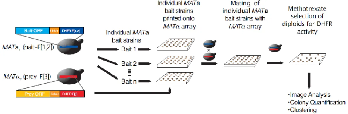
15 Figure 1 In vivo PCA screen of the yeast PIN.
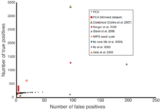
(A) Strategy for high-density array screening of the yeast PIN by DHFR PCA. Both positive [green circles (MATa/α, CDC19 fused to DHFR fragment [1,2] (CDC19-F[1,2]), and MCK1 fused to DHFR fragment [3] (MCK1-F[3]) and negative [red circles (MATa/α, CDC19-F[1,2], and CLN3-F[3])] controls are included on each plate to ensure that each transfer and selection step has occurred correctly. (B) PPV score as a function of raw colony intensity and z score (relative colony intensity on plates). This score represents the ratio of the number of true positive interactions over the sum of the true positive and false positive interactions predicted from the reference sets. (C) The ratio of true positives to false positives in the DHFR PCA network compared with other large-scale data sets [10, 11, 13, 14, 35, 36]. The achieved PPV is indicated above the bars.
16
Successful cassette transformation of S. cerevisiae haploids was achieved for 4326 (75%) open reading frames (ORFs) with the DHFR F[1,2] fragment in MATa and 4804 (83%) ORFs with the DHFR F[3] fragment in MATα strains, with a final combined coverage of 5367 (93%) of all ORFs (table S1, http:// www.sciencemag.org /content /suppl /2008 /05 /08 /1153878.DC1 /1153878s_tables.zip). The entire screening process was performed on solid-phase medium (Fig. 1A and Fig. 2). Briefly, MATa strains (F[1,2] fragment fusions) served as baits and were mated individually with all MATα (F[3]) strains on high-density arrays. The resulting diploids were transferred to a minimal medium [synthetic complete (SC)] plate to select for methotrexate resistance (reconstituted mDHFR activity, with native S. cerevisiae DHFR inhibited), and colony growth was recorded using automated analysis of digital images (Fig. 1A and Figs 3 and 4). PPIs were determined based on the growth of the diploid colonies measured by the pixel intensities on the selection plates (Figs. 2-4). In total, 3247 individual highly reproducible (Fig. 5) bait screens were performed, resulting in more than 15 million individual matings.
1.3.2.Data filtering, quality assessment, and overlap with existing PINs. We experimentally accounted for two potential sources of false positives in a PCA screen: trapping of nonspecific complexes due to irreversible folding of the mDHFR reporter protein, and potential spontaneous complementation (folding) of the DHFR PCA fragments.
17
Figure 2: In vivo PCA screen of the yeast protein interaction network.
Strategy for single bait versus prey array screening of the yeast PIN by DHFR PCA.
18
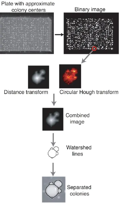
Figure 3: Automated extraction of colony intensities on plates.
The DHFR PCA results were inferred from the growth of diploid colonies on plates containing methotrexate. Images of the plates were taken after a 90-hour growth period with a 4.0 Mega pixel camera (Powershot A520, Canon). Plate images were saved in JPG format at a resolution of 180 dpi and a size of 2,272 × 1,704 pixels. In order to extract the intensity of the colonies, we used available image recognition routines available in the Matlab image analysis toolbox and we modified parameters for it to be able to differentiate colonies that are in proximity to each other. The quality of the position of the grid and the recognition algorithm was examined through visual inspection of all plates.
19
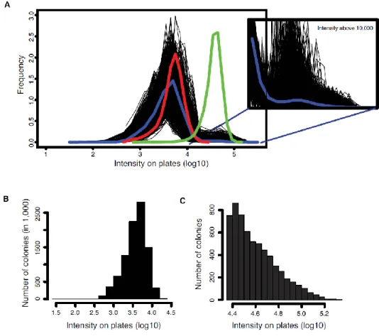
Figure 4: Distribution of colony intensities on plates.
(A) Raw intensities prior to filtering. Black lines represent the intensity distributions on individual plates. The blue line represents the distribution of colony intensities across the entire experiment. The red and green lines represent the intensity distribution of the negative and positive controls respectively. The second panel shows the distribution of colony intensities above 10,000 to illustrate the growth of the methotrexate resistant diploid strains. (B) Intensities of colonies below the threshold. (C) Intensities of colonies above the threshold after filtering positions corresponding to baits and preys that interact with the control fragments.
20
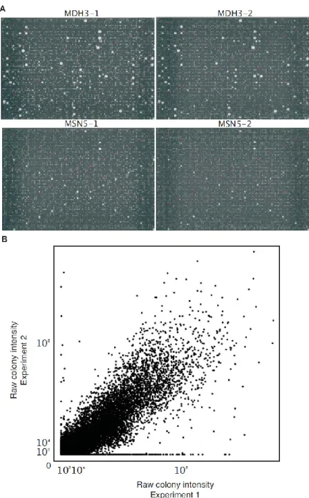
Figure 5: Reproducibility of the screening process.
In order to evaluate the reproducibility of the screening process, we repeated the screen for 48 baits selected for representing the distribution of number of colonies growing above background in the first screen. (A) Example of plates that were repeated. Top row, MDH3 and bottom row, MSN5 used as baits. Green and red circles represent respectively positive and negative controls. (B) Raw colony intensity of experiment two plotted against the raw colony intensity of experiment one (Pearson correlation: 0.86, P < 2.2e-16).
21 Figure 6: The DHFR PCA is reversible.
(A) Upper panel. cAMP-mediated dissociation of the yeast PKA regulatory (Bcy1p) and catalytic (Tpk2p) subunits. Middle panel. Schematic representation of an irreversible PCA for PKA and predicted results of the Bcy1 subunit binding to cAMP-conjugated agarose beads. For the irreversible PCA, Bcy1 and Tpk2 dissociate but remain trapped by the folded PCA reporter protein. Lower panel. For a reversible PCA, reporter protein fragments unfold and dissociate when Bcy1 binds to cAMP-conjugated agarose beads and thus Bcy1 remains bound to the resin while Tpk2 is found in the unbound supernatant fraction. (B) The DHFR PCA is fully reversible. As reported previously [21], the Rluc PCA is reversible; Bcy1 is found in the cAMP-conjugated agarose fraction while Tpk2 is found in the wash. Precisely the same result is found for the DHFR PCA, suggesting that it is reversible, while Venus YFP PCA is irreversible.
22
First, we used the adenosine 3´,5´-monophosphate–dependent dissociation of the yeast protein kinase A complex as a test system [21] to show that the DHFR PCA is fully reversible, and thus the trapping of complexes is unlikely (Fig.6). Second, we screened all the strains against the individual F[1,2] and F[3] complementary fragments or fragment-peptide linker sequences. This allowed us to eliminate 344 promiscuous, highly expressed proteins (Fig. 7 and table S2, http:// www.sciencemag.org /content /suppl /2008 /05 /08 /1153878.DC1 /1153878s_tables.zip), several of which are also often observed as false-positives in affinity purifications. We next identified a threshold of colony intensity above which we could infer PPI. The Munich Information Center for Protein Sequences (MIPS) complexes were used as a standard set of true positives, along with 266,858 true negative interactions between proteins expressed in different cellular compartments or having negatively correlated expression [31, 36]. After several filtering steps describe in section 1.5. and benchmarking on the reference PPIs, we obtained a high-quality data set containing 2770 interactions among 1124 proteins that reach a positive predictive value (PPV) of 98.2% (Fig. 1B and tables S3 and S4, http:// www.sciencemag.org /content /suppl /2008 /05 /08 /1153878.DC1 /1153878s_tables.zip). This resulted in data having precision (number of true positives relative to false positives) comparable to the MIPS small-scale experiments and all previous large-scale data sets (Fig. 1C and Fig. 8).
23 Figure 7. Distribution of protein abundance.
The distribution of protein abundance for cells grown on the same (SC, SD + glucose (from [37])) medium used in the DHFR PCA screen of the entire proteome (black), proteins of the DHFR PCA network (blue) and proteins interaction with the control fragments (yellow).
24
Figure 8: Quality assessment for the PCA networks and other PPI networks.
The curve represents the total number of true positive interactions and the total number of false positive interactions as a function of the score thresholds for defining PPIs in the DHFR PCA screen (ROC curve). Values for published datasets are shown as well as values of the final DHFR PCA networks. Sources for the other networks are described in the section 1.5.13.
25
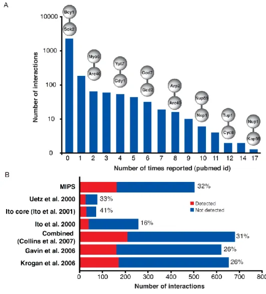
The proteins in the DHFR PCA network are highly enriched in cellular compartments [for example, organelle membranes (P < 10−12), proteasome regulatory particles (P < 10−8), the nucleolus (P < 10−7), and the cell cortex (P < 10−7)] that were less represented in comprehensive TAP-MS results [36] (tables S5 and S6, http:// www.sciencemag.org /content /suppl /2008 /05 /08 /1153878.DC1 /1153878s_tables.zip). The high sensitivity of the DHFR PCA assay is reflected in the abundance of the proteins that populated our network, which are on average only slightly more expressed than the proteome [the median log10(protein abundance) = 2.32 versus 2.28; Wilcoxon rank sum test, P= 0.19] and spanning the whole distribution of protein abundance (Fig. 7). Because this study was performed in vivo, with a technique never used at this scale and in a different medium than previous experiments, we expected that many interactions would be previously undiscovered. An examination of major databases of PPIs reveals that most of the interactions (~80%) we report are among protein pairs for which no data had been previously reported (Fig. 9A). However, when considering only PPIs that could be detected by both DHFR PCA and the other experiments, we confirmed between 16 and 41% of PPIs reported in previous large-scale screens, suggesting excellent concordance between the results of our and very different methods (Figs. 9B and 10). Further, PPIs derived from PCA represent pairwise interactions, which contrast with TAP-MS PINs, which identify clusters and thus complexes of interacting proteins. PPIs detected by PCA are therefore either within, between, or outside these complexes and thus complement these previous studies.
26
Figure 9: Overlap of the DHFR PCA network with other large-scale experiments.
(A) Most DHFR PCA PPIs are new, since they score 0 within the distribution of the number of times a known interaction has been independently deposited in major PPI repositories. Examples of interactions are shown above the bars. (B) The overlap of the DHFR PCA network is substantially increased when only the interactions that could be discovered are considered, i.e. only identified successful baits and preys are considered. Bars indicate the number of PPIs that could have been discovered by PCA. In red is the number of interactions that were discovered. Percentages indicate the percentage of interactions that were discovered by PCA out of the total possible.
27
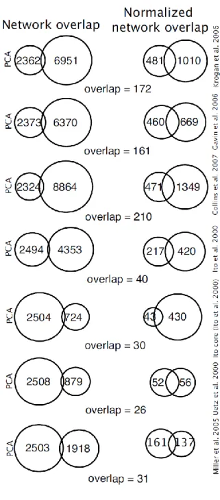
Figure 10: Overlap of the DHFR PCA network with other large-scale experiments.
On the left is the overlap between the different networks. On the right are the same overlaps, but only for those interactions that could have been detected in both experiments; i.e. cases in which the interactions were tested for in both experiments.
28
For instance, 10% of the DHFR PCA PPIs map within specific complexes in the combined analyses of the two TAP-MS data sets [13, 31], and 36 and 38% of the DHFR PCA PPIs are between one protein found in a complex and one protein not in the published data set, or two proteins not in the data set, respectively. We identified several interactions among complexes (15%), which probably mediate the integration of biological processes among PIN modules. For instance, PPIs occur among complexes that are more related in their functional annotations than would be expected to occur by chance [interacting protein pairs had a semantic similarity score of cell compartments (CCs) of 3.44 versus 1.64, P < 10−100; of biological processes (BPs) of 3.48 versus 1.51, P < 10−80; and of molecular functions (MFs) of 3.53 versus 2.3, P < 10−10]. For example, we see interactions between Dhh1p and Lsm4p, both involved in the RNA metabolic process but part of the CCR4 and the RNA-splicing complex, respectively. Another example is the interaction between Reg1p and Snf1p: subunits of the serine/threonine phosphoprotein phosphatase and SNF1 complex, respectively, but both involved in the regulation of carbohydrate metabolic processes (table S7, http:// www.sciencemag.org /content /suppl /2008 /05 /08 /1153878.DC1 /1153878s_tables.zip). Finally, we report 286 interactions involving one uncharacterized protein with proteins of known function (n = 278 interactions) or between two uncharacterized proteins (n = 8) [38], which will aid in their functional annotation.
29
1.3.3.General organization of the yeast DHFR PCA PIN.
Because we detected PPIs as they occurred in intact cells, with the faithful representation of gene expression timing and protein localization, we predicted and observed stronger coregulation of interacting protein pairs (Pearson r = 0.2 versus r = 0.1, P << 0.001) than was expected for random networks of the same size with the same protein connectivity. This is also mirrored in the enrichment of interactions among proteins that share the same BPs, MFs, and CCs and a depletion of interactions among genes of different categories (Fig. 11 and Fig. 12). PPIs among categories are somewhat more enriched in the PCA-determined network as compared with TAP-MS studies. For instance, 64, 56, and 63% of DHFR PCA interactions map to different BPs, CCs, and MFs, whereas these numbers are smaller in the TAP-MS PINs [58, 46, and 57% [14] and 51, 49, and 58% [13]]. Much of this increased enrichment of the cross-cellular components reflects interactions among proteins that the DHFR PCA method covers more of than TAP-MS; these are interactions that appear to represent the natural exchange of proteins between, for instance, the endoplasmic reticulum, Golgi, mitochondrial envelope, and vacuolar proteins, whereas others reflect the organization of complex cellular processes. For example, high enrichments in interactions between proteins localized to the bud and bud neck with those of the cell cortex, cytoskeleton, plasma membrane, and sites of polarized growth reflect the roles of these proteins in several compartments during cell division.
30
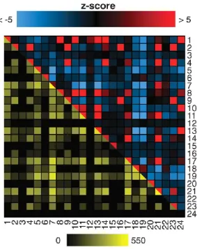
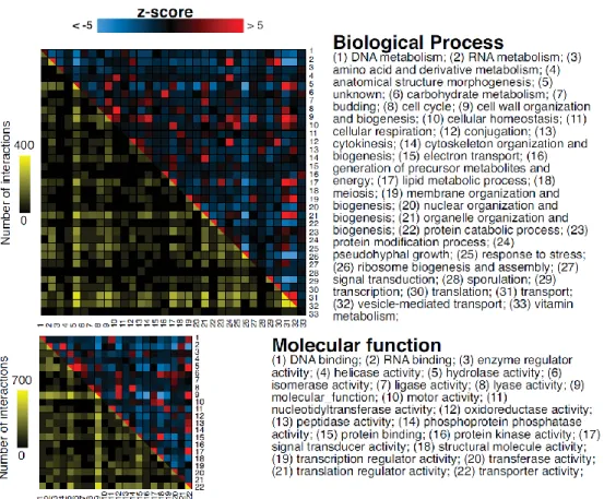
Figure 11. Interactions are enriched within GO categories.
The DHFR PCA network covers several classes of protein function, location, and biological process. The colors above the diagonal represent positive and negative deviations from the expected number of interactions between two cell compartments. A positive z score indicates a larger number of interactions within or between two categories as compared with a random network. A negative z score indicates a smaller number of interactions than expected. A z score of 2 or –2 corresponds to a P value of 0.05, and a z score of 5 or –5 to a P value of 5 × 10⁻⁷. Values below –5 and above 5 were given these minimal and maximal values. Entries below the diagonal indicate the observed numbers of interactions on a log10 scale.
31
Figure 12: Interactions are enriched within Gene Ontology categories.
The DHFR PCA network covers several classes of protein function, location and biological process. The colors above the diagonal represent positive and negative deviations from the expected number of interaction between two categories, Biological Process or Molecular function. A positive z-score indicates a larger number of interactions within or between two categories compared to a random network. A negative z-score indicates a smaller number of interactions than expected. A z-score of 2 or -2 corresponds to a P-value of 0.05 and a z-score of 5 or -5 to a P-value of 5×10⁻⁷. Values below -5 and above 5 were given these minimal and maximal values. z-scores were calculated by generating 10,000 random networks. Entries below the diagonal indicate the observed number of interactions on a log10 scale.
32
We also saw strong compartmentalization of interactions; for example, for nuclear and nucleolar proteins, which show enrichment in interactions between proteins in these two compartments but strong depletions in interactions with those of any other compartment. Equally, patterns of cross-process and molecular function categories reflect differences in complexity and organization (Fig. 12). For example, among molecular functions, RNA binding is specifically enriched in interactions between helicase and translation regulatory functions, whereas the more general transporter activity category shows links to diverse functions. The observation that PCA interactions detect links among functionally related categories is supported by a semantic analysis of the full Gene Ontology (GO) hierarchies. Proteins that show interactions with different GO Slim annotations have higher semantic similarities in their GO terms than expected by chance (CCs, 1.52 versus 0.94, P < 10−231; BPs, 2.04 versus 1.35, P < 10−122; and MFs, 1.89 versus 1.64, P < 10−8), and may thus represent interactions relating information among these processes and CCs that allows their integration into higher-order networks. As we describe below, these interactions reveal specific spatial and topological relationships between known and previously unknown complexes underlying both known and previously unknown cellular processes.
1.3.4.Global structure and topology of the in vivo interactome.
PCA-detected interactions are interpreted differently than purified protein complexes or binary (one-to-one) interactions determined in Y2H
33
screens, allowing us to address how protein complexes and PINs are spatially and topologically organized in living cells. Whether an interaction can be observed depends on the distance between the C termini of two proteins and the length of the polypeptide linker separating bait and prey proteins to the PCA fragments [24, 34] (Fig. 1A). Given that the linkers used in this study were of 10–amino acid residues, for a given protein complex we expected to detect only binary (direct) or near-binary (indirect, C termini within 82Å, but mediated by one or more other proteins) interactions for protein pairs separated by less than this distance. We first tested this prediction by exhaustively screening all pair-wise interactions (n = 45 possible pairs) in the well-known RNA polymerase II complex (Fig. 13). We found that we were 5.7 times more likely to detect an interaction if the C-termini were within 82 Å (Fisher’s exact test, P = 0.01) (Fig. 14B). Interactions that were detected but not predicted could be due to alternative assemblies of this complex in intact cells, to changes in their configuration under different conditions, or to protein dynamics that cannot be interpreted from crystal structures. We then asked whether spatial restraint on observable interactions is reflected in the complete DHFR PCA network.
34
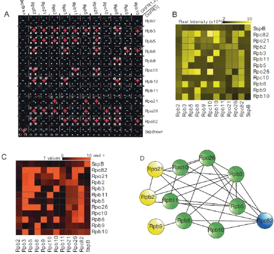
Figure 13. RNA polymerase II complex reconstitution through DHFR PCA.
(A) Results of the RNA polymerase II complex PCA network through an exhaustive screen for interactions among the ten subunits. Colonies for diploid strains that show resistance to methotrexate are indicated with red "+" and for those showing no resistance, with blue "-". (B) Mean colony pixel intensity values extracted from the high-resolution image in (A) by quantification of total colony pixel intensities. (C) t-scores for colony pixel intensities ranging from 0 to 10 and higher (P < 0.0001) resulting from the comparisons with control colonies. (D) Summary of the results of the RNA Pol II DHFR PCA screen where edges represent a physical interactions corresponding to a t-score of 4 and higher (P < 0.05) and nodes are the individual RNA Pol II subunits. Nodes colored in yellow, blue and green are respectively RNA Pol II exclusive proteins, RNA Pol III exclusive protein and RNA Pol II and RNA Pol III shared proteins.
35
Figure 14. The DHFR PCA results provide structural and topological insights.
PCA fragments have to be in proximity to each other in order to fold into the active structure of the reporter protein. (A) PCA PPIs versus protein complexes. Comparison of the PCA network with databases of curated protein complexes (MIPS) and inferred from computational analysis of TAP-MS [31] allows classification of four types of PCA interactions: in which both proteins are found within a complex (type 1), are inferred to be in two separate complexes (type 2), one protein is in a complex and the other is not in the network (type 3), or both are absent from the network (type 4) [31]. Columns of numbers indicate the number of PCA PPIs observed for each data set and each category. (B) A thorough DHFR PCA screen of the RNA polymerase II complex [Protein Data Bank (PDB) number 1I3Q] detects predicted interactions among the 10 subunits. (C) An interaction is 3.5 times more likely to be detected for a pair of proteins known to interact if the C termini of these proteins are within 82 Å of each other in the case of stable crystallized complexes of yeast-homologous proteins deposited in the PDB. (D) Membrane protein topology and PPI detection by PCA. A protein interaction is 12 times more likely to be detected if the C termini are in the same cell compartment.