Acknowledgements
This dissertation would not have been possible without the guidance and the help of several individuals who contributed and extended their valuable assistance in the preparation and completion of this study.
First and foremost, with a deep sense of gratitude, I would like to thank my supervisor Dr. Dominique Tremousaygue for giving me the opportunity to work in his lab and enjoy the most romantic French life. Her guidance throughout the period of my PhD study is greatly acknowledged. Without her help, I cannot finish my PhD project. She helped me a lot. She trained me at the beginning of my project. She continued my experiments when I was on holiday. She was patient to listen to the rehearsals of my presentation again and again.
I am thankful to Dr. Yves Marco, our group leader, for his support, constant encouragement, and careful reading of and helpful comments on the manuscript.
I also thank Dr. Laurent Deslandes, a smart scientist, for helpful discussions. He always patiently explain to me how to do an experiment step by step; which step is a key step; why is the most important step.
Alice Delgas, a cute girl, brought us a lot of fun. When I had problem in my daily life, I always asked Alice. She is a very friendly, and gave me helpful information.
Thanks to all colleagues of DRYM group: Xavier Barlet, Richard Berthomé, Patrick Dabos, Gaëlle Huet, Dominique Roby, Susana Rivas, Claudine Balagué, Carine Huard-Chauveau, Mehdi Khafif, Sylvain Raffaele, Derry Voisin, Mathieu Hanemian, Céline Tasset, Arrighi Jean-François, Marino Daniel, Pata Mickael… although some PhD students were graduate, and some post-doc were leave this team, I also thank for their help and suggestions in seminars throughout my research work.
I wish to extend my warmest thanks to all those who have helped me with my work in LIPM. The greenhouse colleagues help me to grow tobacco, and take care of my plant. The washing room colleagues help me to prepare medium and other thing.
Also, I would like to thank my sweet friends in Toulouse: BIAN Wanping, CHEN Yanling, DENG Xu, Hao Yanwei, HUANG He, GUAN Suhua, JIANG Gaofei, JIANG Wei, KE Yuchun, LI Qiang, LIU Jian, LIU Ran, RAO Man, WEI Lumei, WANG Keke, YU Hong, ZHANG Tiantian, and ZHANG Liping, Ren Xiaoqian, and other friends in the world: HU Hao, HE Ling, MU Dongdong, TIAN Shengchao, WAN Qian, XIA Zhanghui, XU Dabin, XU Mingjiao and ZHOU Songlin. I don’t say much about you guys, because I don’t have enough words to express. With you guys, we talked a lot about science and daily life. You gave me suggestions when I had problem, encouraged me when I was depressed. I feel like I have a family in Toulouse. We shared our happiness and sadness, we comforted each other. With you guys, I am not lonely any more. I will never forget the time we spent together. Thanks again.
Special thanks MA Hang, We know how much effort we did to keep our relationship. I am very happy we made it. I did really enjoy the days we had fun together we when travelled.
In the end, I would like to thank my family. My parents, they bore me, raised me, support me, teach me, and love me. I know how hard they are, how much they love me. Their support is my motivation, and also my sister and her daughter, who is always supportive.
Table of contents
Contents
ABBREVIATIONS ... 1
LIST OF FIGURES AND TABLES ... 5
Chapter I ... 9
General Introduction ... 9
1.1 Plant immunity: efficiency and limits ... 10
1.1.1 Basal defense, a first layer of resistance involved in the recognition of conserved microbial patterns ... 11
1.1.2 A second layer of specific resistance is mediated by resistance proteins ... 12
1.1.3 Signaling pathways at a glance ... 13
1.2 Pathogen effectors: two sided coins. ... 14
1.2.1 Effectors and their mode of action ... 15
1.2.2 Examples of effectors targeting plasma membrane components ... 16
1.2.3 Examples of effectors targeting chloroplast components ... 17
1.2.4 Examples of effectors targeting vesicle trafficking ... 18
1.2.5 Examples of effectors targeting MAPK signaling ... 18
1.2.6 Example of effectors targeting nuclear components ... 20
1.3 Effector recognition mechanisms ... 22
1.3.1 Ligand-receptor model ... 22
1.3.2 Guard model ... 23
1.3.3 Decoy model ... 23
1.3.4 Bait and switch Model ... 23
1.4 Recognition through pairs of resistance proteins. ... 24
1.5 Regulation of plant gene transcription: beyond perception, a major step in immune response ... 24
1.5.1 Hormonal control mediated through transcriptional regulations: What is known about salicylic acid signaling pathways ... 25
1.5.2 Immunity triggered by several PAMPS or by PTI and ETI overlaps at the level of transcriptome responses ... 27
1.5.3 Transcriptional changes associated to the function of resistance proteins ... 28
1.5.4 Role of the Mediator and Elongator transcription complexes in plant immunity ... 29
1.6 The A. thaliana/R. solanacearum pathosystem ... 32
1.6.1 R. solanacearum, a destructive bacterial plant pathogen... 32
1.6.2 Virulence determinants ... 33
1.6.3 Control of disease development ... 34
1.6.4 Identification of an A. thaliana ecotype resistant to R. solanacearum GMI1000 ... 35
RRS1-R, an atypical TIR-NBS-LRR protein ... 35
1.6.5 Pop P2 an avirulence protein of R. solanacearum GMI1000 strain involved in the resistance mediated by RRS1-R. ... 36
1.6.6 Transcriptional reprogramming in response to R. solanacearum. ... 36
1.7 Objectives of the Thesis ... 39
Chapter II ... 40
RRS1 expression profiles and binding to W box cis elements ... 40
2.1 Introduction ... 41
2.2 Results ... 43
2.2.1 RRS1-S and RRS1-R gene expression profiles ... 43
2.2.2 Specific binding of RRS1-R and RRS1-S WRKY domains to W boxes ... 46
2.3 Discussion ... 46
2.4 Conclusion and perspectives ... 48
Chapter III ... 50
In vivo identification of DNA binding sites of RRS1-R proteins ... 50
3.1 Introduction ... 51
3.2 Results ... 52
3.2.1 Set up of the Dam ID approach ... 52
3.2.2 In vivo identification of Fragments Associated to the RRS1-R driven Methylation (FARMs) ... 54
3.3 Discussion ... 58
3.4 Conclusion and perspectives ... 60
Chapter IV ... 63
Hrp mutant bacteria as biocontrol agents ... 63
4.1 Introduction ... 64
Hrp mutant bacteria as biocontrol agents: towards a sustainable approach in the fight against plant pathogenic bacteria ... 65
Chapter V ... 76
Chapter VI ... 80
Material and Methods ... 80
6.1 Materials ... 81 6.1.1 Bacteria ... 81 6.1.2 Bacterial pathogen ... 81 6.1.3 Plant Material ... 81 6.1.4 Oligonucleotides ... 81 6.1.5 Plasmids ... 81
6.1.6 Buffer and solution ... 82
6.1.7 Media and Antibiotics... 82
6.2 Methods ... 82
6.2.1 Plant growth conditions ... 82
6.2.2 A. thaliana floral dip stable transformation ... 83
6.2.3 Transient expression in N. benthamiana ... 84
6.2.4 RNA extraction ... 84
6.2.5 RNA reverse transcription into cDNA ... 84
6.2.6 Extraction of plant genomic DNA ... 85
6.2.7 Plasmid DNA isolation from bacteria ... 85
6.2.8 Protein extraction for SDS-PAGE and western blotting ... 85
6.2.9 Denaturing SDS-polyacrylamide gel electrophoresis (SDS-PAGE) ... 86
6.2.10 Immunoblot analysis (Western blotting)... 86
6.2.11 GATEWAY Cloning Technology ... 86
6.2.12 Histochemical staining for GUS Activity detection ... 87
6.2.13 Electrophoretic mobility shift assays ... 87
6.2.14 Electrophoretic Mobility Shift Assay (EMSA) ... 89
6.2.15 DNA adenine methyltransferase identification (DamID) ... 91
6.2.16 Microplate fluorometric GUS assay ... 94
6.2.17 R. solanacearum inoculation ... 95
6.2.18 Micoscopic analyses ... 96
Chapter VII ... 97
1
ABBREVIATIONS
ABA abscisic acid
ADP adenosine diphosphate ARF ADP-ribosylation factor
BAK1 brassinosteroid insensitive 1-ssociated receptor kinase 1 BCA biological control agent
BEAF boundary element-associated factor BIK1 botrytis-Induced Kinase 1
bZIP basic Leucine Zipper CaM calmodulin
CBP60g calmodulin binding protein 60-like.g CC coiled-coil
cDNA complementary deoxyribonuleic acid CERK1 chitin elicitor receptor kinase1 cGMP cyclic guanosine monophosphate ChIP chromatin immunoprecipitation CKs cytokinins
Col-0 Arabidopsis thaliana ecotype Columbia 0
CRT1 compromised for recognition of Turnip Crinkle Virus CWDEs cell wall-degrading enzymes
DamID DNA adenine methyltransferase identification DNA deoxyribonuleic acid
DREF DNA replication-related element binding factor EAR ERF-associated amphiphilic repression
EDS1 enhanced disease suceptibility 1 EF–Tu elongation factor Tu
EFR EF-Tu receptor
EMSA electrophoretic mobility shift assay EPS extracellular polysaccharide
2 ERF ethylene responsive transcription factor
ET ethylene
ETI effector friggered immunity ETS effector triggered susceptibility
FARM fragments associated to RRS1-R driven Methylation flg22 22-amino acid motif of the bacterial flagellin
FLS2 flagellin sensing 2 GAs gibberellins
GEF guanine nucleotide exchange factor HR hypersensitive response
HLH helix-loop-helix domain hrc hrp-conserved
hrp hypersensitive response and pathogenicity ICS1 isochorismate synthase 1
IkB inhibitor of kB JA jasmonic acid
KO knockout
kb kilobase
LHP1 like heterochromatin protein 1 LPS lipopolysaccharides
LRR leucine rich repeats
LSD1 lesion simulating disease resistance 1 MAMPs microbe associated molecular patterns MAP mitogen activated protein
MPK mitogen activated protein kinase
NB-ARC nucleotide-binding adaptor shared by Apaf1, and CED4 NB-LRR nucleotides binding leucine rich repeats
NBS nucleotides binding site
Nd-1 Arabidopsis thaliana ecotype Niederzens 1
NDR1 non race-specific disease resistance 1 NF-kB nuclear factor kB
3 NLS nuclear localization signal
NO nitric oxide
NPR1 nonexpressor of pathogenesis-related genes 1 PAD4 phytoalexin deficient 4
PAL phenylalanine-ammonia-lyase
PAMP pathogen associated molecular patterns PBS1 avrPphB susceptible 1
PBL PBS1-like
PCR polymerase chain reaction PGN peptidoglycan
Pop pseudomonas outer protein
Pop P2 an R.solanacearum type III effector PR1 pathogenesis-related gene 1
PRR pattern recognition receptors PTI PAMP triggered immunity PSII photosystem II
pv. pathovar
R resistance
RIN4 RPM1-interacting protein 4 RLK receptor-like kinase
RLP receptor-like proteins RNA ribonucleic acid RNAPII RNA polymerase II ROS reactive oxygen species
RPM1 resistance to Pseudomonas syringae pv maculicola 1 RPS resistance to Pseudomonas syringae
RRS1-R Resistance to Ralstonia solanacearum SA salicylic acid
SAG101 senescence associated gene 101 SAR systemic acquired resistance
4 SNC1 suppressor of npr1-1, constitutive 1
SPL6 squamosa promoter binding protein-like 6 SR1 signal responsive 1
STAND signal transduction ATPases with numerous domains SUMO small ubiquitin-like modifier
TAL transcription activator-like T3E type III effector
T3SS type III secretion system TF transcription factor
TGA TGACG-sequence-specific DNA-binding protein TGN/EE trans-Golgi network/early endosome
TIR Toll/interleukin-1 receptor TMV Tobacco mosaic virus TPR1 topless-related 1
UPR Unfolded Protein Response UV ultra-violet
vir Virulence
Yop Yersinia outer protein
5
LIST OF FIGURES AND TABLES
Table C1-1. AVR/ R couples identified in plant/pathogen interaction studies Figure C1-1. A zigzag model of the plant immune system
Figure C1-2. Pattern recognition receptors (PRRs) and signaling adaptors in plants Figure C1-3. Different classes of R protein and their cellular location
Figure C1-4. The Arabidopsis RPM1, a plasma membrane NB-LRR protein is activated by either AvrRpm1 or AvrB effector
Figure C1-5. Schematic representation depicting the virulence and avirulence function of the bacterial cysteine protease AvrPphB
Figure C1-6. Effector HopM1 manipulates components of a putative TGN/endosome-associated proteasome degradation machinery
Figure C1-7. Proposed models for effector recognition
Figure C1-8. Schematic representation of the domain structure of NB-LRR proteins and pathogen isolates
Figure C1-9. Illustration of the overlapping signaling in PTI and ETI
Figure C1-10. The WRKY1 transcription factor interacts directly with the R protein (MLA) Figure C1-11. Transcriptional regulation by promoters and enhancers
Figure C1-12. Bacterial wilt symptoms in plant caused by R. solanacearum
Figure C1-13. Schematic representation of RRS1-R and RRS1-S genes in Col-0 and Nd-1 Figure C1-14. Sequence alignment of different members of the YopJ/AvrRxv effector family from plant and animal bacterial pathogens
Figure C2-1. Homology models of AtWRKY DNA-binding domain Figure C2-2. Phylogenetic tree of the AtWRKY family in A. thaliana Table C2-1. WRKY gene expression in Arabidopsis thaliana
6 Figure C2-3. Promoter structure of RPS4 and RRS1 R-gene pairs
Figure C2-4. GUS reporter gene expression in Arabidopsis plantlets Figure C2-5. Relative RRS1 expression level
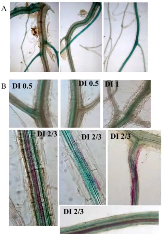
Figure C2-6. P250R:GUS expression in the transition zone from basal hypocotyl to roots Figure C2-7. P250R:GUS expression in transversal sections of Nd-1 plants
Figure C2-8. Details of P250R: GUS expression in vascular tissues
Figure C2-9. RRS1-R gene promoter: GUS reporter gene expression in roots from adult plants Figure C2-10. Down regulation of P250R:GUS expression in plants grown in reduced watering conditions
Figure C2-11. Induction of GUS expression in root tips in response to abiotic stresses
Figure C2-12a. P250R:GUS expression in adults Nd-1 plants grown in soil and root inoculated Figure C2-12b. P250R:GUS expression in adult Col-0 plants grown in soil and root inoculated Figure C2-13. Estradiol induction of PopP2 in plantlets expressing the reporter genes
Figure C2-14. Impact of Pop P2 induction in GUS expression driven by a P250S promoter in Col-0
Figure C2-15. DNA-binding activity of the RRS1 protein WRKY domains Figure C3-1. DNA-binding activity of the WRKY-R::Dam fusion protein Figure C3-2. RRS1-R::Dam methylates plant DNA
Table C3-1. Summary of transgenic lines and genetic background used for DamID Table C3-2. FARMs hits identified from leaves
Table C3-3. FARMs hits identified from Nd-1 plantlets without inducible Pop P2 Table C3-4. FARMs hits identified from Nd-1 plantlets / iPopP2
Figure C3-3. Expression level of Dam/EF-1a in Nd-1 and Nd-1/Estradiol inducible PopP2 lines Figure C3-4. Protocol of DNA adenine methyltransferase identification (DamID)
7 Figure C3-6. PBL1 promoter sequence (Upstream -1000bp)
Figure C3-7. Expression and function of PBL1 Figure C3-8. FARM amplification in plantlets
Figure C3-9. Quantitative overview of FARMS hits in comparison to several data sets Figure C3-10a/b. FARMs within RPS4 and EDS1 coding sequence
Figure C3-11. Model for RSS1-R mode of action. RRS1-R represses expression of genes related to defense
Figure C3-12. Types of epigenetic modifications Table C6-1. Primers used in this thesis
Table C6-2. Plasmids used in this thesis
Figure C6-1. The map of constructions for DamID
Figure C6-2. Construct for stable expression of RRS1-R::Dam in A. thaliana Table C6-3. Buffers and solutions used in this thesis
8
PREFACE
The thesis is presented in 7 sections.
1- Chapter 1: The background and motivation for the project is outlined in the introduction.
2-Chapter 2 presents prerequisite to take on in vivo transcription factor binding sites characterization: The aim of this chapter was to determine the expression profile of RRS1 genes in planta and to test the DNA binding properties of RRS1-R and RRS1-S in vitro.
3-Chapter 3 presents the set-up of the DamID approach in order to determine in vivo binding sites of RRS1-R and RRS1-S proteins. It focuses on the identification of RRS1-R binding sites.
4-Chapter 4 contains a publication as a first co-author in “Plant Signaling & Behaviour”. It is devoted to a side project consisting in reviewing what is known on the molecular mechanisms occurring when bacterial hrp mutants are used as biocontrol agents in protection against bacterial diseases. These mechanisms were particularly well described in the context of the interaction of Ralstonia solanacearum (R. solanacearum) and
Arabidopsis thaliana (A. thaliana).
5-In chapter 5, the results are discussed in terms of perspectives and opening questions. 6-Material and Methods are described in the chapter 6.
9
Chapter I
10
1.1 Plant immunity: efficiency and limits
Plants have several basic needs for survival. They require light, water, air, minerals and nutrients. They also need to be able to reproduce in order to ensure survival of species. Climate disorders, unfavorable geology, pathogen attacks are some of the main threats to plants. Adaptations which develop over time and generations as a response to the ever challenging environment allow an organism to reduce competition for space and nutrients, reduce predation and increase resistance to pathogens. Adaptive traits are particularly well developed in plants that are sessile organisms, and this is highlighted by the extraordinary phenotypic plasticity of plants depending of their growing conditions.
Relationships between plants and neighboring microbes (bacteria, fungi, and oomycetes, nematodes) lasts all along plant life, from seeds falling down on the soil to the death of developed plants. Even if some interactions with these microbes benefit plant development, others affect plant fitness. Therefore, plants are engaged in a battle against many surrounding pathogens and have developed numerous and sophisticated strategies to face these aggressions. These defense mechanisms consist of pre-formed structures, toxic compound production and induced immune reactions. The immune system of a plant is indeed very efficient to resist to the vast majority of pathogens; when all genotypes of a plant species are resistant to all strains of a pathogen, the term non-host resistance is used. Reversely, in cases of host interactions, some pathogens are virulent on some plant genotypes whereas some plant cultivars can resist only to certain strains of a pathogen species. In this context, an interaction is called compatible when the plant is susceptible to a pathogen and develops disease, whereas it is incompatible when the pathogen growth is rapidly limited in the plant which is then resistant.
Plants trigger immune responses to pathogens via a two-layer surveillance system. The first layer is composed of extracellular receptors, or Pattern Recognition Receptors (PRR). These receptors detect pathogens outside plant cells and induce a nonspecific resistance called PTI, for PAMP (Pathogen Associated Molecular Patterns) Triggered Immunity. Indeed, to be able to colonize efficiently plants, pathogens have developed several strategies that enable them to escape the host resistance. Among others strategies, they
Figure C1-1. A zigzag model of the plant immune system.
Pathogen-associated molecular patterns (PAMPs) are detected via plant transmembrane pattern-recognition receptor (PRRs) to trigger basal resistance (PTI). Then, pathogens inject effectors into plant cells. If plants do not recognize effectors, these latter one will interfere with PTI, resulting in effector-triggered susceptibility (ETS). Finally, an effector is specifically recognized by an R protein and the effector triggered immunity is established. PTI is often accompanied with the induction of hypersensitive response leading to a rapid cell death (HR). In last phase, pathogen isolates without a protein effector (red) are selected, and perhaps gained new effectors (in blue), which allows pathogens to suppress ETI. Selection will then again favour the acquisition of a new plant R protein (NB-LRR alleles) that can recognize the newly acquired effectors, resulting again in ETI. (Adapted from Jones and Dangl, 2006)
11 produce proteins, named effectors, which have the capacity to inactivate plant defense systems and cause disease (ETS for effector triggered susceptibility). A second layer of immunity intervenes then in plant defense, thanks to receptors, or nucleotide binding leucine rich repeats proteins (NB-LRR) that are encoded by Resistance genes (R genes). These receptors recognize specifically some pathogen effectors, named avirulence (Avr) protein, and this recognition triggers an induced and specific resistance, also named ETI (Effector Triggered Immunity). Such a co-evolution of host plants and pathogenic microbes resulting in a highly adaptive and rapidly evolving immune system is illustrated by the zigzag model (Dangl e Jones, 2001). (Figure C1-1) (Tiffin e Moeller, 2006). Indeed, the host range of a pathogen can evolve rapidly due for example, to its capacity to synthesize a host-specific toxin or to generate a “novel” effector by mutation or gene transfer from a related organism (Friedman e Baker, 2007).
1.1.1 Basal defense, a first layer of resistance involved in the recognition
of conserved microbial patterns
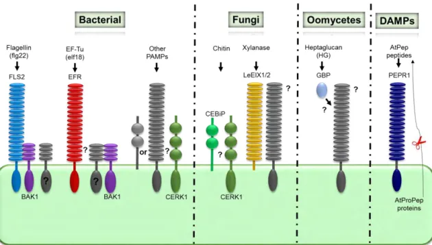
On the cell surface, plant express PRR. These receptors perceive highly conserved molecular signatures of microbes, referred to as Microbe Associated Molecular Patterns (MAMPs) or, when focusing more on pathogenic microbes, PAMPs, for Pathogen Associated Molecular Patterns. So far, most characterized PRRs in plants belong either to the family of receptor-like kinase (RLKs) which have an LRR or LysM extracellular receptor domain and an intracellular kinase domain, or to the family of receptor-like proteins (RLP) which do not possess a kinase domain. Typical examples of PAMPs are bacterial flagellin (flg22), elongation factor Tu (EF–Tu), sulfated peptide Ax21, peptidoglycan (PGN), lipopolysaccharides (LPS), fungal cell wall polysaccharides, chitin, and oomycete glucans (Akerley, Cotter e Miller, 1995; Felix et al., 1999; Dow, Newman e Von Roepenack, 2000; Gust et al., 2007; Erbs et al., 2008). (Figure C1-2) Interaction of PRRs with their corresponding PAMPs initiates a battery of defenses responses, such as the induction of MAP (mitogen-activated protein) kinase (MAPK) signaling, production of reactive oxygen species (ROS), callose deposition at the site of infection and transcriptional activation of defense-related gene.
Figure C1-2. Pattern recognition receptors (PRRs) and signaling adaptors in plants. Bacterial flagellin (flg22) and elongation factor EF-Tu (elf18) are recognized by the Arabidopsis LRR-RKs FLS2 and EFR receptors, respectively. FLS2, and potentially EFR, form a complex with BAK1 and maybe other SERK proteins. The Arabidopsis LysM-RK CERK1 mediates recognition of an unknown PAMP in plant immunity and is also required for chitin responses. The chitin high-affinity-binding site in rice corresponds to CEBiP. In tomato, the RLPs LeEIX1/2 recognizes xylanase and triggers signaling. In legumes, the soluble glucan-binding protein (GBP) directly binds oomycetal heptaglucan. The Arabidopsis LRR-RK PEPR1 recognizes the endogenous AtPep peptides that act as Damage-associated molecular pattern molecules (DAMPs). (Adapted from Zipfel, 2009)
12 The best analyzed plant responses to PAMPs are based on the recognition of bacterial flagellin and bacterial elongation factor Tu by the RLKs FLS2 (Flagellin sensing 2) and EFR (EF-Tu receptor) respectively. The 22 amino acid peptide (flg22) corresponding to the highly conserved amino terminus of flagellin is sufficient to trigger immune responses in Arabidopsis thaliana (A. thaliana), tomato, tobacco and barley (Felix et al., 1999; Peck
et al., 2001; Taguchi et al., 2003; Chinchilla et al., 2006; Hann e Rathjen, 2007; Shen et al., 2007). An N-acetylated peptide comprising the first 18 amino acids, of EF-Tu, termed
elf18, is fully active to trigger PTI (Kunze et al., 2004). Treatment of A. thaliana seedlings with elf18 or flg22 induces a common set of responses including whole genome reprogrammation (Zipfel et al., 2006). Perception of fungal chitin oligosaccharides by the LysM-RLK CERK1 (Chitin Elicitor Receptor Kinase1) is also well documented. N-acetylchitooctaose (GlcNAc)8 induces also PTI in plants as attested by ROS production
(Kaku et al., 2006; Miya et al., 2007).
1.1.2 A second layer of specific resistance is mediated by resistance
proteins
Plants evolved specific R genes sensing pathogen-derived effectors in order to cope with host adapted pathogens, which inject effectors within the plant cell to escape the first extracellular layer of immunity (Dangl e Jones, 2001). These R genes encode NB-LRR proteins structurally related to animal NLR proteins (Nucleotide-binding oligomerization domain-NOD- and LRR containing proteins). The Nucleotide-Binding domain is also known as the NB-ARC (Nucleotide-Binding adaptor shared by Apaf1, and CED4) domain. NB-ARC proteins form a subclass of the STAND super family (signal transduction ATPases with numerous domains), a class of molecular switches that are involved in a variety of processes, including immunity, apoptosis (e.g. Apaf1 and CED4) and transcriptional regulation (Danot et al., 2009). STAND proteins have a modular architecture allowing them to function simultaneously as sensor, switch and response factor. NB-LRR proteins can be further divided into two main subclasses depending on their N-terminal domain (Meyers et al., 2003) (Figure C1-3). One class comprising the TIR-NB-LRR receptors, has homology to the Drosophila Toll and human Interleukin-1 receptor intracellular signaling domains. The second class, CC-NB-LRR receptors, possesses a
Figure C1-3. Different classes of R protein and their cellular location.
Schematic representation of the various domains of R proteins acting either as transmembrane receptors or intracellular NB-LRR receptors. The predicted domains of R proteins are presented as follows: CC (Coiled-Coil); TIR (Toll and Interleukin 1 Receptor-like motif); NB (Nucleotide binding); LRD (Leucine-Rich Domain); LRR (Leucine-Rich Repeat); NLS (Nuclear Localization Signal); WRKY transcription factors. (Adapted from Hammond-Kosack KE and Parker JE, 2003)
13 predicted coiled-coil (CC). R-genes have been isolated from a variety of plants. A collection of plant R genes is available through the PRG data base which register about 112 manually curated R genes in 29 different species (Sanseverino et al., 2013). Some main plant R genes and Avr associated genes are shown in Table C1-1.
1.1.3 Signaling pathways at a glance
MAPK cascades constitute the main signaling pathway from PRRs to downstream components in PTI (Tena, Boudsocq e Sheen, 2011; Hamel et al., 2012). In ETI, signaling pathways from the TIR-NB-LRR and CC-NB-LRR receptors require the CRT1 ATPase (compromised for recognition of Turnip Crinkle Virus) general factor, reported to serve as facilitating the activation of receptors (Kang et al., 2012). Most characterized CC-NB-LRRs then recruit the plasma membrane-associated protein NDR1 (NON-RACE SPECIFIC DISEASE RESISTANCE-1 (Century, Holub e Staskawicz, 1995; Knepper, Savory e Day, 2011) whereas all TIR-NB-LRRs, and one CC-type NB-LRR receptor (HRT), require nucleocytoplasmic EDS1 (Enhanced Disease Suceptibility1) and its partners PAD4 (Phytoalexin Deficient 4) and SAG101 (Senescence associated gene 101) for signal transduction (Venugopal et al., 2009).
Downstream of both PTI or ETI activation, diverse plant hormones act as central players in the triggering of the plant immune signaling network (Howe e Jander, 2008; Bari e Jones, 2009; Pieterse et al., 2009; Katagiri e Tsuda, 2010). Salicylic acid (SA) and jasmonic acid (JA) and their derivatives are recognized as major defense hormones (Browse, 2009; Vlot, Dempsey e Klessig, 2009). Accumulation of SA is important for resistance to biotrophic pathogens. By contrast, JA cooperates with ethylene (ET) to regulate resistance to necrotrophic pathogens. However, the hormones (ET) (Vlot, Dempsey e Klessig, 2009), abscisic acid (ABA) (Ton, J., Flors, V. e Mauch-Mani, B., 2009), gibberellins (GAs) (Navarro et al., 2008), auxins (Kazan e Manners, 2009), cytokinins (CKs) (Walters e Mcroberts, 2006), brassinosteroids (Nakashita et al., 2003), and nitric oxide (NO) (Moreau
et al., 2010) function as well as modulators of the plant immune signaling network and add
another layer of regulation. Changes in hormone concentration or sensitivity triggered during pathogenic interactions mediate a whole range of adaptive plant responses, often at
Table C1-1. AVR/ R couples identified in plant/pathogen interaction studies Avr gene NB-LRR Host/gene name
ATR13 RPP13 A. thaliana RPP13 (Recognition of peronospora parasitica 13); ATP
binding
AvrB, AvrRPP1A RPP1 A. thaliana RPP1 (Recognition of peronospora parasitica 1) AvrPphB RPS5 A. thaliana RPS5 (Resistant to P.syringae 5) (RPS5)
AvrRpm1 RPM1 A. thaliana RPM1 (Resistant to P.syringae pv maculicola 1); protein
binding
AvrRPP4 RPP4 A. thaliana RPP4 (Recognition of peronospora parasitica 4) AvrRPP5 RPP5 A. thaliana RPP5 (Recognition of peronospora parasitica 5) AvrRPP8 RPP8 A. thaliana RPP8 (Recognition of peronospora parasitica 8) AvrRps4 Rps4 A. thaliana RPS4 (Resistant to P.syringae 4)
AvrRpt2 Rps2 A. thaliana RPS2 (Resistant to P.syringae 2) Coat protein HRT A. thaliana viral resistance protein (HRT) gene Coat protein RCY1 A. thaliana RCY1 gene for R-protein
Pop P2 RRS1 A. thaliana RRS1 (Resistant to R.solanacearum 1) AvreBs3 Bs3 Capsicum annuum cultivar ECW-30R Bs3 (Bs3) gene AvreBs2 Bs2 Capsicum chacoense disease resistance protein BS2
AVRA MLA10 Hordeum vulgare MLA10 (Mla10)
AvreRpg1 RPG1 Hordeum vulgare subsp. vulgare RPG1 gene, Rpg1-Swiss Hv 489 allele
Avr3 Dm3
(RGC2B) Lactuca sativa resistance protein RGC2B (RGC2B) gene AvrM M Linum usitatissimum rust resistance protein M gene
AyrL L6 Linum usitatissimum alternatively spliced rust resistance (L6) gene AvrN AvrL567 N Nicotiana glutinosa virus resistance (N) gene
AvreXa1 XA1 Oryza sativa mRNA for XA1
AvreXa21 xa21 Oryza sativa Indica Group Xa21 gene for receptor kinase-like protein,
cultivar:II you 8220
Avr-Pita Pi-ta Oryza sativa (japonica cultivar-group) pi-ta protein (Pi-ta) gene Coat protein Rx2 Solanum acaule Rx2.ac15 gene for NBS-LRR protein, exons 1-3 Avrblb1 Rpi-blb1 Solanum bulbocastanum putative disease resistant protein RGA2 gene Avr1 R1 Solanum demissum late blight resistance protein (R1) gene
Avr4 Cf-4 Lycopersicon hirsutum Cf-4 resistance gene cluster 30 kD movement
protein Tm-2a Lycopersicon esculentum ToMV-resistance locus, Tm-2^2 resistance allele Avr1 I-2 Lycopersicon esculentum disease resistance protein I2 (I2) gene
Replicase Tm-2 Lycopersicon esculentum ToMV-resistance locus, Tm-2 resistance allele Avr5 Cf-5 Lycopersicon esculentum disease resistance protein (Cf-5) gene
Avr2 Cf-2 Lycopersicon pimpinellifolium leucine rich repeat protein Cf-2.1 gene Avr9 Cf-9 Lycopersicon pimpinellifolium Cf-9 resistance gene cluster
AvrPto Pto Lycopersicon pimpinellifolium Rio Grande-PtoR protein kinase Avr3a R3a Solanum tuberosum potato late blight resistance protein R3a gene Coat protein Rx Solanum tuberosum rx gene
Nla proteae RY-1 Solanum tuberosum subsp. andigena ry-1 gene for resistance gene-like,
exons 1-6, splice variants C38 and C19
14 the cost of growth and development (Walters e Heil, 2007). Antagonistic and synergistic interactions between diverse hormone signal transduction pathways provides the plant with a powerful capacity to finely tune its immune response according to the invader encountered and to utilize its resources in a cost-efficient manner (Mundy, Nielsen e Brodersen, 2006; Jaillais e Chory, 2010). Transcriptome analyses are powerful tools to decipher the changes accompanying plant defense and transcription dynamics is emerging as an important theme in plant resistance or susceptibility establishment.
This introduction aims at the description of pathogens attack strategies and plant mechanisms known to play a role in induced immunity, highlighting more specifically the steps of pathogen perception and transcriptional changes associated to pathogen attacks.
1.2 Pathogen effectors: two sided coins.
Based on their lifestyles, phytopathogenic microbes can be divided into biotrophs, hemibiotrophs, and necrotrophs. While biotrophs feed on living cells and actively maintain host cell viability, necrotrophs kill host cells before feeding on dead tissues. Hemibiotrophs adopt an early biotrophic phase followed by a necrotrophic phase. In this chapter, we will focus on pathogens with a biotrophic step in their infectious cycle.
Pathogens can deliver effectors into the apoplast or directly inside the host cell. Apoplastic effectors include cell wall-degrading enzymes (CWDEs), toxins, and various cysteine-rich proteins. CWDEs and toxins are important virulence factors for necrotrophs but are thought to play a less important role for biotrophs and hemibiotrophs (Barras, Vangijsegem e Chatterjee, 1994; Cantu et al., 2008). Intracellular effectors are secreted mainly by biotrophic and hemibiotrophic pathogens. These intracellular effectors will retain our attention in this introduction.
Bacterial pathogens can deliver these effectors into the host cell through different secretion systems (type III, type IV, and type VI secretion systems). Among these, the type III secretion system (T3SS) plays a crucial role for several plant bacterial pathogens such as
Pseudomonas syringae (P. syringae), Xanthomonas spp., and Ralstonia solanacearum (R. solanacearum). The N terminus of type III effectors (T3Es) displays biased amino acid
15 composition, which serves as a signal recognized by the type III secretion machinery (Collmer et al., 2002; Vinatzer, Jelenska e Greenberg, 2005).
Some biotrophs (e.g., Blumeria graminis) or hemibiotrophs (e.g., Phytophthora infestans) fungal and oomycete pathogens develop into the host cell a specialized structure called haustorium (Kamoun, 2006; Horbach et al., 2011). Haustoria are not only responsible for nutrient uptake but also represent the main site of effector secretion (Mendgen and Hahn, 2002). Intracellular effectors of fungal and oomycete pathogens are presumably targeted to extracellular spaces by their N-terminal signal peptide before being translocated into the plant cell. How these effectors are translocated into the plant cell is an active area of investigation. (Dou e Zhou, 2012).
Extensive genome sequencing programs coupled with robust computational predictions of sequence motifs characteristic of effector proteins, allowed the description of complete sets of putative T3Es for a significant number of bacterial pathogens and the identification of their host targets is therefore well documented. We are going to concentrate on bacterial T3Es from different phytopathogenic bacteria in order to present the various cellular and molecular processes which they target.
1.2.1 Effectors and their mode of action
Plant components targeted by effectors are located in several plant cell compartments including, plasma membrane, nucleus, chloroplast or vesicle compartments in the cytoplasm. Even if host components are manipulated by effectors to favor disease, as previously mentioned, this manipulation in ETI can also provide an efficient alarm mechanism that can turn on plant immunity. Molecular mechanisms are tangled in different ways for ETS and ETI. Intricate molecular interactions need then to be deciphered in order to pinpoint important clues of the interaction outcome. Furthermore, some plant resistance-signaling components appear to be targeted by multiple bacterial effectors and a growing body of evidence supports the idea that plants have evolved a highly sophisticated surveillance system, involving molecular sensors that are able to perceive multiple effector activities. These observations may reflect the molecular co-evolution between host plants and invading bacteria. Some illustrative examples have been chosen to present our current
16 knowledge about the molecular dialogue resulting from the co-evolution of pathogens and host plants (Deslandes e Rivas, 2012).
1.2.2 Examples of effectors targeting plasma membrane components
1.2.2.1 AvrPto and AvrPtoB target multiple plant kinases
AvrPto and AvrPtoB, two effectors from Pseudomonas syingae (P. syringae) have been shown to suppress ETI or PTI by targeting multiple plant kinases. AvrPto was indeed reported to interact with the kinase domain of FLS2 and EFR and appeared to block PAMP signaling by inhibiting their kinase activity. AvrPtoB may also interact with the kinase domain of FLS2 and thereby facilitate its degradation by the proteasome. In addition, both AvrPto and AvrPtoB induce ETI in tomato by interacting with the intracellular serine/threonine kinase Pto in concert with the NB-LRR protein PRF. Furthermore, AvrPtoB is recognized by Fen, another kinase from the Pto family. This leads to effector triggered susceptibility (ETS) through the degradation of Fen and the suppression of ETI. 1.2.2.2 AvrB, AvrRpm1, AvrRpt2, and HopF2 target RIN4
Multiples effectors of P. syringae were shown to suppress PTI in A. thaliana through the targeting of RIN4 (RPM1-interacting protein 4), a key regulator of plant immunity that provides a link between PTI, ETI and ETS responses. Phosphorylation of RIN4 by AvrB results in the blocking of PTI when the resistance protein RPM1 is not present or in ETI in the presence of RPM1 that will sense the RIN4 phosphorylation. This is also the case for another effector of P. syringae, AvrRpm1. AvrRpt2 is a third effector from P. syringae and is a cysteine protease that will also target RIN4 and disrupt both ETI by abrogating detection by RPM1 of RIN 4 modifications induced by other effectors and PTI. Moreover RIN4 has been identified as a virulence target of another P. syringae effector (Figure C1-4).
1.2.2.3 AvrPphB targets PBS1 and related kinases
In A. thaliana, the P. syringae T3E AvrPphB targets and cleaves the RLCK (receptor-like cytoplasmic kinase) PBS1, which triggers ETI responses when the NB-LRR R protein
Figure C1-4. The Arabidopsis RPM1, a plasma membrane NB-LRR protein is activated by either AvrRpm1 or AvrB effector. AvrRpm1 enhances the virulence of some P. syringae strains on Arabidopsis as does AvrB on soybeans. Both proteins are delivered into cells by the type III secretion system and targeted to the plasma membrane. One of their targets is RIN4, which is phosphorylated (+P), and activates RPM1. In the absence of RPM1, AvrRpm1 and AvrB presumably act on RIN4 and other targets to contribute to virulence. RPS2 is a plasma membrane NB-LRR protein and is activated by the AvrRpt2 cysteine protease. AvrRpt2 also targets RIN4. Cleavage of RIN4 by AvrRpt2 leads to RPS2-mediated ETI. In the absence of RPS2, AvrRpt2 may cleave RIN4 and other targets as part of its virulence function. Light blue motives represent RAPs (Rin-4 associated proteins) and unidentified proteins. (Adapted from Jones and Dangl, 2006)
17 RPS5, which guards PBS1, is present (Shao et al., 2003; Ade et al., 2007). Interestingly, AvrPphB is also capable to inhibit PTI signaling by cleaving a number of PBS1-like (PBL) kinases, including BIK1 and PBL1 that play a general role as integrators of immune signaling responses triggered by multiple immune receptors (Zhang, J. et al., 2010) (Figure C1-5).
1.2.3 Examples of effectors targeting chloroplast components
1.2.3.1 HopI1 targets Hsp70
HopI1 proteins from P. syringae are localized in chloroplasts where salicylic acid (SA), an important hormone for immune signaling, is synthesized. This effector suppresses SA accumulation as well as the related plant defences and affects thylakoid stack structure within chloroplasts (Jelenska et al., 2007). HopI1 is a virulence factor required for bacterial growth and symptom development in a variety of crop plants. HopI1 forms large complexes in association with an heat shock protein, Hsp70 and recruits cytosolic Hsp70 to chloroplasts (Jelenska, Van Hal e Greenberg, 2010). At high temperature, the Hsp70 pool appears to be diverted to deal with heat stress functions at the expense of the defense response. In agreement with this observation, HopI1 is dispensable for virulence at high temperature suggesting that this effector reduces plant defenses by subverting an Hsp70 defense-promoting function (Jelenska, Van Hal e Greenberg, 2010). Finally, Hsp70 is essential for mediating HopI1 virulence in response to a non-pathogenic strain of P.
syringae, supporting the idea that Hsp70 plays a role in basal resistance.
1.2.3.2 HopN1 targets PsbQ
HopN1, a cysteine protease T3E protein from P. syringae, is associated with the hypersensitive response (HR), that prevents spreading of the pathogen, in non-host tobacco plants and disease in host tomato plants (López-Solanilla et al., 2004; Rodríguez-Herva et
al., 2012). In tomato, HopN1 co-localizes in chloroplastic thylakoids with PsbQ, a member
of the oxygen evolving complex of Photosystem II (PSII), and is able to degrade PsbQ, thereby inhibiting the PSII activity in chloroplast preparations. Interestingly, PsbQ induces ROS production and programmed cell death upon infection (Rodríguez-Herva et al., 2012).
Figure C1-5. Schematic representation depicting the virulence and avirulence function of the bacterial cysteine protease AvrPphB. The AvrPphB effector is delivered into plant cells by P. syringae via the type III secretion system. Multiple innate immune signaling pathways are targeted by AvrPphB including: PTI, via the cleavage of BIK1 kinase; ETI, via the cleavage of the kinase PBS1, guarded by the resistance protein RPS5. (Adapted from Porteret et al, 2012)
18 These results highlight a general role of PsbQ in maintaining photosynthetic activity during infection by plant pathogens and underline the contribution of the photosynthetic pathway during plant defense responses. The identification of PsbQ as a natural target of HopN1 uncovers a virulence strategy aimed at the subvertion of host defenses by repressing the generation of potentially harmful ROS.
1.2.4 Examples of effectors targeting vesicle trafficking
1.2.4.1 HopM1 targets AtMIN7
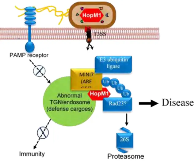
The P. syringae virulence protein HopM1 accumulates in the trans-Golgi network/early endosome (TGN/EE) of host cells where it interacts with and mediates degradation of AtMIN7 (A. thaliana HopM1 Interactor7) by the host 26S proteasome (Nomura et al., 2006; Nomura et al., 2011). AtMIN7 belongs to the adenosine diphosphate (ADP) ribosylation factor (ARF) guanine nucleotide exchange factor (GEF) protein family, whose members are key components of vesicle trafficking and may play a role in plant immunity by mediating callose deposition on the plant cell wall. This illustrates a strategy of suppression of cell wall-associated host defense, thereby promoting bacterial infection (Nomura et al., 2006) (Figure C1-6).
Interestingly, AtMIN7 is required not only for PTI, but also for ETI and SA-regulated immunity (Nomura et al., 2011). Indeed, activation of ETI by three different P. syringae effectors (AvrRpt2, AvrPphB and HopA1) blocks HopM1-dependent AtMIN7 destabilization without affecting translocation of HopM1 into the host cell. Thus, blocking pathogen-mediated degradation of AtMIN7 in the TGN/EE is a critical step of the establishment of ETI. Furthermore, this finding provides an illustration of a mechanism by which plants are able to re-establish pathogen resistance during ETI in the context of pathogen suppression of ETI-associated components via effector proteins. Indeed, this work suggests a competition between HopM1-mediated degradation of AtMIN7 and plant defence-induced AtMIN7 stabilization inside the plant cell.
Figure C1-6. HopM1 effector manipulates components of a putative TGN/endosome-associated proteasome degradation machinery. (In blue: E3 ubiquitin ligase; Ub, ubiquitin; Rad23; and 26S proteasome) in order to remove the ARF-GEF protein MIN7, leading in dysfunctional TGN/endosomes, immune suppression, and disease. (Adapted from website of Sheng Yang HE Lab)
19 Targeting of MAPK signaling is a conserved virulence strategy used by a wide range of animal and bacterial pathogens. As a result of the inhibition of PAMP-induced phosphorylation of MKKs, an effector interferes with PAMP-triggered defenses and promotes pathogen virulence inside the plant. In A. thaliana, at least two MAPK signaling cascades are activated upon PAMP perception. The first one involves MPK3 and MPK6, whereas the second one leads to the activation of MPK4, which was previously described to be able to negatively regulate PTI through modulation of multiple hormone pathways (Petersen et al., 2000) and requires the MAP kinase kinases MEKK1 and MKK1/MKK2 (Nakagami et al., 2006; Suarez-Rodriguez et al., 2007; Gao et al., 2008; Qiu, Zhou, et al., 2008).
1.2.5.1 HopAI1 targets MPK3, MPK4 and MPK6
The P. syringae effector HopAI1 displays phosphothreonine lyase activity that results in dephosphorylation of MAP kinases MPK3 and MPK6 in a way that prevents their rephosphorylation. HopAI1-mediated inactivation of MAPK proteins results in the suppression of PAMP-induced gene transcription and cell wall-associated host defenses (Zhang, J. et al., 2007).
1.2.5.2 HopF2 targets MKK5
HopF2 is able to suppress PTI signaling through the attenuation of multiple MAP kinase kinases (MKKs). For example, HopF2 ADP-ribosylates MKK5 in vitro and inhibits its kinase activity (Wang et al., 2010). Beyond its ability to interfere with MAPK signaling, HopF2 expression diminishes flagellin-induced phosphorylation of BIK1, whose activation results in the phosphorylation of the FLS2/BAK1 receptor complex (Wu et al., 2011).
1.2.5.3 AvrB targets RAR1 and MPK4
AvrB was shown to enhance plant susceptibility to P. syringae by perturbing jasmonic acid (JA) hormone signaling when the resistance protein RPM1 is absent (Shang et al., 2006). In addition to its ability to induce RIN4 phosphorylation (see above), AvrB enhances also phosphorylation of MPK4. MPK4 was shown to be able to interact with and
20 phosphorylate RIN4 (Cui et al., 2010), which negatively regulates resistance to P. syringae, and positively modulate JA responses. Thus, AvrB may induce plant susceptibility by promoting MPK4-mediated perturbation of hormone signaling (Cui et al., 2010). Indeed, AvrB mediates suppression of PTI responses through its interaction with RAR1, a cochaperone of HSP90 that is required for ETI signaling. (Shang et al., 2006). Interestingly, both RAR1 and HSP90 are required for AvrB-induced plant susceptibility and up regulation of JA responses (Shang et al., 2006).
1.2.6 Example of effectors targeting nuclear components
1.2.6.1 TAL effectors target plant promoters
TAL (Transcription Activator-Like) effectors are virulence determinants found in plant pathogenic Xanthomonas spp. and R. solanacearum. These effectors present a central DNA-binding domain and a C-terminal region comprising nuclear localization signals (NLSs) and an acidic activation domain typical of transcription factors (TFs) (Scholze e Boch, 2011). According to their architecture, TAL effectors mimic eukaryotic TFs and are able to activate transcription in the plant nucleus after binding to their host target promoters (Kay et al., 2007; Römer et al., 2007).
The role of most TAL effectors in virulence is still rather elucidated. Some of them may be involved in the activation of genes encoding sugar transporters. Indeed, the X. oryzae pv. oryzae (Xoo) TAL effector PthXo1 induces expression of OsSWEET11 that is defined as a susceptibility gene because its expression facilitates rice infection by Xoo. OsSWEET11 has been proposed to mediate sugar efflux in plants in order to feed bacteria, although its ability to transport sugars remains to be demonstrated (Yang, Sugio e White, 2006). A second study suggests that OsSWEET11 may act as a copper transporter in the plasma membrane in order to decrease the copper content of the xylem sap and facilitate vascular infection by Xoo (Yuan et al., 2010). Interestingly, distinct TAL effectors appear to target different types of functionally interchangeable SWEET genes. For example, two additional TAL effectors from Xoo (AvrXa7 and PthXo3) (Antony et al., 2010) as well as the TalC protein from strain BAI3 of the African Xoo strain (Yu et al., 2011) trigger induction of OsSWEET14 expression.
21 AvrBs3 from Xanthomonas campestris pv. vesicatoria, one of the best characterized TAL proteins, induces cellular hypertrophy in susceptible pepper (Capsicum annuum) varieties, which probably promotes bacterial proliferation and dispersal (Marois, Van Den Ackerveken e Bonas, 2002). AvrBs3 directly binds to a conserved element (called UPA box) in the UPA20 promoter and induces the expression of UPA20, a gene encoding a basic helix-loop-helix-type TF that acts as a master regulator of cell enlargement (Kay et
al., 2007). Notably, resistant pepper varieties evolved to deceive TAL recognition
specificities in order to trap the effector and trigger defence responses. Indeed, the promoter of the pepper resistance gene Bs3, which encodes a protein homologous to flavine-dependent mono-oxygenases, contains a UPA box that is recognized and bound by AvrBs3, resulting in transcription of Bs3. As a result, HR (instead of mesophyll hypertrophy) is triggered (Römer et al., 2007; Römer et al., 2009).
1.2.6.2 XopD targets AtMYB30
XopD from Xanthomonas campestris is a modular T3E targeted to the nucleus of host cells. XopD exhibits small ubiquitin-like modifier (SUMO) protease activity thanks to the presence of a cysteine protease domain at its C-terminus (Hotson et al., 2003). In addition, two tandem repeated transcriptional repressor EAR (ERF-associated Amphiphillic Repression) motifs confer to XopD the ability to repress transcription of defence- and senescence-related plant genes (Kim et al., 2008). Finally, an intact helix-loop-helix domain (HLH) is necessary for XopD nuclear targeting (Canonne et al., 2011) and the ability to display non-specific DNA-binding (Kim et al., 2008). It has been suggested that a XopD N-terminal domain of unknown function may confer specificity for DNA-binding, but this hypothesis remains to be demonstrated (Canonne et al., 2010). A recent study showed that XopD from strain B100 of X. campestris pv. campestris is able to target the R2R3-type MYB TF AtMYB30 in A. thaliana (Canonne et al., 2011), a positive regulator of plant defence and cell death associated responses (Raffaele et al., 2008).
1.2.6.3 HopAI1 and AvrRps4 target EDS1
The P. syringae effector AvrRps4 is recognized by the Toll-interleukin-1 receptor- (TIR)-NB-LRR protein RPS4 in A. thaliana (Gassmann, Hinsch e Staskawicz, 1999).
RPS4-22 mediated immune responses require EDS1, a lipase-like protein considered as a crucial regulator of immunity. EDS1 nucleo-cytoplasmic shuttling and coordination of its cytoplasmic and nuclear activities are required for immunity-related transcriptional reprogramming (García et al., 2010). Like EDS1, RPS4 and AvrRps4 display a nucleo-cytoplasmic distribution. Depending on its subcellular localization, AvrRps4 was shown to trigger distinct, but coordinated defence-related responses. Indeed, restriction of bacterial growth relies on AvrRps4 nuclear localization whereas programmed cell death and transcriptional reprogramming of defence related-genes require nucleo-cytoplasmic pools of the bacterial effector (Heidrich et al., 2011). Consistent with the fact that nuclear pools of EDS1 and RPS4 are also essential for AvrRps4-triggered immunity (Wirthmueller
et al., 2007; García et al., 2010), EDS1-AvrRps4 and EDS1-RPS4 complexes have been recently detected within the nucleus (Heidrich et al., 2011). Taken together, these data indicate that EDS1 associates with RPS4 to form an RPS4-EDS1 receptor signaling module that is able to shuttle between the cytoplasm and the nucleus and intercept AvrRps4.
1.3 Effector recognition mechanisms
Recognition of an effector by an R protein constitutes the first step of ETI activation, in agreement with the “gene for gene” theory proposed by Flor in 1942. Current evidence suggests that NB-LRRs behave as molecular switches that are in an auto-inhibited but primed conformation by intra-molecular interactions between the different domains and associations with co-factors (Collier e Moffett, 2009; Lukasik e Takken, 2009). Release from inhibition by an effector triggers a series of conformational changes that allows the R protein to activate downstream defenses (Collier e Moffett, 2009; Lukasik e Takken, 2009; Takken e Goverse, 2012). Four different molecular models have been proposed to explain different recognition processes of Avr proteins by R proteins (Figure C1-7).
1.3.1 Ligand-receptor model
Initially, it was thought that products of R genes act as receptors by directly interacting with the products of Avr genes (Keen, 1990). This ligand-receptor model was supported by the fact that some Avr gene products are small and co-localize with R gene products. Indeed, direct binding of a few R-Avr combinations was found, consistent with a
receptor-Figure C1-7. Proposed models for effector recognition.
The various models are represented schematically: guard hypothesis; Decoy model; bait and switch models. The interaction is represented either in the absence of R gene (top), or presence of R gene (bottom). (Adapted from Hann and Boller, 2012)
23 ligand mode of action (Jia et al., 2000; Deslandes et al., 2003; Dodds et al., 2006; Ueda, Yamaguchi e Sano, 2006). However, for a number of R-Avr combinations, physical interactions have not been detected, and perception in these cases is thought to be indirect.
1.3.2 Guard model
The idea that effectors have specific targets in the host inspired another indirect mechanism of effectors recognition by R proteins. The guard model predicts that R proteins act by monitoring (guarding) the effector target and that modification of this target by the effector results in the activation of the R protein, which triggers disease resistance in the host (Van Der Biezen e Jones, 1998; Dangl e Jones, 2001).
The Guard model was originally proposed to explain the perception of P. syringae AvrPto by the tomato proteins Pto and Prf (Van Der Biezen e Jones, 1998) and was later generalized to other models (Dangl e Jones, 2001). Classical examples of these presumed guardees are A. thaliana RIN4 and PBS1 and tomato RCR3 and Pto (Jones e Dangl, 2006). The indirect effector perception mechanism postulated by the guard model explains how multiple effectors could be perceived by a single R protein, thus enabling a relatively small R gene repertoire to perceive a broad diversity of pathogens (Dangl e Jones, 2001). Support for the guard model has been accumulating with the identification of a number of guarded effector targets (see below).
1.3.3 Decoy model
The decoy concept is based upon the observation that some host targets of effectors act as decoys to detect pathogen effectors via R proteins (Zhou e Chai, 2008; Zipfel e Rathjen, 2008). This concept emerged as many alterations of the decoy by effectors did not result in enhanced pathogen fitness in plants that lack the R protein and triggered innate immunity in plants that carry the R protein. In addition, this model is compatible with the fact that many pathogen effectors have multiple targets in the host.
24 This model proposes that the use of recognition cofactors as baits is a mechanism employed by R proteins to sense Avr proteins and activate a molecular switch that results in the induction of resistance responses. As such, the bait and switch model provides a mechanistic explanation of how NB-LRR proteins translate pathogen recognition into defense activation (Collier e Moffett, 2009).
1.4 Recognition through pairs of resistance proteins.
Another concept arose from an increasing number of reports on dual NB-LRR genes conferring resistance to pathogens. However, in this mechanism, the function of each one of the R protein is not established yet (Figure C1-8).
The first evidence that a pair of NB-LRR genes function together in disease resistance against a pathogen isolate was the finding that both RPP2A and RPP2B R proteins are required for disease resistance to an oomycete pathogen isolate (Sinapidou et al., 2004). Characterization of N-NRG1 and RPM1-TAO1 revealed that disease resistance to viral and bacterial pathogens expressing a single avr product (p50-Tobacco Mosaic Virus, AvrB- P.
syringae, respectively) can be mediated by an NB-LRR pair encoding proteins of the TIR
and CC subclasses (Peart et al., 2005; Eitas, Nimchuk e Dangl, 2008). The existence of CC-NB-LRR-encoding gene pairs mediating disease resistance to fungal pathogens came from the identification of Lr10-RGA2 and Pi5-1-Pi5-2 (Lee et al., 2009; Loutre et al., 2009). Finally, characterization of Pikm1-TS and Pikm2-TS demonstrated that two NB-LRR genes encoding non-TIR domains are required for disease resistance against a fungal pathogen isolate (Ashikawa et al., 2008). Recent investigation of RRS1 and RPS4 demonstrated that this TIR-NB-LRR pair is required for disease resistance against multiple pathogen isolates (Gassmann, Hinsch e Staskawicz, 1999; Deslandes et al., 2002; Narusaka
et al., 2009). In this case, heterodimerization of the TIR domains of both proteins was
demonstrated by immunoprecipitation (Williams et al., XV Congress on Molecular Plant-Microbe Interactions, Kyoto, 2011, Japan).
1.5 Regulation of plant gene transcription: beyond perception, a major
step in immune response
Figure C1-8. Schematic representation of the domain structure of NB-LRR proteins and pathogen isolates (black italic).
Top row: R proteins NB-LRR identified in Arabidopsis and Tobacco. The Avr gene products are represented with blue lettering. Bottom row: NB-LRR proteins present in wheat and rice. The various pathogen isolates are represented with black italic lettering. (Adapted from Eitas and Dangl, 2010)
25 Following perception, signal transduction pathways result, in the end, in the induced production of defense proteins that directly or indirectly inhibit pathogen proliferation. Many transcription factors (TFs) are involved in the various defense pathways leading to these responses. On overview of the most important classes of transcription factors, (AP/ERF, MYB, MYC, bZIP and WRKY transcription factor families) engaged in plant defense is available in a review from van Verk and collaborators (Van Verk, 2009). In the last few years, the diversity of transcription factor families involved in defense increased largely. For example, TCP and NAC transcription factors appeared to be important actors in these processes (Muktar et al., 2011; Nuruzzaman, Sharoni e Kikuchi, 2013). Although this study is not exhaustive, the following examples will highlight important mechanisms by which TFs contribute to plant immunity. Emphasis will be related to this study.
1.5.1 Hormonal control mediated through transcriptional regulations: What is known about salicylic acid signaling pathways
Transcription regulation, related to hormonal immunity control, is exemplified by using salicylic acid (SA), as this hormone is an important player in induced defense of the plant against invading biotrophic pathogens.
Several levels of transcriptional regulation have been described concerning the mode of action of SA (hormone synthesis or downstream pathways). In A. thaliana, the EDS1/PAD4 couple controls SA biosynthesis and is essential for the activation of the SA signaling (Wiermer, Feys e Parker, 2005; Wang et al., 2008). The observation that EDS1 and PAD4 expression are induced by SA suggests the existence of a feedback loop that amplifies the signal.
It was shown that transcription of EDS1 and PAD4 is negatively regulated by SR1 ,a Ca2+/calmodulin-dependent binding transcription factor, leading to a pathogen-controlled accumulation of SA (Du et al., 2009). SID2 constitutes another critical component in the biosynthesis of SA in response to biotic challenges; SID2 encodes indeed an isochorismate synthase (ICS1) capable of catalysing the formation of isochorismate, the SA precursor from chorismate (Wildermuth et al., 2001). Several transcriptional regulators influence
26 and induces SID2 expression in transfection assays, have been identified. Two other genes involved in SID2 regulation are CBP60g and SARD1. CBP60g is a member of a family of calmodulin (CaM) binding proteins identified as being strongly induced in response to MAMPs treatment. Chromatin immunoprecipitation experiments established that, following pathogen attack, the binding of these proteins is increased in ICS1 promoters and they were demonstrated as potent activators of ICS1 transcription (Wang et al., 2009; Zhang, Y. et al., 2010). Negative regulators of SID2 expression have also been characterized; EIN3, a key transcription factor involved in the ethylene signal transduction pathway (ref) is capable of binding to the SID2 promoter and combined mutations of ein3 and of its close homolog eil1, a EIN3-related transcription factor gene, lead to elevated
SID2 expression, SA accumulation and increased resistance to bacterial infection (Chen et al., 2009). Similarly, three related NAC transcription factors (ANAC019, ANAC055 and
ANAC072) were found to inhibit SID2 expression, SA accumulation and resistance to bacterial infection (Zheng et al., 2012).
The transcriptional regulator NPR1 (NONEXPRESSOR of PR GENES 1), is a major actor in the downstream pathway, and controls approximately 95% of SA-dependent genes (Wang, Amornsiripanitch e Dong, 2006). In the absence of SA, NPR1 is localized in the cytoplasm, where it forms multimers. SA treatment induces a redox change in the cell, leading to the dissociation of the NPR1 complex and migration of NPR1 monomers into the nucleus where they behave as positive regulators (Kinkema, Fan e Dong, 2000; Mou, Fan e Dong, 2003; Tada et al., 2008). Once inside the nucleus, NPR1 binds indeed to TGA transcription factors, enhancing their binding to SA-responsive promoters. Upon SA treatment, NPR1 is phosphorylated in the nucleus and this modification facilitates the interaction between NPR1 and CULLIN, a hydrophobic protein providing a scaffold for ubiquitin ligases (E3). This interaction enhances NPR1 degradation required for the full induction of target genes (Spoel et al., 2009). A recent study has indicated that NPR3 and NPR4, two NPR1-like proteins, act as SA receptors and regulate NPR1 functions (Fu et
al., 2012). NPR1 and TGAs directly regulate PATHOGENESIS-RELATED 1 (PR1)
expression, which results in PR1 protein production and secretion into the apoplast, where this protein exerts its antimicrobial activity on proliferating pathogens. NPR1 also
27 positively regulates TBF1 (a TL1-binding factor) expression and, in turn, TBF1 promotes SA-dependent BiP2 expression. The BiP2 protein prevents activation of the Unfolded Protein Response (UPR) in the absence of biotic stress. Another actor, nitric oxid (NO) initiates SA biosynthesis and nitrosylates key cysteines on TGA- class transcription factors to aid in the initiation of SA-dependent gene expression. Against this, S- nitrosylation of NPR1 promotes the NPR1 oligomerization within the cytoplasm to reduce TGA activation (Mur et al., 2013).
A complex interplay between different hormone systems contributes to the fine tuning of SA biosynthesis/signaling. For example, an antagonism between SA-JA/ET exists and several transcriptional regulators play key roles in this process. SA has a negative effect on the accumulation of the AP2/ERF-type transcription factor ORA59 (for OCTADECANOID-RESPONSIVE ARABIDOPSIS AP2/ERF domain protein59), an important factor in the JA signaling pathway (Van Der Does et al., 2013).
1.5.2 Immunity triggered by several PAMPS or by PTI and ETI overlaps at the level of transcriptome responses
Analyses of A. thaliana transcriptional responses triggered by various MAMPs are very similar in the early stages after treatment with flg22, elf26, and chitin, suggesting that the induced PTI responses are similar (Wan et al., 2008). In contrast, late responses to oligo-galacturonides (degradation products of the plant cell wall typically produced by fungal pathogens) and flg22 diverged (Denoux et al., 2008). This may allow plants to ensure the appropriate immune response according to the nature of the pathogen.
Genome-wide transcriptional profiling and analysis of various signaling mutants in A.
thaliana suggest the existence of a highly overlapping signaling network in PTI and ETI
(Figure C1-9) (Tsuda et al., 2009). A significant overlap between genes induced by flg22 and genes induced by effector recognition was also observed (Navarro et al., 2004). Accordingly, several transcription factors were identified as regulators both of ETI and PTI. For instance, OsWRKY62 is a negative regulator of basal and Xa21-mediated defense against Xanthomonas oryzae pv. oryzae in rice (Peng et al., 2008). MYB6, a barley
Figure C1-9. Illustration of the overlapping signaling in PTI and ETI.
The recognition of the MAMP by a PRR triggers basal resistance (PTI) and leads to the activation of a signaling pathway which affects plant gene transcription. Then, pathogen injects effectors into the cell. An effector is detected by an R protein, which triggers strong immune responses (ETI). Plant gene transcription is also altered. In addition, transcriptional profiling suggests an overlap between the signaling pathways of PTI and ETI. (Adapted from Tsuda and Katagiri, 2010)