HAL Id: tel-01542781
https://tel.archives-ouvertes.fr/tel-01542781
Submitted on 20 Jun 2017
HAL is a multi-disciplinary open access archive for the deposit and dissemination of sci-entific research documents, whether they are pub-lished or not. The documents may come from teaching and research institutions in France or abroad, or from public or private research centers.
L’archive ouverte pluridisciplinaire HAL, est destinée au dépôt et à la diffusion de documents scientifiques de niveau recherche, publiés ou non, émanant des établissements d’enseignement et de recherche français ou étrangers, des laboratoires publics ou privés.
immature blood-brain barrier
Ricardo Viana Soares
To cite this version:
Ricardo Viana Soares. Study of the antiepileptic drugs transport through the immature blood-brain barrier. Human health and pathology. Université Sorbonne Paris Cité, 2015. English. �NNT : 2015USPCB087�. �tel-01542781�
PRES Sorbonne-Paris-Cité
Doctoral School 563 MTCI – Médicament, Toxicologie, Chimie et Imageries Year 2015
PhD dissertation presented
At the Pharmacy Faculty of the Paris-Descartes University
Under the supervision of Professor Gérard PONS and Doctor Aloïse MABONDZO
In support for the PhD degree on Pharmacology by the Paris-Descartes University By
Ricardo VIANA SOARES
Study of the Antiepileptic Drugs Transport through the Immature Blood-Brain Barrier
Presented on October the 8th 2015
Jury:
Professor Robert FARINOTTI Reporter
Doctor Chantal BARIN-LE GUELLEC Reporter
Doctor Pascal CLAYETTE Examiner
Doctor Astrid NEHLIG Examiner
Professor Gérard PONS Thesis supervisor
3
Abstract (English)
Université Paris Descartes PRES Sorbonne-Paris-Cité
Thesis Title: Study of the Antiepileptics Transport through the Immature Blood-Brain Barrier Ricardo VIANA SOARES
Thesis Supervisors: Pr. Gérard PONS and Dr. Aloïse Mabondzo Associated Laboratories:
1 - U 1129 Inserm/Université Paris-Descartes/CEA (Infantile Epilepsies and Brain Plasticity) 149 rue de Sèvres, 75015 Paris, France
2 – CEA/DSV/iBiTEC-S/SPI/LEMM (Laboratoire d’Etudes du Métabolisme des Médicaments) CEA de Saclay, 91191 Gif-sur-Yvette
Abstract:
Resistance to Antiepileptic Drugs (AEDs) has been a major concern in infantile epilepsies such as for example the Dravet Syndrome. One hypothesis concerning the pharmacoresistance in epilepsy is that a decreased delivery of these drugs to the brain may occur in relation to changes in the Blood-Brain Barrier (BBB) function. BBB exhibits ATP-binding cassette (ABC) and SoLute Carrier (SLC) transporters at the surface of endothelial cells that control the blood-brain transport. Pharmacoresistance in epilepsy may be linked to changes in the functions of these transporters since some AEDs are substrates of the P-glycoprotein (P-gP) and Breast Cancer Resistance Protein (BCRP) transporters. The increased expression of efflux transporters in epileptogenic tissue and the identification of polymorphisms in the efflux transporters genes of resistant patients further support this potential relationship. The interaction of endothelial cells with astrocytes and neurons during brain development could change the pattern of transporters in the BBB. AEDs are also known as either inducers or inhibitors of drug metabolic enzymes and membrane transporters. Taken together, these facts led us to test the following hypothesis: 1) the developing BBB in immature animals presents a different pattern of transporters that could change AEDs disposition in the brain of immature subjects; and 2) the chronic pharmacotherapy used in infantile epilepsies such as the Dravet Syndrome may change the transporters phenotype of the BBB. Our work showed that the expression of P-gP and
4
BCRP increases during development as a function of age. We also showed the maturation of the BBB has an impact on brain disposition of the studied AEDs. We finally observed an increase in the expression of various ABC and SLC transporters induced by the pharmacotherapy of the Dravet Syndrome in immature animals. One of the drugs used, valproic acid, appeared nonetheless to reduce the efflux activity of P-gP in developing and adult animals, which was confirmed in an in-vitro model of the immature BBB. Taken together, these results demonstrated that the interaction between the developing BBB and the AEDs chronic treatment may lead to differences in brain disposition of the AEDs that may impact on the response to AEDs.
Keywords: Pharmacoresistance, Antiepileptic drugs, Dravet syndrome, Blood-Brain Barrier, Efflux Transporters.
5
Abstract (French)
Université Paris Descartes PRES Sorbonne-Paris-Cité
Titre: Etude du Passage des Médicaments Antiépileptiques à travers la Barrière Hémato-Encéphalique Etudiant : Ricardo VIANA SOARES
Directeurs de Thèse: Pr. Gérard PONS and Dr. Aloïse Mabondzo Equipes d’Accueil:
1 - U 1129 Inserm/Université Paris-Descartes/CEA (Epilepsies de l’Enfant et Plasticité Cérébrale) 149 rue de Sèvres, 75015 Paris, France
2 – CEA/DSV/iBiTEC-S/SPI/LEMM (Laboratoire d’Etudes du Métabolisme des Médicaments) CEA de Saclay, 91191 Gif-sur-Yvette
Résumé:
La résistance aux médicaments antiépileptiques (MAEs) est un des problèmes majeurs des épilepsies infantiles, comme par exemple le syndrome de Dravet. La pharmacoresistance de l’épilepsie pourrait s’expliquer par une diminution du passage des MAEs dans le cerveau, à travers la Barrière Hémato-Encéphalique (BHE). La BHE comporte des transporteurs des familles « ATP-binding cassette » (ABC) et « SoLute Carrier » (SLC) localisés au niveau de la membrane des cellules endothéliales qui contrôlent leur passage entre le sang et le cerveau. La pharmacoresistance des épilepsies a été associée à ces transporteurs car des MAEs ont été identifiés comme substrats de transporteurs comme la glycoprotéine-P (P-gP) et la « Breast Cancer Resistance Protein » (BCRP). L’hypothèse de cette relation est confortée par l’observation de l’augmentation de l’expression de ces transporteurs d’efflux dans le foyer épileptogène et par l’identification des polymorphismes dans les gènes des transporteurs chez des patients pharmacorésistants. L’interaction au cours du développement cérébral entre les cellules endothéliales et les neurones et astrocytes pourrait modifier le profil des transporteurs de la BHE. Les MAEs sont aussi connus pour être soit des inducteurs, soit des inhibiteurs des enzymes du métabolisme des médicaments et des transporteurs membranaires. Ces données nous permettent de faire les hypothèses suivantes: 1) La BHE en développement présente un profil de transporteurs
6
différent de la BHE mature qui pourrait modifier le passage des MAEs vers le cerveau ; et 2) le traitement chronique administré au cours du syndrome de Dravet pourrait changer le phénotype des transporteurs de la BHE en développement. Nous résultats ont montré que la P-gP et la BCRP augment leur expression au cours du développement. La maturation de la BHE a aussi un impact sur le passage des MAEs étudiés. Nous avons constaté une augmentation de l’expression des différents transporteurs ABC et SLC étudiés pendant le développement de la BHE, suite au traitement chronique avec la thérapie du Syndrome de Dravet. L’acide valproïque, un des MAEs utilisé dans ce traitement, diminue l’activité d’efflux de la P-gP chez les rats en développement et adultes, ce qui a été confirmé dans un modèle in-vitro de BHE immature. Ces résultats mettent en évidence l’interaction entre la BHE en développement et le traitement chronique par les MAEs peut modifier leur distribution au niveau du cerveau et la réponse aux MAEs.
Mots-clés: Pharmacorésistance, Médicaments Antiépileptiques, Syndrome de Dravet, Barrière Hémato-Encéphalique, Transporteurs d’Efflux.
7
AcknowledgementsI would like to thank my thesis supervisors Pr. Gérard Pons and Dr. Aloïse Mabondzo for having accepted me as their PhD student and their roles as supervisors during this work. This is a once in a life opportunity. I am deeply grateful for allowing me to work with your research teams and for the very interesting subject of this work
I would like to thank the Agence National de la Recherche for the finantial support through the Neuratris project.
I would like to thank Dr. Catherine Chiron for the opportunity to work in the Inserm unity UMR 1129, for her precious help in the revision of this work and articles, and also for the finantial support of the UMR 1129
I would like to thank Dr. Christophe Junot and Dr. Christophe Creminon for the opportunity to work at the Pharmacology and Immunoanalysis Service with a very good team.
I would like to thank Pr. Robert Farinotti and Dr. Chantal Barin-Le Guellec for accepting to be revisors of this work and make part of the jury. Also, I would like to thank Dr. Astrid Nehlig and Dr. Pascal Clayette for accepting to participate as jury members during the PhD dissertation.
I would like to thank Dr. Stéphanie Chhun and Dr. Vincent Jullien for their continous help and advising throughout the experiments. Their comments and revision of the articles are also very important.
I would like to thank my closer partners at Pharmacology and Immunoanalysis Service for their support and help during this work: Charlotte Leuxe, Clemence Disdier, Anne-Cécile Guyot, Emilie Jaumain, Tuan Minh Do, Jerôme Telliet, Gael Noyalet and Gabriella Ullio.
This work is dedicated in first place to my family, my mother and my sister who helped and continuously supported me throughout the days of work in France. I am deeply thankfull for their support.
This work is also dedicated to my closest friends, especially Ana Luísa Amorim for our long friendship and the continous support throughout the years. The friends I have met in France are also thanked: Carina Silva, João Pedro Cabral, Anabela Costa, Jorge Corsino and Ademar Pozzatti Junior.
8
List of FiguresFigure 1. Types of transport through the BBB
Figure 2. Types of SLC transporters
Figure 3. Phases of BBB development
Figure 4. Development of brain vasculature between embryonic day 9.75 (E9.72) and E10.5
Figure 5. Effects of CBZ on the expression of ABC (A) and SLC (B) efflux transporters genes in the microvessels of P14, P21 and P56 rats
Figure 6. Impact of the CBZ treatment on P-gP efflux activity measured by Kp,brain/plasma of DGX in post-natal day 14 (P14), 21 (P21) and 56 (P56) rats.
Figure 7. Impact of the CBZ treatment on BCRP efflux activity measured by Kp,brain/plasma of PRZ in post-natal day 14 (P14), 21 (P21) and 56 (P56) rats.
Figure 8. Schematic representation of the in vitro blood-brain barrier model
9
List of TablesTable 1. Pediatric Epileptic Syndromes
Table 2. Current AEDs and their main molecular targets
Table 3. Current SLC families
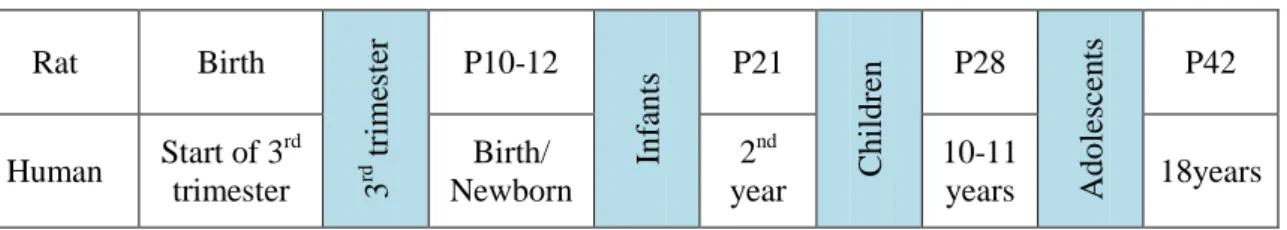
Table 4. Comparison between human and rat brain development.
Table 5. Impact of CBZ treatment on the mRNA levels of selected ABC and SLC transporters in isolated rat brain microvessels at post-natal days 14, 21 and 56.
Table 6. Impact of the CBZ treatment on the efflux activity of P-gP and BCRP efflux transporters in the BBB at post-natal days 14, 21 and 56.
10
AbreviationsABC - Adenosine triphosphate binding cassette AC - Astrocyte
AED – Antiepileptic drug Ang - Angiopoetin
BCRP – Breast cancer resistance protein BBB – Blood-Brain Barrier
CAR – Constitutive androstane receptor CBZ – Carbamazepine
cDNA – Complementary deoxyribonucleic acid CLB – Clobazam
CNS – Central nervous system CSF – Cerebrospinal fluid DGX – Digoxin DS – Dravet syndrome EC – Endothelial cell EEG - Electroencphalogram ER – Efflux ratio
FCD – Focal cortical dysplasia GABA - -aminobutyric acid
GEFS+ - Generalized epilepsies with febrile seizures plus GLUT – Glucose transporter
ILAE – International league against epilepsy IS – Infantile spasms
JAM - Junctional adhesion molecules
Kp,brain/plasma – Brain-plasma partition coefficient
LC-MS – Liquid chromatography coupled to mass spectroscopy MCT – Monocarboxylic acid transporter
11
mRNA – Messenger ribonucleic acidMRP – Multidrug resistance-associated protein nCLB – N-desmethylclobazam
NMDA – N-methyl-D-aspartate NPC – Neuropercursor cell NPR – Neuropilin receptor NVU – Neurovascular unit OAT – Organic anion transporter PDGF – Platelet derive growth factor
PECAM - Platelet Endothelial Cell Adhesion Molecules PET – Positron emission tomography
P-gP – P-glycoprotein PRZ – Prazosin
PXR - Pregnane X Receptor
RT-PCR - Real-Time Polymerase Chain Reaction SMEI - Severe Myoclonic Epilepsy of Infancy SNP – Single nucleotide polymorphisms SSeCKs - Src-Suppressed C-Kinase Substrate STP – Stiripentol
SUDEP - Sudden unexplained death in epilepsy TEER - Transendothelial Electrical Resistance TLE – Temporal lobe epilepsy
TPM – Topiramate TS – Tuberous sclerosis VBL – Vinblastine
VCAM – Vascular cell adhesion molecule VEGF – Vascular endothelial growth factor VPA – Valproic acid
12
P14 – Post-natal day 14 P21 – Post-natal day 21 P56 – Post-natal day 56 SLC – Solute Carrier ZO – Zona occludens13
DisseminationThe results obtained in this work have been presented in international congresses and meetings and are the subject of articles submitted or to be submitted.
5.1 – Resulsts of the exhaustive review of the published in vitro BBB models
This work is to be submitted to “Moelcular Pharmaceutics” journal under the tittle:
“In vitro models of the immature blood brain barrier for optimizing drug disposition” List of authors: R. Soares, A. Mabondzo, C. Chiron, S. Chhun, G. Pons
5.2 – Ontogeny of the ABC and SLC transporters in the microvessels of developing rat brain This work was submitted to “Fundamental and Clinical Pharmacology” on the 15th
of June 2015 under the title:
“Ontogeny of the ABC and SLC transporters in the microvessels of developing rat brain”
List of authors: Ricardo V. Soares, Tuan M. Do, Aloïse Mabondzo, Gérard Pons, Stéphanie Chhun 5.3 - Impact of the Antiepileptic Drugs used in the Dravet Syndrome on the Blood-Brain Barrier in the Immature Rat
This work is to be submitted to “Molecular Pharmaceutics” journal under the title:
“Impact of the Antiepileptic Drugs used in the Dravet Syndrome on the Blood-Brain Barrier in the
Immature Rat”
List of authors: Ricardo Viana Soares, Stéphanie Chhun, Vincent Jullien, Tuan Minh Do, Anne-Cécile Guyot, Catherine Chiron, Gérard Pons, Aloïse Mabondzo
The results assembled in this section have been partly presented as an oral communication at the 11th European Congress on Epileptology in july 2014 in Stockholm, Sweden; as an oral communication at the 19th Annual Meeting of the French Society of Pharmacology and Therapeutics in april 2015 in Caen, France; and as a poster in 11th International Conference on Cerebral Vascular Biology in june 2015 in Paris, France.
14
Table of Contents1. Introduction ... 16
2. State of the Art ... 18
2.1. Child Epilepsies ... 19
2.1.1. Epileptic seizures and Epilepsy ... 19
2.1.2. Childhood Epilepsies ... 20
2.1.3. Treatment of Epilepsy ... 22
2.1.4. Dravet Syndrome. ... 24
2.1.5. Focal Epilepsies ... 26
2.2. Resistance to antiepileptic drugs ... 28
2.2.1. Intrinsic epilepsy severity ... 28
2.2.2. Modifications on drug targets ... 29
2.2.3. Drug transport related mechanisms of resistance ... 30
2.3. Blood-Brain Barrier Drug Transport ... 36
2.3.1. Blood Brain Barrier ... 37
2.3.2. Types of Transport ... 38
2.4. Blood-Brain Barrier Development ... 44
2.4.1. Brain Development ... 46
2.4.2. Development of the Blood-Brain Barrier’s Functions ... 47
2.5. Methods for the study of drug transport through the Blood-Brain Barrier ... 50
2.5.1. In vivo Models ... 50
2.5.2. In vitro Models ... 52
3. Objectives ... 55
4. Methods ... 57
4.1. Methods of literature review: “In vitro models of the immature blood brain barrier for optimizing drug disposition” ... 58
4.2. Methods for the study of the BBB ontogeny: “Ontogeny of ABC and SLC transporters in the microvessels of developing rat brain” ... 58
4.3. Methods for the study of the impact of AEDs treatment in the developing BBB:” Impact of the antiepileptic drugs used in the Dravet syndrome on the Blood-Brain Barrier in the immature rat brain” ... 59
4.4 Impact of carbamazepine on the pattern of ABC and SLC transporters of the developing BBB... 61
15
4.5. Interaction of VPA with the efflux transporters P-gP and BCRP in an in vitro model of
the developing BBB constructed with primary cultures of rat brain endothelial cells ... 62
5. Results... 65
5.1. Results of the exhaustive review of the literature on the topic of in-vitro BBB models 66 5.2. Ontogeny of ABC and SLC transporters in the microvessels of developing rat brain . 95 5.3. Impact of the Antiepileptic Drugs Used in the Dravet Syndrome on the BBB phenotype in the Developing Rat Brain ... 125
5.4. Impact of carbamazepine on the pattern of ABC and SLC transporters of the developing BBB... 166
5.5 Interaction of VPA with the efflux transporters P-gP and BCRP in an in vitro model of the developing BBB constructed with primary cultures of rat brain endothelial cells ... 170
6. Discussion ... 172
6.2. Discussion on results ... 175
6.2.1. Animal models of immature blood brain barrier for optimizing drug disposition . 175 6.2.2. Ontogeny of ABC and SLC transporters in the microvessels of developing rat brain. 175 6.2.3. Impact of the antiepileptic drugs used in the Dravet syndrome on the Blood-Brain Barrier in the immature rat brain ... 176
6.2.4. Interaction of VPA with the efflux transporters P-gP and BCRP in an in vitro model of the developing BBB constructed with primary cultures of rat brain endothelial cells ... 177
7. Perspectives ... 178
8. Conclusion ... 180
16
17
Epilepsy is a disease of the central nervous system characterized by the occurrence of seizures which affects all ages and may have different causes. Antiepileptic Drugs (AEDs) have been developed, which act by different mechanisms to reduce the frequency of seizures. Nonetheless, one third of the patients do not respond to this therapy, especially children who represent 62% of the resistant patients. Dravet Syndrome is an example of infantile epilepsies that is frequently resistant to AEDs.
The causes of this resistance to antiepileptics are not well known. In this work we focused on the hypothesis that the pharmacoresistance in Epilepsy is related to reduced concentrations of the AEDs in the epileptic brain. This may be caused by the brain-blood interface, also named Blood-Brain Barrier (BBB). This structure is composed of cells from the brain parenchyma, such as pericytes astrocytes and neurons, and most importantly the endothelial cells of the brain capillaries. The BBB has special characteristics that may be responsible for the resistance to AEDs. Endothelial cells form tight junctions that reduce the permeability of this barrier. They express many types of transporters on their surfaces that control the passage of blood-borne molecules towards the brain.
The work presented in the present PhD thesis is composed of three main parts.
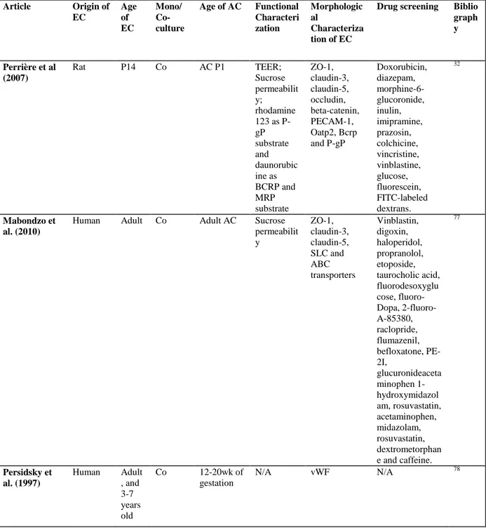
1 – An exhaustive review of the published in-vitro BBB models, based on the culture of endothelial cells, focused on the impact of brain maturation in these models;
2 – An experimental work on the ontogeny of the surface transporters belonging to the ATP-binding cassette (ABC) and SoLute Carrier (SLC) families of genes, in the microvessels of the developing rat brain;
3 – An experimental work where it was studied the impact of the AEDs used to treat the Dravet Syndrome on the efflux transporters present in the endothelial cells of the developing rat BBB. each of these three parts corresponding to a manuscript either already submitted or to be submitted for publication.
18
19
2.1. Child Epilepsies
2.1.1. Epileptic seizures and Epilepsy
Epilepsy is a disease of the Central Nervous System characterized by the existence of seizures which can affect all ages from newborns to the elderly. Seizures are related to the abnormal and excessive synchronous activity of neurons within neuronal networks, which can be localized in a small region of the brain cortex or spread out to both hemispheres. The ILAE defines an epileptic seizure as “a transient occurrence of signs and/or symptoms due to abnormal excessive or synchronous neuronal activity in the brain” (Fisher et al., 2005), which are recurrent and transiently interrupt normal brain function. Nonetheless, the exhaustive mechanisms by which neurons develop the synchronous activity characteristic of seizures are still unknown.
Epilepsy is defined by the International League Against Epilepsy (ILAE) as “a disorder of the brain characterized by an enduring predisposition to generate epileptic seizures and by neurobiological, cognitive, psychological and social consequences of this condition. The definition of epilepsy requires the occurrence of at least one epileptic seizure.” (Fisher et al., 2005). Epilepsy is characterized by criteria such as etiology, age of onset, seizure type, responsiveness to pharmacological treatment and other concomitant disorders (Forsgren et al., 2005). Specific associations of these criteria to specific types of epileptic seizures have been identified as “epilepsy syndromes” by the ILA (Berg et al., 2010); each syndrome carries its own diagnostic criteria, prognosis and drug response.
Seizures are the hallmark of epilepsy; however humans can have seizures without having epilepsy. One good example for this is febrile seizures in children: they may appear during an age-related period (1 to 4 years) after an infectious insult when body temperature rises, but most often this is an isolated episode without further affecting the child normal development (Offringa and Newton, 2012). Therefore, these patients usually do not require any chronic drug treatment.
Seizures are electro-clinical events. They result in non voluntary clinical features which correspond to the forced activity of the subsequent cortical regions involved from the onset to the end of the discharge. The diagnosis of seizures is essentially made by the description of symptoms from the patient itself or from a witness. Electroencephalography (EEG), a technique by which the electric activity of the brain is routinely recorded using electrodes on the scalp, is mandatory to precisely identify the region(s) of the brain involved in the epileptic discharge. Between seizures, during the so-called interictal periods, EEG may be normal or show various patterns of spikes and/or slow waves, which may occasionally be specific for a given epilepsy syndrome.
20
Seizures are mainly classified as either being focal (also called partial) or generalized. Focal seizures arise from neuronal activity from a discrete region of the brain in only one hemisphere. Affected individuals may be completely conscious of their symptoms, even perceive them before the discharge becomes clinically apparent, a phenomenon called aura. Consciousness can be however impaired to a varying degree. Generalized seizures imply bilateral neuronal discharges. They may be primarily or secondarily generalized. In the latter they correspond to focal seizures which evolve to affect both hemispheres, whereas the hypersynchronous activity is supposed to involve the whole cortex from the onset in the former. Generalized seizures are mainly characterized by their motor signs: tonic, clonic, tonic-clonic, atonic, or myoclonic seizures, also called “convulsive” seizures. Alternatively, absences seizures are non-convulsive generalized seizures, characterized by sudden and seconds lasting impairments of conscience that present with typical electroencephalographic features (Panayiotopoulos, 2008).
2.1.2. Childhood Epilepsies
The occurrence of seizures can begin from the first year of life. These episodes are nonetheless different from the adults: seizures in the neonatal period may have unclear symptoms, being difficult to define even in electroencephalographic records. Later on, they may appear as clear events, occasionally age-specific, such as epileptic spasms (Berg et al., 2010).
Beside the occasional seizures which are symptoms of many infectious or traumatic events occurring in childhood, but are not considered as epilepsies, pediatric epilepsies are more frequent than adult epilepsies. The incidence of epilepsy in the pediatric ages is higher than in adults. Estimations of incidence of epilepsy during the first year of life in the European population report between 153-256 new cases per 100.000 inhabitants per year. The incidence of epilepsy then decreases with age, with a minimum around 20-39 years-old individuals, to increase again after the 6th decade of life (Dulac et al., 2007; Forsgren et al., 2005).
Similarly, pediatric epilepsies are more various than adult epilepsies. Focal epilepsies (also called in therapeutic trials “epilepsies with partial onset seizures”) represent about the half of pediatric epilepsies, whereas the proportion is higher in adulthood. In addition, a non-lesional form of focal epilepsy is frequent in children (benign epilepsy with centro-temporal spikes), but does not exist in adults. Among the remaining epilepsies, the majority can be classified into epilepsy syndromes, based on distinct signs and symptoms (Chapman et al., 2012).
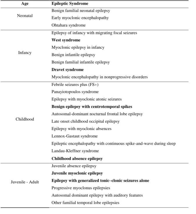
The classification of Pediatric Epileptic Syndromes has been recently updated by the ILAE classification commission (Berg et al., 2010). They are reported in Table 2 according to the period of
21
brain development when the first clinical manifestations appear. This list includes very severe epileptic syndromes, such as Dravet syndrome, West syndrome or Lennox-Gastaut syndrome, but also syndromes with better prognosis such as childhood absence epilepsy. Syndromes are defined by the age at onset, type of seizures, electroclinical characteristics, etiology and comorbidities.
Table 1. Pediatric Epileptic Syndromes (Berg et al., 2010), adapted (in bold the most frequent syndromes according to age).
Age Epileptic Syndrome
Neonatal
Benign familial neonatal epilepsy Early myoclonic encephalopathy Ohtahara syndrome
Infancy
Epilepsy of infancy with migrating focal seizures West syndrome
Myoclonic epilepsy in infancy Benign infantile epilepsy Benign familial infantile epilepsy Dravet syndrome
Myoclonic encephalopathy in nonprogressive disorders
Childhood
Febrile seizures plus (FS+) Panayiotopoulos syndrome
Epilepsy with myoclonic atonic seizures Benign epilepsy with centrotemporal spikes Autosomal-dominant nocturnal frontal lobe epilepsy Late onset childhood occipital epilepsy
Epilepsy with myoclonic absences Lennox-Gastaut syndrome
Epileptic encephalopathy with continuous spike-and-wave during sleep Landau-Kleffner syndrome
Childhood absence epilepsy
Juvenile - Adult
Juvenile absence epilepsy Juvenile myoclonic epilepsy
Epilepsy with generalized tonic–clonic seizures alone Progressive myoclonus epilepsies
Autosomal dominant epilepsy with auditory features Other familial temporal lobe epilepsies
These syndromes have frequently well defined genetic causes, which develop their consequences at different times of brain development, or are related to malformations that occur in the brain during the
22
mutations in genes SCN1A and SCN1B) or Tuberous Sclerosis associated seizures (mutations in genes
TSC1 and TSC2) which are usually part of West syndrome, Lennox-Gastaut syndrome or focal
epilepsy (Curatolo et al., 2015).
2.1.3. Treatment of Epilepsy
The management of patients with epilepsy is mainly performed by pharmacological treatment with Antiepileptic Drugs (AEDs). These drugs act through various mechanisms of action and most drugs act through more than one mechanism. The action of AEDs can be simplified as to inhibiting excitatory synapses, such as glutamatergic synapses, inducing inhibitory synapses such as gabaergic synapses, or modifications in ionic conductances. Such mechanisms include the inhibition of sodium and calcium channels, induction of -aminobutyric acid (GABA) chloride channels, inhibition of the metabolism of inhibitory neurotransmitters such as GABA, or inhibition of glutamate-dependent excitatory receptors. The main mechanisms of action of the AEDs are summarized in table 2.
Table 2. Current AEDs and their main molecular targets (Camfield et al., 1996; Perucca, 2005), adapted
Antiepileptic Drug Abbreviation Mechanism of action
Carbamazepine CBZ Voltage-gated Na+ channel inhibition
Clobazam CLB GABAA receptor agonist
Eslicarbazepine ESL Voltage-gated Na+ channel inhibition
Ethosuximide ESX Voltage-gated Ca2+ channels inhibition
Felbamate FLB NMDA receptor inhibition
Gabapentin GBP Voltage-gated Ca2+ channels inhibition Lamotrigine LTG Voltage-gated Ca2+ and Na+ channels inhibition
Levetiracetam LEV SV2A protein inhibitor
Oxcarbazepine OXC Voltage-gated Na+ channel inhibition
Perampanel PER AMPA receptor inhibition
Phenobarbital PB GABAA receptor agonist
Phenytoin PHT Voltage-gated Na+ channel inhibition
Stiripentol STP GABAA receptor agonist
Tiagabine TGB GABA transporter inhibition
Topiramate TPM Voltage-gated Na+ channel inhibition and increased GABA levels Valproic acid VPA Voltage-gated Na+ channel inhibition and increased GABA levels
Vigabatrin VGB GABA transporter inhibition
Zonisamide ZNS Voltage-gated Na+ channel inhibition
The development of AEDs has led to a considerable list of options to treat seizures. However, many patients do not respond completely to treatment and other therapeutic approaches have been used to
23
treat Epilepsy. Surgery is a treatment option in essentially two clinical settings: in focal epilepsy resistant to antiepileptic drugs and with a clear identified region where seizures develop, such is the case of cortical dysplasia or to some extent tuberous sclerosis; or when a complete brain hemisphere is injured whereas the other is not, e.g. Rasmussen Syndrome (Ryvlin et al., 2014). Another interesting non-pharmacological approach to treat epilepsy, in particular pediatric epilepsy, is the ketogenic diet. This therapy consists on shifting the child food intake towards a higher consumption of fat and low consumption of carbohydrates. This leads to a high rate of formation of ketone bodies, which are important energy substrates for the developing brain, with significant improvement on seizure control (Neal et al., 2008). Furthermore, vagus nerve stimulation has been applied in some pharmacoresistant children who are not eligible for epilepsy surgery (Joshi et al., 2013).
24
2.1.4. Dravet Syndrome.
Dravet Syndrome (DS) was first described by Charlotte Dravet as Severe Myoclonic Epilepsy of Infancy (SMEI) in 1978 and then recognized as an epilepsy syndrome by ILAE and updated several times (Dravet, 2011; Dravet and Oguni, 2013). Its incidence in epileptic children is estimated to be between 3% and 7% (Caraballo and Fejerman, 2006).
Since the first descriptions of the syndrome already a genetic basis had been recognized, because about 50% of patients had familial antecedents of seizures, particularly febrile seizures (Benlounis et al., 2001). It became apparent that a common genetic background existed between the patients with milder epilepsies such as generalized tonic-clonic seizures, febrile seizures and the more severe symptoms such as Dravet syndrome. This led to the creation of the Generalized Epilepsies with
Febrile Seizures Plus (GEFS+) assembling all these epileptic disorders (Scheffer and Berkovic, 1997). DS was then included in this group of disorders as its most severe form after a major mutation on
SCN1A gene had been identified in families with this syndrome (Scheffer et al., 2009; Singh et al.,
2001).
The clinical features of DS appear at about six months of age, when normally developing children initially present generalized long lasting febrile clonic or tonic-clonic seizures. Further non-febrile seizures will also as well as partial seizures such as hemiclonias, though in alternating body sides (Scheffer, 2012). Children usually present with a normal EEG at onset (Caraballo and Fejerman, 2006). As the child further grows up, other types of seizures emerge, such as myoclonic seizures and atypical absences. These are accompanied by changes in EEG (e.g. spike and waves) (Caraballo and Fejerman, 2006). Triggering factors for seizures are very common and consist mainly of temperature changes (fever, infection or hot baths) but also light or sound (Dravet, 2011). As the child grows older, seizure frequency tends to decrease and occur predominantly during sleep (Guerrini, 2012).
Further extending the neurological symptoms, DS children have a normal cognitive development rate until the second year of age, where it stagnates or even degrades so that children severely mentally retarded are frequent after the age of 6 years (Guerrini, 2012). Main deficits include attention disorders, poor motor coordination and gait abnormalities (Dravet, 2011). In addition DS carries one of the highest mortality risk in pediatric epilepsy, due to accidents or SUDEP (Sudden Unexplained Death in Epilepsy) (Dravet and Oguni, 2013).
The major mutation associated with the DS was first identified in 2001: they were de novo mutations on the α-subunit NaV1.1 (SCN1A gene), coding for missense, frameshift or truncation mutations (Claes
et al., 2001; Ohmori et al., 2002). Mutations on the same channel were also identified in GEFS+ patients (Escayg et al., 2000) as well as in β-subunit NaVβ1 (encoded by gene SCN1B) of the
voltage-25
dependent sodium channel (Wallace et al., 1998). The mutations in SCN1A presented in a group of DS patients seemed to cause a loss of function of the voltage-gated sodium channel (Sugawara et al., 2003), whereas in GEFS+ patients, mutations apparently increased sodium channel function (Lossin et al., 2002; Rhodes et al., 2005). Further analysis identified mutations in about 80% of DS patients and cases of mosaicism in parents with SCN1A mutations (Gennaro et al., 2006; Morimoto et al., 2006). Since then, mutations in PCDH19, a gene coding for procadherine, have been found in 5% of the DS females negative for SCN1A (Depienne et al., 2009), so that PCDH19 is now considered as the second gene involved in DS. Mutations on the γ2-subunit of the GABAA receptor were also detected in few
Dravet patients (Baulac et al., 2001).
Pharmacological treatment of DS has been addressed in very few clinical trials. The mainstay of the treatment is the combination of VPA, STP and CLB (Wilmshurst et al., 2015). STP is the only AED approved for treatment of DS after an exploratory open trial and two placebo-controlled randomized trials were conducted in French and Italian patients: these trials showed more than a 50% reduction in seizures frequency in about 70% of patients concomitantly treated with VPA and CLB (Chiron et al., 2000; Kassaï et al., 2008; Perez et al., 1999). These results were further confirmed in observational studies on DS patients in Japan and US; moreover response to STP was observed even in patients who did not receive co-medication with VPA or CLB (Inoue et al., 2009, 2014; Wirrell, 2013). The mechanism of action of STP appears to be related not only to direct activation of GABAA receptors
but also through a pharmacokinetic drug-drug inhibition of CLB metabolism (Quilichini et al., 2006). TPM can also be useful to decrease seizure frequency in children with DS, but only observational studies were performed (Chiron, 2011). On the opposite, sodium channel blocker AEDs such as LTG, CBZ and PHT tend to actually exacerbate seizures in DS patients and are not recommended (Wilmshurst et al., 2015). Finally, ketogenic diet has also been tried in DS with interesting results including in patients already treated with VPA, STP and CLB: 66% of the patients in ketogenic diet had a reduction in seizure frequency of at least 75% after 3 months of ketogenic diet (Nabbout et al., 2011). Other authors also reported a reduction of at least 50% in seizure rate in 70% of the patients after 3 months of diet and 60% after one year (Dressler et al., 2015).
DS is nonetheless still considered a pharmacoresistant syndrome. It has a very low probability to get completely control of the seizures and the likelihood of remission is also very low. DS summarizes the most significant predicting risk factors of pharmacoresistance: the early onset, high initial seizure frequency and the intellectual impairment (Wirrell, 2013). Furthermore, surgery is not suitable for DS children because DS does not present as focal epilepsy.
26
2.1.5. Focal Epilepsies
Focal (or partial) seizures are caused by the firing of neurons in a discreet area of the brain cortex. As previously mentioned, signs and symptoms which are displayed in seizures will vary depending on the affected region, and focal seizures may also spread locally or occasionally become secondarily generalized. The incidence of focal seizures in pediatrics is higher than generalized seizures: they account for 52% to 68% of the new cases, with an estimated incidence of 63.5 cases per 100000 persons each year (Camfield et al., 1996; Wirrell, 2013).
Epilepsies with partial onset seizures are called focal epilepsies. Contrary to the other pediatric epilepsies which are mostly age-related, focal epilepsies may occur at any age from birth to adulthood, although occipital epilepsies usually occur earlier in life than frontal epilepsies, according to the sequential brain maturation (Chiron and Duchowny, 2013; Chiron et al., 1992). Beside pure focal epilepsies with only focal seizures, several “non-determined whether focal or generalized” epilepsy syndromes have been described in pediatric patients, which associate focal and generalized seizures in an age-related profile (Berg et al., 2010). Among the most illustrating examples are West syndrome (or Infantile spasms), frequently associating focal seizures to epileptic spasms and an hypsarrhythmic EEG pattern in infants before 1 year of age (Hrachovy and Frost, 2013), and Lennox-Gastaut syndrome which associates focal seizures to generalized tonic, atonic and absence seizures with specific slow spike and waves on interictal EEG (Arzimanoglou et al., 2009). Focal epilepsy, West syndrome (Infantile spasms, IS) and Lennox-Gastaut syndrome can occasionally affect the same patient at different ages, especially in case of tuberous sclerosis (TS): TS is the most frequent cause of IS, focal seizures are most often associated with IS due to TS, and Lennox-Gastaut syndrome used to be the epilepsy outcome of the non-controlled IS due to TS (Curatolo et al., 2015).
First line AED treatment of focal seizures still remains on the administration of CBZ in children in Europe, except under the age of 2 years where CBZ may worsen IS and is therefore not recommended as long as the proof of pure focal seizures has not been established (Chiron and Duchowny, 2013). Contrary to adults, only OXC is approved for first line monotherapy among the new AEDs. Therefore second line most often consists in adjunctive therapy to CBZ, either clobazam or one of the new AED. The choice among them mostly depends on age (versus AED approval), seizure frequency (versus titration schedule), and potential interactions (versus pharmacokinetic profile). OXC, LTG, GBP, TPM, LEV are currently approved in children over 4 years, perampanel also is approved over 12 years, but only LEV is approved in infants and lacosamide, pregabaline, retigabine, zonisamide and eslicarbazepine are only approved in adults to date. In TS, vigabatrin is the drug of choice because of its demonstrated efficacy as first line monotherapy in IS due to TS (Chiron and Duchowny, 2013; Chiron et al., 1997). Due to the relationships between TSC1/TSC2, the TS genes, and the mTOR
27
pathway, mTOR inhibitor everolimus has been approved for treatment of SEGA in children and is currently under trial for focal seizures in TS over 4 years of age (Krueger et al., 2013). In refractory cases, surgery is an option to consider in focal epilepsy, at any age including the youngest ones. Except the Benign epilepsy with centro-temporal spikes previously mentioned, which is probably genetically determined and vanishes before adulthood in all cases, pure focal epilepsies are due to a cortical brain lesion. Lesion may be acquired (tumor, cerebral traumatism, cerebral infarct, and hippocampal sclerosis) or developmental and therefore present before birth (malformation, anoxo-ischemia, and antenatal infection). By contrast with adults, developmental lesions are the most frequent cause of pediatric focal epilepsies, mostly malformations of cortical development, with Focal Cortical Dysplasia (FCD) as the major etiology. FCD is detected in up to 7.5% of patients submitted to epilepsy surgery, moreover this percentage can reach up to 26% when considering exclusively the pediatric population submitted to surgery (Chassoux et al., 2000). FCD appears to be responsible for higher seizure frequencies, younger onset, and a higher proportion of patients with daily seizures than other lesions (Hauptman and Mathern, 2012; Semah et al., 1998).
In 1971 Taylor and colleagues identified a group of patients with intractable focal epilepsies submitted to brain surgery that presented marked histological disturbances in the removed tissue (Taylor et al., 1971). These findings clearly distinguished this group of patients: although they presented seizures similar to tuberous sclerosis, the microscopic findings of the removed tissue suggested an independent etiology. FCD is characterized by the disruption of the normal neuronal laminar distribution in the cortical grey matter, with blurring of the typical clear frontier between grey and white matter (Taylor et al., 1971). According to the ILAE updated classification, FCD type II remains the most typical form, with the classical giant dysmorphic neurons eventually associated to the balloon cells, whereas type I is less characteristic and often more extended. A new type III is now considered which dual pathology, that is FCD type II and remote hippocampal sclerosis, glial tumors, or other brain malformations (Blümcke et al., 2011). FCD type II is the perfect model of focal epilepsy since this lesion proved to be intrinsically epileptogenic: spontaneous seizures were directly recorded from the FCD using intracranial EEG with depth electrodes (Chassoux et al., 2000) and lesion brain tissue from adult patients operated on from FCD showed long-lasting epileptiform activity when exposed to 4-aminopyridine in an in vitro culture (Mattia et al., 1995). Although a proportion of patients up to 80% can reach seizure freedom after epilepsy surgery, there is still a proportion of patients that continue to have seizures after resection of the dysplastic area (Dhamija et al., 2011).
28
2.2.Resistance to antiepileptic drugs
Resistance to AEDs is of currently importance problem in the management of epileptic disorders. Despite the large number of AEDs that are available today to the neurologist and the development of consensus guidelines for the treatment of seizures, still a proportion of patients do not respond to the AEDs.
A practical definition of pharmacoresistant epilepsy has been proposed by the International League Against Epilepsy in 2010. Accordingly, “Drug resistant epilepsy may be defined as failure of adequate
trials of two tolerated and appropriately chosen and used AED schedules (whether as monotherapies or in combination) to achieve sustained seizure freedom.” (Kwan et al., 2010).
The development of pharmacoresistance in epileptic patients can differ individually, with patients who never respond appropriately to any AEDs and other patients who may have a fluctuating pattern, with periods with lack of response to the therapy and other periods of time where seizures are controlled (Gorter and Potschka, 2012). About one third of the epilepsy patients treated with AEDs will never respond to treatment, and fewer that 50% will have a good response to the first AED (Kwan and Brodie, 2000). This phenomenon is even more important in the pediatric population because after the failure of the first administered AED the probability of seizure is reduced to less than 30% (Ramos-Lizana et al., 2012). Moreover, 62% of the pharmacoresistant children are less than 4 years-old and this is associated with impaired cognitive and motor development (Ramos-Lizana et al., 2012). The main risk factors of pharmacoresistance in epilepsy population are early seizure onset (before 1 year of age), high number of seizures at onset, and the existence of a brain lesion (Berg et al., 2014).
The exact mechanisms through which resistance to the AEDs appear are not thoroughly discovered. The two main hypotheses that have been the subject of research in the past years are related to modifications in the targets of the AEDs and the reduced brain access to the AEDs. Recently a third hypothesis, related to the severity of the disease has been developed but more evidence is still needed. Drug transport related mechanisms will be discussed in more detail.
2.2.1. Intrinsic epilepsy severity
The hypothesis that resistance to AEDs is linked to the severity of the epilepsy was founded on the observation that patients who have high frequency of seizures are more commonly resistant to drugs. This phenomenon would be related to the reorganization of neuronal networks in the brain or changes in individual neurons that became very sensitive to triggering events and would not respond to the AEDs (Rogawski and Johnson, 2008). The actual mechanisms through which this resistance could
29
developed are still speculative, and more information concerning the neurobiological factors that impact on seizure severity must be obtained to further develop this hypothesis (Potschka et al., 2002).
2.2.2. Modifications on drug targets
The AEDs exert their effects by inhibiting or stimulating receptors in neurons. These receptors can be voltage-gated ionic channels, neurotransmitters receptors or enzymes involved in the metabolism of neurotransmitters. The hypothesis that resistance to AEDs was caused by modifications in the drug targets postulates that either intrinsic modifications (such as polymorphisms or other types of mutations) or acquired modifications (e.g. induced by drugs) are present in pharmacoresistant epilepsy.
Direct evidence of reduced antiepileptic efficacy was observed in sodium channels of temporal lobe epilepsy patients submitted to surgery. These channels are the major receptors of AEDs such as carbamazepine or phenytoin, and in these patients the pharmacoresistance was associated with a reduced capacity of carbamazepine to induce protective hyperpolarization in neurons (Gorter and Potschka, 2012). One study also revealed that in an animal model of epilepsy, alternative splicing of the rat sodium channel rNav1.2/rNav1.3 was observed after 4 hours induced status epilepticus (Aronica et al., 2001). The authors could not however analyze if there was a different sensibility to AEDs in this splicing variant.
GABA receptors are also interesting examples of target modifications that could be related to pharmacoresistant epilepsy. Apparently, the activation of these receptors causes a reduction in its sensibility towards benzodiazepines that could be related to internalization of these receptors (Gorter and Potschka, 2012). Moreover, GABA receptors are chloride channels that transport chloride down a concentration gradient: when chloride intracellular concentrations are lower than extracellular, the net movement of chloride is towards the cell cytoplasm inducing hyperpolarization. This mechanism is tightly regulated by other chloride channels, such as the NKCC1 or KCC2 channels that keep intracellular chloride concentrations lower than in the extracellular fluid (Maa et al., 2011). This chloride gradient is inverted during early stages of brain development and most importantly it was also observed in epileptic patients (Dzhala et al., 2005). Activation of the GABA receptor by benzodiazepines could lead to an actual outflow of chloride ions, causing depolarization of the neuron and induce seizures (Ben-Ari et al., 2007; Yamada et al., 2004).
These examples corroborate to some extent the possibility that drug target modifications are responsible for resistance to antiepileptics. Nonetheless increasing evidence concerning the reduction in brain concentrations of the AEDs are more strongly in favor of the drug transport hypothesis.
30
2.2.3. Drug transport related mechanisms of resistance
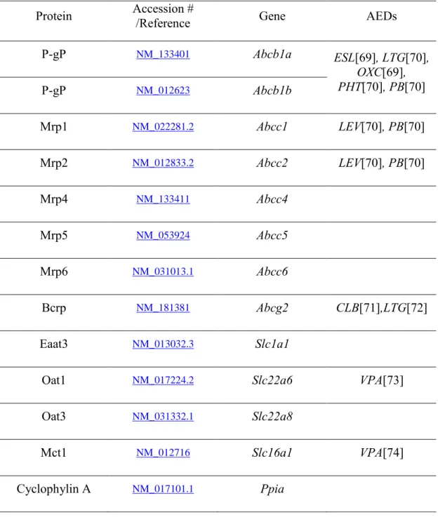
The brain is protected from potentially toxic substances present in the blood by a unique structure, the Blood-Brain Barrier (BBB). This barrier controls the passage of substances towards the brain and could reduce the AEDs concentrations in the brain, causing pharmacoresistant epilepsy (Gorter and Potschka, 2012). Patients resistant to AEDs frequently do not respond to a broad range of drugs, which act in different targets on neurons, suggesting that the resistance mechanisms may be unspecific. The ATP-binding cassette (ABC) efflux transporters and certain Solute Carriers (SLC) transporters that are present in the BBB seem to be a good explanation because they can transport molecules that have very different chemical structures. Resistance to AEDs should be linked therefore to high levels of ABC transporters in the epileptic brain and AEDs should also be substrates of these transporters.
2.2.3.1.
Alterations of the expression of efflux transporters expression in
the epileptic brain
The hypothesis that the resistance to AEDs is caused by the BBB appeared after the study of drug resistance in cancer. The decrease in the efficacy of certain anticancer drugs led to the discovery of the efflux transporter glycoprotein (gP) (Juliano and Ling, 1976). The first identification of higher P-gP gene expression in the capillaries of brain tissue of patients operated on for pharmacoresistant epilepsy led to the formulation of this hypothesis (Tishler et al., 1995). These results were later confirmed and more genes related to drug resistance, such as ABCC1, ABCC2 and ABCC5 were upregulated in brain specimens of temporal lobe epilepsy patients (Dombrowski et al., 2001). It should be stressed that these clinical conditions are associated with severe resistant epilepsy: they are therefore the typical candidates for epilepsy surgery and good examples of pharmacoresistant epilepsy. The upregulation of resistance genes in the brain of epilepsy patients was also found in other brain cells. Astrocytes showed increased immunoreactivity to P-gP in tissue from patients with temporal lobe epilepsy with hippocampal sclerosis and focal cortical dysplasia (Aronica et al., 2012). This expression was not only observed in astrocytes close to the capillary endothelial cells, but also in other astrocytes further away. The transporter BCRP (Breast Cancer Resistance Protein) has also been detected in endothelial cells, astrocytes and neurons (Aronica et al., 2012). This suggests that P-gP may have also an important role in protecting glial cells in pathological conditions. Moreover, an increase in P-gP and MRP1 immunoreactivity was observed in neurons of brain tissue from patients operated for focal cortical dysplasia (Sisodiya et al., 2002). This immunoreactivity was not observed in healthy neurons. Apparently, this increased expression of efflux transporters in brain parenchyma
31
and brain endothelial cells allows a coordinated elimination of eventual toxic substances from brain cells towards the cerebrospinal fluid and the blood.
The relationship between P-gP expression and decreased brain concentrations of AEDs was analyzed in patients treated with oxcarbazepine (Marchi et al., 2005). Its active metabolite 10-hydroxy-10,11-dihydrocarbazepine (10-OHCBZ) was quantified in the brain tissue of patients operated on for temporal lobe epilepsy. These concentrations were further correlated with the expression levels of P-gP (mRNA levels), where a negative correlation was observed: higher expression of P-P-gP correlated with low concentrations of 10-OHCBZ.
The study of the efflux activity of P-gP in the brain of epilepsy patients has recently been developed with PET-scan studies. A pilot test using [11C]-verapamil as a P-gP substrate showed that this radioligand had an apparent lower diffusion in the epileptogenic tissue of patients with temporal lobe epilepsy when compared with the non epileptogenic tissue of the same patient (Langer et al., 2007). These results where further confirmed in a recent study: [11C]-verapamil brain levels in patients with pharmacoresistant TLE were compared with non-pharmacoresistant TLE patients and with healthy controls. [11C]-verapamil uptake into the brain was negatively correlated with seizure frequency, and inhibition of P-gP with tariquidar (recognized as P-gp inhibitor) was most effective in healthy controls, which suggests higher expression of P-gP in pharmacoresistant patients (Feldmann et al., 2013). Moreover, in a subset of pharmacoresistant patients that were later submitted to surgery to remove the epileptogenic foci, immunostaining of P-gP was higher when [11C]-verapamil brain uptake was lower. Overall, these results pointed out that the higher expression of P-gP in pharmacoresistant patients that could increase the AEDs brain efflux.
The association between pharmacoresistant epilepsy and higher expression of efflux transporters was analyzed in animal models of epilepsy. By inducing seizures in rodents, either by injection of epileptogenic substances such as kainate or pilocarpine, or inducing status epilepticus through electrical stimulation, the increased expression of P-gP, MRP1 and MRP2 was observed (Aronica et al., 2012; Löscher and Potschka, 2005; van Vliet et al., 2004). This increased expression was observed not only in endothelial cells, but also in brain parenchyma cells such as astrocytes or neurons (van Vliet et al., 2005). Interestingly, in the epilepsy models induced by electrical stimulation, the higher expression of P-gP was associated with reduced brain concentration of phenytoin (van Vliet et al., 2006). Furthermore, when regional brain concentrations of phenytoin were measured, higher concentrations were achieved in brain regions with lower expression of P-gP (van Vliet et al., 2007). Whenever selective inhibition of P-gP was induced by treatment with tariquidar, the brain concentrations of either phenobarbital or phenytoin were increased (Brandt et al., 2006; van Vliet et al., 2007).
32
The link between pharmacoresistant epilepsy and increased efflux transporters expression led intuitively to the search of single nucleotide polymorphisms (SNP) in these transporters genes. These potential polymorphisms could be related to a reduction on the absorption, distribution of the AEDs, or an increase in the elimination of these drugs (Löscher et al., 2009). Polymorphisms on the ABCB1 gene were first analyzed in pharmacoresistant patients. The 3435C>T SNP, which was associated with increased expression and activity of P-gP, was more frequent in pharmacoresistant patient than in non-pharmacoresistant or control individuals (Siddiqui et al., 2003). Other polymorphisms in P-gP were also associated with increased resistance to AEDs, such as the 2677G>T or the 1236C>T, in fact the existence of haplotypes combining these three SNPs were discovered in resistant patients (Löscher et al., 2009). However, studies reported that there was a lack of association between these polymorphisms and the reduced activity of valproic acid, carbamazepine or phenytoin in pharmacoresistant patients (Grover et al., 2010; Haerian et al., 2011). This may be related to the lack of evidence concerning the possibility of VPA and CBZ being substrates of P-gP. Moreover, other AEDs appear to be weak substrates of efflux transporters, therefore limiting the impact of these polymorphisms in the clinical efficacy of the studied AEDs (Löscher et al., 2009). However, in-vitro assays where made with the genes containing these SNP that were transfected in cells later used for transport studies with P-gP substrates. This study showed that some of these SNP either alone or in combinations (1236T-2677T-3435T, 1236T-3435T, or 2677A alone) had an impact on P-gP efflux activity when AEDs such as CBZ, PHT, PB or LTG were present (Hung et al., 2008). Conflicting results have shown that in some group of patients SNP in the ABCB1 and ABCC2 genes seem to be associated with pharmacoresistance whether in other patients this association was not observed (Bruhn and Cascorbi, 2014; Subenthiran et al., 2013; Ufer et al., 2009). Finally, exploratory studies in order to search for other SNPs were made in pharmacoresistant patients of the pediatric population. Several SNPs were identified in a group of patients from Mexican origin and significant associations were observed for SNPs in the ABCC2 gene (Escalante-Santiago et al., 2014).
2.2.3.2.
Interactions of AEDs with the efflux transporters
To further substantiate that pharmacoresistant epilepsy is linked to the expression of efflux transporters in the BBB, the AEDs should be evaluated as substrates of these transporters. Many studies have analyzed the ability of the two major ABC transporters expressed in the BBB, P-gP and BCRP, to promote the efflux of AEDs.
Several major AEDs have been identified as P-gP substrates mostly in in-vitro assays using cells overexpressing the efflux transporters. Phenytoin, phenobarbital, lamotrigine and levetiracetam were all found to be effluxed by P-gP, although to a lesser extent when compared with a prototypical P-gP
33
substrate, digoxin (Luna-Tortós et al., 2008). These results were obtained in equilibrium conditions in order to detected weaker P-gP substrates, and were confirmed by inhibition assays using specific P-gP inhibitors such as tariquidar or MK571. Carbamazepine was studied but it was not found any interaction with P-gP, although a previous study using microdialysis in mice showed that inhibition of P-gP increased brain concentrations of CBZ (Potschka et al., 2001). Studies concerning the intestinal absorption of CBZ in mice also found no effect of P-gP (Fortuna et al., 2012). By contrast, AEDs with similar structures such as oxcarbazepine, eslicarbazepine or licarbazepine were identified as P-gP substrates in two cell lines (MDCKII and LLC-MDR1 cells), showing that small differences in structure can change dramatically the interactions with efflux transporters (Zhang et al., 2011).
The major AEDs were also studied as potential substrates of BCRP. In in-vitro assays with cell lines overexpressing BCRP, it was not observed any interaction between this transporter and phenobarbital, phenytoin, ethosuximide, primidone, valproic acid, carbamazepine, clonazepam, or lamotrigine (Cerveny et al., 2006). These AEDs were also analyzed as potential BCRP inhibitors, by studying its effects on the efflux of BCRP substrates such as prazosin and mitoxantrone. Only valproic acid at high concentrations (500μM) induced a small but statistically significant reduction in BCRP. A study with knock-out rats for genes both Abcb1a/b and Abcg2 (P-gP and BCRP, respectively), found out that the absence of BCRP originated increases in brain concentrations of phenobarbital, clobazam, zonisamide, gabapentin and levetiracetam (Nakanishi et al., 2013). Other drugs such as topiramate and tiagabine showed to be influenced by both transporters, indicating an overlap in the selectivity of these transporters. Topiramate was also shown to be substrate of P-gP in one study using knock-out mice for
Abcb1a gene (Sills et al., 2002).
Because other ABC transporters such as MRP1, MRP2 and MRP5 are overexpressed in epileptic tissue, cell-lines overexpressing these transporters were used to identify potential substrates amongst AEDs. Levetiracetam, valproic acid, carbamazepine, lamotrigine and phenobarbital were not shown to be substrates of these transporters (Luna-Tortós et al., 2010). However, the authors understate that this method can lead to false negative results, particularly with high permeability drugs that can cross easily biological membranes. For instance, in rats knockout for MRP2 phenytoin was shown to reach higher concentrations in the brain when compared with wild-type rats (Potschka et al., 2003).
Most of the studies have analyzed the interaction between AEDs and ABC transporters, and only a few have looked on the interaction with other types of transporters such as SLC carriers. However, the interaction of VPA with SLC transporters has been the subject of several studies. Initial studies showed that VPA influx towards the brain was higher than efflux (Cornford et al., 1985). This was also the first study to find an asymmetry between blood-to-brain and brain-to-blood transport of a substance. VPA competitively inhibited the brain uptake of acetate in primary brain endothelial cells derived from capillaries of calves, putting forward the possibility that VPA transport is carrier
34
mediated (Terasaki et al., 1991). Work from Adkinson and colleagues showed that transport of VPA through the BBB was dependent on organic anion transporters sensitive to probenecid and also to carriers of the monocarboxylic acids (Adkison and Shen, 1996; Adkison et al., 1994, 1996). These studies were essentially performed in rodents. Interestingly, studies comparing the effects of probenecid in the transport of VPA and E-2-VPA (a VPA metabolite) showed increased concentrations of both molecules in the brains of rabbits (Scism et al., 1997). Overall, this study brought further evidence that VPA is effluxed from the brain through a transporter sensitive to probenecid.
In microvessels of temporal lobe epilepsy patients with hippocampal sclerosis it was revealed a decrease in the expression of the monocarboxylic acid transporter 1 (MCT1) (Lauritzen et al., 2011). These samples were compared to autopsy specimens from individuals without epilepsy and to patients with temporal lobe epilepsy without hippocampal sclerosis. The authors claimed that this reduction on MCT1 transporter may reduce VPA concentrations in the epileptic foci. Furthermore, this reduction in MCT1 expression may be caused by the inflammation observed in the hippocampal tissue. The reduction in brain capillaries of the MCT1 transporter was also observed in induced animal models of temporal lobe epilepsy that was accompanied by increase in the expression of MCT1 in astrocytes (Lauritzen et al., 2012). This further suggests that the permeability of VPA in the epileptic tissue may be conditioned by this transporter in pharmacoresistant epilepsies. Moreover, an unidentified monocarboxylate transporter was shown to be important for the uptake of VPA in trophoblast cell lines (Utoguchi and Audus, 2000).
The organic anion transporters were identified as carriers of VPA. The most important transporters, OAT1 and OAT3 transporters, belong to the Solute Carriers family (SLC transporters), specifically the SLC22 sub-family. They are involved in the excretion of xenobiotics in the proximal convoluted tubule of the kidney but their expression is also detected in the brain (Burckhardt and Burckhardt, 2003). VPA was used as a competitive inhibitor of the common OAT substrate, p-aminohippurate, and specific cloning of the rat Oat1 transporter in Xenopus oocytes showed that Oat1 transported VPA (Sekine et al., 1997, 2000). It was further shown that the mouse Oat2 transporter mediated the uptake of VPA in Xenopus oocytes overexpressing this transporter (Scism et al., 2000). Uptake of fluorescein, a substrate of the anion transporter present in the mitochondrial membrane was inhibited by VPA (Kobayashi et al., 2002). Although VPA in humans is majorly eliminated through hepatic metabolism, these transporters could be important on VPA disposition in other tissues such as the brain.
The study of the interaction of VPA with the SLC transporters also showed that VPA can act as an inhibitor. The L-carnitine transporter OCTN2, expressed in the brush-border of the placental membrane, was inhibited by VPA (Lahjouji et al., 2004).
35
The interaction of VPA with ABC transporters seems to be less probable. Studies with VPA showed that P-gP, BCRP, MRP1, MRP2 and MRP5 do not transport VPA (Baltes et al., 2007; Cerveny et al., 2006; Luna-Tortós et al., 2010). One study in bovine brain capillary endothelial cells suggested that VPA was effluxed by MRPs using probenecid as a MRP inhibitor. Nonetheless the authors did not discard the potential effects of other organic anion transporters that might be inhibited by probenecid: higher concentrations of probenecid actually decreased the uptake of VPA, suggesting other mechanisms regulating VPA transport through endothelial cells (Gibbs et al., 2004).
VPA may however be important in the regulation of the ABC transporters expression and function. VPA was shown to induce a 4-fold induction in the expression and function of P-gP in tumor cell lines and also in rat liver tissue (Eyal et al., 2006). This induction seemed to be dependent on the activation of nuclear receptors such as the constitutive androstane receptor (CAR) and the progesterone X receptor (PXR) (Cerveny et al., 2007). VPA also appeared to induce P-gP expression in primary cultures of rat astrocytes (Lü et al., 2004).
The data concerning the drug transport hypothesis showed that it may potentially be the answer to the problem of pharmacoresistance in epilepsy. Not only should this information be useful to counteract the diminished activity of the AEDs but can also reveal new targets for the development of newer AEDs. Particularly to this work, the lack of information concerning transport of AEDs in the children should be the focus of future work in order to prevent the deleterious effects of uncontrolled seizures in the pediatric population.