i
Université de Montréal
Survival of the Fittest: Understanding the Role of eIF4E in
Cancer Invasion and Treatment Evasion
par
Hiba Ahmad Zahreddine
Département de Pathologie et Biologie Cellulaire
Faculté de Médicine
Thése présentée à la Faculté de Médicine
en vue de l’obtention du grade de Philosophiae Doctorat (Ph.D.) en Biologie Moléculaire
Option Biologie des Systémes
Mai 2017
ii
Université de Montréal
Faculté des études supérieures Cette thèse intitulée
Survival of the Fittest: Understanding the Role of eIF4E in Cancer Invasion and Treatment Evasion
Présentée par Hiba Ahmad Zahreddine
à été évaluée par un jury compose des personnes suivantes:
Dr. Lea Harrington Président-rapporteur Dr. Katherine LB Borden Directeur de thèse Dr. Daniel Zenklusen Membre du Jury Dr. Aaron Schimmer Examinatuer Externe Université de Toronto Dr. Richard Bertrand Représentant du doyen la FES
iii
Déclaration
Direction des bibliothèques AVIS
L’auteur a autorisé l’Université de Montréal à reproduire et diffuser, en totalité ou en partie, par quelque moyen que ce soit et sur quelque support que ce soit, et exclusivement à des fins non lucratives d’enseignement et de recherche, des copies de ce mémoire ou de cette thèse.
L’auteur et les coauteurs le cas échéant conservent la propriété du droit d’auteur et des droits moraux qui protègent ce document. Ni la thèse ou le mémoire, ni des extraits substantiels de ce document, ne doivent être imprimés ou autrement reproduits sans l’autorisation de l’auteur. Afin de se conformer à la Loi canadienne sur la protection des renseignements personnels, quelques formulaires secondaires, coordonnées ou signatures intégrées au texte ont pu être enlevés de ce document. Bien que cela ait pu affecter la pagination, il n’y a aucun contenu manquant.
NOTICE
The author of this thesis or dissertation has granted a nonexclusive license allowing Université de Montréal to reproduce and publish the document, in part or in whole, and in any format, solely for non-commercial educational and research purposes.
The author and co-authors if applicable retain copyright ownership and moral rights in this document. Neither the whole thesis or dissertation, nor substantial extracts from it, may be printed or otherwise reproduced without the author’s permission.
In compliance with the Canadian Privacy Act some supporting forms, contact information or signatures may have been removed from the document. While this may affect the document page count, it does not represent any loss of content from the document.
iv
Declaration
The thesis presented is the work of the author except when clearly stated otherwise by reference and/or acknowledgement. Any work performed in collaboration or conducted by others is explicitly acknowledged. The work has not been submitted for any other degree or
professional qualification.
May 10
th, 2017
v
RESUME EN FRANÇAIS ET MOTS CLES FRANÇAIS
La métastase et la chimiorésistance sont les principales causes de mortalité chez les patients atteints d’un cancer. La compréhension des mécanismes moléculaires régissant ces deux processus devient donc un domaine de recherche important pour la conception de nouvelles stratégies thérapeutiques. Dans ma thèse, je me concentre sur la compréhension du rôle du facteur d’initiation de la traduction chez les eucaryotes 4E (eIF4E) dans l’invasion du cancer, et je décris un nouveau mécanisme de résistance que nous avons découvert en étudiant le développement de la résistance à un inhibiteur connu d’eIF4E, la ribavirine.
eIF4E est un puissant oncogène qui est connu pour être élevé dans une multitude de cancers comprenant entre autres les sous-types M4 / M5 de la leucémie myéloïde aiguë (AML). Il fonctionne dans la traduction et l'exportation nucléocytoplasmique d'ARNm en se liant à la coiffe m7G des ARNm possédant des codes USER spécifiques dans leur région UTR 5' et/ou 3'. En reconnaissant ces codes USER, le complexe dans lequel se trouve eIF4E régule de manière coordonnée l'expression de gènes essentiels à la croissance, à la prolifération et à la survie, et ainsi, eIF4E a été placée en tant que nœud central d'un régulon d'ARN régissant la prolifération. En analysant les voies dans lesquelles l’export est régulé de façon coordonnée par eIF4E et les effets physiologiques qui en découlent, j'ai trouvé un enrichissement de la voie biosynthétique de l'acide hyaluronique (HA) et de son principal récepteur CD44 qui sont des médiateurs clés connus des métastases cancéreuses. J’ai également démontré que l'élévation d’eIF4E modifie la surface des cellules cancéreuses en les recouvrant de protrusions riches en HA de type microvillus et enrichies d'armes de destruction métastatique. Heureusement, en dégradant le manteau HA ou en utilisant des inhibiteurs de CD44 en combinaison avec la ribavirine, nous pouvons alors nous défendre.
Compte tenu de l'avantage prolifératif que confère la surexpression d’eIF4E, il est devenu un talon d'Achille attrayant pour le traitement de cancers ayant un niveau élevé d'eIF4E. En effet, lors d'un essai clinique de phase II parmi des patients atteints de leucémie myéloïde aiguë M4 / M5 réfractaire et récidivante, la ribavirine a conduit au ciblage d'eIF4E et a donné lieu à des réponses cliniques significatives, incluant des réponses complètes ou partielles. Cependant, tel qu’attendu lors d’un traitement monothérapique, les patients ayant répondu finissent par développer une résistance au médicament. Mon analyse a révélé que cette résistance est due à
vi
un mécanisme nouveau caractérisé par l'élévation du facteur de transcription Sonic Hedgehog GLI1 qui conduit à la glucuronidation du médicament et donc à la perte de l'interaction entre la drogue et sa cible. Heureusement, ce mécanisme peut être inversé en utilisant des inhibiteurs de la voie Hedgehog.
En conclusion, ces découvertes fournissent de nouvelles cibles thérapeutiques pour le traitement des cellules cancéreuses agressives et résistantes.
Mots clés : eIF4E, Cancer, Résistance multi-drogue, Invasion, GLI1, UGT1A, Acide
vii
RESUME EN ANGLAIS ET MOTS CLES ANGLAIS
Metastasis and chemoresistance are the leading cause of mortality among cancer patients. The discovery of molecular mechanisms governing these two processes is becoming an important area of research for the design of novel therapeutic strategies. In my thesis, I focus on understanding the role of the eukaryotic translation initiation factor 4E (eIF4E) in cancer invasion and describe a novel mechanism of resistance that we discovered while studying the development of resistance to a known eIF4E inhibitor, ribavirin.
eIF4E is a potent oncogene that is known to be elevated in a multitude of cancers including M4/M5 subtypes of acute myeloid leukemia (AML). It functions in mRNA translation and
nucleocytoplasmic export by binding to the m7G cap of mRNAs possessing specific USER
codes in their 5’ and/or 3’ UTRs. By recognizing these USER codes, eIF4E complex coordinately regulates the expression of genes essential for growth, proliferation and survival and as such has been placed as a central node of an RNA regulon governing proliferation. When analyzing which pathways have their export coordinately regulated by eIF4E and what physiological effects arise from it, I found an enrichment in the hyaluronic acid (HA) biosynthetic pathway as well as its major receptor CD44 which are known key mediators of cancer metastasis. I demonstrate that eIF4E elevation changes the surface of cancer cells sugar-coating them with HA-rich microvillus-like protrusions that are enriched with weapons of metastatic destruction. Luckily, through degrading the HA-coat or using inhibitors of CD44 in combination with ribavirin we can strike back.
Given the proliferative advantage that eIF4E overexpression conveys, this rendered it as an attractive Achilles heel for the treatment of cancers where eIF4E levels are high. Indeed, in a phase II clinical trial in refractory and relapsed poor prognosis M4/M5 AML patients, ribavirin led to eIF4E targeting and resulted in significant clinical responses including complete and partial remissions. However, as it is expected for monotherapy treatment, all responding patients eventually developed resistance to the drug. My analysis revealed that resistance is due to a novel mechanism characterized by elevation of the Sonic Hedgehog transcription factor GLI1 which leads to drug glucuronidation and the subsequent loss of drug-to-target interaction. Fortunately, this mechanism can be reversed using Hedgehog pathway inhibitors.
viii
Taken together, these findings provide novel therapeutic venues for the treatment of aggressive and resistant cancer cells.
Keywords: eIF4E, Cancer, Multidrug Resistance, Invasion, GLI1, UGT1A, Hyaluronic acid,
ix
List of Publications
This thesis is based on the following:
A-Main Research Papers:
1-The sonic hedgehog factor GLI1 imparts drug resistance through inducible
glucuronidation.
Zahreddine HA, Culjkovic-Kraljacic B, Assouline S, Gendron P, Romeo AA, Morris SJ, Cormack G, Jaquith JB, Cerchietti L, Cocolakis E, Amri A, Bergeron J, Leber B, Becker MW, Pei S, Jordan CT, Miller WH, Borden KL.
Nature. 2014 Jul 3;511(7507):90-3. doi: 10.1038/nature13283.
2-A phase I trial of ribavirin and low-dose cytarabine for the treatment of relapsed and
refractory acute myeloid leukemia with elevated eIF4E.
Assouline S, Culjkovic-Kraljacic B, Bergeron J, Caplan S, Cocolakis E, Lambert C, Lau CJ, Zahreddine HA, Miller WH Jr, Borden KL.
Haematologica. 2015 Jan;100(1):e7-9. doi: 10.3324/haematol.2014.111245.
3-The eukaryotic translation initiation factor eIF4E harnesses hyaluronan production to
drive its malignant activity
Hiba Ahmad Zahreddine1, Biljana Culjkovic-Kraljacic1, Lucy Skrabanek2, Ronald Midura3, Mark Lauer3, Sonia Del Rincon4, Audrey Emond4, Valbona Cali3, Leandro Cerchietti5, Wilson H Miller4, Vincent Hascall3, Katherine LB Borden1
Nature. 2017 (Submitted)
B-Extra Research Papers:
4-Conformational changes induced in the eukaryotic translation initiation factor eIF4E by a
clinically relevant inhibitor, ribavirin triphosphate.
Volpon L, Osborne MJ, Zahreddine H, Romeo AA, Borden KL.
Biochem Biophys Res Commun. 2013 May 10;434(3):614-9. doi: 10.1016/j.bbrc.2013.03.125.
5-Combinatorial targeting of nuclear export and translation of RNA inhibits aggressive
B-cell lymphomas.
Culjkovic-Kraljacic B, Fernando TM, Marullo R, Calvo-Vidal N, Verma A, Yang S, Tabbò F, Gaudiano M, Zahreddine H, Goldstein RL, Patel J, Taldone T, Chiosis G, Ladetto M, Ghione P, Machiorlatti R, Elemento O, Inghirami G, Melnick A, Borden KL, Cerchietti L.
x
Blood. 2016 Feb 18;127(7):858-68. doi: 10.1182/blood-2015-05-645069.
C-Reviews:
1-Mechanisms and insights into drug resistance in cancer.
Zahreddine H, Borden KL.
Front Pharmacol. 2013 Mar 14;4:28. doi: 10.3389/fphar.2013.00028.
2-Inducible drug modification: a new form of resistance.
Culjkovic-Kraljacic B, Zahreddine HA, Borden KL.
Cell Cycle. 2014;13(16):2485-6. doi: 10.4161/15384101.2014.946372.
3-Sonic Hedgehog factor Gli1: As good as resistant.
Zahreddine HA, Culjkovic-Kraljacic B, Borden KL.
Mol Cell Oncol. 2015 Feb 24;2(1):e961827. doi: 10.4161/23723548.2014.961827.
4-Molecular Pathways: GLI1-Induced Drug Glucuronidation in Resistant Cancer Cells.
Zahreddine HA, Borden KL.
xi
Table of Contents
CHAPTER 1 ... 1
INTRODUCTION... 1
1.1 The eukaryotic translation initiation factor 4E: a multifaceted protein ... 4
1.1.1 Cytoplasmic eIF4E: Initiates mRNA translation ... 5
1.1.2 Cap-Dependent Eukaryotic Translation Initiation ... 5
1.1.3 Nuclear eIF4E: mediates mRNA export ... 6
1.1.4 eIF4E dependent mRNA export goes through a pathway distinct from bulk mRNA export ... 7
1.1.5 eIF4E mRNP traversing the nuclear pore complex ... 9
1.1.6 eIF4E remodels the nuclear pore ... 10
1.2 eIF4E is regulated at multiple levels ... 11
1.2.1 Regulation of eIF4E transcription (Myc, NF-kB) ... 11
1.2.2 Regulation of eIF4E mRNA stability ... 12
1.2.3 Regulation of eIF4E activity by protein interactions ... 12
1.2.4 Regulation of eIF4E activity by post-translational modifications ... 13
1.3 Dysregulation of eIF4E levels in Cancer ... 14
1.3.1 Acute Myeloid Leukemia (AML) ... 14
1.3.2 The French-American-British classification of AML ... 14
1.3.3 The World Health Organization (WHO) classification of AML ... 15
1.3.4 AML: Aberrant eIF4E expression and activity ... 15
1.4 Targeting eIF4E with Ribavirin in Cancer ... 15
1.4.1 Ribavirin suppresses eIF4E mediated transformation by physical mimicry of m7G cap ... 16
1.4.2 Ribavirin treatment targets eIF4E and leads to clinical benefit in poor prognosis AML patients ... 17
1.4.3 Ribavirin combination therapy for AML treatment ... 18
1.5 Ribavirin resistance and non-responding patients ... 18
1.5.1 The Hedgehog Signalling Pathway in Cancer Treatment Resistance ... 19
1.5.1.1 Molecular Mechanism: ... 19
1.5.1.2 HH signalling in cancer ... 20
1.5.2 Glucuronidation mediated by UGT enzymes ... 22
xii
1.6 eIF4E in cancer invasion and metastasis ... 23
1.6.1 The metastatic cascade ... 24
1.6.2 Hyaluronic Acid is required for EMT of cancer cells ... 24
1.6.2.1 HA structure and synthesis ... 25
1.6.2.2 HA-induced signal transduction ... 25
1.6.2.3 HA mediates that formation of cell surface protrusions ... 27
Hypotheses and Aims ... 28
CHAPTER 2 ... 41
Mechanisms and insights into drug resistance in cancer ... 41
CHAPTER 3 ... 51
The Sonic Hedgehog Factor GLI1 Imparts drug resistance through inducible glucuronidation ... 51
3.6 Patent: ... 70
CHAPTER 4 ... 72
A Phase I trial of Ribavirin and Low-Dose Cytarabine for the Treatment of Relapsed and Refractory Acute Myeloid Leukemia with Elevated eIF4E ... 72
CHAPTER 5 ... 77
GLI1-Induced Drug Glucuronidation in Resistant Cancer Cells ... 77
CHAPTER 6 ... 83
Inducible Drug Modifications: A New Form of Resistance ... 83
CHAPTER 7 ... 87
Sonic Hedgehog Factor GLI1: As good as Resistance ... 87
CHAPTER 8 ... 92
The eukaryotic translation initiation factor eIF4E harnesses hyaluronan production to drive its malignant activity... 92
CHAPTER 9 ... 133
Combinatorial Targeting of Nuclear Export and Translation of RNA Inhibits Aggressive B-cell Lymphomas ... 133
CHAPTER 10 ... 163
Conformational Changes Induced in the Eukaryotic Translation Initiation Factor eIF4E by a Clinically Relevant Inhibitor, Ribavirin Triphosphate ... 163
CHAPTER 11 ... 180
xiii
List of Tables
-Chapter 3:
Extended Data Table 1: RNA-seq results of genes with different expressions in FRII versus
parental and FRI cells (page 68)
-Chapter 4:
Table 1: Patients characteristics (page 74)
Table 2: Response and ribavirin plasma levels (page 75) -Chapter 9:
xiv
List of Figures
-Chapter 1:
Figure 1: eIF4E regulates mRNA translation and mRNA export. (page 4)
Figure 2: Model for the eIF4E-dependent mRNA export and re-entry of the machinery via
importin 8. (page 8)
Figure 3: eIF4E is regulated at multiple levels. (page 13) -Chapter 2:
Figure 1: A major impediment in the treatment of cancer is the development of resistance (page
45)
-Chapter 3:
Figure 1: Ribavirin resistance in patients and cell lines (page 54) Figure 2: GLI1 resistance underpins resistance (page 55)
Figure 3: Targeting GLI1 activity (page 56)
Figure 4: Link between GLI1, UGT1A and drug glucuronidation (page 56)
Extended Data Figure 1: Ribavirin resistance in some ribavirin monotherapy clinical trial
patients (page 61)
Extended Data Figure 2: Characterization of FaDu- and THP-1 derived resistant cell lines
(page 62)
Extended Data Figure 3: Pro-drug metabolism is impaired in type I resistance (page 63) Extended Data Figure 4: Confocal micrographs of leukemic blasts isolated from bone marrows
of responding and non-responding patients before treatment, at response or at EOT (page 64)
Extended Data Figure 5: Higher GLI1 expression is found in poor cytogenetic risk group and
predicts a trend of worse survival outcome in AML (page 65)
xv
Extended Data Figure 7: MS analysis of ribavirin and Ara-C glucuronidation (page 67) Extended Data Figure 8: Investigation into why GLI1 levels are elevated in FRII cells (page
68)
-Chapter 4:
Figure 1: Percentage of bone marrow blasts following treatment (page 79) -Chapter 5:
Figure 1: Summary of means to regulate glucuronidation enzymes (page 80) -Chapter 6:
Figure 1: How cancer cells resist drugs (page 86) -Chapter 7:
Figure 1: Novel mechanism of multidrug resistance driven by GLI1 (page 90) -Chapter 8:
Figure 1: eIF4E regulates the expression of HA synthesizing enzymes and HA receptor CD44
(119)
Figure 2: eIF4E overexpression correlates with increased HA synthesis (page 121) Figure 3: eIF4E elevates HA in cancer cell lines and primary specimens (page 122)
Figure 4: Surface HA is required for eIF4E-mediated invasion and migration of cancer cells
(page 124)
Figure 5: HA biosynthesis is required for eIF4E-mediated lung metastasis in mice (page 125) Extended Figure 1: eIF4E regulates HA synthesis (page 126)
Extended Figure 2: eIF4E elevates HA in cancer cell lines and primary specimens (page 127) Extended Figure 3: eIF4E concentrates in HA rich protrusions and correlates with sites of
xvi
Extended Figure 4: Surface HA is required for eIF4E-mediated invasiveness of cancer cells
(page 129)
Extended Figure 5 part 1: CD44 is required for the invasion of eIF4E cells (page 130) Extended Figure 5 part 2: CD44 is required for the invasion of eIF4E cells (page 131) -Chapter 9:
Figure 1: eIF4E is expressed in DH/TH DLBCL (page 137) Figure 2: MYC, BCL2, and BCL6 are eIF4E targets (page 138)
Figure 3: Nuclear eIF4E regulates the export of lymphomagenic transcripts (page 140)
Figure 4: Ribavirin is active a TH DLBCL patient-derived xenograft (PDX) model (page 141) Figure 5: Antilymphoma effect of combined inhibition of eIF4E and TEHsp90 (page 142) Figure 6: Ribavirin decreases TEHsp90 inhibition–induced Hsp70 upregulation (page 144) Supplemental Figure 1: TEHsp90 proteomics pathway visualization (page 150)
Supplemental Figure 2: Nuclear and cytosolic fractionation of SU-DHL6, DoHH2 and
OCI-Ly1 cells (page 151)
Supplemental Figure 3: EIF4E is expressed in DLBCL (page 151)
Supplemental Figure 4: Total mRNA levels in U2OS overexpressing eIF4E plasmids or
siRNA (page 152)
Supplemental Figure 5: Control mRNA levels in U2OS cells (page 153)
Supplemental Figure 6: eIF4E regulates protein levels of lymphomagenic transcripts (page
154)
Supplemental Figure 7: STRING visualization of eIF4E-RIP sequencing top 1000 most
significant transcripts with 0.70 confidence (page 155).
Supplemental Figure 8: Polysomal profiling and input levels in DLBCL cell lines treated with
xvii
Supplemental Figure 9: Establishment of a triple-hit DLBCL patient-derived xenograft (page
157)
Supplemental Figure 10: Effect of combined inhibition of eIF4E and TEHp90 in oncoproteins
expression (page 158)
Supplemental Figure 11: Polysomal profiling of DLBCL cell lines treated with PU-H71 (page
159)
Supplemental Figure 12: Polysomal loading of genes in DLBCL cell lines treated with
PU-H71 (page 160)
-Chapter 10:
Figure 1: Analysis of RTP binding and protein samples (page 166)
Figure 2: NMR analysis of the low micromolar eIF4E-RTP complex is characterized by
intermediate and slow exchange (page 167)
Figure 3: NMR analysis of the high micromolar eIF4E-RTP complex is characterized by fast
exchange (page 168)
Figure 4: Comparison of the residues affected in the RTP and m7GTP eIF4E complexes (page 169)
Supplementary Figure 1: 1H-15N HSQC spectra of human eIF4E or apo eIF4E (page 173) Supplementary Figure 2: NMR analysis of the low and high micromolar eIF4E-RTP complex
(page 174)
Supplementary Figure 3: Comparison of the residues affected on the dorsal surface in the RTP
and m7GTP eIF4E complexes (page 175)
Supplementary Figure 4: The concentration dependence of eIF4E and its cap ligand (page
176)
xviii
Abbreviations
4E-BP eIF4E Binding Protein
4E-SE eIF4E-Sensitivty Element
4GI 4E/4G Inhibitor
ADK Adenosine Kinase
AhR Aryl Hydrocarbon Receptor
AKT Serine/Threonine-Specific Protein Kinase
AMAC 2-Aminoacridone
AML Acute Myeloid Leukemia
AraC Cytarabine
ARE AU Rich Element
AUF1 ARE Binding Factor
AZA Azacitidine
BC-CML Blast Crisis Chronic Myelogenous Leukemia
BCL2 B-Cell Lymphoma 2
BM Bone Marrow
BSA Bovine Serum Albumin
β–TrCP F-box/WD Repeat-Containing Protein 1A (FBXW1A)
C-MET Receptor Tyrosine Kinase
CAR Constitutive Androstane Receptor
CD34 Hematopoietic Progenitor Cell Antigen
CD44 Indian blood group
CML Chronic Myelogenous Leukemia
CO2 Carbon Dioxide
CRM1 Chromosome Region Maintenance 1
CUL Cullin
DEPC Diethyl Pyrocarbonate
xix
DTT Dithiothreitol
DDX DEAD-box Helicases
DMEM Dulbecco’s Modified Eagle Medium
DNA Deoxyribonucleic Acid
ECM Extracellular Matrix
EDTA Ethylenediaminetetraacetic Acid
eIF3 Eukaryotic Initiation Factor 3
eIF4A Eukaryotic Translation Initiation Factor 4A
eIF4E Eukaryotic Translation Initiation Factor 4E
eIF4G Eukaryotic Translation Initiation Factor 4G
EGF Epidermal Growth Factor
EGFR Epidermal Growth Factor Receptor
EMT Epithelial-Mesenchymal Transition
ENT Equilibrative Nucleoside Transporter
ER Endoplasmic Reticulum
ERBB4 erb-b2 Receptor Tyrosine Kinase 4
ERM Ankyrin and Ezrin, Radixin and Moesin proteins
EWS-FLI1 Ewing's Sarcoma - Friend Leukemia Integration 1 Transcription Factor Fusion protein
FBS Fetal Bovine Serum
FDA Food and Drug Administration
FITC Fluorescein Isothiocyanate
FKBPL FK506 binding protein like
GA Glucuronic Acid
GAG Glycosaminoglycan
GAPDH Glyeraldehyde 3-Phosphate Dehydrogenase
GATA Erythroid Transcription Factor
xx
GLI Glioblastoma Associated Protein
GRB2 Growth Factor Receptor-Bound Protein 2
GTP Guanosine Triphosphate
HA Hyaluronan
H&E Hematoxylin and eosin
HAS Hyaluronan Synthase
HEPES (4-(2-Hydroxyethyl)-1-piperazineethanesulfonic Acid )
HEX Hematopoietically Expressed Homeobox
HH Hedgehog Protein
HNF Hepatic Nuclear Factor
hnRNPA1 Heteronuclear Ribonuclear Protein A
HOXA9 Homeodomain Box Protein A9
HPG L-Homopropargylglycine
HuR Human Antigen R
IGF Insulin Growth Factor
ITCH E3 Ubiquitin-Protein Ligase Itchy Homolog
LRP/PRC Leucine-Rich Pentatricopeptide Repeat Containing
m7G Methyl-7-Guanosine
M-MLV Moloney murine leukemia virus
MCL1 Myeloid Cell Leukemia Protein 1
MDM2 Mouse Double Minute 2 Homolog
MDR Multidrug Resistance
MDS Myelodysplastic Syndrome
MMP Matrix Metalloproteinase
mRNA Messenger Ribonucleic Acid
mRNP Messenger Ribonucleoprotein
MYC Myelocytomatosis Protein
xxi
NaCl Sodium Chloride
NB Nuclear Body
NBS1 Nibrin
NFAT Nuclear Factor of Activated T-cells
NF-κB Nuclear Factor Kappa-light-chain-enhancer of activated B-cells
NMR Nuclear Magnetic Resonance
NP-40 Nonyl Phenoxypolyethoxylethanol
NPC Nuclear Pore Complex
NUMB Endocytic Adaptor Protein
NUP Nucleoporin
ODC Ornithine Decarboxylase
P53 Tumour Protein p53
PAX Paired Box Protein
PBS Phosphate-buffered saline
Pen/Strep Penicillin Streptomycin
PET Polyethylene Terephthalate
PFA Paraformaldehyde
PIM-1 Proto-Oncogene, Serine/Threonine Kinase
PML Promyelocytic Leukemia
PPAR Peroxisome Proliferation-Activated Receptor
PRH Proline-Rich Protein HaeIII Subfamily
PTCH PATCHED
PTEN Phosphatase and Tensin Homolog
PU.1 Purine Rich (PU) box Binding Protein
RANBP Ran-Binding Protein
RPMI Roswell Park Memorial Institute medium
RBV Ribavirin
xxii
RIP RNA Immunoprecipitation
RNA Ribonucleic Acid
RTP Ribavirin- 5’-Triphosphate
SDF-1 Stromal Cell-Derived Factor 1
SMO SMOOTHENED SNAIL Zinc Finger Protein SNAI1
SP1 Specificity Protein 1
SPOP Speckle Type BTB/POZ Protein
STAT Signal Transducer and Activator of Transcription
SUFU Suppressor of Fused
TAP/NXF1 Nuclear RNA Export Factor 1
TBS Tris-Buffered Saline
TGFβ Transforming Growth Factor β
TK Thymidine Kinase
UAP56 ATP-dependent RNA helicase
UDPGA Uridine 5’-diphosphoglucuronic acid
UGT UDP-Glucuronosyltransferases
USER Untranslated Sequence Elements for Regulation
UTR Untranslated Regions
VEGF Vascular Endothelial Growth Factor
VpG Viral Protein Genome-linked
WHO World Health Organization
xxiii
Dedicated to my father, Ahmad Zahreddine My mother, Mariam Dib And all my family and friends,
xxiv
Acknowledgements
The research resulting in this thesis was conducted at the Institute for Research in Immunology and Cancer (IRIC), Faculty of Medicine, University of Montreal, CANADA, from September 2011 to January 2017.
I am sincerely indebted to all the people who encouraged and supported me through out my Ph.D. It is my pleasure to pay tribute to them.
Foremost, I would like to extend my sincere gratitude to Dr. Katherine LB Borden for being the best supervisor I could have asked for. Thank you for your patience, guidance, encouragement and positive spirit over the course of this research. Without your time, energy and enthusiasm this work would not have been accomplished. My sincerest thank you for all your effort to help me grow as an independent researcher.
I genuinely acknowledge all the funding agencies that had supported my research: The Cole Foundation, and the National Council for Scientific Research _Lebanon (CNRS).
Thank you to the members of my thesis committee for their scientific knowledge and input. Dr. Lea Harrington (IRIC-UdeM), Dr. Koren Mann (LDI-McGill University), Dr. Aron Schimmer (University of Toronto), Dr. Daniel Zenklusen (UdeM), Dr. Claude Perreault (IRIC-UdeM) as well as Dr. Trang Hoang (IRIC-UdeM). I am forever grateful for the excellent advice.
Dr, Biljana Culjkovic-Kraljacic, our research associate; What would I have done without you? Thank you for your intellectual input, collaborative efforts and guidance. You are the principal source of motivation that has kept me focused and moving forward. Your endless support has
xxv
allowed me to work tirelessly. The time I have spent on this thesis was filled with joy and happiness because of your companionship.
I would also like to acknowledge my past and present lab mates (Laurent Volpon, Mike Osborne, Lotfi Amri Abdellatif, Maggie Davis, Luciana Coutinho de Oliveira, Jadwiga Gasiorek, Daria Krutauz, Mildred Delaleau and Sahil Mann) for their help, support and suggestions throughout my Ph.D. Furthermore, I would like to thank all the staff at the histology, genomics and imagery platforms at IRIC. A special tribute to Dr. Sylvie Mader’s lab members for always being there and for the fun times we have spent together. Mohammad El Ezzy, your friendship and conversations have gone long ways to preserve my sanity!
My parents Ahmad and Mariam who have given me a lifetime of love and support. Thank you for being so patient throughout this period of my life. You have sacrificed so much in order for me to have amazing life opportunities. Special thanks to my Aunt Fatima Muldowney, without you I would not have accomplished anything. You have shaped me in to a better person with your life advice and knowledge.
Last but not least, I want to thank my siblings (Lama, Zainab, Marwa, Mohammad and Issa)
and friends (Mano Jbai, Houssam Ismail, Samir Jlassi, AhmadRaza, Ahmad Koleilat, Alex
Pilon, and Caroline Cote!) for their endless friendship and encouragement. You guys have helped me to remain clear-headed and confident. I love you guys!
Hiba Zahreddine
1
CHAPTER 1
2
It was probably 2,500 years BC when the first evidence of cancer had been documented in The Papyrus of Edwin Smith, “a bulging tumour in the breast… like touching a ball of wrappings”. Discussing treatment, the ancient scribe noted: “There is none” (1). From the fire drill and natural remedies recommended by ancient Egyptian medicine to our era’s most recent concepts of “magic bullet” and “molecular-targeted therapies” (2), the cancer matrix remains unlocked. Even though targeted therapy has achieved encouraging results in the treatment of specific types of neoplasms, primary or acquired resistance to chemotherapeutics as well as metastasis of cancer cells remain as two of the main causes of failure in the treatment of patients. It might be that our failure to eradicate most tumours is as fundamental as our lack of understanding to their origin. Had it been the consequence of multicellularity and longer lifespans, then how come not all whales have cancer- given that the more cells we have and the longer we live the higher the risk of developing tumours- a question raised by Peto R. and his colleagues back in 1975 (3). Peto’s
Paradox depicts that no correlation exists between body size and age with increased cancer risk; rather it is the mere fact that age equals duration of exposure to carcinogenic stimuli. Since then, resolving Peto’s Paradox has become the focus for many researchers, as it might hold the key to curing or even preventing cancer. Perhaps evolution of these large organisms had selected for intrinsic cancer suppression mechanisms, such as decreased rates of somatic mutations, redundancy of tumour suppressor genes, shorter telomeres, the presence of hypertumours that grow on and destroy their parent tumour, more efficient immune system and suppression of inflammation, higher resistance to oncogenic viruses, …etc. (4-6). Which of these mechanisms, if any, is/are likely to contribute to the observed suppression remains elusive (3). What is clear is that resolving this paradox might direct towards a better understanding of the molecular mechanisms governing cancer drug resistance and metastasis.
The new era of targeted therapy has proven that the complexity of human cancers can be broken down by targeting a single hijacked oncogene, a phenomenon referred to as “oncogene addiction”. This phenomenon allowed initial steps towards a breakthrough in the treatment of tumour cells without affecting their normal counterparts. In my thesis, I aim at understanding the oncogenic dependency of cancer cells on the roles of a protein known as the eukaryotic translation initiation factor 4E (eIF4E) for their survival and metastasis. I demonstrate how eIF4E changes the surface of tumour cells arming them with microvillus-like protrusions that
3
are enriched with molecules required for invasion, including Hyaluronic acid (HA), CD44, and matrix metalloproteinases, among others. Results from this study provide first evidence of an oncogene that post-transcriptionally regulates the biosynthesis of HA to harness its oncogenic potential. It also suggests that direct targeting of HA-positive tumour cells with available FDA-approved Hyaluronidases could be a valuable addition to treatment regimens for high-eIF4E, and potentially other cancers. In another chapter, I discuss the serendipitous discovery of a novel multidrug resistance mechanism while investigating the molecular underpinnings of the development of resistance to a given eIF4E inhibitor, Ribavirin. I show that elevation of the Sonic Hedgehog transcription factor GLI1 correlates with increased drug glucuronidation which in turn disrupts drug-to-target interaction. Fortunately, I further demonstrate that this mechanism can be reversed using FDA approved Sonic Hedgehog signalling pathway inhibitors.
4
1.1 The eukaryotic translation initiation factor 4E: a multifaceted protein
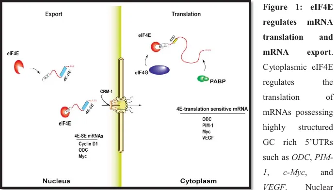
In response to environmental signals, cells regulate gene expression and protein synthesis in a coordinated fashion. Compared to corn and fruit fly, the human genome encodes approximately the same or nearly twice the number of genes, respectively. In addition, it has become increasingly clear that the majority of eukaryotic proteins function in different cellular processes. This multitasking phenomenon suggests that, as the number of genes does not proportionally increase with size and complexity of organisms, the eukaryotic regulatory mechanisms therefore act to diversify the proteome by shuffling the limited number of genes to use them in multiple combinations. Indeed, many eukaryotic proteins have been shown to multitask as components of different cellular complexes, the roles of which might differ with subcellular localization. This multitasking phenomenon holds true for the eukaryotic translation initiation factor 4E (eIF4E), which is traditionally known for its function in translation initiation of 5’-capped mRNA in the cytoplasm (7-9). Recent studies have highlighted a novel role of eIF4E in the nucleus where it regulates mRNA export (Figure 1). Accordingly, eIF4E provides an example of a factor that differentially regulates coordinated gene expression patterns.
Figure 1: eIF4E regulates mRNA translation and mRNA export. Cytoplasmic eIF4E regulates the translation of mRNAs possessing highly structured GC rich 5’UTRs such as ODC,
PIM-1, c-Myc, and VEGF. Nuclear
eIF4E mediates the export of mRNA molecules containing a unique ~ 50 nucleotides sequence element known as eIF4E-senstivity element (4E-SE), including Cyclin D1, ODC and c-Myc.
5
1.1.1 Cytoplasmic eIF4E: Initiates mRNA translation
In 1976, Witold Filipowicz discovered eIF4E as a cap binding protein involved in cytoplasmic mRNA translation (10, 11). Here, eIF4E binds the 7-methyl guanosine dinucleotide cap structure m7GpppN (where N is any nucleotide) located on the 5’ end of mRNA (10, 12-14). Consequently,
protein synthesis commences. Sensitivity of given mRNAs to eIF4E’s translational activity does not only rely on the binding of eIF4E to the m7G cap (8, 10, 13, 15). Overexpression studies have
shown that eIF4E does not lead to global increase in protein expression (12, 16). Analyses
indicated that only a given subset of mRNAs possessing highly structured GC rich 5’UTRs have their translation regulated by eIF4E (8, 10, 13, 15). These mRNAs usually encode for growth and survival factors. Thus when overexpressed, eIF4E disproportionally and dramatically induces the translation of mRNAs encoding for cancer-related proteins that control cell proliferation (8,
13).
1.1.2 Cap-Dependent Eukaryotic Translation Initiation
mRNA translation is the process in which mRNA molecules are decoded to produce specific sequences of amino acids, or proteins (17-19). It proceeds in three stages: initiation, elongation and termination; with initiation being the rate-limiting step and as such is subject to tight control
(20-23). Translation initiation consists of the recruitment of ribosomes to target mRNA. At least
two different mechanisms of ribosome binding have been identified in eukaryotic cells: the cap-dependent and the cap-incap-dependent scanning (21, 24, 25). In this thesis, I focus on the eukaryotic
translation initiation factor 4E which is an inherent component of the cap-dependent translation initiation. eIF4E mediates binding of the 40S ribosomal subunit to the 5’end of capped mRNAs in the cytoplasm (18, 20, 21, 23). Together with eIF4A (an RNA-dependent ATPase/ATP-dependent RNA helicase) and eIF4G (a high molecular weight scaffold protein that binds eIF4E and eIF4A) it forms the eukaryotic translation initiation factor 4F (eIF4F) (18, 20, 21, 23). Through interaction of eIF4G with the 40S ribosomal binding factor eIF3 and the poly(A)-binding protein, the eIF4F complex forms a critical link between mRNAs and ribosomes (18, 20, 21, 23). Given that expression of the various eIF4F factors in most cells is readily different, with eIF4E being the least abundant, formation of the aforementioned link therefore relies on the availability of latter (19, 26, 27). A family of small translation repressor molecules known as eIF4E-binding
6
proteins (4E-BPs) have been shown to interact with eIF4E and modulate the assembly of eIF4F
(28-30). Binding of 4E-BPs to eIF4E is modulated by phosphorylation (17, 31, 32). In the presence of
hormonal or nutritional stimuli, 4E-BPs are hyperphosphorylated and do not interact with eIF4E allowing for protein synthesis to occur. On the contrary, environmental or nutritional stress elicit hypophosphorylation of 4E-BPs which can now bind strongly to eIF4E thus rendering it unavailable for initiation of mRNA translation. In addition to regulation by signalling pathways via 4E-BPs, eIF4E expression and activity is controlled by transcriptional and posttranscriptional mechanisms, which will be discussed in this chapter.
1.1.3 Nuclear eIF4E: mediates mRNA export
Discovery of the role of eIF4E in catalyzing nucleocytoplasmic export of mRNA has been a major advance. Up to 68% of eIF4E is found in the nucleus of most eukaryotic cell types either in distinct mulitprotein structures known as eIF4E nuclear bodies (NBs) or distributed throughout the nucleoplasm (33, 34). Electron microscopy studies in Saccharomyces cerevisiae, and later on in Drosophila melanogaster S2 cells and Xenopus laevis embryos, also showed nuclear eIF4E localization (35-37); suggesting that nuclear eIF4E is conserved across eukaryotes. eIF4E-dependent mRNA export was first reported by Rousseau et al. where eIF4E overexpression increased nucleocytoplasmic export but not translation of cyclin D1 (38). Later studies showed that treatment of eIF4E nuclear bodies with cap but not RNases results in complete release of eIF4E and dispersal of these bodies (39). As such, similar to its cytoplasmic counterpart, nuclear eIF4E also binds the m7G cap and that the cap, but not the mRNA per se, is essential for nuclear localization and function of eIF4E (35). However, cap binding is not the sole determinant of mRNA export mediated through eIF4E; as indicated by the increased export of cyclin D1 but not the housekeeping mRNAs (such as GAPDH) following eIF4E overexpression (38, 40, 41). Mapping of the 3’ and 5’-UTRs identified that the basis for this
discriminatory interaction is an approximately 50-nucleotide sequence in the 3’-UTR, which is referred to as eIF4E sensitivity element (4E-SE) (40). Further, differential display analysis of
nuclear eIF4E-associated mRNAs following immunoprecipitation of endogenous eIF4E from nuclear lysates, revealed that among the hundreds of mRNAs many of the identified genes are involved in cell cycle progression and survival, including c-Myc, cyclin E1, Mdm2 and NBS1
7
(41). Accordingly, in addition to its role in regulating translation initiation, eIF4E-mediated
nuclear export of a specific subset of mRNA also contributes to oncogenesiss.
The functions of eIF4E in mRNA export and translation appear to be decoupled as they rely on different USER codes, i.e. mRNAs require both the 3’ 4E-SE and the 5’ USER code to be modulated by eIF4E at both levels (12, 41). For instance, some mRNAs are said to be export targets only (such as cyclinD1), others are translational targets (such as VEGF), and in some cases, mRNAs could possess both USER codes and thus have their export and translation regulated by
eIF4E (such as ODC, c-Myc and Pim-1) (12, 41). According to the RNA regulon model, gene
expression is combinatorial, i.e. related to the different USER codes found in a given mRNA (42,
43). Therefore, through promotion of mRNA export and translation, the eIF4E regulon provides
a coordinated and combinatorial way for the cell to more finely tune gene expression. It is one of the earliest examples of an RNA regulon which through coordinately modulating proliferation and survival signal gene expression networks is positioned to directly impact human diseases, including oncogenic transformation; necessitating tight control over its expression and activity.
1.1.4 eIF4E dependent mRNA export goes through a pathway distinct from bulk mRNA export
The molecular basis for the mRNA nuclear export function of eIF4E is less well understood than its role in translation. eIF4E only associates with its target mRNAs post-splicing and the eIF4E-4E-SE RNA complexes are found in the soluble export competent fraction within the nucleus. While the majority of messenger ribonucleoproteins (mRNPs) exit the nucleus using
bulk mRNA export pathway, catalyzed by TAP/NXF1 factors, eIF4E mRNPs do not (41, 44).
Treatment of cells with leptomycin B, a specific inhibitor of the nuclear pore receptor chromosome region maintenance 1 (CRM1), inhibited the export of these mRNAs; indicating, that eIF4E-dependent mRNA export is CRM1 dependent (Figure 2) (41, 44). Recently, our lab identified the leucine-rich pentatricopeptide repeat protein C (LRPPPC) as platform for the
assembly of eIF4E, 4E-SE mRNA, and CRM1 (44). Following release of mRNA cargo in the
cytoplasm, cap-free eIF4E and 4E-SE-free LRPPRC are recycled back in to the nucleus via Importin 8, a member of the karyopherin family of transporters (unpulished data). Interestingly,
8
the fact that both LRPPRC and eIF4E use Importin 8 to re-enter the nucleus suggests that their import could be coordinated in a way to increase the efficiency of future export cycles. The molecular mechanisms underlying eIF4E-mediated mRNA export and the specific composition of the eIF4E mRNA export complex are becoming increasingly clear. As such, LRPPRC could be one of several assembly platform proteins involved in the selective export of mRNA subgroups whose expression could be context/cell type specific. Further, a better understanding of the physiological role of the eIF4E-4E-SE RNA complexes is an important area of future work.
Figure 2: Model for the eIF4E-dependent mRNA export and re-entry of the machinery via importin 8. LRPPRC binds to both eIF4E and 4ESE RNA using distinct N- and C-terminal
motifs, and binds CRM1 supporting transit though the nuclear pore complex (NPC). Once the complex arrives at the cytoplasmic side and after the dissociation of 4ESE RNA cargoes, eIF4E and LRPPRC return to the nucleus via Importin 8. Here, Importin 8 only binds cap-free
9
eIF4Eand RNA-free LRPPRC thereby reducing futile export cycles. (Taken from Volpon L. et
al. RNA 2016)
1.1.5 eIF4E mRNP traversing the nuclear pore complex
In addition to LRPPRC, eIF4E export mRNPs contain some factors shared with the bulk export pathway, including UAP56, hnRNPA1, and DDX3 (44-46). Also, in the nucleus CRM1 binds its
cargo in the presence of the bound form of Ran. Accordingly, as the CRM1-Ran GTP-eIF4E mRNP complexes are formed, these are targeted to traverse the nuclear pore complex (NPC) by virtue of the transport receptor interaction with specific NPC-proteins lining the central transport channel, namely Nucleoporins (Nups) (45, 47). Reaching the cytoplasmic face of NPCs, loading and release of the CRM1-cargo complexes occurs by either one of two mechanisms; (1) docking of the CRM1-cargo complex on cytoplasmic fibrils mainly composed of Nup358 proteins anchored to the NPC via two nucleoproteins Nup88 and Nup214; thus sequestering the CRM1-cargo complex; or (2) binding of CRM1-cargo complexes to a soluble form of nucleoporins known as RanBP1 which provides a fast release mechanism (13, 45). Either way, association is then followed by the recruitment of Ran GTPase-activating protein (RanGAP) enabling GTP hydrolysis by Ran (13, 45). Once this step is completed, CRM1-cargo complexes dissociate, permitting the RNA to enter the cytoplasm and the export factors to be recycled back to the nucleus (13, 45, 47). Note that, in nuclear import, RanGTP acts as a dissociation factor since import factors cannot bind both cargo and RanGTP simultaneously (13, 45, 47). Hence, RanGTP gradient across the nuclear envelope results in the activation several proteins including the cytoplasmic GTPase-activating protein RanGAP, and is considered the major driving force for nuclear transport in both directions (47, 48). Importantly, endogenous 4E-SE mRNAs could be targets of both bulk and eIF4E-dependent processes, where 3’-UTRs can be 1000s of
nucleotides in length and contain many USER codes (41). Thus, eIF4E competes with bulk
mRNA export pathway to enhance preferentially the export of specific subset of transcripts (13, 45, 46).
10 1.1.6 eIF4E remodels the nuclear pore
The loading and release options that CRM1 has at the cytoplasmic face of the NPC question whether the link between eIF4E’s transforming ability and its mRNA export activity is due to the favoring of RanBP1-mediated fast release over the rate limiting RanBP2-mediated process; and whether eIF4E could be driving this preference. Consequently, would eIF4E upregulation alter the expression and/or localization of NPC components.
Indeed, work presented by Culjkovic B. et al. revealed that eIF4E overexpression correlates with changes in the composition of the cytoplasmic face of the NPC (13, 47). The authors identified
RanBP1 but not RanBP2 as a direct export target of eIF4E. As such, the expression of these two proteins is inversely correlated following eIF4E overexpression. While RanBP1 levels are upregulated, eIF4E overexpression indirectly decreases RanBP2 proteins; with the remaining RanBP2 being more dispersed throughout the nucleoplasm. Further, the RanBP2 partner, Nup214, is not a direct eIF4E target but its localization is also altered from rim-concentrated to a more dispersed form throughout the nucleoplasm. Accordingly, these results indicate that eIF4E overexpression does favor a RanBP1 release pathway which enables enhanced mRNA export by promoting release and/or recycling of export complexes. This is consistent with data showing that RanBP2 hypomorph mice, which have genetically lower RanBP2 levels, do not have bulk mRNA export defects but have increased export of specific mRNAs.
Interestingly, eIF4E overexpression may not only change the CRM1-mediated export of 4E-SE mRNAs, but it can also alter the export of a subset of mRNAs using bulk export pathway (mediated by TAP/NXF1) (13, 47). This occurs via upregulating the expression of DDX19 and its cofactor Gle1 which catalyze the loading and release step of the TAP/NXF1 pathway. These data indicate the presence of a cross talk between RNA export pathways or that the helicase activity of DDX19 is required for remodeling 4E-SE export RNPs. On the other hand, both DDX19 and Gle1 have been shown to play independent roles in the initiation and termination steps of translation of certain mRNAs, indicating that eIF4E can impact on translation in an indirect way, in addition to its direct effects (13, 47). Thus, eIF4E not only is exported by CRM1
via its interactions with LPRPRC, but also modulates proteins acting in the CRM1 pathway to likely maximize its mRNA export potential.
11
In conclusion, these observations indicate that eIF4E has the capacity to alter the composition of NPC in favor of increasing its mRNA export and oncogenic activities.
1.2 eIF4E is regulated at multiple levels
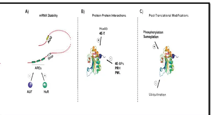
Clearly, eukaryotic cells evolved means to regulate different nodes in RNA regulons in order to alter gene expression and prevent deleterious outcomes. Studies aiming to identify regulators of eIF4E expression and activity are still ongoing and regulation can be divided into four levels: transcriptional regulation and regulation through mRNA stability, protein interactions and post-translational modifications (Figure 3).
1.2.1 Regulation of eIF4E transcription (Myc, NF-kB)
Dysregulation of eIF4E in cancer is correlated with increased RNA and protein levels (~ 3-to 10-fold in M4/M5 AML specimens relative to normal samples). While c-Myc has long been thought as the sole transcriptional regulator of eIF4E (49-51), recent studies are unveiling novel mediators. Analysis of eIF4E promoter demonstrates enrichment with binding sites for various
transcription factors including NF-κB, PU.1, NFAT, GATA, STAT, PAX and SP1 (52-54). For
instance, NF-κB has four putative binding sites (termed κB 1 to 4) in eIF4E promoter (52). These sites are occupied by a heterodimer comprising two members of the NF- κB family, namely cRel and p65. Binding results in the recruitment of p300 and phosphorylated RNA polymerase II to the κB sites and the coding region, respectively, thus marking transcription activation (52).
This is consistent with the presence of more NF-κB complexes on eIF4E promoter in primary M4/M5 myeloid samples as compared to normal primary hematopoietic cells and other myeloid subtypes (52). As such, genetic and pharmacological inhibition of NF-κB in this population
abrogates eIF4E transcription resulting in downregulation of eIF4E targets (52). Interestingly,
there is a significant overlap between eIF4E target genes and those of NF-κB (such as c-Myc and cyclin D1) which suggests that these two pathways cooperate forming a nexus between transcriptional and posttranscriptional gene expression networks to drive proliferation (Hariri, F unpublished data). Consequently, targeting both nodes, i.e. eIF4E and NF-κB, could have significant clinical utility in cancer patients where both proteins are elevated.
12 1.2.2 Regulation of eIF4E mRNA stability
Independent of the transcript levels, eIF4E expression is also regulated post-transcriptionally. The 3’UTR of eIF4E mRNA contains three AU rich elements (AREs) that dictate binding of competing proteins, HuR and p42 AUF1, to enhance or reduce eIF4E transcript stability, respectively (44). As such, HuR is found to be elevated in cancers with high eIF4E levels and its
knockdown is associated with downregulation of eIF4E (44).
1.2.3 Regulation of eIF4E activity by protein interactions
eIF4E is subject to additional level of regulation where its activity is tightly controlled via inhibitory or activating binding partners. So far, four classes of direct binding partners have been identified:
i. Proteins containing conserved eIF4E binding site, such as 4E-BPs, eIF4G, HOXA9 and PRH/Hex (14, 35). These proteins share a short conserved amino acid motif
YXXXXLϕ, where X is any residue and ϕ is a hydrophobic amino acid. As a result, their binding to eIF4E is said to be competitive to inhibit or enhance its mRNA export and/or translation activity. More than 200 homeodomain proteins containing this consensus binding motif have been identified; suggesting a redundancy in eIF4E regulators as 4E-BP knockout mice, for instance, were not more prone to developing cancers than their normal counterparts.
ii. RING domain containing proteins including PML, Z-protein, and HHAR1 (55). This
class of binding partners lacks the eIF4E binding motif but possess a RING domain instead. Direct binding of these proteins via the RING domain to eIF4E reduces its affinity to the m7G cap (~ 100 fold by PML and Z-protein).
iii. Amphipathic helix strategy used by VpG proteins (56, 57). Binding of VpG to eIF4E also
reduces its affinity to the m7G cap but the mechanism by which it does that is still not known.
iv. Importin 8 binding to eIF4E at its cap-binding site (48). This binding selects only
RNA-free eIF4E for nuclear entry and represents a new class of interacting partners.
In addition to the aforementioned regulatory modes, novel structural and biochemical functions of an eIF4E family member, eIF4E3, implicating it as a repressor of eIF4E activity via an mRNA
13
competition mechanism has been identified (58-60). Here, eIF4E3 competes for the same mRNA pools through a novel cap binding activity which inhibits expression of both mRNA export and translation targets of eIF4E.
1.2.4 Regulation of eIF4E activity by post-translational modifications
Lastly, regulation of eIF4E activity also occurs via post-translational modifications, including
phosphorylation, sumolyation and ubiquitination (61-65). While phosphorylation has been
implicated in enhanced eIF4E-mediated mRNA export and increased cell transformation capacity, the role of ubiquitination and sumolyation on eIF4E activity is still ambiguous.
Figure 3: eIF4E is regulated at multiple levels. (A) Regulation of mRNA stability via HuR
and AUF proteins which bind to AU rich elements (AREs) in the 3’UTR of the eIF4E transcript
(B) Regulation of eIF4E activity through protein-protein interactions. For instance, HOXA9
promotes the export and translation functions of eIF4E, while PML and PRH negatively regulate eIF4E-mediated mRNA export (C) Regulation by post-translational modifications including phosphorylation, sumolyation and ubiquitination.
14
1.3 Dysregulation of eIF4E levels in Cancer
Aberrant regulation of eIF4E expression and/or activity has been linked to malignancies as well as cell transformation. The pro-survival properties of eIF4E have been underscored by various evidence. For instance, its overexpression blocks apoptosis upon serum starvation, inhibits c-Myc-driven apoptosis, promotes DNA synthesis, decreases cell cycle transit time and represses differentiation (41, 66-68). Of which, downregulation of eIF4E reverts these phenotypes. Further,
moderate overexpression of eIF4E results in increased production of growth factors that are essential for malignant transformation (10, 69). And high eIF4E levels have been correlated with oncogenic transformation in cell lines, cancers in animal models and poor prognosis in several human cancers (10, 69). Approximately 30% of human cancers including: breast, prostate, lung, colon, squamous head and neck carcinoma, Hodgkin and non-Hodgkin lymphomas, as well as M4 and M5 subtypes of acute myeloid leukaemia (AML), have high eIF4E expression (10, 69, 70).
1.3.1 Acute Myeloid Leukemia (AML)
Acute myeloid leukemia is a hematologic malignancy characterized by aberrant proliferation of immature cells (myeloblasts) of the myeloid lineage. It is a particularly challenging malignancy since the majority of patients diagnosed are older than 60 years of age and often cannot receive intensive chemotherapy (70, 71). The overall survival of such patients has been estimated to be around 4 months, with a five-year survival rate of less than 10%, highlighting the need for new treatments and increasing the interest in developing them (71-73).
1.3.2 The French-American-British classification of AML
According the French-American-British (FAB) system, AML is subdivided into 9 distinct types based on the cell of origin and its level of maturity (74, 75). It includes: M0 (undifferentiated acute myeloblastic leukemia), M1 (Acute myeloblastic leukemia with minimal maturation), M2 (Acute myeloblastic leukemia with maturation), M3 (Acute promyelocytic leukemia (APL)), M4 (Acute myelomonocytic leukemia), M4eos (Acute myelomonocytic leukemia with eosinophilia) M5a (Acute monoblastic leukemia), M5b (Acute monocytic leukemia), M6 (Acute erythroid leukemia) and M7 (Acute megakaryoblastic leukemia).
15
1.3.3 The World Health Organization (WHO) classification of AML
A new classification for hematopoietic and lymphoid malignancies has been published recently by the World Health Organization (WHO) (76, 77). This new system classifies AML into three clinical prognosis groups based on not only morphologic findings but also on available clinical features, and genetic, biologic and immunophenotypic data. These subgroups include: (1) AML with recurrent genetic abnormalities, (2) AML with multilineage dysplasia, and (3) AML and MDS (myelodysplastic syndrome), therapy related.
1.3.4 AML: Aberrant eIF4E expression and activity
eIF4E levels are upregulated in various genetically distinct leukemias (10, 69). Strikingly, eIF4E overexpression is observed in M4/M5 poor prognosis AML but not in most M1/M2 specimens examined (78). Inline with this observation, inhibition of eIF4E using low concentrations (10 times less than physiological levels) of the m7G cap physical mimic, Ribavirin, leads to growth inhibition in M4/M5 but not normal or M1/M2 AML specimens which are inhibited at much higher levels (78, 79). Interestingly, not only the levels of eIF4E are upregulated in M4/M5 AML cells but also its nuclear localization is predominantly augmented (47, 48, 71, 73). This correlates with increased eIF4E-dependent mRNA export (40, 69, 79). Accordingly, both the mRNA export
and translation initiation activities are required for eIF4E-mediated malignant transformation. And these findings suggest that AML cells have developed an oncogenic dependency to eIF4E for their proliferation and survival.
1.4 Targeting eIF4E with Ribavirin in Cancer
Collectively, targeting eIF4E should have a major impact on tumorigenesis and cancer progression and as such has derived interest in identifying therapeutic agents that could directly or indirectly target its aberrant activation. Indeed, several preclinical and clinical methods have been defined including (i) anti-sense oligos targeting eIF4E in xenograft models (80-82), (ii)
synthetic peptides inhibiting the interaction of eIF4E with proteins involved in translation, such as eIF4G using 4EGI-1 to block formation of the eIF4F complex in T-cell leukemia and non-small-cell lung cancer cells (83), (iii) suicide gene therapy characterized by fusing a complex
16
eIF4E overexpressing cells in mouse xenograft models (84), and (iv) using a cap mimetic, known as Ribavirin (71, 73, 79, 85, 86).
1.4.1 Ribavirin suppresses eIF4E mediated transformation by physical mimicry of m7G cap
Ribavirin (RBV) is an FDA approved antiviral drug used for the treatment of patients with hepatitis C virus (71, 73, 79). It belongs to a family of nucleoside drugs and has a structure that is
closely related to guanosine. RBV is imported into cells using the equilibrative nucleoside transporter 1 (ENT1) which mediates facilitated bidirectional diffusion of nucleosides across cell membranes. In order to stay in the cell, ribavirin is phosphorylated in a three-steps reaction to its major intracellular metabolite, ribavirin- 5’-triphosphate (RTP); where the rate-limiting enzyme is adenosine kinase (ADK) catalyzing step one (87). In vitro biophysical assays identified RTP as a physical mimic of m7G cap and ultimately as a direct inhibitor of eIF4E (79, 86). Direct binding of RTP to eIF4E has been shown by fluorescence, circular dichroism, mass spectrometry, nuclear magnetic resonance (NMR) and cap chromatography with affinities for eIF4E similar to that of cap (79, 86). 1H-15N chemical shift mapping studies at low eIF4E and RTP concentrations are consistent with the high affinity binding previously reported. Chemical shift perturbations indicate that binding is located around the m7G cap binding site, which is consistent with the model that RTP competes for m7G cap binding. Further evidence for this is supported by the W56A cap-binding mutant which reduces RTP affinity for eIF4E by ~ 15-fold similar to the effects for cap; additionally, amide chemical shifts for W56A eIF4E mutant are not perturbed upon addition of RTP, indicating loss of interaction. Similar to m7G cap, RTP binding perturbs peaks at the dorsal surface: although different residues are perturbed. Whether RTP binding can affect proteins binding at the dorsal surface different to m7G cap has to be determined but these data suggest this is possible. Finally, although NMR data indicate binding is close to the m7G cap binding site: it is likely not identical. Other binding sites in the eIF4E
cap binding site have recently been exploited by structure based drug design efforts. These indicate that there are sites deeper in the cap binding pocket available for binding. Unfortunately, efforts to solve the eIF4E-RTP structure are hindered by the concentration dependence in this interaction. Both X-ray and NMR techniques require high eIF4E/RTP concentrations, yet NMR data indicate that only at lower concentrations the high affinity complex can be observed.
17
In living cells, immunoprecipitation studies show that 3H-ribaivirin directly binds eIF4E in treated cells versus IgG controls (69, 87). Note that low micromolar levels of RTP are readily achievable intracellularly which is within the range for its dissociation constant for eIF4E and achievable clinically. Further, treatment is associated with reduced association of endogenous eIF4E with its mRNA export targets in nuclear lysates (79). These experiments strongly suggest that ribavirin directly binds eIF4E, in or near the cap-binding site, successfully competing for cap binding in vitro and in living cells. Effects of ribavirin treatment are similar to those observed by genetic knockdown of eIF4E. Phenotypically, it impedes growth of eIF4E dependent xenografts, impairs growth of eIF4E mediated foci formation and eIF4E mediated apoptotic rescue of serum-deprived fibroblasts and leads to cell cycle arrest, rather than apoptosis, at least in the contexts examined thus far (71, 73, 79, 85, 88). At the molecular level,
inhibition of eIF4E with ribavirin correlates with reduced expression of eIF4E target genes, re-localization of nuclear eIF4E to the cytoplasm and inhibition of the nuclear functions of eIF4E
(35, 71, 79). Biochemically, ribavirin reduces the translation efficiency of transcripts that are
enhanced by eIF4E such as VEGF and ODC and inhibits eIF4E dependent nuclear mRNA export (71, 79, 87, 88).
Treatment of M4/M5 AML specimens with 1-10 µM ribavirin leads to significant impairment of colony growth in methylcellulose (79, 85). Importantly, ribavirin, at this concentration range, does not substantially affect the growth of normal CD34+ cells or blasts isolated from M1/M2 AML patients which had normal eIF4E levels as verified by western and/or RNA analysis. Ribavirin affects growth of these latter groups only in the 100+ micromolar range (79). These studies strongly suggest that M4/M5 AML specimens have developed an oncogene addiction to, or dependency on, eIF4E. This is consistent with findings that prostate cancer cells with elevated eIF4E were more sensitive to knockdown of eIF4E than normal cells (81).
1.4.2 Ribavirin treatment targets eIF4E and leads to clinical benefit in poor prognosis AML patients
Ribavirin treatment beneficially impacted M4/M5 AML patients in clinical trials. A phase II clinical trial was carried out to monitor the response to ribavirin monotherapy in refractory and relapsed patients as well as patients unable to undergo traditional chemotherapy regimens (71).
18
In this trial, ribavirin achieved significant clinical responses including one complete remission, two partial remissions, two blast responses and four stable diseases out of 11 evaluable patients. No therapy related toxicities were observed for any patients in the trial, even after 9 months of treatment (71). Molecularly, beside dramatic re-localization of eIF4E to the cytoplasm in specimens from patients that responded, striking reduction in eIF4E mRNA and protein levels after 28 days are observed. Both reduction of eIF4E and its re-localization led to a phenotype nearly indistinguishable from normal cells (in terms of eIF4E) (71). Continuous culturing of FaDu or THP-1 cell lines in ribavirin for over 200 days does not lead to any reduction in eIF4E levels
(87) nor was this observed in shorter treatment times in cell lines tested. Thus, the reduction of
eIF4E is surprising but likely yields unexpected clinical benefit. Further, substantial reduction in cyclin D1 and NBS1 proteins levels, reduced phospho-Akt levels, as well as inhibition of NBS1 and cyclin D1 mRNA export in specimens from responding patients is observed, which is consistent with inhibition of eIF4E activity and reduction in its levels. In summary, ribavirin targets eIF4E within the first 28 days of treatment and this correlates with clinical response.
1.4.3 Ribavirin combination therapy for AML treatment
Although substantial clinical benefit to ribavirin monotherapy is observed in patients; it is important to increase the frequency and duration of clinical responses. Accordingly, combination of ribavirin with known chemotherapy regimens is examined. Results of this trial are presented in chapter four of this thesis. In vitro studies of combination therapies suggest that ribavirin will cooperate with a wide variety of commonly used agents (85). Thus, ribavirin may become a commonly added adjuvant to many treatment regimens.
1.5 Ribavirin resistance and non-responding patients
Unfortunately, despite the significant clinical responses that ribavirin has achieved in poor prognosis M4/M5 AML patients, loss of clinical response around 4 months of treatment was observed for most of the patients (with the exception of one patient which continued to respond for 9 months). As such, one of the aims of my thesis was to investigate the molecular mechanism(s) underlying ribavirin resistance. This work is presented in details in chapter
three. Briefly, we have identified a novel mechanism of resistance whereby upregulation of the