T
T
H
H
E
E
S
S
I
I
S
S
To obtain the title
P
P
h
h
.
.
D
D
.
.
Delivered by Semmelweis University and Paul Sabatier University Discipline or speciality : Experimental pharmacology
JURY
Chairman: Prof. Dr. György Nagy Ph.D., D.Sc. Referee: Dr. Khadija El Hadri Ph.D., D.Sc. Referee: Prof. Dr. Anikó Somogyi Ph.D., D.Sc.
Members:
Dr. András Kristóf Fülöp Ph.D., Prof. Dr. Philippe Valet Ph.D., D.Sc., Dr. Bruno Fève Ph.D., D.Sc., Dr. Christian Carpéné Ph.D., D.Sc., Dr. Gábor Márk Somfai Ph.D.
Final exam committee:
Prof. Dr. Éva Lemberkovics Ph. D. (Chair)
Prof. Dr. Péter Mátyus Ph.D., D.Sc., Prof. Dr. György Falkay Ph.D., D.Sc.
Doctoral schools : Pharmaceutical Sciences/ Biology-Health-Biotechnology Research unit : Department of Pharmacodynamics/INSERM U858
Tutors :
Presented by Zsuzsa Iffiú-Soltész dr. Pharm. Viva voce examination : Budapest, 15 February 2010.
Title :
Semicarbazide-sensitive amine oxidase substrates in
obesity and diabetes
1.
Content
1. Content 1
2. Abbreviations 5
3. Introduction 8
3.1. Amine oxidases 10
3.1.1. Semicarbazide-sensitive amine oxidases 12
Genes and protein structure 12
SSAO substrates 15
SSAO inhibitors 19
The physiological and pathophysiological role of semicarbazide sensitive amine
oxidases 21
3.1.2. The oxidative deamination 21
Membrane-bound SSAOs 22
Glucose metabolism 23
The role of SSAO/VAP-1 in the regulation of leucocyte adhesion 23
Organisation of the arterial wall 25
Soluble SSAOs 26
Obesity, diabetes and atherosclerosis 26
Other diseases 27 3.2. Adipose tissue 29 3.2.1. Metabolic function 29 Lipogenesis 29 Lipolysis 31 3.2.2. Endocrine function 31 3.2.3. Obesity 32 3.2.4. Hydrogen peroxide 33
3.2.5. Expression of SSAO in adipocytes and in white adipose tissue 33
3.2.6. Insulin-like actions of SSAO substrates 35
3.3. Diabetes induced oxidative stress and vasculopathy 36
4. Research Objectives 40
5.1. Chemicals 42
5.2. Animals 42
5.2.1. Housing 42
5.2.2. Rat models 43
Streptozotocin-induced diabetes model 43
Fasting conditions 43
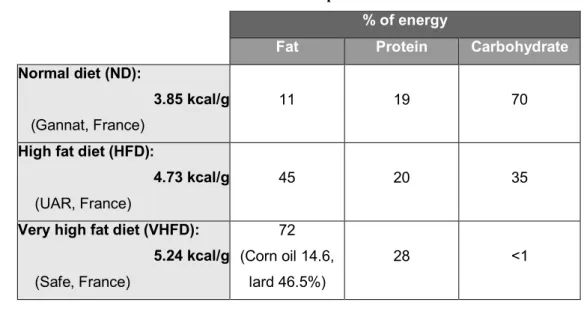
5.2.3. Mouse models 43
VHFD model 43
Characteristics of the VHFD model 44
HFD model 45
Characteristics of the HFD model 46
db-/? mice 46
Characteristics of the db-/? model 47
Mice invalidated for the AOC3 gene 47
Characteristics of the model 48
5.3. In vivo non invasive studies 48
5.3.1. Intraperitoneal glucose tolerance tests (IPGTT) 48
5.3.2. In vivo metabolic follow up 48
5.3.3. Determination of fat and lean mass 49
5.4. Blood and tissue sampling 49
5.4.1. Plasma/serum markers 49
5.4.2. RNA Extraction, Reverse Transcription, and Real-Time PCR 50
5.4.3. Human adipose tissue 50
5.5. Ex vivo and in vitro determinations 51
5.5.1. Determination of DNA content from adipose tissue 51
5.5.2. Immunohistochemistry 51 5.5.3. Immunoblotting assays 52 5.5.4. Isolation of adipocytes 52 5.5.5. Lipolysis 53 5.5.6. Hexose Transport 53 5.5.7. Lipogenesis 53
Radioactive method 54
Amplex Red method 54
5.5.9. Measurement of tissue nitrite and nitrate concentration 55
5.6. Statistical analysis 55
6. Results 56
6.1. Investigation of SSAO activity in adipose tissue 56
6.1.1. Determination of SSAO activity in subcutaneous adipose tissue 56 6.1.2. Influence of the adipose mass on the SSAO activity in different adipose
tissues 58
High fat diet induced obesity 58
Genetic obesity 58
Influence of fasting on INWAT SSAO activity 59
6.2. In vitro insulin-like effect of benzylamine 60
Lipolysis in rodent fat cells 61
Glucose uptake in rodent adipocytes 61
Lipogenesis in rodent adipocytes 62
6.3. In vivo insulin-like effect of benzylamine 63
6.3.1. Effect of single injection of benzylamine on glucose handling 63 6.3.2. Effect of chronic benzylamine injection in streptozotocin-induced
diabetes in rats 65
Blood glucose, HbA1C and serum AGE levels 65
NO availability in diabetic animals 66
6.3.3. Effect of oral benzylamine treatment in various animal models of insulin
resistance 67
Oral administration of benzylamine in VHFD-fed mice 67
Glucose handling in VHFD mice 67
Water intake and body weight gain 68
Plasma markers of metabolic disturbances 68
Lipolytic and glucose uptake activities in adipocytes 69
Oxidative stress and aorta NO bioavailability 69
Oral administration of benzylamine in HFD-fed mice 70
Water intake and body weight gain 71
Plasma markers of metabolic disturbances 72
Lipolytic and glucose uptake activities in adipocytes 73
Oxidative stress and aorta NO bioavailability 74
Oral administration of benzylamine in db-/- mice 75
Glucose handling in db-/- mice 75
Water and food intake, body weight gain 77
Plasma markers of metabolic disturbances 80
Lipolytic and glucose uptake activities in adipocytes 80
Oxidative stress and aorta NO bioavailability 82
6.4. In vitro tests for screening SSAO substrate drug candidates 83
Lipolysis in human adipocytes 85
Glucose uptake in human adipocytes 86
7. Discussion 87
7.1. Investigation of SSAO activity in adipose tissue 87
7.2. In vitro insulin-like effect of benzylamine 89
7.3. In vivo insulin-like effect of benzylamine 90
7.3.1. Effect of a single injection of benzylamine on glucose handling 90 7.3.2. Effect of chronic benzylamine injection in streptozotocin-induced
diabetic rats 92
7.3.3. Effect of oral benzylamine treatment in various animal models of insulin
resistance 93
7.4. In vitro tests for screening SSAO substrate drug candidates 100
8. Conclusions 103 Summary 105 Összefoglalás 106 Résumé 107 9. References 109 10. List of publications 127
Publications related to Thesis work 127
Publications not related to Thesis work 127
2.
Abbreviations
2-DG 2-Deoxyglucose
3-MTPPA 3-(4-Methylthiophenyl)propylamine 4-PBA 4-Phenylbuthylamine
5'-AMP Adenosine 5'-Monophosphate
AC Adenyl Cyclase
ACC Acetyl-CoA Carboxylase Acetyl-CoA Acetyl-Coenzyme A
ACS Acetyl-CoA Synthase
AGE Advanced Glycation Endproduct AMPK AMP-activated Protein Kinase
AO Amine Oxidase
AOC3 This gene codes SSAO
APJ 7 transmembrane domains receptor of apelin
AR Amplex Red
ATGL Adipocyte Triglyceride Lipase ATP Adonosine Triphosphate AUC Area Under the Curve
B6V10 Benzylamine-Vanadate complex salt by Gene Medica
BSA Bovine Serum Albumin
BzA Benzylamine
cAMP Cyclic Adenosine Monophosphate
CAO Copper Amine Oxydase
cd31 Adipocyte differentiation marker
cd45 Leucocyte marker
cGMP Cyclic Guanosine Monophosphate
CRP C-Reactive Protein
CTRL Control
DAG Diacyl-glycerol
DAO Diamine Oxidase
db -/- Mice having null mutation for db gene
db -/? Heterozygotes and mice having null mutation
db -/+ Heterozygote mice for db gene mutation
DG Diglyceride
DNA Deoxyribonucleic Acid ECD Endothelial Cell Dysfunction ECM Extracellular Matrix
eNOS Endothelial NOS
ER Endoplasmic Reticulum
ERK1/2 Extracellular signal-regulating Kinase F 4/80 Adipocyte differentiation marker
FAD Flavin Adenine Dinucleotide FAS Fatty Acid Synthase
FATP FFA transporter
FFA Free Fatty Acid
GAPDH Glycerynaldehyde-phosphate Dehydrogenase Gi Inhibiting G-protein
GLUT-1 Glucose Transporter-1 GLUT-4 Glucose Transporter-4 GPCR G-protein Coupled Receptor
Gs Stimulating G-protein H2O2 Hydrogene peroxide
HbA1c Hemoglobine A 1C
HFD High Fat Diet
HOMA index Homeostasis Model Assessment Index HSL Hormone Sensitive Lypase
IKK Inhibitor KappaB Kinase IL-6 Interleukine-6
Ins Insulin
INWAT Intra-abdominal White Adipose Tissue IPGTT Intraperitoneal Glucose Tolerance Test IRS-1 Insulin Receptor Substrate 1
IRS-2 Insulin Receptor Substrate 2 IRS-3 Insulin Receptor Substrate 3
Iso Isoprenaline
JNK Janus Kinase
Km Michaelis-Menten konstant
KO AOC3 Knock Out for AOC3 gene
LO Lysyl Oxidase
LPL Lipoprotein Lipase
LTQ Lysine Tyrozylquinone Malonyl-CoA Malonyl-Coenzyme A
MAO Monoamine Oxydase
MAPK MAP-Kinase
MG Monoglyceride
MG Methylglyoxal
MGL Monoglyceride Lipase
MRI Magnetic Resonance Imaging
NC-IUBMB
Nomenclature Comittee of the International Union of Biochemistry and Molecular Biology
NO Nitric Oxide, Nitrogen Monoxide
NOS NO Synthase
NS Not Significant
OH • Hydroxyl Radical
PAGE Polyacrlyamide Gel Electrophoresis PAI-1 Plasminogen Activator Inhibitor 1
PAO Polyamine Oxidase (FAD-containing AOs) PBS Phosphate Buffer Saline
PDE-3B Phosphodiestherase 3B
PGWAT Perigonadal White Adipose Tissue PI-3 kinase Phosphatidyl Inositol 3 Kinase
PKA Protein Kinase A
PKB Protein Kinase B
PKC Protein Kinase C
PRWAT Perirenal White Adipose Tissue RNS Reactive Nitrogen Species ROS Reactive Oxygen Species
SCWAT Subcutaneous White Adipose Tissue
SDS Sodium Disulphide
SOCS Supressors Of Cytokine Signaling
SOD Superoxide Dismutase
STAT Signal Transducers and Activators of Transcription
STZ Streptozotocin
TBS TRIS Buffer Saline
TBST TRIS Buffer Saline Tween
TE TRIS EDTA Buffer
TGs Triglycerides
Tie 2 Endothelial cell differentiation marker
TNF-α Tumor Necrosis Factor α
TPQ Topaquinone
UCP Uncoupling Protein
GlcNAc N-Acetyle Glucoseamine
V Vanadate
VAP-1 Vascular Adhesion Protein 1 VHFD Very High Fat Diet
VLDL Very Low Density Lipoprotein Vmax Maximal Reaction Velocity WAT White Adipose Tissue
WT Wild Type
α2-AR α2-Adrenergic Receptor β-AR β-Adrenergic Receptor
3.
Introduction
The number of obese individuals increases worldwide, leading to consider obesity as a global epidemic. It has been estimated that, in 2005, approximately 1.6 billon adults were overweight and more than 400 million adults were obese. Nowadays, around 15 % of the world’s population is overweight or obese, according to the International Association for the Study of Obesity (website: http://www.iaso.org). Obesity is recognized as a major risk factor for type 2 diabetes, cardiovascular diseases, non-alcoholic fatty liver diseases and certain types of cancers. Insulin resistance (namely the deficiency of response to insulin, leading to very blunted glucose uptake into cardiac or skeletal muscles and adipose tissues) is a metabolic disturbance that firmly links excess of white adipose tissue (WAT) with the obesity-related complications.
When considering the number of diabetic patients, type 1 diabetes represents a minor part, approximately 10% of all patients with diabetes. However, taking into account that these patients suffer from their disease from early on in their lives, they also merit serious attention because of late complications. Today's modern lifestyle with greatly reduced physical activity resulted in the increasing number of obese and thus insulin-resistant subjetcs, also among type 1 diabetic patients. Moreover, diabetic patients have very high risk for vascular complications. Therefore new therapeutic targets to mimic the effetcs of insulin, but acting independently of insulin are of great interest.
Some biogenic amines are well-established neurotransmitters, having a key role in central appetite regulation. Other amines are modulators or pertubators of neuronal functions, not only in mammals but also in insects or crustaceans where they are naturally present. They are also abundant in plants as products of secondary metabolism. Therefore, foods (meat, fish, vegetables), food additives or various medicinal plants that are world-wide ingested bring substantial amounts of alimentary amines. Once ingested, these dietary amines are mainly metabolised by amine oxidation. In spite of a great amount of amines metabolized in the gut, some of them may reach the circulation and affect different peripheral or central receptors or may also be degraded by different enzymes.
Benzylamine (BzA), a synthetic exogenous amine, has never been used in clinical studies or proposed as nutraceutic by para-medical companies, but has been recently described that it is capable to reduce hyperglycemia in diverse animal models of diabetes. The effect of this amine can be extended to diverse amines which are substrates of monoamine oxidases (MAO) and mainly of semicarbazide sensitive amine oxidases (SSAO). Since the enzymes MAO and SSAO are abundantly expressed in adipose tissue, this anatomical location is a key feature in the action of BzA and other amine oxidase (AO) substrates. Further studies are necessary before extraplolating to man the promising anti-hyperglycemic effects of this family of agents.
3.1. Amine oxidases
AOs are widely used by both autotrophic and heterotrophic life forms. They catalyse the metabolism of different mono- and polyamines. Their physiological importance is still not entirely understood, though their investigation grew more and more intensive in recent years. Their substrates are endogenous or exogenous, both decomposing by oxidative deamination that can be described by the following equation:
R-CH2-NR’R” + O2 + H2O → R-CHO + H2O2 + R’R”NH
During the process aldehyde, hydrogen peroxide and an amine or ammonium (in case of a primary amine) are formed. AOs can be classified by the chemical structure of their substrates: monoamine oxidases (MAOs), diamine oxidases (DAOs), polyamine oxidases (PAOs) and lysyl oxidases (LOs).
Table 1. Classification of amine oxidases. Adapted from Jalkanen et al., 2001. For abbreviations see the
text.
Amine oxidases (AOs)
FAD-containing AOs CAOs
MAO PAO renalase DAO SSAO RAO LO
Cofactor FAD FAD FAD TPQ TPQ TPQ LTQ
Coding human gene MAO-A, MAO-B, chromosome X AOC1, chromo-some 17 AOC3, chromosome 17 AOC2, chromosome 17 LOX1-4, chromosome 10
Presence mitochondria intracell. soluble intracell. extracell.,
soluble extracell. extracell.
Substrates noradrenaline dopamine adrenaline β-phenylethyl -amine tyramine tryptamine BzA octopamine spermine spermi-dine nor-adrenaline dopamine adrenaline putrescine, cadeverine histamine BzA, methylamine aminoacetone β- phenylethyl-amine tryptamine p-tyramine lysine Inhibitors pargyline clorgyline selegyline carbonyl reactive agents not known semi-carbazide, hydroxyl-amine semicarbazide, hydroxylamine semi-carbazide semicarbazide hydroxylamine Effect neuro-transmission cell growth cardiac function, blood pressure Histamine-degradation cell division amine oxidation, glucose uptake, leucocyte- adhesion
The traditional classification of AOs divides them into two main groups, based on the chemical nature of the attached cofactor (Table 1.). The FAD- and topa-quinone containing (TPQ) AOs not only differ in their cofactors, but are also distinct in terms of their subcellular distribution, substrates, inhibitors and biological functions. A third classification based on the enzyme reaction is proposed by the Nomenclature Committee of the International Union of Biochemistry and Molecular Biology (NC-IUBMB).
The flavin adenine dinucleotide (FAD)-contaning enzymes are also classified as amine:oxygen oxidoreductase (deaminating) EC 1.4.3.4 by NC-IUBMB:
MAO-A and –B are well known mitochondrial enzymes that have firmly established roles in the metabolism of neurotranssmitters and other biogenic amines (Shih et al., 1999).
FAD-containing PAOs use secondary amines spermine and spermidine as their preferred substrates, and thereby possibly regulate cell growth (Seiler, 1995).
Renalase is the most recently found FAD-containing AO (Xu et al., 2005). It is released by the kidney, oxidises chatecholamines and regulates cardiac function and blood pressure, however independently of its enzyme ctivity (Boomsma et al., 2007).
The TPQ containing enzymes are collectively designated as SSAOs due to their characteristic sensitivity of inhibition by a carbonyl-reactive compound, semicarbazide. They were also cited as copper amine oxidases (CAOs), however NC-IUBMB has recently divided them to EC 1.4.3.21 - primary-amine: oxygen oxidoreductase (deaminating) and EC 1.4.3.22 -histamine:oxygen oxidoreductase (deaminating).
The TPQ-containing enyme DAO prefers diamines putrescine, cadaverine and histamine as its substrate. Diamine oxidase is mainly an intracellular enzyme preferentially synthetised in the placenta, kidney and intestine. The secreted form binds in a heparin-dependent manner to endothelial cells. It is important in regulating infammation and allergic reactions.
The other SSAOs are mostly soluble or expressed on the cell surface, have methylamine, aminoaceton, BzA as substrates, are insensitive or only weakly sensitive to classical MAO inhibitors, like clorgyline or deprenyl, and mediate different biological functions.
The retina specific amine oxidase (RAO) is a cell surface enzyme, present in the retina. It prefers β- phenylethylamine, tryptamine and p-tyramine as substrates. Its proper function is yet to be understood (Imamura et al., 1997; Kaitaniemi et al., 2009).
LO, another copper containing enzyme is also inhibited by SSAO inhibitors, but only to a limited extent, compared to other SSAOs. Its classification is still disputed, hence there are several points where it differs from other SSAOs: its structure misses some conservative motifs and its cofactor lysine tyrozylquinone (LTQ) is also slightly different. The ε-amino groups are its excellent substrates. It is able to establish cross-links between collagen and elastin during the formation of the extracellular matrix (ECM) (Smith-Mungo et al., 1998). It is also classified as EC 1.4.3.13-protein-L-lysine:oxygen 6-oxidoreductase (deaminating).
3.1.1. Semicarbazide-sensitive amine oxidases
SSAOs are widely present in nature: they are expressed by plants, microorganisms, animals and men. SSAOs convert primary amines to aldehydes (the only exception is histamine). During the process the concomitant hydrogen peroxide and ammonium are formed. There are two mammalian isoforms known: the membrane-bound and soluble (plasma) isoforms. The majority of the tissular SSAOs can be found in the vascular walls, particularly in smooth muscle, but endothelial cells, adipocytes, chondrocytes, fibroblasts, the retina, the sclera, the kidney, the spleen, the placenta and the bone marrow also show membrane-bound SSAO activity.
TPQ containing SSAOs were discovered some decades ago. The enzyme was cloned and its primary structure was determined. The results were surprising: the structure was identical with that of another protein, mapped before, the vascular adhesion protein (VAP-1) (Smith et al., 1998). The deaminating enzyme activity was also proven in case of the VAP-1 and it also turned out that its distribution in the body is also similar to other SSAOs.
Genes and protein structure
Most SSAOs are dimeric glycoproteins with molecular masses of 140-180 kDa, containing two atoms of copper per dimer (Klinman et al., 1994; Lyles, 1996). An unknown amino acid 6-hydroxydopa (2,4,5-tridydroxyphenylalanine quinone or TPQ) was identified (Janes et al., 1990; Klinman, 1996) being the cofactor. TPQ is generated
from an intrinsic tyrosine molecule by a self-processing event that only requires bound copper ion and molecular oxygen (Mu et al., 1992).
SSAO enzyme has been cloned from several species: cow (Klinman et al., 1994; Mu et al., 1992), mouse (Bono et al., 1998a; Bono et al., 1998b; Moldes et al., 1999), rat (Morris et al., 1997; Ochiai et al., 2005), human (Smith et al., 1998; Zhang et al., 1996). At the moment full length cDNA sequences are available from seven mammalian SSAOs. The human SSAO contains 762 amino acids.
The products of two genes AOC3 and AOC4 have been identified to be responsible for SSAO activity. In man AOC3 and AOC4 are situated in cluster with AOC2, the gene coding the RAO, all in the long arm of chromosome 17. AOC3 gene codes membrane-bound SSAO. So far it is not clear if soluble SSAO is derived from AOC3 or it is encoded by a separate gene. The large extracellular part of the SSAO protein was shown to be released by proteolytic cleavage from endothelial (Stolen et al., 2004b), vascular smooth muscle (Göktürk et al., 2003), liver endothelial cells or adipocytes using transgenic and cell culture models (Abella et al., 2004; Stolen et al., 2004b). At least it is the case in humans and rodents where plasma SSAO activity is very low compared to other mammalian species like cow, dog or pig. In pig, the product of AOC4 gene, situated in a cluster with AOC3 was found to be responsible for such high plasma SSAO activity (Schwelberger, 2006). The mature protein derives from the primary translation product by cleavage of a 19 or 22 amino acid signal peptide and addition of N-linked carbohydrate residues. However, in human AOC4 a single base change converts a codon for a conserved tryptophan at position 225 to a stop codon thus leading to a truncated and non-functional protein (Schwelberger et al., 2008). It is intriguing that a functional AOC4 gene is also present in cow and dog, which, similary to the pig, have a high serum SSAO activity, whereas humans and rodents, which have a defective or absent AOC4 gene, respectively, have only a very low serum SSAO activity. Proteolytic AOC3 gene product release may also contribute to a minor part of plasma SSAO activity in cow, dog and sheep.
Conserved regions of the protein
Although we know that SSAOs are not exacly the same depending on the species, the isoform (membrane-bound or soluble) and even the age, there are regions that are the same regardless of these factors.
Figure 1. Conserved regions of SSAO protein. Adapted from Jalkanen 2001.
It has been proven that the active site of the enzyme remained practically unchanged during the evolution (Klinman et al., 1994). Figure 1 shows the conserved main caracteritics:
The protein consists of 763-765 amino acids, 90 kDA, foming a homodimer where the subunits are attached in a bond disulphur.
5 amino-acids at N terminal part are positioned intracellular and a transmembrane domain, which is a secretion signal is to 5-20 amino acids from N terminal part.
The carbon atom has been shown to be in the second position of TPQ is situated 6 Å away from the Cu2+ ion.
There is a Asn-TPQ-Asp/Glu-Tyr sequence in the active site of the enzyme. Tyr is situated at 470 or 471 amino acids from N terminal which is
post-trancriptionally modified to TPQ.
The protein contains three conserved histidine-binding Cu2+ ion (there is a His-X-His motif situated approximately 50 residues C-terminal from the cofactor and the third one 20-30 residues N-terminal from it).
Asp is approximately 100 residues N-terminal from TPQ, the catalytic base of the deamination in its reductive half-reaction.
6 sites of N-glycosilation (Asn)
Arg-Gly-Asp motive for binding with integrines.
Generally, amino acid identity among SSAOs isolated from different species but being of the same cellular origin is higher than that of different isoforms of the same species.
3D protein structure
Bovine serum amine oxidase was the first mammalian SSAO characterised 3D (Lunelli et al., 2005). The 3D structure of human SSAO has recently been described (Jakobsson et al., 2005). The SSAO is a homodimer. The TPQ and the Cu2+ ion are situated in the active site. The protein is composed of 4 domains:
N terminal D1 domain of each monomer is a transmembrane helix. D2-D4 are located extracellularly.
The monomers are bound by the D4 domain.
Leucine residue (Leu 469) located adjacent to the active site has been revealed to function as a gate controlling its accessibility.
An Arg-Gly-Asp motif is displayed on the surface, where it could be involved in integrin binding and possibly play a role in the shedding of SSAO from the membrane. Carbohydrate moieties are observed at five of six potential N-glycosylation sites. Carbohydrates attached to Asn 232 flank the active-site entrance and might influence substrate specificity.
SSAO substrates
SSAOs accept primary amines as substrates, although there may be exceptions to this rule (like histamine). Nevertheless, different species show a wide variety of substrate preferences. Therefore, some caution must be taken when concluding results with SSAOs of different species. In 1996, Lyles summarized that BzA, phenethylamine, tyramine, dopamine, which are also MAO substrates, tryptamine and histamine are good SSAO substrates in rat or human tissue (Figure 2.) (Lyles, 1996). Generally, it would appear that SSAO plays only a small role in metabolizing substrates that are also oxidised by MAO, although it could be more important when MAO is inhibited, or in the rare genetic condition where the MAO genes are deleted (Fitzgerald et al., 2002). Although serotonin is not a substrate for SSAO from most sources, it is a good substrate for the enzyme from pig and human dental pulp (O'Sullivan et al., 2002). The enzyme may also play a role in the metabolism of some xenobiotics, such as mescaline and primaquine (O'Sullivan et al., 2004; Tipton et al., 2004). Among all, BzA has been the preferred substrate for most SSAOs for a long time.
Methylamine Allylamine Aminoacetone
Benzylamine Phenylethylamine Tyramine
Dopamine Triptamine Histamine
Methylamine Allylamine Aminoacetone
Benzylamine Phenylethylamine Tyramine
Dopamine Triptamine Histamine
Figure 2. Well-caracterized substrates of SSAO.
SSAO also recognises aliphatic amines. Allylamine, an industrial chemical has been described to cause serious vascular and myocardial damage in several species (Boor et al., 1982; Boor et al., 1979). Vascular SSAO oxidises allylamine to acrolein, acting as a distal toxin, (Boor et al., 1987) responsible for the lesions caused by allylamine intoxication (Boor et al., 1980).
Figure 3. Role of endogenous substrates in the development of angiopathy.
Aminoacetone and methylamine do not have affinity to MAOs (Lizcano et al., 1998). The major source of methylamine is endogenous with contributions from the diet. The role of gut microflora, which varies both within and between individuals, in
processing several chemicals or food supplements to methylamine or suitable precursors should also be considered. Methylamine is produced in several catabolic reactions, including the breakdown of adrenaline (catalysed by MAO) sarcosine, creatine, lecithin and choline. It is also a common component of some foods and beverages, and is an atmospheric pollutant therefore some sources suggested the high levels of SSAO in the lung as a protection against inhaled methylamine and other volatile amines (Lizcano et
al., 1998). Methylamine is also a constituent of cigarette smoke and is considered as the
major end product of nicotine metabolism. Smoking or nicotine can induce the release of adrenaline, which is in turn deaminated by monoamine oxidase, also producing methylamine (Yu, 1998b). Plasma levels of methylamine vary between 11.5 and 123.4 ng/ml (Li et al., 2004), and may be altered in some pathological conditions such as diabetes, typhoid fever, and liver or renal insufficiency or uremia (Baba et al., 1984; Mitchell et al., 2001).
Methylamine has in fact been shown to be converted to formaldehyde in vivo by SSAO (Yu et al., 1997):
CH3NH2 + O2 + H2O → HCHO + H2O2 + NH3.
The deaminated product formaldehyde is extremely reactive, forming adducts with lysyne side chain of proteins and nucleic acids (Gubisne-Haberle et al., 2004). It is potentially carcinogenic and is a subject of major environmental concern. Some reactive free radicals can be generated from formaldehyde in the presence of hydrogen peroxide:
2 HCHO + H2O2 -» 2 H-*C=O + 2 H2O.
Formaldehyde and hydrogen peroxide simultaneously generated from deamination of methylamine may act synergistically causing cellular damage (Yu et al., 2003b). Yu et al. found that methylamine by itself is relatively nontoxic towards cultured endothelial cells obtained from both human umbilical vein and calf pulmonary artery. It becomes very toxic, however, in the presence of SSAO. In some pathologies there is an increased risk for formaldehyde formation and consequent oxidative stress (Yu, 1998a; Yu et al., 1998).
It has been proposed that excessive deamination of methylamine (e.g. due to increased occurrence or impaired disposal of the amine) may become a potential risk factor responsible for initiation of endothelial injury of diabetic complications (Yu et
behaviour (Yu et al., 1997). The association of nicotine or disease-mediated increase of methylamine can contribute to the potential adverse health effects. Selective SSAO inhibitors, e.g. MDL-72974A or aminoguanidine, can effectively protect the cells from SSAO-methylamine induced damage (Yu et al., 1993).
Aminoacetone, which may be formed as a result of threonine or glycine metabolism, is oxidised by SSAO to form methylglyoxal:
CH3COCH2NH2 + O2 + H2O → CH3COCHO + H2O2 + NH3.
Methylglyoxal (MG) is cytotoxic and mutagenic. Although there are other pathways of aminoacetone breakdown, SSAO makes a significant contribution to the process (Mathys et al., 2002). MG is suggested to induce formation of oxygen free radicals and chemical modification of essential proteins by reacting with arginine, lysyne and cystein residues. MG modifies cell proteins non-enzymatically through the Maillard reaction, in which aldehydes and ketones react with ε-amino groups of lysine residues and guanidino groups of arginine residues resulting in stable chemical adducts in proteins known as advanced glycation end products, or AGEs (Monnier et al., 1992). AGEs have been implicated in diverse long-term complications of diabetes, including atherosclerosis, retinopathy, nephropathy, and cataract formation. Because MG appears to be a major precursor of AGEs in vivo (Brownlee, 2001), any reaction that increases MG levels in tissue or plasma could ultimately lead to these complications.
There is a growing number of evidence proving the existence of in vivo SSAO-dependent oxidation of amonoacetone and methylamine:
Urinary formaldehyde was in correlation with plasma SSAO activity when testing different mouse models (Yu et al., 1998).
Methylamine and aminoacetone administration resulted in higher urinary excretion of formaldehyde and methylglyoxal in diabetic rats (Deng et al., 1998).
Increased urinary levels of methylamine were found following SSAO inhibition in different rodent models (Yu et al., 1998).
Some studies report an increase of urinary excretion of methylamine following administration of nicotine or creatine and the subsequent production of formaldehyde and cross-linkage with tissue constituents (Poortmans et al., 2005; Yu, 1998b).
4-aminomethyl-benzenesulphonamide 2,3-dimethoxy-benzylamine C-naphtalen-1-yl-methylamine PD 0119035 PD 0125999
Arylalkylamine substrates 3-MTPPA R1=F,CH3 or R2=H,F or n=4 4-aminomethyl-benzenesulphonamide 2,3-dimethoxy-benzylamine C-naphtalen-1-yl-methylamine PD 0119035 PD 0125999
Arylalkylamine substrates 3-MTPPA R1=F,CH3
or R2=H,F
or n=4
Figure 4. Novel subtrates identified after Dunkel et al., 2008 and Yraola et al., 2009.
The characterisation of the 3 D structure of the protein promoted the chemical design of new SSAO substrates. Homology modeling of the catalytic site of the mouse SSAO was performed by the group of Marti and pharmacophoric motif based on BzA was constructed (Marti et al., 2004). Their model resulted in 5 substrates having arylalkylamine or aminomethyl-pyrrolidine structure. Yraola has then published structure-activity relationship of arylalkylamines in human and mouse enzyme preparations (Yraola et al., 2006), the best substrates of the screening are illustrated in Figure 4. Unzeta and coworkers synthesised 3-(4-methyltiophenyl) propylamine (Gallardo-Godoy et al., 2004).
SSAO inhibitors
The enzyme is characterised by its sensitivity towards semicarbazide, however, this hydrazine derivative has a relatively weak inhibitor capacity. Inhibitors can be conveniently subdivided into the main groups of hydrazine derivatives, arylalkylamines, propenyl- and propargylamines, oxazolidinones, and haloalkylamines.
Table 2. Some important inhibitors of SSAO.
Pharmacons having SSAO-inhibiting capacity
chemical
structure/group Compounds
SSAO inhibition
MAO
inhibition other characteristics
Hydrazine
Derivatives semicarbazide
weak (1mM),
irreversible no inhibits LO and DAO aminoguanidine potent,
irreversible weak weak NOS inhibitor isoniazid Weak (1mM) weak antituberculoticum hydralazine
(neprezole)
irreversible
(μM) reversible antihypertensive drug
phenelzine irreversible
MAO antidepressant
carbidopa anti-Parkinson drug
benserazide anti-Parkinson drug
procarbazine selective reversible strong no anticancer drug LJP-1207 potent (nM) competitive, irreversible Arylalkylamine Derivatives B24 potent (μM), reversible no
mexiletine antiarrythmic drug
Propenylamines MDL 72974A potent (10 nM) MDL 72145 potent (10 nM) MAO-B irreversible, competitive MAO-A anti-Parkinson drug candidate Propargylamine Derivatives propargylamine potent (μM), irreversible weak β-aminopropionitrile weak (200 μM),
competitive reversible LO inhibitor at μM Haloalkylamine Derivatives 2-bromoethylamine potent, irreversible no Oxazolidinone Derivatives MDL 220662 potent (μM) Antidepressants with different chemical structures imipramine antidepressant maprotiline antidepressant zimeldine antidepressant
nomi fensine antidepressant
Hydrazinoalcohols BTT-2052 potent amikacin tobramycin puromycin gentamicin aminoglycoside antibiotic
Of them, aryl(alkyl)hydrazines, and 3-halo-2-phenylallylamines are generally very strong SSAO inhibitors. Most of these inhibitors of SSAO have been originally
developed for other purposes, or they are simple chemical reagents with highly reactive structural element(s); these compounds have not been able to fulfil all criteria of high potency, selectivity, and acceptable toxicity. New potent compounds have recently been developed with the structures: oxazolidinone; hydrazinoalcohols; 4,5,6,7-tetrahydroimidazo[4,5-c]pyridines; thiocarbamoyl derivatives; carboxamides and sulfonamides; tetraphenylphosphonium analogues; 1,3,4-oxadiazines. There are also alternative ways to inhibit SSAO. Some biomolecules with a free NH2 group, such as peptides with lysine side-chain, aminohexoses or aminoglycoside antibiotics can interact with the enzyme. Function-blocking antibodies to hVAP-1, as well as small interfering RNAs could be applied for modulation of the adhesion molecule. There is a demand for novel selective and non-toxic inhibitors, which may be useful tools for further understanding the roles and function of SSAO. SSAO inhibitors may even be valuable substances for the treatment of various diseases (Dunkel et al., 2008; Matyus et
al., 2004). Table 2. presents the most important SSAO inhibitors.
The physiological and pathophysiological role of
semicarbazide sensitive amine oxidases
The physiological role of SSAOs is not fully understood yet. Until recently it was assumed that their most important effect is to decrease the level of monoamines by oxidative deamination. The elevated level of the known endogenous substrates, methylamine and aminoacetone, is harmful, therefore it seems reasonable to keep them under control. Some novel functions of SSAO have been characterised: they may play a role in inflammatory processes, in the regulation of the glucose transport of adipocytes and in the maturation of collagen and elastin.
3.1.2. The oxidative deamination
SSAOs catalyse oxidative deamination of mainly primary amines. The reaction consists of two parts. In the first step the enzyme is reduced by the substrate, itself being oxidated to the corresponding aldehyde (Figure 5, 1-4). In the second half-reaction the enzyme is reoxidated by molecular oxygen with conformant release of hydrogen peroxide and ammonium (Figure 5, 5-6).
During the reductive half-reaction multiple transition stages exist among others a transient but covalent Schiff base is formed, thus substrate is ad interim trapped in a covalent bond. During the oxidative half-reaction, the reduced cofactor returns in its former oxidised TPQ form, and hydrogen peroxide and ammonium are released.
O O OH CH2 CH N H Asp O Asp Tyr O OH CH2 OH CH N H Asp O Asp Tyr O OH CH2 N CH2R CH N H Asp O Asp Tyr NHCH2R Asp/Glu O O OH CH2 N C H R O H CH N H Asp O Asp Tyr OH CH2 NH2 O H CH N H Asp O Asp Tyr O O OH CH2 CH N H Asp O Asp Tyr -RCHO + RCH2NH2 + O2 +H2O - H2O2 -H2O +H2O -NH3 H2O Cu(II) (1) (2) (3) (4) (5) (6)
Figure 5. The oxidative deamination catalysed by SSAO. After Matyus et al., 2004.
Membrane-bound SSAOs
The biological role of SSAOs, with the exception of LO, has remained enigmatic for decades in mammals. In bacteria and yeast, the SSAO reaction provides a source of nitrogen (and carbon) when growing. On the other hand the hydrogen peroxide is used
for wound healing in plants. In mammals novel functions of SSAO has been revealed besides amine oxidation. Although the significance of all SSAO mediated biological effects is far from clear.
Mammals express SSAO in a wide variety of tissues (Jaakkola et al., 1999; Lewinsohn, 1984; Lyles, 1996; Salmi et al., 1993). The most prominent synthesis takes place in smooth muscle (both vascular and non-vascular) and adipocytes, endothelial cells and follicular dendritic cells. Leucocytes, epithelial and fibroblastoid cells lack SSAO. Brain is also devoid of SSAO (except microvessels). Unlike in other mammals, in man, the protein is also absent from chondrocytes and odontoblasts.
Membrane-bound SSAOs may be involved in the regulation of glucose metabolism in adipose cells, in the regulation of leucocyte trafficking in endothelial cells as well as vascular elasticity.
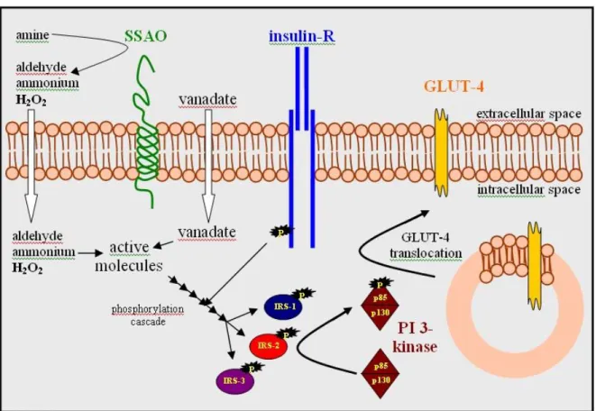
Glucose metabolism
SSAO is involved in glucose metabolism in adipocytes and smooth muscle cells, with H2O2 being responsible for the effect. The mechanism involves insulin-responsive glucose transporter (GLUT-4) translocation to the plasma membrane in fat cells, while GLUT-1 accumulation on the surface of smooth muscle cells (El Hadri et al., 2002). The involvment of adipocyte SSAO in the glucose metabolism is a major topic of this thesis. For more details see chapter 3.2.
The role of SSAO/VAP-1 in the regulation of leucocyte adhesion
As mentioned previously, SSAO has been found identical to VAP-1 adhesion molecule. SSAO induces cell adhesion and regulates leucocyte trafficking through the endothelial barrier. Circulating leucocytes reversibly tether to and roll on the endothelial cells under conditions of blood flow as a first step of a well-orchestrated cascade (Salmi
et al., 1997a). Reception of appropriate activation signals is followed by firm adhesion
to the vessel wall and transmigration into the tissue by leucocytes. Multiple adhesion and activation molecules on the leucocytes and endothelial cells regulate the molecular execution of the process (Figure 6.) (Jalkanen et al., 2001).
SSAO is constitutively present in intracellular granules within endothelial cells (Salmi et al., 1993). However, with in vivo inflammation models, SSAO seems to be translocated lumenally from intracellular storage granules only upon elicitation of
inflammation (Salmi et al., 1997b). SSAO mediates leucocyte subtype specific adhesion of CD8+ T-cells, NK-cells and granulocytes. SSAO is also present on sinusoidal endothelial cells in liver (McNab et al., 1996). The SSAO molecule seems to be important for the rolling phase and most likely later at the transmigration step of leucocyte extravasation/transmigration (Tohka et al., 2001).
Figure 6. Role of VAP-1 and other molecules in the extravasation of leucocytes. Adapted from
Jalkanen 2001.
The protein was also shown to oxidise BzA and methylamine. Moreover, the reactions catalysed by SSAO have been shown to be important in the leucocyte adhesion (Salmi et al., 2001b). It is interesting that artificial SSAO inhibitors decrease rolling and adhesion of leucocytes by about 50% and anti-VAP-1 antibodies inhibit lymphocyte rolling but do not affect enzyme activity. On the other hand products of the catalytic reactions, like aldehydes or hydrogen peroxide do not alter the lymphocyte-endothelium adhesion. Leucocyte adhesion can also be decreased by SSAO substrate pretreatment suggesting that during the multistep extravasation cascade there is a transitional covalent bond between the enzyme (the endothelial VAP-1) and a membrane-bound substrate of leucocyte origin. It is therefore the membrane-bound isoform and not the soluble one that plays the important role in the adhesion to the endothelial cells. This hypothesis was substantiated by an experiment using a synthetic peptide blocking the catalytic cavity of VAP-1. It is possible that surface bound amines (like N-termini of proteins, NH2-containing amino acid side chains, amino sugars etc.) may be SSAO substrates in addition to soluble amines (Salmi et al., 2001b). Moreover, aminosugars, also promoting the adhesion, bind reversibly to SSAO in the presence of
H2O2. The enzyme catalysed oxidation of substrates may thus considerably contribute to leucocyte binding to the endothelial cell (O'Sullivan et al., 2003; O'Sullivan et al., 2004). SSAO, in this case an endothelial ectoenzyme, can directly and indirectly regulate the leucocyte extravasation. VAP-1 is a dual function molecule: it binds lymphocytes to the endothelium and utilises the catalytic reaction between the surface-bound amine and VAP-1 to transiently link the interacting cells. The VAP-1-catalysed reaction may further contribute to cross-linking if the protein-bound aldehydes formed on lymphocytes interact with molecules on the endothelial cell surface (Jalkanen et al., 2001).
Organisation of the arterial wall
SSAO may be involved in vascular smooth muscle cell differentiation (El Hadri
et al., 2002), organization of the extracellular matrix (Vidrio, 2003) and regulation of
vascular tone (Langford et al., 1999; Langford et al., 2002; Vidrio, 2003). Langford and his coworkers have shown that pharmacological inhibition of SSAO induced by semicarbazide in a growing rat model led to striking elastic fiber disorganisation. Semicarbazide, however, inhibits both SSAO and LO and its effect on collagen cross-linking is mainly attributed to LO inhibition (Mercier et al., 2007; Mercier et al., 2006). Alternatively, it has been suggested that SSAO could influence arterial vascular tone, although contradictory results have been reported: Vidrio and his group have proposed that SSAO-mediated H2O2 production could increase vascular tone and enhance hydralazine-induced vasodilation (Vidrio et al., 2003); In contrast, Conklin et al. have recently suggested that SSAO activation by methylamine mediated vasorelaxation in isolated human arteries (Conklin et al., 2004). At a molecular level, it is conceivable, in view of previous results using molecular modeling (Salmi et al., 2001a) that in addition to soluble primary amines, such as methylamine or aminoacetone, SSAO may act on amino acids, including matrix proteins. SSAO may, thus, contribute to physiological cross-linking of elastic and collagen fibers. Formaldehyde-induced protein cross linkage (Gubisne-Haberle et al., 2004) might also be involved in the process. Nevertheless, SSAO-deficient mice have normal elastic fiber network, elastin cross-linking processes, and vascular endothelial or smooth muscle function (Mercier et al., 2006). These results do not support a major role of SSAO in the rodent arterial wall, even if unknown compensatory mechanisms may contribute to the phenotype in this model. However, the
only detectable alteration in KO AOC3 mice was an increase in carotid diameter, in line with a potential involvement of SSAO in arterial growth and/or the pathophysiology of aneurysms, as suggested (Langford et al., 1999; Sibon et al., 2004).
Soluble SSAOs
Soluble SSAOs may have an important function in several biological mechanisms. First of all, increased levels of soluble serum SSAO were found in specific diseases, for instance in diabetes and liver diseases. Soluble SSAO may also be involved in the production of non-enzymatic addition of oligosaccharides to proteins during formation of advanced AGEs, typical of diabetic lesions. Finally, it has been proven, that soluble SSAOs also modulate lymphocyte adhesion to endothelial cells, presumably by triggering positive signals on the lymphocyte (Kurkijarvi et al., 1998).
Plasma SSAO activity varies considerably in mammals (Boomsma et al., 2000b). It is high in species with a functional AOC4 gene and low in humans and rodents that lack a functional AOC4 gene. Despite that human plasma amine oxidase activity is lower than in tissues, the easier plasma accessibility has resulted in a substantial number of clinical reports in various pathologies (Boomsma et al., 2003).
In healthy adults with BMI ≤ 25, SSAO activity varies in a narrow range. It is interesting, that the activity is much higher in children, with a sharp decrease at the age of 16 to finally settle at the adult values. There is a slight increase in the activity from the age of 50. Enzyme activity is independent from gender, posture (standing, sitting, lying) or circadian rhythm. A higher activity can be measured during pregnancy (Boomsma et al., 2003).
Obesity, diabetes and atherosclerosis
Obesity is not a condition associated with clear-cut increase in plasma SSAO, despite adipose tissue has been proposed as a source of soluble SSAO, essentially in diabetes (Stolen et al., 2004b). While plasma SSAO clearly increases in diabetic conditions, its association with obesity is less consistent (Carpéné, 2009). Therefore, the correlation found between plasma SSAO activity and BMI in type 2 diabetic patients (Mészáros et al., 1999b) might be the consequence of a parallel increase in insulin resistance and adiposity. An elevation in SSAO activity has been found in non-diabetic, obese patients and has been proposed as a cardiovascular risk factor (Weiss et al.,
2003). However, such an association between plasma SSAO and BMI was not confirmed by other studies conducted with non-diabetic (Dullaart et al., 2006) or young obese (Visentin et al., 2004) subjects. When morbidly obese patients (BMI of 38.8) received vertical banded gastroplasty and lost weight after the surgery (BMI lowering to 30.8), there was only a weak tendency to correlate the individual changes in plasma SSAO with the decrease in different anthropometric measurements of adiposity (Li et
al., 2005). A more substantial increase in plasma SSAO than those reported in the above
mentioned studies should be expected in obese subjects, regarding to the dramatic increase in TNF-α in obesity and owing to in vitro TNF-α stimulation of SSAO shedding from adipocytes (Abella et al., 2004).
High plasma SSAO level was measured in type 1 (Boomsma et al., 1995), type 2 (Mészáros et al., 1999b), and type 1 juvenile diabetes. This phenomenon has also been observed in animal models of diabetes (Hayes et al., 1990). It has been proven that oxidative stress is important in the development of diabetic complications. It is assumed that diabetic vascular complications are caused partially by elevated blood level of the toxic formaldehyde, MG and hydrogen peroxide, all deriving from the SSAO mediated oxidative deamination of methylamine and aminoacetone. SSAO activity is elevated in patients with diabetic complications such as retinopathy (Garpenstrand et al., 1999; Gronvall-Nordquist et al., 2001), hypertension and atherosclerosis (Mészáros et al., 1999a), therefore plasma SSAO has been suggested to be a possible clinical marker of diabetic complications.
Serum SSAO levels correlate with Crouse score and intima media thickness, both reliably marking the progression of atherosclerosis (Mészáros et al., 1999a). Experiments done on human umbilical vein showed that SSAO catalyses LDL oxidation by endothelial cells (Exner et al., 2001). A more intensive oxidative stress can help to progress the disease. Oxidation of LDL increases the atherogenity of the lipoprotein and this can hasten the formation of atherosclerotic plaques.
Other diseases
Since there is a significant amount of SSAO in the vascular wall, great interest has been given to patients suffering from all types of vascular diseases. In patients with congestive heart failure plasma SSAO has been found considerably increased (Boomsma et al., 2000a) and was in correlation with severity of the disease. According
to certain observations, SSAO activity of individuals with essential and renovascular hypertension is normal (Lyles, 1996), SSAO levels of patients with chronic renal insufficiency are lower compared to controls. There is a very high level in stroke and malignant hypertension, which can be a consequence of the cardiac insufficiency. SSAO activity is increased in pre-eclampsia (Boomsma et al., 2003) or in cerebral autosomal dominant arteriopathy with subcortical infarcts as well as in leukoencephalopathy (Ferrer et al., 2002), inflammatory liver diseases (Kurkijarvi et al., 1998), multiple sclerosis with ongoing inflammatory activity (Airas et al., 2006), psoriasis (Madej et al., 2007), atopic eczema (Madej et al., 2006), Alzheimer’s disease (del Mar Hernandez et al., 2005), myopathies (Olive et al., 2004) and also bone cancer with skeletal metastases (Ekblom et al., 1999).
SSAO activity is unchanged in Sjögren syndrome or colitis ulcerosa. Decreased levels are rare, e.g. in children born with hernia diaphragmatica, in cases of severe burns, in different malignomes and in lung cancer, and decreased serum levels have also recently been reported in highly depressed (Roessner et al., 2006) and schizophrenic patients treated with second-generation “diabetogenic” antipsychotics (a possible link to the role of SSAO in glucose metabolism was suggested) (Roessner et al., 2007).
In pathologies with high plasma SSAO activity some of the enzyme substrates are also elevated: methylamine and aminoacetone in diabetes, creatinine in uremia and adrenaline in stress-related diseases. It is possible that the substrates mentioned above cause the increase of SSAO activity.
It is not clear how a moderate increase of SSAO activity observed in a number of human diseases could possibly cause any damage (Boomsma et al., 2003; Yu et al., 2003b). However, the increase in released SSAO could just be indicative of an increase of the membrane-associated SSAO at the cell surface where the actual damage is done.
The regulation of plasma SSAO levels is yet to be defined. Although several correlations have been set up between plasma SSAO activity and other clinical markers like ACE activity (Boomsma et al., 2005b), thyroid hormone (Boomsma et al., 2005a), TNF-α, insulin and blood glucose levels, glycated haemoglobin (Dullaart et al., 2006; Salmi et al., 2002) etc, only TNF-α and insulin seems to be involved in the regulation of plasma SSAO activity. TNF-α promoting insulin resistance, causes an increase in the release of SSAO by adipocytes (Abella et al., 2004; Garcia-Vicente et al., 2005). On the
other hand insulin appears to hamper shedding from tissues or increase the clearance of plasma SSAO levels. Finally, some recent trials showed that acute glucose load itself may increase plasma SSAO activity in non-diabetic individuals (Li et al., 2009a; Li et
al., 2009b).
3.2. Adipose tissue
In vertebrates white adipose tissue (WAT) is the primary site of energy storage. The lipid droplets of the adipocytes contain mostly triglycerides (TG). When energy expenditure exceeds energy intake WAT releases free fatty acids (FFA). WAT is also a secretory organ as it produces several adipocytokines permitting self-regulation (autocrine effect), a cross-talk with local (paracrine effect) and distant adipocytes and other cells in the brain, the liver, the muscles, the pancreas, etc. (endocrine effect) (Kim
et al., 2000).
WAT contains different cell types. Only one third of the cells are adipocytes and the rest is a mixture of stromal vascular fraction: fibroblasts, macrophages, stromal cells, monocytes and preadipocytes. Adipose tissue seems to derive from an embryonic stem cell precursor (Pittenger et al., 1999). The development of white adipose tissue begins early during embryogenesis and continues throughout life resulting in an increase in fat cell size (hypertrophy) as well as in fat cell number (hyperplasia) depending on the energy status and the storage needs of the body.
Fat develops in many different sites and has different functions: subcutaneous adipose tissue (SCWAT) is an effective insulating layer located between the muscles and dermis while visceral adipose tissue fills in space gaps between organs, like gonads (PGWAT), heart, kidneys (PRWAT), or gut, protecting them against mechanical impacts and maintaining them in the adequate position.
3.2.1. Metabolic function
Lipogenesis
Figure 7 shows the lipogenic processes grouped on the left hand side. In postprandial state adipocytes produce TG. Lipid accumulation in adipose tissue depends on circulating FFA and glucose uptake. Fatty acids may be synthesised from carbohydrate precursors (de novo lipogenesis). Insulin stimulates glucose uptake by
stimulating the translocation of GLUT-4 to the plasma membrane. Glucose undergoes glycolysis resulting in acetyl-coenzyme A (acetyl-CoA). Acetyl-CoA is transformed to Malonyl-CoA by the Acetyl-CoA carboxylase (ACC). Further chain prolongation is catalysed by the fatty acid synthase (FAS). In human, only a fraction of the TG stock is formed by de novo lipogenesis. Insulin also favours fatty acid uptake from circulating lipoproteins by stimulating lipoprotein lipase (LPL) activity. FFAs are released by lipoprotein lipase hydrolysing TGs from chylomicrons or VLDLs and are finally taken up by FFA transporters (FATP). Be the source of FFAs carbohydrates or TGs, reesterification is necessary to form TGs suitable for lipid storage (Vazquez-Vela et al., 2008).
Figure 7. Major biochemical pathways involved in the regulation of energy homeostasis in the adipocyte (Vazquez-Vela et al., 2008). For abbreviations see the text.
Lipolysis
During fasting WAT hydrolyses TGs to supply other organs with FFAs (Figure 7. right hand side). TGs stored in the lipid droplet are first hydrolysed by the adipose triglyceride lipase (ATGL), resulting in diacylglycerol moiety and FFA, then further hydrolysed sequentially by the hormone-sensitive lipase (HSL) and monoglyceride lipase (MGL) producing FFAs and glycerol. Receptors enhancing cyclic adenosine monophosphate (cAMP) formation stimulate lipolysis. β-adrenergic stimulation (Gs) of adipocytes and the subsequent protein kinase A-dependent (PKA) phosphorylation of HSL and perilipin trigger the translocation of HSL from the cytoplasm to the lipid droplet and induce neutral lipid hydrolysis. Opposingly, molecules decreasing intracellular cAMP inhibit lipolysis. Insulin induces PDE-3B (phosphodiesterase 3B) which degrades cAMP. α2-adrenergic agonists are also anti-lipolytic, as they inhibit adenyl cyclase (AC) enzyme activity.
3.2.2. Endocrine function
Beyond its essential function in lipid storage and the supply of free fatty acids, WAT produces numerous bioactive molecules (lipids or peptides). Among these molecules, we can find hormones, like leptin, adiponectin, resistin and apelin (Table 3.). Leptin is considered as a metabolic signal for energy sufficiency by directly acting on the hypothalamus. Adiponectin enhances insulin-sensitivity, decreases FFA influx, increases β-oxidation in liver and muscle. Adiponectin decreases atherogenic risk via depressing the expression of adhesion molecules within the vascular wall, as well. Resistin acts on skeletal muscle and WAT and its high plasma levels are associated with insulin resistance. Apelin increases cardiac contractility, decreases blood pressure and hyperglycemia (Dray et al., 2008; Vazquez-Vela et al., 2008). Obesity might reduce physiological response of some adipokines (resistance).
In addition to the above mentioned molecules, adipose tissue also produces and secretes a variety of other peptides, cytokines and complement factors, including tumour necrosis factor α (TNF-α), interleukine 6 (IL-6), plasminogen activator inhibitor 1 (PAI-1), angiotensinogen, etc. TNF-α is involved in the pathophysiology of insulin resistance via modifying insulin sensitivity which induces abnormal phosphorylation of
insulin receptor substrate (IRS)-1. IL-6 comes from the stromal vascular fraction and it controls the hepatic production of inflammatory proteins (Antuna-Puente et al., 2008).
Table 3. Some biologically active molecules of WAT. See abbreviations in chapter 2.
Source Receptors Pathway Main effects In obesity
TNF-α adipocytes membrane bound and soluble cytokine receptors
JNK, IKK block insulin's action
(IRS-1) ↑ macrophages lymphocytes IL-6 fibroblasts cytokine receptor JNK
induce liver inflammatory protein production (CRP)
↑ endothelial
cells alters tyr phosphatase
activity and SOCS monocytes Leptin adipocytes long, short, soluble cytokine receptors JNK/ STAT β-oxidation ↑ ↑ RESISTANCE UCP expression↑ hypothalamus-food intake↓ insulin secretion↑ Resistin adipocytes not
known not known
WAT, skeletal muscle-
insulin resistance (IRS-2↓) ↑ macrophages
liver-glycogen synthesis ↑ Adiponectin adipocytes GPCR AMPK/MAPK
liver and skeletal muscle-insulin sensitivity, β-oxidation ↑
↑ RESISTANCE hypothalamus-food intake↑
Apelin adipocytes GPCR AMPK/PKB blood pressure↓ ↑ RESISTANCE? blood glucose↓
3.2.3. Obesity
The abundance of nutrients and the decreased physical activity coupled with modern western life, result in excessive accumulation of adipose mass. Obesity, the excessive storage of energy as fat caused by the imbalance between energy intake and expenditure, has become a prevalent health hazard in industrialised countries.
Disturbed energy balance alters WAT activity especially that of adipocytes or macrophages, inducing low-grade chronic inflammation which is supposed to play a central role in obesity-linked insulin resistance. Insulin resistance may be featured in many pathologies complications such as hypertension, hyperlipidemia, atherosclerosis, metabolic syndrome, polycystic ovarian disease, certain cancers, and type 2 diabetes mellitus. Especially, increase in visceral fat mass seems to contribute to such complications.
3.2.4. Hydrogen peroxide
The majority of the bioactive H2O2 is derived from spontaneous or superoxide dismutase-catalyzed (SOD) dismutation of superoxide, a side product of several enzyme reactions: mitochondrial electron transport chain, lipoxygenase, cycloxygenase, cytochrome P450s, xanthine oxidase, NADPH oxidases, uncoupled eNOS, etc. Some other enzymes, like xantine oxidase, glucose oxidase or amine oxidases produce H2O2 directly.
H2O2 has long been known to mimic several of insulin’s actions in adipocytes (Czech et al., 1974; May et al., 1979). H2O2 acts through insulin signaling: it enhances insulin receptor phosphorylation and kinase activity (Hayes et al., 1987) and subsequent tyrosine phosphorylation of intracellular proteins. Insulin can induce NADPH oxidase-dependent H2O2 release, H2O2 thus can be regarded as a second messenger of insulin (Mukherjee et al., 1977). The membrane-bound NADPH oxidase is coupled to the insulin receptor via a Gi/Go heterotrimeric G protein (Krieger-Brauer et al., 1997) regulating insulin action (Moxham et al., 1996).
Despite its unquestionable biological function, it cannot be denied that H2O2, as a reactive oxygen species (ROS), may also have deleterious effects on the cells. As a matter of fact, H2O2 is poorly reactive in chemical terms. Its danger comes mainly from its conversion into hydroxyl radical (OH•) either in the Fenton or the Haber-Weiss reaction. Insulin resistance, as well as its complications have also been associated with high levels of H2O2. This might be the consequence of impaired antioxidant systems (catalase, glutathione peroxidase) and/or increased ROS production (Eriksson, 2007).
3.2.5. Expression of SSAO in adipocytes and in white adipose tissue
An increase of SSAO during adipocyte differentiation in rodents has been described as early as 1990 by Raimondi and co-workers on cells isolated from the stroma-vascular fraction of rat adipose tissue and cultured under adipogenic conditions (Raimondi et al., 1990). Afterwards, an impressive increase of SSAO mRNA, protein and activity was detected during adipocyte differentiation in the murine lineages 3T3-F442A and 3T3-L1 (Moldes et al., 1999). More recently, it has been evidenced that the product of the AOC3 gene dramatically increases during adipogenesis of human
preadipocytes (Bour et al., 2007a) resulting in high SSAO activity in mature adipocytes (Bour et al., 2007a; Morin et al., 2001). The increase in SSAO activity found during adipogenesis has still a poorly defined role.
In human adipose tissue, there is no doubt that fat cells exhibit higher levels of SSAO than any other cells belonging to the stromal vascular fraction (Bour et al., 2007a). This observation confirms previous findings made on rat brown adipose tissue (Barrand et al., 1984). The very high expression of SSAO in adipocytes has been confirmed by independent approaches studying protein expression profile in different subcelullar compartments: SSAO has been detected in plasma membrane (Barrand et
al., 1984), glucose transporter-containing vesicles (Enrique-Tarancon et al., 2000),
caveolae (Souto et al., 2003) and in diverse microvesicles of adipocytes (Morris et al., 1997).
The variations of obesity-induced changes in SSAO expression of WAT are still unknown and need further investigation. In the subcutaneous adipose tissue, no change in SSAO was found when comparing young obese subjects with age-matched controls. (Visentin et al., 2004). Since the visceral, intra-abdominal adipose depots are especially involved in the serious complications of massively obese patients, it would be worth to obtain information on pathology-related SSAO expression in these tissues, as well.
Our limited knowledge about the regulation of SSAO in human obesity is hardly compensated by animal studies (Carpéné, 2009). An upregulation of the protein has been detected in omental adipose tissue of dogs after 9 weeks of high-fat feeding: both SSAO mRNA and activity increased. Intriguingly simultaneous upregulation in subcutaneous WAT has not been observed (Wanecq et al., 2006). On the contrary, high-fat diet did not induce changes of SSAO activity or expression in mice (Visentin et al., 2005). To further complicating the puzzle, a decrease in SCWAT was showed in obese Zucker rats (activity and mRNA) (Moldes et al., 1999), as well as a decreased SSAO mRNA and increased SSAO activity were found in PGWAT of db-/- animals when compared to lean littermates (Cioni et al., 2006). Although, some in vitro experiments proposed TNF-α, forskolin and isoproterenol to decrease SSAO activity of adipocytes by releasing the protein to the culture medium (Abella et al., 2004; Garcia-Vicente et
al., 2005; Mercier et al., 2003; Moldes et al., 1999). Among all, only TNF-α has been proven in vivo to downregulate SSAO activity in WAT (Mercier et al., 2003). Despite