HAL Id: dumas-01496476
https://dumas.ccsd.cnrs.fr/dumas-01496476
Submitted on 27 Mar 2017
HAL is a multi-disciplinary open access
archive for the deposit and dissemination of sci-entific research documents, whether they are pub-lished or not. The documents may come from teaching and research institutions in France or abroad, or from public or private research centers.
L’archive ouverte pluridisciplinaire HAL, est destinée au dépôt et à la diffusion de documents scientifiques de niveau recherche, publiés ou non, émanant des établissements d’enseignement et de recherche français ou étrangers, des laboratoires publics ou privés.
Une élévation du quotient respiratoire est-elle prédictive
des complications post opératoires après chirurgie
cardiaque ? Étude observationnelle
Juliette Piot l’Emeillet
To cite this version:
Juliette Piot l’Emeillet. Une élévation du quotient respiratoire est-elle prédictive des complications post opératoires après chirurgie cardiaque ? Étude observationnelle. Médecine humaine et pathologie. 2017. �dumas-01496476�
AVERTISSEMENT
Ce document est le fruit d'un long travail approuvé par le
jury de soutenance et mis à disposition de l'ensemble de la
communauté universitaire élargie.
Il n’a pas été réévalué depuis la date de soutenance.
Il est soumis à la propriété intellectuelle de l'auteur. Ceci
implique une obligation de citation et de référencement
lors de l’utilisation de ce document.
D’autre part, toute contrefaçon, plagiat, reproduction illicite
encourt une poursuite pénale.
Contact au SID de Grenoble :
bump-theses@univ-grenoble-alpes.fr
LIENS
LIENS
Code de la Propriété Intellectuelle. articles L 122. 4
Code de la Propriété Intellectuelle. articles L 335.2- L 335.10
http://www.cfcopies.com/juridique/droit-auteur
UNIVERSITÉ GRENOBLE ALPES FACULTÉ DE MÉDECINE DE GRENOBLE
Année : 2017 N°
UNE ÉLÉVATION DU QUOTIENT RESPIRATOIRE EST-ELLE PRÉDICTIVE DES COMPLICATIONS POST-OPÉRATOIRES APRÈS CHIRURGIE CARDIAQUE ?
ÉTUDE OBSERVATIONNELLE
THÈSE
PRÉSENTÉE POUR L’OBTENTION DU DOCTORAT EN MÉDECINE DIPLÔME D’ÉTAT
JULIETTE PIOT L’EMEILLET
THÈSE SOUTENUE PUBLIQUEMENT A LA FACULTÉ DE MÉDECINE DE GRENOBLE* Le : 20 mars 2017
DEVANT LE JURY COMPOSÉ DE
Président du jury : M. le Professeur Pierre ALBALADEJO Directeur de Thèse : M. le Docteur Michel DURAND Membres : M. le Professeur Jean-François PAYEN
M. le Professeur Nicolas TERZI
*La Faculté de Médecine de Grenoble n’entend donner aucune approbation ni improbation aux opinions émises dans les thèses ; ces opinions sont considérées comme propres à leurs auteurs.
Mis à jour le 8 septembre 2016 Page 1 sur 4
...
Doyen de la Faculté : M. le Pr. Jean Paul ROMANET
Année 2016-2017
ENSEIGNANTS A L’UFR DE MEDECINE
CORPS NOM-PRENOM Discipline universitaire
PU-PH ALBALADEJO Pierre Anesthésiologie réanimation
PU-PH APTEL Florent Ophtalmologie
PU-PH ARVIEUX-BARTHELEMY Catherine Chirurgie générale
PU-PH BALOSSO Jacques Radiothérapie
PU-PH BARONE-ROCHETTE Gilles Cardiologie
PU-PH BARRET Luc Médecine légale et droit de la santé
PU-PH BAYAT Sam Physiologie
PU-PH BENHAMOU Pierre Yves Endocrinologie, diabète et maladies métaboliques
PU-PH BERGER François Biologie cellulaire
MCU-PH BIDART-COUTTON Marie Biologie cellulaire
MCU-PH BOISSET Sandrine Agents infectieux
PU-PH BONAZ Bruno Gastro-entérologie, hépatologie, addictologie
PU-PH BONNETERRE Vincent Médecine et santé au travail
PU-PH BOREL Anne-Laure Endocrinologie, diabète et maladies métaboliques
PU-PH BOSSON Jean-Luc Biostatistiques, informatique médicale et technologies de communication
MCU-PH BOTTARI Serge Biologie cellulaire
PU-PH BOUGEROL Thierry Psychiatrie d'adultes
PU-PH BOUILLET Laurence Médecine interne
PU-PH BOUZAT Pierre Réanimation
PU-PH BRAMBILLA Christian Pneumologie
MCU-PH BRENIER-PINCHART Marie Pierre Parasitologie et mycologie
PU-PH BRICAULT Ivan Radiologie et imagerie médicale
PU-PH BRICHON Pierre-Yves Chirurgie thoracique et cardio- vasculaire
MCU-PH BRIOT Raphaël Thérapeutique, médecine d'urgence
MCU-PH BROUILLET Sophie Biologie et médecine du développement et de la reproduction
PU-PH CAHN Jean-Yves Hématologie
MCU-PH CALLANAN-WILSON Mary Hématologie, transfusion
PU-PH CARPENTIER Françoise Thérapeutique, médecine d'urgence
PU-PH CARPENTIER Patrick Chirurgie vasculaire, médecine vasculaire
PU-PH CESBRON Jean-Yves Immunologie
PU-PH CHABARDES Stephan Neurochirurgie
PU-PH CHABRE Olivier Endocrinologie, diabète et maladies métaboliques
Mis à jour le 8 septembre 2016 Page 2 sur 4
PU-PH CHARLES Julie Dermatologie
PU-PH CHAVANON Olivier Chirurgie thoracique et cardio- vasculaire
PU-PH CHIQUET Christophe Ophtalmologie
PU-PH CINQUIN Philippe Biostatistiques, informatique médicale et technologies de communication
PU-PH COHEN Olivier Biostatistiques, informatique médicale et technologies de communication
PU-PH COUTURIER Pascal Gériatrie et biologie du vieillissement
PU-PH CRACOWSKI Jean-Luc Pharmacologie fondamentale, pharmacologie clinique
PU-PH CURE Hervé Oncologie
PU-PH DEBILLON Thierry Pédiatrie
PU-PH DECAENS Thomas Gastro-entérologie, Hépatologie
PU-PH DEMATTEIS Maurice Addictologie
MCU-PH DERANSART Colin Physiologie
PU-PH DESCOTES Jean-Luc Urologie
MCU-PH DETANTE Olivier Neurologie
MCU-PH DIETERICH Klaus Génétique et procréation
MCU-PH DOUTRELEAU Stéphane Physiologie
MCU-PH DUMESTRE-PERARD Chantal Immunologie
PU-PH EPAULARD Olivier Maladies Infectieuses et Tropicales
PU-PH ESTEVE François Biophysique et médecine nucléaire
MCU-PH EYSSERIC Hélène Médecine légale et droit de la santé
PU-PH FAGRET Daniel Biophysique et médecine nucléaire
PU-PH FAUCHERON Jean-Luc Chirurgie générale
MCU-PH FAURE Julien Biochimie et biologie moléculaire
PU-PH FERRETTI Gilbert Radiologie et imagerie médicale
PU-PH FEUERSTEIN Claude Physiologie
PU-PH FONTAINE Éric Nutrition
PU-PH FRANCOIS Patrice Epidémiologie, économie de la santé et prévention
MCU-MG GABOREAU Yoann Médecine Générale
PU-PH GARBAN Frédéric Hématologie, transfusion
PU-PH GAUDIN Philippe Rhumatologie
PU-PH GAVAZZI Gaétan Gériatrie et biologie du vieillissement
PU-PH GAY Emmanuel Neurochirurgie
MCU-PH GILLOIS Pierre Biostatistiques, informatique médicale et technologies de communication
MCU-PH GRAND Sylvie Radiologie et imagerie médicale
PU-PH GRIFFET Jacques Chirurgie infantile
PU-PH GUEBRE-EGZIABHER Fitsum Néphrologie
MCU-PH GUZUN Rita Endocrinologie, diabétologie, nutrition, éducation thérapeutique
PU-PH HAINAUT Pierre Biochimie, biologie moléculaire
PU-PH HENNEBICQ Sylviane Génétique et procréation
PU-PH HOFFMANN Pascale Gynécologie obstétrique
PU-PH HOMMEL Marc Neurologie
PU-MG IMBERT Patrick Médecine Générale
PU-PH JOUK Pierre-Simon Génétique
Mis à jour le 8 septembre 2016 Page 3 sur 4
PU-PH KAHANE Philippe Physiologie
PU-PH KRACK Paul Neurologie
PU-PH KRAINIK Alexandre Radiologie et imagerie médicale
PU-PH LABARERE José Epidémiologie ; Eco. de la Santé
MCU-PH LANDELLE Caroline Bactériologie - virologie
MCU-PH LAPORTE François Biochimie et biologie moléculaire
MCU-PH LARDY Bernard Biochimie et biologie moléculaire
MCU-PH LARRAT Sylvie Bactériologie, virologie
MCU - PH LE GOUËLLEC Audrey Biochimie et biologie moléculaire
PU-PH LECCIA Marie-Thérèse Dermato-vénéréologie
PU-PH LEROUX Dominique Génétique
PU-PH LEROY Vincent Gastro-entérologie, hépatologie, addictologie
PU-PH LEVY Patrick Physiologie
MCU-PH LONG Jean-Alexandre Urologie
PU-PH MAGNE Jean-Luc Chirurgie vasculaire
MCU-PH MAIGNAN Maxime Thérapeutique, médecine d'urgence
PU-PH MAITRE Anne Médecine et santé au travail
MCU-PH MALLARET Marie-Reine Epidémiologie, économie de la santé et prévention
MCU-PH MARLU Raphaël Hématologie, transfusion
MCU-PH MAUBON Danièle Parasitologie et mycologie
PU-PH MAURIN Max Bactériologie - virologie
MCU-PH MC LEER Anne Cytologie et histologie
PU-PH MERLOZ Philippe Chirurgie orthopédique et traumatologie
PU-PH MORAND Patrice Bactériologie - virologie
PU-PH MOREAU-GAUDRY Alexandre Biostatistiques, informatique médicale et technologies de communication
PU-PH MORO Elena Neurologie
PU-PH MORO-SIBILOT Denis Pneumologie
PU-PH MOUSSEAU Mireille Cancérologie
PU-PH MOUTET François Chirurgie plastique, reconstructrice et esthétique ; brûlologie
MCU-PH PACLET Marie-Hélène Biochimie et biologie moléculaire
PU-PH PALOMBI Olivier Anatomie
PU-PH PARK Sophie Hémato - transfusion
PU-PH PASSAGGIA Jean-Guy Anatomie
PU-PH PAYEN DE LA GARANDERIE Jean-François Anesthésiologie réanimation
MCU-PH PAYSANT François Médecine légale et droit de la santé
MCU-PH PELLETIER Laurent Biologie cellulaire
PU-PH PELLOUX Hervé Parasitologie et mycologie
PU-PH PEPIN Jean-Louis Physiologie
PU-PH PERENNOU Dominique Médecine physique et de réadaptation
PU-PH PERNOD Gilles Médecine vasculaire
PU-PH PIOLAT Christian Chirurgie infantile
PU-PH PISON Christophe Pneumologie
PU-PH PLANTAZ Dominique Pédiatrie
Mis à jour le 8 septembre 2016 Page 4 sur 4
PU-PH POLACK Benoît Hématologie
PU-PH POLOSAN Mircea Psychiatrie d'adultes
PU-PH PONS Jean-Claude Gynécologie obstétrique
PU-PH RAMBEAUD Jacques Urologie
PU-PH RAY Pierre Biologie et médecine du développement et de la reproduction
PU-PH REYT Émile Oto-rhino-laryngologie
PU-PH RIGHINI Christian Oto-rhino-laryngologie
PU-PH ROMANET Jean Paul Ophtalmologie
PU-PH ROSTAING Lionel Néphrologie
MCU-PH ROUSTIT Matthieu Pharmacologie fondamentale, pharmaco clinique, addictologie
MCU-PH ROUX-BUISSON Nathalie Biochimie, toxicologie et pharmacologie
MCU-PH RUBIO Amandine Pédiatrie
PU-PH SARAGAGLIA Dominique Chirurgie orthopédique et traumatologie
MCU-PH SATRE Véronique Génétique
PU-PH SAUDOU Frédéric Biologie Cellulaire
PU-PH SCHMERBER Sébastien Oto-rhino-laryngologie
PU-PH SCHWEBEL-CANALI Carole Réanimation médicale
PU-PH SCOLAN Virginie Médecine légale et droit de la santé
MCU-PH SEIGNEURIN Arnaud Epidémiologie, économie de la santé et prévention
PU-PH STAHL Jean-Paul Maladies infectieuses, maladies tropicales
PU-PH STANKE Françoise Pharmacologie fondamentale
MCU-PH STASIA Marie-José Biochimie et biologie moléculaire
PU-PH STURM Nathalie Anatomie et cytologie pathologiques
PU-PH TAMISIER Renaud Physiologie
PU-PH TERZI Nicolas Réanimation
MCU-PH TOFFART Anne-Claire Pneumologie
PU-PH TONETTI Jérôme Chirurgie orthopédique et traumatologie
PU-PH TOUSSAINT Bertrand Biochimie et biologie moléculaire
PU-PH VANZETTO Gérald Cardiologie
PU-PH VUILLEZ Jean-Philippe Biophysique et médecine nucléaire
PU-PH WEIL Georges Epidémiologie, économie de la santé et prévention
PU-PH ZAOUI Philippe Néphrologie
PU-PH ZARSKI Jean-Pierre Gastro-entérologie, hépatologie, addictologie
PU-PH : Professeur des Universités et Praticiens Hospitaliers
MCU-PH : Maître de Conférences des Universités et Praticiens Hospitaliers PU-MG : Professeur des Universités de Médecine Générale
SOMMAIRE
I
Résumé
7
1 Résumé . . . 8 2 Abstract . . . 9II
Article
10
1 Introduction . . . 112 Materials and methods . . . 12
2.1 Study protocol . . . 12 2.2 Data collection . . . 13 2.3 Statistical analysis . . . 15 3 Results . . . 15 3.1 Population . . . 15 3.2 Primary endpoint . . . 16 3.3 Secondary endpoint . . . 19 4 Discussion . . . 21 4.1 Primary endpoint . . . 21
4.2 Anaerobic metabolism and RQ . . . 23
4.3 Limitations . . . 24
5 Conclusion . . . 24
Partie I
Résumé
1. Résumé 8
1 Résumé
Introduction Après une chirurgie cardiaque l’élévation de la lactatémie, en lien avec un métabolisme anaérobie, est associée à une augmentation de la morbi-mortalité. Nous avons montré dans une étude précédente que l’élévation du quotient respiratoire QR était prédictive de la survenue d’un métabolisme anaérobie. Dans cette étude nous voulons montrer qu’une élévation du QR est associée à une augmentation de la morbi-mortalité après chirurgie cardiaque.
Matériels et Méthodes Dans cette étude monocentrique, prospective, observationnelle, nous avons inclus consécutivement les patients majeurs admis en réanimation après une chirurgie cardiaque sous CEC et équipés d’une sonde de Swan Ganz. Le pro-tocole a été validé par le Comité d’Ethique régional (CECIC). Les données étaient recueillies à l’admission en réanimation (H0) et 1 heure plus tard (H1) : consomma-tion d’oxygène (VO2), production de CO2 (VCO2), QR (
V CO2
V O2
) ; lactates artériels ; SvO2 ; données hémodynamiques et caractéristiques démographiques standards.
Le critère de jugement principal était défini par le décès à 30 jours.
L’analyse statistique a été réalisée par comparaison des aires sous la courbe ROC (AUC-ROC) du QR, de la lactatémie et de la SvO2 pour prédire la survenue de
complications post-opératoires.
Résultats 151 patients ont été inclus dans l’étude du 20 mai 2015 au 19 février 2016. Parmi ces patients, 78 (52%) ont présenté des complications en post-opératoire : 7 patients (4%) sont décédés, 56 (37%) ont eu une durée de séjour en réanima-tion supérieure à 3 jours et 15 (10%) une durée de séjour hospitalière supérieure à 14 jours. Le QR à H1 des patients décédés (0,83 ± 0,08) était significativement supérieur au QR des survivants (0,75 ± 0,09 ; p<0,05).
L’AUC-ROC du QR à H1 pour prédire le décès était de 0,77 (IC95[0,70-0,84]). Pour une valeur seuil de QR à 0,76, la sensibilité était de 64% et la spécificité de 100%. Comparativement, l’AUC-ROC à H1 était significativement supérieure pour le lac-tate (AUClact 0,89 (IC95[0,83-0,93], p=0,02)) mais pas pour la SvO2 (AUCSvO2 0,71
(IC95 [0,63-0,78]), p=0,62)).
Conclusion Dans cette première étude, l’élévation du quotient respiratoire apparaît pré-dictive de la survenue du décès chez les patients en post-opératoire de chirurgie cardiaque.
Mots Clés chirurgie cardiaque ; post-opératoire ; quotient respiratoire ; lactate ; SvO2 ;
2. Abstract 9
2 Abstract
Introduction After cardiac surgery, hyperlactatemia due to anaerobic metabolism is as-sociated with an increase in both morbidity and mortality. We demonstrated in a previous study that an elevated RQ predicts anaerobic metabolism. In this study we aim to demonstrate that an increased RQ is associated with bad outcome after cardiac surgery.
Materials and methods In this single institution, prospective, observational study we included consecutively all patients more than 18 years old admitted in ICU after a cardiac surgery with cardiopulmonary bypass (CPB) and monitored with a pul-monary artery catheter (Swan-Ganz). Study protocol had been validated by the local ethic committee (CECIC). We recorded data at admission (H0) and after one hour (H1) : oxygen consumption (VO2), CO2 production (VCO2), RQ (
V CO2
V O2
) ; lactate ; SvO2 ; standard hemodynamic and demographic data.
Primary endpoint was defined by 30 day mortality.
Secondary endpoints were defined by a length of stay higher than 3 days in ICU, higher than 14 days in hospital, and organ dysfunction. Statistical analysis were performed by comparison of ROC curves area (AUC-ROC) for RQ, lactate and SvO2
to predict bad outcome after surgery.
Results We included 151 patients between May 20th 2015 and February 19th 2016. 78
patients (52%) had bad outcome in the post operative period : 7 patients (4%) died, 56 (37%) stayed more than 3 days in ICU and 15 (10%) stayed more than 14 days in hospital. RQ at H1 in non surviving patients (0.83 ± 0.08) was higher than in surviving patients (0.75 ± 0.09 ; p<0.05).
AUC-ROC for RQ to predict death was 0.77 (IC95 0.70-0.84) with a threshold value
of 0.76 (sensibility 64%, specificity 100%). AUC-ROC for lactate was significantly superior (AUClact 0.89 (IC95 [0.83-0.93], p=0.02)), but there was no difference for
SvO2 (AUCSvO2 0.71 (IC95 [0.63-0.78], p=0.62)).
Conclusion In this study, an increasing RQ appears to be predictive of mortality after cardiac surgery with CPB.
Keywords cardiac surgery ; post-operative ; respiratory quotient ; lactate ; SvO2 ;
Partie II
Article
1. Introduction 11
1 Introduction
The first cause of mortality and morbidity after cardiac surgery is the occurrence of a low cardiac output syndrome (LCOS) due to myocardial dysfunction [1, 2]. Hyperlactatemia and low SvO2 are often used as indicators of anaerobic metabolism and tissue hypoxia
due to LCOS after cardiac surgery [3, 4]. Indeed, hyperlactatemia is an important prog-nostic factor : studying 325 patients after cardiac surgery, Maillet et al. [5] found that a lactate level above 3 mmol/L at the ICU admission was associated with a higher risk of morbidity and mortality. Consistently, other studies suggested a shorter stay in ICU and better outcome when lactate was maintained under 2 mmol/L and/or SvO2above 70% [6].
During the post-operative period, the metabolic demand in oxygen gradually increases [7] due to several factors : emergence from anesthesia, pain, increase in body temperature and shivering, recovery of spontaneous ventilation.
Per-operative cardiopulmonary bypass (CPB) is also responsible of an important neu-roendocrine reaction [8]. Organism reacts by increasing firstly cardiac output (CO) and oxygen delivery (DO2), and secondly oxygen extraction (EO2) [9]. These necessary
adap-tive mechanisms may nevertheless be insufficient and result in a mismatch between oxygen demand and supply. Studies found that morbidity was rather due to an increase in EO2
and low SvO2 at admission than to conventional risk factors [10, 11] ; to corroborate
it, goal-directed therapy aimed to increase EO2 reduced organ failure in post operative
patients [12]. In contrast, other studies [13] questioned the reliability of SvO2 ; Futier
et al. [14] demonstrated that normal SvO2 can be falsely reassuring, in case of EO2
ab-normalities (tissue hypoxia with normal SvO2).
During hypoxia, anaerobic metabolism creates H+ ions and lactic acid. H+ ions are buffered by bicarbonate, which increases the tissue carbon dioxide. This phenomenon is undetected if cardiac output is sufficient to eliminate CO2 excess. Consequently, study of
venous-arterial CO2 difference alone cannot totally eliminate anaerobic metabolism onset
2. Materials and methods 12
In contrast, the increase in CO2 production and the ratio between this production and
the O2 consumption (respiratory quotient RQ) are more significant parameters. During a
septic choc, Mekontso-Dessap et al. [17] found that the ratio between venous-arterial CO2
difference and O2 extraction had the highest correlation with the arterial lactate level. In
cardiac surgery, increase in CO2 production and RQ during extra corporeal circulation
predict hyperlactatemia [18]. Interestingly, calculation of RQ is possible continuously and without blood sampling during mechanical ventilation using indirect calorimetry to mea-sure VO2 and VCO2 (RQ =
V CO2
V O2
).
We have shown in a previous study [19] that an increased RQ predicted hyperlactatemia after cardiac surgery.
This study aimed to evaluate the relation between RQ and mortality after cardiac surgery. Secondary endpoints were to assess if a high RQ was associated with organ dysfunction, and to compare it with the usual criteria SvO2 and arterial lactate.
2 Materials and methods
2.1 Study protocol
After the validation of the protocol by the local ethic committee, we conducted a single institution, prospective, observational study in a cardiothoracic intensive care unit. Over a 10 months period, we included all consecutive patients admitted in the ICU after cardiac surgery with CPB, and monitored with a pulmonary artery catheter (Swan Ganz, CCO, EdwardsLife Sciences, Irvine, CA, USA).
The use of a pulmonary artery catheter was decided by the anesthesiologist according to our protocol : Swan-Ganz catheter was usually used for patients with a low left ventricle ejection fraction (<40%), pulmonary hypertension, recent myocardial infarction, or un-dergoing a redo or a combined surgery. Patients unun-dergoing an emergency surgery (aortic aneurysm, heart transplantation), under ECMO or under 18 years old were excluded. All patients had a radial artery catheter and a central venous catheter. During surgery, induction and maintenance of anesthesia were obtained with a continuous infusion of
2. Materials and methods 13
propofol and remifentanil using a Target Controlled Infusion (TCI). We used a neuromus-cular blocking agent (cisatracurium) to facilitate tracheal intubation. Monitoring used during the surgery was not specific for the study. After surgery, a continuous infusion of propofol and remifentanil was maintained during transfer to ICU.
In ICU, patients were sedated with propofol (1-2 mg/kg/h) and remifentanil (0.05-0.10 µg/kg/min) and connected to an Engström-Carestation ventilator (General Electric HealthCare) with a CAiVOX R metabolic module. This module includes a fast response
paramagnetic oxygen analyzer, an infrared CO2 measurement tool and a
pneumotacho-graph to measure inhaled and exhaled volumes, allowing the measurements of VO2 and
VCO2 at each respiratory cycle along with a RQ calculation via indirect calorimetry [20].
The measured respiratory gases were sampled via a connector near the Y-shaped section of the respiratory circuit. We used a controlled-volume ventilation mode with a tidal vol-ume (Vt) between 6 and 8 mL/kg of ideal theoretical weight, a respiratory rate (RR) of 15 adapted to the results of the arterial gazometry for normocapnia, a fraction of inspired oxygen (FiO2) set at 40% then adjusted to preserve normoxia. Anesthesia was maintained
until the patient was stable and normothermic, without excessive bleeding.
As it was an observational study, we did not need to calculate a number of subjects required. However, for a correct description of abnormalities, a number of 150 patients has been adopted.
Every patient included received oral information about the study and could decline to participate.
2.2 Data collection
The following data were recorded :
Demographic characteristics : age ; sex ; weight ; height ; calculated body surface area ; type of surgery ; left ventricular ejection fraction (LVEF, %) ; Euroscore II.
2. Materials and methods 14
Hemodynamic parameters : heart rate ; systolic, diastolic and mean blood pressure ; car-diac index ; central venous pressure (CVP) ; systolic, diastolic and mean pulmonary artery pressure ; pulmonary artery occlusion pressure (PAOP) ; systemic vascu-lar resistance and pulmonary vascuvascu-lar resistance (SVR and PVR) ; support from inotropes and vasopressors, both qualitative and quantitative.
Ventilation parameters : Vt, FiO2, end-tidal CO2 (EtCO2).
Metabolic parameters : VO2, VCO2 and RQ.
Biological parameters : arterial and mixed venous blood gases ; arterial lactate ; hemoglobin ; arterial and venous O2 content (CaO2 and CvO2) using a RapidLab R series 1265,
Siemens HealthCare Diagnostics.
The following equations were used to calculate these parameters :
CaO2 = (1.34 × SaO2× Hb) + (0.003 × P aO2) CvO2 = (1.34 × SvO2× Hb) + (0.003 × P vO2) Ca − vO2 = CaO2− CvO2 EO2 = Ca − vO2 CaO2 DO2 = CaO2× CO ∆P CO2 = P vCO2− P aCO2
We collected hemodynamic, ventilation, metabolic and biological data at the admission in ICU (H0) and one hour after (H1). In addition, biological data (renal and liver func-tion, hemoglobin, electrolyte panel) were recorded at the admission (day 1) and the day after (day 2), in order to calculate the SOFA score.
The patients were followed during 14 days after the surgery ; we collected at this time outcome (died, at hospital, at home or in a reeducation center) and renal function. All data were anonymized and collected in the database ; the first author who collected
3. Results 15
the data was not in charge of the patient care.
Patients with missing data concerning RQ, respiratory gases, lactate level or SvO2 were
not included in the statistical analysis.
The primary endpoint of this study was defined by 30 day mortality.
Secondary endpoints included outcome (ICU length of stay higher than 72 hours or hos-pital length of stay over 14 days) and organ failure : more than 12 hours of mechanical ventilation, renal failure (increase of creatinine more than 1,5 basal value at day 14 or necessity of dialysis, corresponding to classification KDIGO 1 or more), hepatic failure (transaminase level over 10 normal value), cardiac failure (needs of dobutamine higher than 3 µg/kg/min more than 12 hours), and SOFA score higher than 2 at day 1 or 2.
2.3 Statistical analysis
Data were analyzed using t test for continuous variables and χ2 for categorical variables.
Diagnosis performances for RQ, lactate and SvO2 were described using AUC under ROC
curves. We used the Youden index to select the optimal threshold value.
Results are expressed as mean and standard deviation. A p-value under 0.05 was consid-ered significant.
Statistical analysis were performed using MedCalc R software for Windows, version 15.0
(Medcalc, Ostend, Belgium).
3 Results
3.1 Population
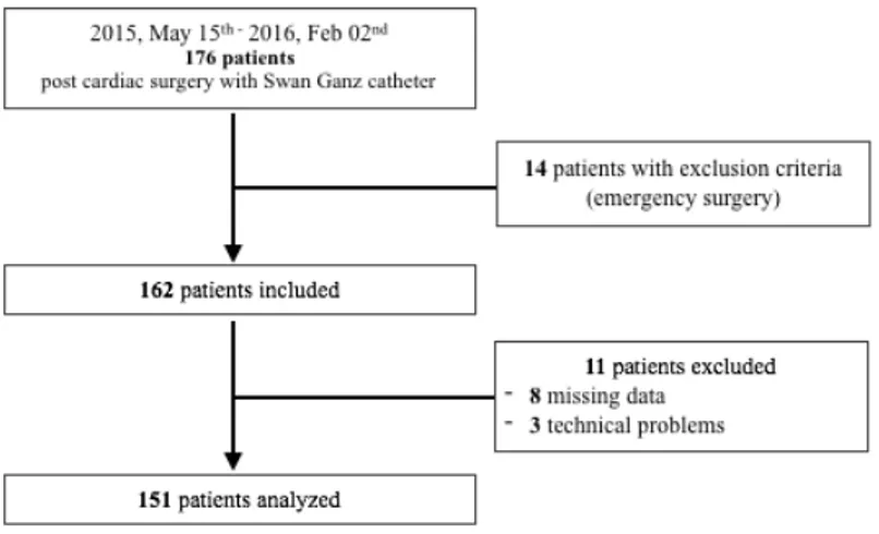
Between May 20th 2015 and February 19th 2016, we included consecutively 162 patients
admitted in the ICU after cardiac surgery. 11 patients were excluded because of missing data (3 RQ record failure, 8 gases analysis missing). The flowchart is described Figure 1. Patients characteristics are described in Table 1.
3. Results 16
Figure 1. Flow Chart
78 patients (52%) presented at least one complication : 7 patients (5%) died while in the ICU, 56 (37%) stayed more than 3 days in ICU and 15 (10%) stayed more than 14 days in hospital.
3.2 Primary endpoint
Univariate analysis
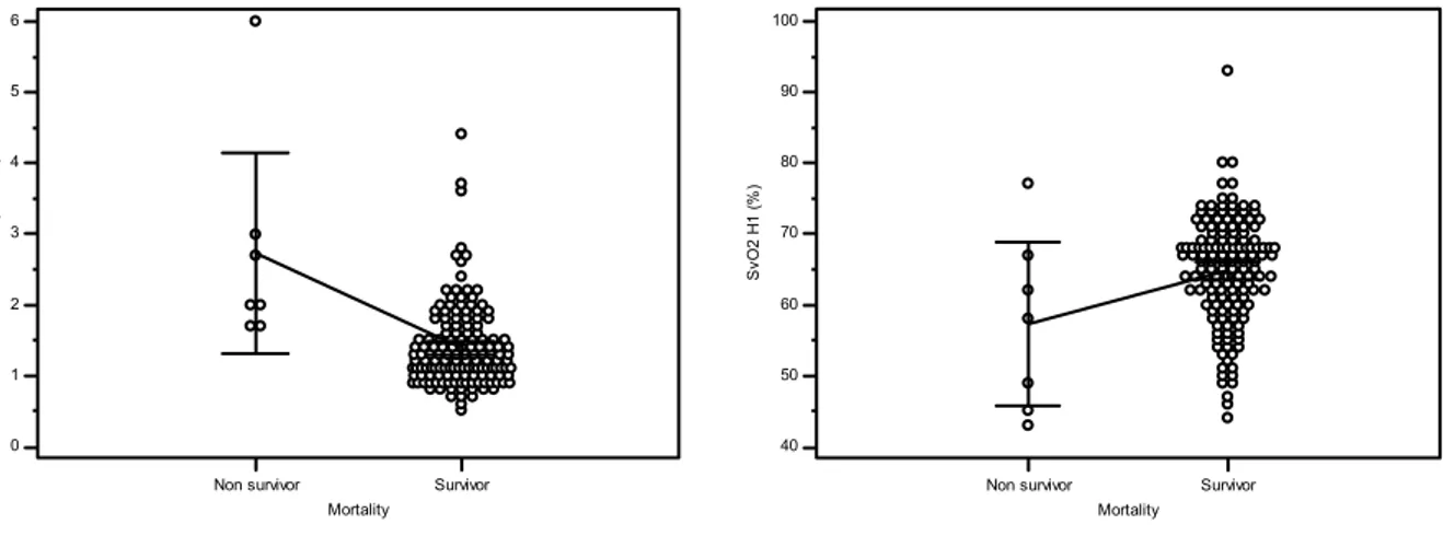
RQ at H1 was significantly higher in non survivors than in survivors (0.83 ± 0.08 vs 0.75 ± 0.09 ; p<0.05) (see Figure 2a).
Lactate and SvO2 in non survivors were respectively significantly higher (2.7 ± 0.5 vs 1.4
± 0.6 ; p<0.001) and lower (57.3 ± 12.4 vs 64.6 ± 7.6 ; p=0.03) than in survivors (see Figure 2b - 2c).
ROC curves
Concerning mortality, AUC ROC curve for RQ was 0.77 (IC95% [0.69-0.84])and for lactate 0.89(IC95% [0.83-0.93]). Diagnosis performances of lactate were significantly higher that of RQ (p=0.02). There was no difference between RQ and SvO2 (AUC of SvO2 0.71 (IC95% [0.63-0.78]) ; p=0.62). Results are shown in Figure 3.
3. Results 17
Table 1. Population characteristics
Characteristics (med) N (151) Age (yrs) 72 [30-88] Sex(%) Male 70 Female 30 Euroscore (%) 2.8[0.5-15.9] Pre-op. characteristics LVEF (%) 58 [25-80]
Pre op. renal failure1(%) 82
Surgery (%) Valve 60 Bypass 16 Combined surgery 17 Other 7 Duration CPB (min) 117 [45-300]
Aortic clamping(min) 84 [0-205]
Total(min) 300 [80-630] Hemodynamic MAP H1(mmHg) 71 [44-120] CVP H1(mmHg) 9[1-19] CI H1 (mL/min/m2) 1.8[0.9-3.6] Biological characteristics pH H1 7.32[7.19-7.48] Lactate H1 (mmoL/L) 1.3[0.5-6.0] SvO2 H1 (%) 66 [43-93] RQ H1 0.75[0.60-1.29]
SOFA score(day 1) 7 [2-16]
ICU duration (days) 3 [1-25]
LVEF : Left Ventricular Ejection Fraction CPB : Cardio Pulmonary Bypass
MAP : Mean Arterial Pressure CVP : Central Venous Pressure CI : Cardiac Index
RQ : Respiratory Quotient
3. Results 18
(a) RQ data on survival and non survival pa-tients at H1 0 1 2 3 4 5 6 Mortality la ct at es H 1 (m m ol /L )
Non survivor Survivor
(b) Lactate data and mortality at H1
40 50 60 70 80 90 100 Mortality S vO 2 H 1 (% )
Non survivor Survivor
(c) SvO2 data and mortality at H1
Figure 2. RQ, lactate and SvO2 data for mortality at H1
3. Results 19
3.3 Secondary endpoint
Outcome
71 patients (47%) had a negative post operative outcome : 56 (37%) stayed in ICU more than 3 days, and 15 (10%) stayed in hospital more than 14 days.
Patients with negative outcome had a higher RQ (0.77 ± 0.08) than others patients (0.75 ± 0.09), but without statistical significance (p=0.056) (see Figure 4).
Figure 4. RQ data and outcome at H1
Moreover, there was no significant difference between AUC ROC curves for RQ, lactate and SvO2 concerning outcome, as shown in Table 2.
Table 2. AUC ROC curves for outcome
Variable AUC [IC95%] p-value
RQ 0.63[0.54-0.70]
Lactate 0.59[0.51-0.67] 0.55
3. Results 20
Organ failure
Regarding dialysis, 4 patients needed extra renal replacement therapy. There was no significant difference between AUC ROC curves for RQ, lactates and SvO2 (see Table 3).
Table 3. AUC ROC curves for dialysis
Variable AUC [IC95%] p-value
RQ 0.69[0.61-0.76]
Lactate 0.65[0.57-0.73] 0.82
SvO2 0.85[0.78-0.90] 0.14
7 patients over 151 presented renal failure at day 14, according to the KDIGO classifica-tion previously defined. AUC for SvO2 was higher than for RQ to predict renal failure at
day 14, without any statistical difference (see Table 4). There was no difference between RQ and lactate.
Table 4. AUC ROC curves for renal failure at day 14
Variable AUC [IC95%] p-value
RQ 0.62[0.54-0.70]
Lactate 0.62[0.54-0.70] 0.97
SvO2 0.77[0.69-0.83] 0.19
Concerning ventilation, 6 patients stayed ventilated more than 12 hours. AUC for lactate was significantly higher than for RQ ; there was no difference between RQ and SvO2 (see Table 5)
Table 5. AUC ROC curves for mechanical ventilation
Variable AUC [IC95%] p-value
RQ 0.73[0.65-0.79]
Lactate 0.95[0.91-0.98] 0.005
4. Discussion 21
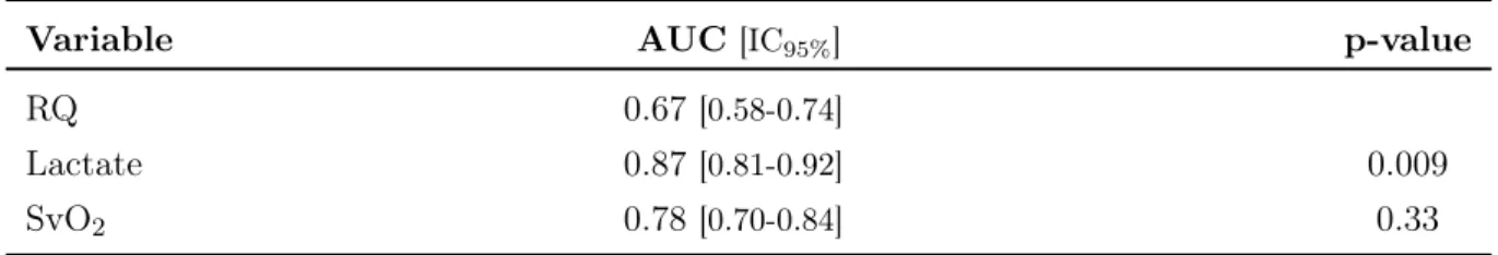
AUC for lactate was also significantly higher than RQ in patients with hepatic failure at day 14 (6 patients over 151). SvO2 and RQ were not significantly different (see Table 6).
Table 6. AUC ROC curves for hepatic failure at day 14
Variable AUC [IC95%] p-value
RQ 0.67[0.58-0.74]
Lactate 0.87[0.81-0.92] 0.009
SvO2 0.78[0.70-0.84] 0.33
Results concerning cardiac failure (needs of dobutamine) and SOFA score (day 1 as well as day 2) were not significant for RQ, lactate or SvO2.
4 Discussion
4.1 Primary endpoint
The main result of this study is that after cardiac surgery, an elevated RQ one hour after arrival in ICU was significantly associated with mortality and, to a lesser extent, morbid-ity. The same results were found for lactate and SvO2.
Lactate is a well known factor associated with mortality after cardiac surgery. Smith et al. [21] found that lactate predicts mortality in ICU, with a AUC of 0.78, instead of 0.89 (IC95% [0.83-0.93]) in our study. The same results are reported in several previous
studies [3,9], regardless of the liver function and lactate clearance [22].
The occurrence of tissue hypoperfusion after cardiac surgery has been assessed using SvO2
and lactate criteria (< 70% and > 2mmol/L respectively) [23] : patients who developed severe tissue hypoperfusion defined by a ScvO2 < 70% and lactate > 4 mmol/L had
significantly prolonged ICU stays. The authors noticed that at ICU admission, 32% of these patients did not have clinical signs of shock (mean blood pressure > 65mmHg and urine output > 0.5mL/kg/h). They concluded that SvO2and lactate after cardiac surgery
4. Discussion 22
may identify occult hypoperfusion and guide hemodynamic optimization before clinical adverse effects.
However, these criteria have several limitations. First, blood lactate level is a discon-tinuous monitoring. Although serial tests are possible, it may therefore be insufficient to conduct an early and efficient hemodynamic optimization. Valenza et al. [4] showed that hyperlactatemia is an early marker for tissue hypoxia, but that after a prolonged anaer-obic metabolism cellular and mitochondrial functions are too impaired to be reverted to a normoxic state, despite hypoxia correction. The existence of this early and reversible period may explain the results obtained by Gattinoni et al. [24] : this study showed no difference in mortality and morbidity between critically ill patients, even after CO or SvO2 correction. In this protocol hemodynamic optimization was initiated 3 days after
patients’ admission in ICU, and energy failure probably occurred even after restauration of oxygen supply, due to cells’ damages.
RQ appears as a promising additional parameter because of its non invasive and continu-ous character. Hemodynamic is unstable after cardiac surgery ; variations occur rapidly, and we need an effective and responsive monitoring to optimize our care.
SvO2 is another useful tool to monitor tissue oxygenation, but it is important to
remem-ber its determinants : its value depends of hemoglobin, cardiac output (CO), oxygen saturation (SaO2) and oxygen consumption, but also of dissolved oxygen. Actually this
criteria is often overlooked because the largest part of oxygen is bound to the hemoglobin. In critically ill patients, with stable and monitored CO and hemoglobin, an increase in SvO2 is reported after increasing FiO2 [25]. Most of this elevation was related to dissolved
oxygen. Therefore, before interpreting a SvO2 result, it is primordial to observe FiO2. In
our study, patients were ventilated with FiO2 40% to 50% for normoxia and it was stable
during SvO2 investigation.
SvO2 reflects the global balance between oxygen supply and demand in the entire
organ-ism ; it can be falsely reassuring in case of regional tissue hypoxia [26, 27]. Lastly, safety of pulmonary artery catheter is still discussed [28, 29].
4. Discussion 23
4.2 Anaerobic metabolism and RQ
RQ elevation during anaerobic metabolism can be explained by an increase in CO2
produc-tion [30,17]. RQ elevation is used to determine the anaerobic threshold in sport training [31]. During normoxia, CO2 production (VCO2) is correlated with O2consumption (VO2).
When tissue oxygen demand increases, VO2 rises until it reaches its maximum. If oxygen
delivery (DO2) can not adapt by increasing, a mismatch between O2 demand and supply
occurs and tissue hypoxia can be observed. In this condition, cells produce lactate and H+ ions via the anaerobic pathway, which increases the CO
2 production. Due to these
variations, VCO2 and RQ rise.
Several studies [32, 33] validated RQ by calorimetry, instead of calculation by the Fick’s methods. We chose to use calorimetry because we consider Fick calculation presents a bias : it tends to underestimate VO2 as it does not integrate the pulmonary oxygen
con-sumption. In our study we employed calorimetry with Haldane equations to ensure a good reliability to our measures.
Other studies have found a relationship between RQ and mortality in critically ill patients. Ranucci et al. [18] studied the predictive value of carbon dioxide derived param-eters during cardiac surgery. They found that an increase in VCO2 and RQ during CPB
(threshold value 0.9) was correlated with hyperlactatemia. In our study, we observed similar results, with RQ also correlated with mortality, for a threshold value of 0.76.
In another context Monnet et al. [34] compared SvO2 and anaerobic markers (lactate
and ratio between veno-arterial carbon dioxide tension difference ∆PCO2 and
arterio-venous oxygen content difference Ca-vO2,
∆P CO2
Ca − vO2
) in septic patients. They demon-strated that among volume responders, patients with an increased VO2 after fluid
expan-sion were characterized by a higher lactate level and a higher ∆P CO2 Ca − vO2
ratio, than those who did not increase their VO2 after fluid expansion. SvO2 did not appear to predict this
increase in VO2. They concluded that we should used anaerobic markers instead of SvO2
to start hemodynamic resuscitation in septic patients.
5. Conclusion 24
and Ca-vO2. In consideration of Haldane effect and relation between CvCO2, SvO2 and
PCO2, their ratio appears therefore to be correlated with RQ. Both can predict tissue
hypoxia and its reversibility (due to the VO2 dependence), allowing to start an early
hemodynamic resuscitation.
4.3 Limitations
Our study presents several bias. Firstly, caregivers were not blinded to RQ values. Al-though RQ was not supposed to be used during patients management as it is not included in our decision algorithms, it appeared on the ventilators screen. Therefore its value might have influenced physicians decision during the patient care.
Secondly we conducted a single institution study : we can not exclude a site effect con-cerning specific management of cardiac patients in our hospital. Moreover the selected population is not totally representative of all the post cardiac surgery patients.
In addition, the interpretation of some results (in particular for secondary endpoints) might be limited by the small number of patients who presented complications.
Lastly, we did not obtain significant results at the admission (H0). This might be due to the calibration of the RQ module : it needs a constant FiO2 during 20 minutes to
mea-sure a reliable RQ. The patients were transfered from the operating room with a portable ventilator, and therefore minute ventilation and FiO2 could vary at the arrival in ICU.
These modifications may explain the absence of results at H0.
5 Conclusion
In this first study, we found that a high RQ was correlated with mortality and morbidity after cardiac surgery, with a threshold value of 0.76 to predict mortality. We found the same results for lactate and, less importantly, for SvO2.
These results suggest that RQ may be a part of the multimodal management to help predicting high risk patients in ICU. This work needs to be extended with other studies, particularly to help define a reliable threshold for RQ.
BIBLIOGRAPHY
[1] Giezeman, A., Bosman, R.J., Oudemans-van Straaten, H.M., Van der Spoel, H.I., Wester, J.P.J., and Zandstra, D.F.: Incidence of low CI immediately post-cardiac surgery in ICU, and its relation to hospital mortality. Intensive care medicine. 15th ESICM Annual Congress - Barcelona, 28 (1): S155–S186, 29 September - 2 October 2002.
[2] Håkanson, E., Svedjeholm, R., and Vanhanen, I.: Physiologic aspects in postopera-tive cardiac patients. Ann. thorac. surg. 59 (2 Suppl): S12–14, 1995.
[3] Cecconi, M. et al.: Consensus on circulatory shock and hemodynamic monitoring. Task force of the European Society of Intensive Care Medicine. Intensive care med, 40 (12): 1795–1815, 2014.
[4] Valenza, F., Aletti, G., Fossali, T., Chevallard, G., Sacconi, F., Irace, M., and Gat-tinoni, L.: Lactate as a marker of energy failure in critically ill patients: hypothesis. Crit care, 9 (6): 588–593, 2005.
[5] Maillet, J-M, Le Besnerais, P, Cantoni, M, Nataf, P, Ruffenach, A, Lessana, A, and Brodaty, D: Frequency, risk factors, and outcome of hyperlactatemia after cardiac surgery*. Chest, 123 (5): 1361–1366, 2003.
[6] Pölönen, P., Ruokonen, E., Hippelainen, M., Poyhonen, M., and Takala, J.: A Prospective, Randomized Study of Goal-Oriented Hemodynamic Therapy in Car-diac Surgical Patients: Anesthesia & analgesia, 90 (5): 1052–1059, 2000.
[7] Chiara, O., Giomarelli, P., Biagoli, B., Rosi, R., and Gattinoni, L.: Hypermetabolic response after hypothermic cardiopulmonary bypass. Crit. care med. 15 (11): 995– 1000, 1987.
[8] Pezzella, A: Care of the adult cardiac surgery patient: part II. Clinical pharmacology & therapeutics, 76 (1): 526–574, 2004.
Bibliography 26
[9] Routsi, C., Vincent, J. L., Bakker, J., De Backer, D., Lejeune, P., d’Hollander, A., Le Clerc, J. L., and Kahn, R. J.: Relation between oxygen consumption and oxygen delivery in patients after cardiac surgery. Anesth. analg. 77 (6): 1104–1110, 1993. [10] Pölönen, P., Hippeläinen, M., Takala, R., Ruokonen, E., and Takala, J.: Relationship
between intra- and postoperative oxygen transport and prolonged intensive care af-ter cardiac surgery: a prospective study. Acta anaesthesiologica scandinavica, 41 (7): 810–817, 1997.
[11] Pearse, R., Dawson, D., Fawcett, J., Rhodes, A., Grounds, R.M., and Bennett, E.D.: Changes in central venous saturation after major surgery, and association with outcome. Critical care, 9 (6): R694, 2005.
[12] Donati, A., Loggi, S., Preiser, J-C., Orsetti, G., Münch, C., Gabbanelli, V., Pelaia, P., and Pietropaoli, P.: GOal-directed intraoperative therapy reduces morbidity and length of hospital stay in high-risk surgical patients*. Chest, 132 (6): 1817–1824, 2007.
[13] Boylan, J. F. and Teasdale, S. J.: Con: perioperative continuous monitoring of mixed venous oxygen saturation should not be routine in high-risk cardiac surgery. J car-diothorac anesth, 4 (5): 651–654, 1990.
[14] Futier, E., Robin, E., Jabaudon, M., Guerin, R., Petit, A., Bazin, J-E, Constantin, J-M, and Vallet, B.: Central venous O2 saturation and venous-to-arterial CO2 dif-ference as complementary tools for goal-directed therapy during high-risk surgery. Crit care, 14 (5): 1–11, 2010.
[15] Lamia, B., Monnet, X., and Teboul, J. L.: Meaning of arterio-venous PCO2 differ-ence in circulatory shock. Minerva anestesiol, 72 (6): 597–604, 2006.
[16] Vallet, B., Teboul, J-L., Cain, S., and Curtis, S.: Venoarterial CO2 difference during regional ischemic or hypoxic hypoxia. Journal of applied physiology, 89 (4): 1317– 1321, 2000.
[17] Mekontso-Dessap, A., Castelain, V., Anguel, N., Bahloul, M., Schauvliege, F., Richard, C., and Teboul, J-L.: Combination of venoarterial PCO2 difference with arteriove-nous O2 content difference to detect anaerobic metabolism in patients. Intensive care med, 28 (3): 272–277, 2002.
[18] Ranucci, M., Isgrò, G., Romitti, F., Mele, S., Biagioli, B., and Giomarelli, P.: Anaer-obic metabolism during cardiopulmonary bypass: predictive value of carbon dioxide derived parameters. Ann. thorac. surg. 81 (6): 2189–2195, 2006.
[19] Hébrard, A. Un quotient respiratoire élevé est prédictif d’un métabolisme anaérobie après chirurgie cardiaque sous circulation extra-corporelle. PhD thesis. 2013. [20] Brandi, L. S., Bertolini, R., and Calafà, M.: Indirect calorimetry in critically ill
Bibliography 27
[21] Smith, I., Kumar, P., Molloy, S., Rhodes, A., Newman, P. J., Grounds, R. M., and Bennett, E. D.: Base excess and lactate as prognostic indicators for patients admitted to intensive care. Intensive care med, 27 (1): 74–83, 2001.
[22] Kruse, J.A., Zaidi, S.A.J., and Carlson, R.W.: Significance of blood lactate levels in critically III patients with liver disease. The american journal of medicine, 83 (1): 77–82, 1987.
[23] Hu, B.Y., Laine, G.A., Wang, S., and Solis, R.T.: Combined Central Venous Oxygen Saturation and Lactate as Markers of Occult Hypoperfusion and Outcome Following Cardiac Surgery. Journal of cardiothoracic and vascular anesthesia, 26 (1): 52–57, 2012.
[24] Gattinoni, L., Brazzi, L., Pelosi, P., Latini, R., Tognoni, G., Pesenti, A., and Fu-magalli, R.: A trial of goal-oriented hemodynamic therapy in critically ill patients. SvO2 Collaborative Group. N. engl. j. med. 333 (16): 1025–1032, 1995.
[25] Legrand, M., Vallée, F., Mateo, J., and Payen, D.: Influence of arterial dissolved oxygen level on venous oxygen saturation: don’t forget the Pao2! Shock (augusta, ga.) 41 (6): 510–513, 2014.
[26] Pearse, R. M. and Rhodes, A. Mixed and Central Venous Oxygen Saturation. en. In: Yearbook of Intensive Care and Emergency Medicine 2005. Ed. by Vincent, Prof Jean-Louis. EN]Yearbook of Intensive Care and Emergency Medicine 2005. Springer New York, 2005: 592–602.
[27] Vallée, F., Vallet, B., Mathe, O., Parraguette, J., Mari, A., Silva, S., Samii, K., Fourcade, O., and Genestal, M.: Central venous-to-arterial carbon dioxide difference: an additional target for goal-directed therapy in septic shock? Intensive care med, 34 (12): 2218, 2008.
[28] Connors, A F., Speroff, T., Dawson, N V., Thomas, C., Harrell, F. E., Wagner, D., Desbiens, N., Goldman, L., Wu, A.W., Califf, R.M., et al.: The effectiveness of right heart catheterization in the initial care of critically III patients. Jama, 276 (11): 889–897, 1996.
[29] Ramsey, S.D., Saint, S., Sullivan, S.D., Dey, L., Kelley, K., and Bowdle, A.: Clini-cal and economic effects of pulmonary artery catheterization in nonemergent coro-nary artery bypass graft surgery. Journal of cardiothoracic and vascular anesthesia, 14 (2): 113–118, 2000.
[30] Faisy, C. and Taylor, S. J.: Dépense énergétique en réanimation. Réanimation, 18 (6): 477–485, 2009.
[31] Solberg, Geir, Robstad, Bjørn, Skjønsberg, Ole Henning, and Borchsenius, Fredrik: Respiratory Gas Exchange Indices for Estimating the Anaerobic Threshold. J sports sci med, 4 (1): 29–36, 2005.
Bibliography 28
[32] Bizouarn, P., Soulard, D., Blanloeil, Y., Guillet, A., and Goarin, Y.: Oxygen con-sumption after cardiac surgery —a comparison between calculation by Fick’s prin-ciple and measurement by indirect calorimetry. Intensive care med, 18 (4): 206–209, 1992.
[33] Straaten, H. M. Oudemans-van, Scheffer, G. J., Eysman, L., and Wildevuur, Ch R. H.: Oxygen consumption after cardiopulmonary bypass — implications o of dif-ferent measuring methods. Intensive care med, 19 (2): 105–110, 1993.
[34] Monnet, X., Julien, F., Ait-Hamou, N., Lequoy, M., Gosset, C., Jozwiak, M., Per-sichini, R., Anguel, N., Richard, C., and Teboul, J_L.: Lactate and venoarterial carbon dioxide difference/arterial-venous oxygen difference ratio, but not central venous oxygen saturation, predict increase in oxygen consumption in fluid respon-ders. Crit. care med. 41 (6): 1412–1420, 2013.
Partie III
Annexes
LISTE DES FIGURES
1 Flow Chart . . . 16
2 RQ, lactate and SvO2 data for mortality at H1 . . . 18
3 ROC curves for mortality at H1 . . . 18
LISTE DES TABLEAUX
1 Population characteristics . . . 17
2 AUC ROC curves for outcome . . . 19
3 AUC ROC curves for dialysis . . . 20
4 AUC ROC curves for renal failure at day 14 . . . 20
5 AUC ROC curves for mechanical ventilation . . . 20
Clermont-Ferrand, le 25/03/2015 Cher Monsieur,
Nous vous prions de prendre connaissance de l’évaluation de votre projet présenté au CECIC en date du 24/02/2015, après relecture. Cette décision a été rendue après revue de votre projet selon la loi Française sur la Recherche Biomédicale [1] et la déclaration de Déclaration d'Helsinki de l'Association Médicale Mondiale [2].
[1] Chapitre Ier du titre II du livre Ier de la première partie du Code de la Santé Publique relatif aux recherches biomédicales.
[2] Déclaration d'Helsinki de l'Association Médicale Mondiale. Principes éthiques applicables aux recherches médicales sur des sujets humains [http://www.wma.net/f/policy/b3.htm].
Avec nos sentiments les meilleurs.
Dr Christian DUALÉ, Médecin Délégué Pr Claude DUBRAY, Médecin Coordonnateur
Date de la réunion 24/02/2015
N° IRB 5891
Membres présents Abergel A, Dualé C, Dubray C, Guttmann A, Laclautre L, Pereira B, Richard R.
Membres excusés Bernard L, Durando X, Langlois J, Mourgues C, Ouchchane L.
Expertise extérieure NA
Titre du projet soumis Une élévation du quotient respiratoire est-elle prédictive des complications postopératoires après chirurgie cardiaque sous CEC ? Etude observationnelle.
Porteur du projet Durand M.
N° de dossier IRB 2015-17
Service Anesthésie-Réanimation
CHU de rattachement Grenoble Autres destinataires CECIC
Pr Hervé Decousus
CIC Saint Etienne cic@chu-st-etienne.fr Tél. 04 72 12 08 26 Fax 04 77 12 78 20 Dr Christian Dualé Chair Centre de Clermont-Ferrand IRB n°00005891 cduale@chu-clermontferrand.fr Tél. 04.73.17.84.18 Fax 04.73.17.84.12 Dr Jean-Luc Cracowski Chair Centre de Grenoble IRB n°00005921 cic@chu-grenoble.fr Tél. 04 76 76 92 60 Fax 04 76 76 92 62 Dr Behrouz Kassaï CIC Lyon bk@upcl.univ-lyon1.fr Tél. 04 72 35 72 31
Comité d’Ethique des Centres d’Investigation Clinique
de l’inter-région Rhône-Alpes-Auvergne
Et h iq u e con ce rn an t le p roj et : la r ec he rc he pe ut -e ll e êt re c ons idé ré e com m e obs er vat ionne ll e ? 1 Ou i N on ? (cf . n ot e) R ec ue il de s cr itè re s de jug em en t U ti lis at io n de s m éd ic am en ts / di spo si ti fs m éd ic au x (A M M / m ar qua ge C E ) M oda lit és de s ur ve il la nc e pr év ue s (c on su lta tio ns , vi si te s. ..) A ut re pr at ique L e re n se ign em en t e n « n on » d’ u n it em c on du it à u n r ec las se m en t e n R B M / R S C / C E B 2 In fo rm at ion d u p ar ti ci p an t : le par ti ci pant à l’ ét ude e st -i l l oy al em ent in for m é ? 3 Ou i N on ? (c f. n ot e) N at ur e et de la f ina lit é de la r ec he rc he M ét ho de e m pl oy ée po ur pr en dr e co nt ac t a ve c lu i C at égor ie de do nné es t ra ité es P os si bi lit é d’ oppo si ti on à la pa rt ic ipa ti on o u à l’ exp lo ita tio n de s do nn ée s G ar an ti e de qua lit é de s so ins e n ca s de r ef us à la p ar tic ipa ti on o u à l’ ex pl oi ta ti on de s do nn ée s D ro it d’ ac cè s et de r ec ti fi ca ti on po ur le s inf or m at io ns le c on ce rn an t P rot ec ti on d es d on n ée s : l’ ét ude e st -e ll e con for m e à la loi I nf or m at ique e t L ibe rt és ? Ou i N on ? (c f. n ot e) U n tr an sf er t de d on né es in te r-ét abl is se m en t es t-il pr év u ? L e fi chi er o ri gi na l c on te na nt l’ inf or m at io n no m ina ti ve e st -i l pr ot égé s el on une pr oc édur e dé cr ite ? L ’a no nym is at io n de s do nn ée s sur le f ic hi er d’ ex pl oi ta tio n es t-el le pr év ue s el on u ne pr oc édur e dé cr ite ? L a pr ot ec tio n du se cr et m éd ic al e st -e lle ga ra nt ie ? Y a- t-il de s do nné es s ens ib le s au se ns de la lo i ? 1 Ce s re ch erc he s so nt , se lo n l’ ar tic le L 1121 -1 du CS P, c el le s « da ns l es que ll es t ous l es a ct es s on t pra tiqué s et l es p ro dui ts ut il is és de m ani èr e ha bi tue ll e, s an s auc un e pr oc édur e supp lé m en ta ir e ou inha bi tue ll e de d ia gn os ti c ou de s ur ve il la nc e » (à jus tif ie r au b es oi n pa r re co m m an da tio n va li dé e). 2 Co ll ec ti on d’é ch ant ill on s bi ol ogi que s (CE B ) : « R éun io n à de s fi ns s ci en ti fi que s, de p ré lè ve m en ts b io lo gi que s effe ct ué s sur un g ro upe de pe rs onn es i de nt if ié es e t sé le ct io nn ée s en f on ct io n de s c ara ct éri st iq ue s cl in iq ue s ou bi ol ogi que s d’un ou pl us ie ur s m em br es d u gr oupe a in si que d es dé ri vé s de c es pr él ève m en ts . » ( Lo i n °2004 -800 du 6 ao ût 2004 r el at iv e à la b io ét hi que ) 3 Po ur le s ét ude s re pr ena nt de s do nn ée s is sue s du so in in tra -ho spi ta li er, u ne in fo rm at io n in fo rm el le pa r af fi ch age e st po ss ib le .
Recommandations « Informatique et Libertés » Non applicable : autorisation déjà obtenue et conforme
Pas de nécessité de déclaration de fichier nominatif
Déclaration simple de fichier nominatif X
Demande d’autorisation + avis du CCTIRS
RBM / CEB : non applicable (méthodologie de référence) Décision du CECIC
Validé X
Réserves de modifications à soumettre au CECIC Réserves majeures en termes d’éthique
Reclassement en recherche biomédicale (RBM) avis CPP requis
Reclassement en collection d’échantillons biologiques (CEB) avis CPP requis Reclassement en recherche sur les soins courants avis CPP requis
En cas de publication de ce travail, voici une suggestion de formulation pour attester de l’obtention de l’avis favorable du CECIC Clermont-Ferrand :
« Un avis éthique consultatif favorable a été obtenu le 25/03/2015 (CECIC Rhône-Alpes-Auvergne, Clermont-Ferrand, IRB 5891) ».
« Study ethics approval was obtained on 25 March 2015 (CECIC Rhône-Alpes-Auvergne, Clermont-Ferrand, IRB 5891) ».
SERMENT D’HIPPOCRATE
En présence des Maîtres de cette Faculté, de mes chers condisciples et devant l’effigie d’HIPPOCRATE,
Je promets et je jure d’être fidèle aux lois de l’honneur et de la probité dans l’exercice de la Médecine.
Je donnerais mes s oi n s gratui tement à l ’i ndigent et n’exi gerai jamais un salaire au dessus de mon travail. Je ne participerai à aucun partage clandestin d’honoraires.
Admis dans l’intimité des maisons, mes yeux n’y verront pas ce qui s’y passe ; ma langue taira les s ecr ets qui me s eront conf iés et mon ét at ne servira pas à corrompre l es mœurs , ni à favori ser l e cri me.
Je ne permettrai pas que des considér ations de rel igion, de nation, de race, de part i ou de classe sociale viennent s’interposer entre mon devoir et mon patient.
Je garderai l e res pect abs olu de la vie humaine.
Même sous la menace, je n’admettrai pas de faire usage de mes connaissances médicales contre les lois de l’humanité.
Respectueux et reconnaissant envers mes Maîtres, je rendrai à leurs enfants l’instruction que j’ai reçue de leurs pères.
Que les hommes m’accordent leur estime si je suis fidèle à mes promesses. Que je sois couvert d’opprobre et méprisé de mes confrères si j’y manque.