Oncogenic Activity
Ste´phanie Delval1, Arnaud Taminiau1, Juliette Lamy1, Ce´cile Lallemand2, Christine Gilles2, Agne`s Noe¨l2, Rene´ Rezsohazy1*
1 Molecular and Cellular Animal Embryology Group, Life Sciences Institute (ISV), Universite´ Catholique de Louvain, Louvain-la-Neuve, Belgium, 2 Laboratory of Biology of Tumors and Development, GIGA-Cancer, University of Lie`ge and Centre Hospitalier Universitaire, Lie`ge, Belgium
Abstract
Hoxa1 belongs to the Hox family of homeodomain transcription factors involved in patterning embryonic territories and governing organogenetic processes. In addition to its developmental functions, Hoxa1 has been shown to be an oncogene and to be overexpressed in the mammary gland in response to a deregulation of the autocrine growth hormone. It has therefore been suggested that Hoxa1 plays a pivotal role in the process linking autocrine growth hormone misregulation and mammary carcinogenesis. Like most Hox proteins, Hoxa1 can interact with Pbx proteins. This interaction relies on a Hox hexapeptidic sequence centred on conserved Tryptophan and Methionine residues. To address the importance of the Hox-Pbx interaction for the oncogenic activity of Hoxa1, we characterized here the properties of a Hoxa1 variant with substituted residues in the hexapeptide and demonstrate that the Hoxa1 mutant lost its ability to stimulate cell proliferation, anchorage-independent cell growth, and loss of contact inhibition. Therefore, the hexapeptide motif of Hoxa1 is required to confer its oncogenic activity, supporting the view that this activity relies on the ability of Hoxa1 to interact with Pbx.
Citation: Delval S, Taminiau A, Lamy J, Lallemand C, Gilles C, et al. (2011) The Pbx Interaction Motif of Hoxa1 Is Essential for Its Oncogenic Activity. PLoS ONE 6(9): e25247. doi:10.1371/journal.pone.0025247
Editor: Anton Wutz, Wellcome Trust Centre for Stem Cell Research, United Kingdom Received December 3, 2010; Accepted August 30, 2011; Published September 21, 2011
Copyright: ß 2011 Delval et al. This is an open-access article distributed under the terms of the Creative Commons Attribution License, which permits unrestricted use, distribution, and reproduction in any medium, provided the original author and source are credited.
Funding: This work was supported by the Belgian Fund for Scientific Research (FNRS, http://www1.frs-fnrs.be/, FRSM grant nu3.4.536.06.F), the ‘‘Direction Ge´ne´rale des Technologies, de la Recherche et de l’Energie’’ of the Walloon Region (http://recherche-technologie.wallonie.be/, WALEOII grant nu516054), and the Fonds Spe´ciaux de Recherche (FSR) of the Universite´ catholique de Louvain (UCL). SD held a Te´le´vie fellowship from the FNRS. The funders had no role in study design, data collection and analysis, decision to publish, or preparation of the manuscript.
Competing Interests: The authors have declared that no competing interests exist. * E-mail: rene.rezsohazy@uclouvain.be
Introduction
Hox genes define a subset of the homeobox gene family coding for homeodomain transcription factors involved in mammalian embryogenesis and organogenesis [1,2,3]. They contribute to pattern the main body axis and the limbs and they control cell fate determination in several organs and cell lineages [4,5,6]. Misregulation of Hox genes has been reported to be associated with the development of a variety of human cancers, including those of skin [7], breast [8], lung [9], prostate, and blood cells [10]. Whether this association between tumorigenesis and Hox gene misexpression reveals that Hox genes actually contribute to the transformation process, is an issue that remains largely unresolved. Only a few Hox proteins have actually been proved to act on cancer progression, either as oncoproteins or tumor suppressors [11,12,13].
In the normal mammary gland, distinct Hox genes exhibit specific expression patterns and functions along its successive development phases, from prenatal stages to lactation at adulthood [14]. Hoxc6 is expressed during mammary development and this expression declines during pregnancy [15] while Hoxa9, Hoxb9 and Hoxd9 are required for the expansion and/or differentiation of the mammary epithelial ductal system in response to pregnancy [16] and targetted disruption of Hoxd10, leads to a failure in alveolar expansion in late pregnancy and concomitant lactation defect [17].
In addition to their involvement in the normal mammary gland biology, studies have shown that some Hox genes are repressed or overexpressed in mammary carcinomas and therefore influence cancer progression. For example, when HOXA10 is expressed in both benign and malignant breast tissue in adult women, it impacts on tumor cell phenotype by decreasing cell invasiveness and upregulating the tumor suppressor gene p53 [18]. HOXA5 is also a positive regulator of p53 in the normal breast tissue. In human breast tumors, p53 expression can be dramatically decreased by a compromised HOXA5 function [19], and expression of HOXA5 in epithelial cancer cells displaying wild-type p53 led to apoptotic cell death. HOXD10 also has a tumor suppressor function. Its expression is progressively reduced in epithelial cells as malignancy increases in breast tumors and restored Hoxd10 activity inhibits tumor development in mouse xenografts and impairs migration of tumor cells [20]. HOXA9 positively regulates BRCA1 expression and represses breast tumor growth and malignancy [21]. While several Hox proteins act as tumor suppressors, HOXB7 is overexpressed in primary breast carcinoma and metastasis, and it stimulates tumor progression by promoting epithelial-mesenchymal transition [22].
Hoxa1 is one of the first Hox genes to be expressed during embryonic development [23]. Gene inactivation has demonstrated its functional importance for hindbrain segmentation, hindbrain patterning, inner and middle ear organogenesis and skull basis morphogenesis [24]. While Hoxa1 is not expressed in the adult
mammary gland, several studies revealed that it can be upregulated in mammary carcinomas [8,15,17,25]. Hoxa1 can be activated in mammary epithelial cells in response to an increased autocrine growth hormone (hGH) stimulation which leads to cell transformation as well as cancer progression and invasiveness [26,27,28]. Forced expression of Hoxa1 is sufficient to provoke the oncogenic transformation of immortalized human mammary epithelial cells and formation of tumors in vivo after cell grafting in mice [29].
Several Hoxa1 target genes have been identified to take part in carcinogenesis. Genes coding for signal tranducing proteins active in the p44/42 mitogen-activated protein (MAP) kinase pathway (GRB2, MEK1, SDFR1) are downstream targets of Hoxa1 [30]. Some p44/42 MAP kinase-regulated genes (IER3, EPAS1, PCNA, catalase) can also be modulated by Hoxa1 [30]. Hoxa1 has further been demonstrated to stimulate oncogenicity by activating STAT3, STAT5B [31] and the anti-apoptotic gene BCL-2, with the consequence to dramatically reduce the apoptotic cell death [29]. Another gene directly regulated by Hoxa1, EphA2, has also been reported to transform mammary epithelial cells and to promote tumor formation in vivo [32]. Expression of EphA2 and its ligand ephrin-A1 has been observed in the vasculature of human primary breast cancer and of breast-tumor-cell-line-derived tumors in nude mice. Thus, EphA2 has been proposed to be involved in tumor-induced angiogenesis [33]. Furthermore, Hoxa1 promotes the activation of Cyclin-D1 required for the autocrine hGH-mediated cell cycling stimulation in mammary carcinoma [29,34]. Finally, an increased Hoxa1 expression is not only observed upon autocrine hGH stimulation but can also occur as a consequence of E-cadherin-mediated signalling. Hoxa1 activation is required for E-cadherin-dependent anchorage-independent proliferation and decreases apoptotic cell death of human mammary carcinoma cells [35].
As transcription factors, Hox proteins cooperate with other transcription regulators or coregulators [36,37,38,39]. Such interactions affect the DNA binding specificity and/or the transcriptional activity of the Hox proteins [40,41,42,43,44]. Among the best characterized Hox cofactors are the Three-Amino-acid-Loop-Extension (TALE) family of homeodomain proteins [45,46], which can be subdivided into four groups according to sequence similarities: PBC (Pbx, ceh-20, exd), TGIF, MEIS (Meis, ceh-25, hth, Prep) and IRO [47,48]. The Pbx proteins belong to the PBC group of TALE proteins able to cooperatively bind to DNA with Hox proteins of paralogy groups 1–10. In vitro studies have shown that Hox/Pbx heterodimers display a greater affinity and specificity for cognate DNA sequences than the Hox monomers [41,49]. The interaction between Hox proteins of paralog groups 1–8 and Pbx relies on a conserved hexapeptide sequence located N-terminal to the Hox homeodomain and sharing core Tryptophan and Methionine residues. Hox proteins of paralog groups 9 and 10 do not contain this hexapeptide, they only present a conserved Tryptophan allowing their interaction with Pbx [50,51,52,53].
Mutational analysis of Hoxa1 has revealed that the Tryptophan and Methionine residues of the conserved hexapeptide are critical for the cooperative interaction between Hoxa1 and Pbx1 [42]. Moreover, the mutant Hoxa1 protein was found to be inactive on cognate target enhancers in live cells [54]. Finally, in vivo studies have demonstrated that knock-in mice for mutations resulting in a WM-to-AA substitution in the hexapeptide of Hoxa1 display hindbrain, cranial nerve and skeletal defects corresponding to the phenotype of the Hoxa1 knock-out [55]. Together, these data support that the embryonic function of Hoxa1 requires the
integrity of its hexapeptide motif, which in turn suggests that the activity of the protein critically relies on its partnership with Pbx. Considering the requirement for an intact hexapeptide for the normal activity of Hoxa1, we have addressed here its importance for the oncogenic potential of the protein. Proliferation, anchorage-independent growth and foci assays have been performed to compare the cellular responses to wild-type or hexapeptide mutant Hoxa1. Our data demonstrate that the WM-to-AA substitution in the Hoxa1 hexapeptide severely impairs its oncogenic properties, which therefore suggests the Hoxa1/Pbx partnership to be involved in its ability to transform mammary epithelial cells. Possible implications in terms of therapeutic applications are discussed. Results
The Hoxa1 protein mutated in its hexapeptide has lost the ability to stimulate mammary cells proliferation
Hoxa1 has previously been shown to affect the phenotype of the epithelioid mammary tumor cell line MCF7 in a way that is indicative of its pro-oncogenic activity, as its forced expression enhanced cell proliferation and anchorage-independent growth [29,35]. To address the importance of the Hoxa1 hexapeptide for its mammary carcinogenic activity, we generated stable MCF7 cell clones for the expression of distinct Hoxa1 variants. The Hoxa1 gene encodes two alternatively spliced mRNA. A 2.2 kb long mRNA resulting from a single splicing event encodes the full-length protein. A shorter mRNA is obtained when a second, alternative splicing event takes place, generating a frameshift and coding for a truncated protein devoid of homeodomain and hexapeptide sequences [56]. The truncated Hoxa1 variant has been shown to interact with Hoxa1 and Pbx1 and to interfere with the activity of the full-length Hoxa1 [57]. cDNA based expression vectors derived from the long Hoxa1 mRNA could theoretically generate two mRNA species as the alternative splicing event can take place. A first expression vector was designed based on the full length wild-type cDNA (Hoxa1WT). A second vector was generated in which the alternative splice site was mutated leading to an Isoleucine-to-Valine substitution at position 115 in the Hoxa1 sequence (Hoxa1I-V) which does not affect the Hoxa1 activity in transcription assays [58]. Finally, based on this second construct an expression vector was generated for the Hoxa1 mutant with the core Tryptophan and Methionine residues of the hexapeptide substituted for Alanines (Hoxa1WM-AA).
To control the relative activity of the variant Hoxa1 proteins, transient co-transfection experiments were first carried out involving a luciferase reporter construct as well as expression vectors for both Pbx1a and Prep1. The pML-EphA2-r42B-luc reporter plasmid contains a cognate Hoxa1 target enhancer derived from the EphA2 gene, a well-known mammary oncogene. Prep1 is a TALE homeodomain protein which stimulates the nuclear entry of Pbx and which enhances the ability of Hox-Pbx complexes to activate transcription [38]. Cotransfection experi-ments revealed that the EphA2-r42B-luc reporter was significantly activated in MCF7 cells expressing Hoxa1WT and Hoxa1I-V proteins, but not in Hoxa1WM-AAexpressing cells (Figure 1A). To exclude that the loss of transcriptional activation ability observed for Hoxa1WM-AAwas due to a loss in protein stability, the relative abundance of Hoxa1 proteins in transfected cells was evaluated by western blots. Although these western blots are not quantitative, it clearly appeared that the Hoxa1WM-AAwas properly expressed, at a similar level as the Hoxa1WTand Hoxa1I-Vvariants (Figure 1B). Two stable MCF7 clones were obtained for each expression vector in addition to control clones transfected with the empty vector (CTL). Hoxa1 expression in the selected clones was verified by RT-PCR.
(Figure 2A). One amplified fragment corresponding to the full length Hoxa1 mRNA was detected in the MCF7 clones for the Hoxa1WT, -Hoxa1I-Vand -Hoxa1WM-AAvectors (630 bp, Figure 2A), while no Hoxa1 expression was detected in CTL clones or non-transfected MCF7 cells (not shown). In addition, the fragment expected for the short length Hoxa1 mRNA was never detected in the MCF7-Hoxa1WTcells, suggesting that the alternative splicing does not take place and that the truncated Hoxa1 is not expressed. Therefore, the MCF7 clones for the Hoxa1WTand Hoxa1I-Vconstructs both express only the full length protein and only differ by the fact that the Hoxa1
I-V
clones express a single amino-acid variant of Hoxa1. Finally, we also verified that the PBX1 gene is endogenously expressed in all cell clones (Figure 2A), so that in all clones the Hoxa1 protein can potentially interact with its cofactor. Quantitative RT-PCR con-firmed that Hoxa1 expression level is not significantly different between the Hoxa1 clones ensuring that cell phenotype changes which could be observed are not due to differences in Hoxa1 expression (data not shown).
To check that the constitutively expressed Hoxa1 variants appropriately reach the cell nucleus to achieve gene regulatory roles, immuno-cytochemical assays were performed (Figure 2B). As expected, the CTL clones did not show Hoxa1 expression. As a positive control, transiently transfected MCF7 cells displayed a strong signal for Hoxa1 in cell nuclei. Nuclear staining of Hoxa1 was detected in all stable clones (Figure 2B). Immuno-cytodetection assay revealed that the endogenously expressed PBX1 protein was the PBX1B isoform and that it also localized into the nucleus of the MCF7 cells and stably transfected derivatives (Figure 2C and data not shown). To evaluate if the Hoxa1 variants expressed in the stably transfected clones are transcriptionally active, the pML-EphA2-r42B-luc reporter construct was transiently co-transfected in the stable clones in combination with expression vectors for both Pbx1a and Prep1. Cotransfection experiments revealed that the EphA2-r42B-luc reporter was significantly activated in the clones expressing Hoxa1WTand Hoxa1I-Vproteins, but not in Hoxa1
WM-AA
expressing clones (Figure 3). That the Hoxa1WM-AAvariant was unable to activate the target reporter was confirmed by transient transfection which allows a strong overexpression of the protein [54] (data not shown). These results therefore confirm that MCF7-Hoxa1WT and MCF7-Hoxa1I-V clones express active Hoxa1 proteins, whereas the Hoxa1WM-AAvariant has lost the ability to transactivate target genes.
We then addressed the effect of the hexapeptide substitution on cell growth stimulation provided by Hoxa1. Cell proliferation rate was twice higher for the MCF7-Hoxa1WTand MCF7-Hoxa1I-V clones than for control clones or clones expressing Hoxa1WM-AA mutant (Figure 4A). Interestingly, clones transfected for Hoxa1 WM-AA
grew at the same rate as the control cells transfected with the empty vector. Complementary to proliferation assays, cell growth was recorded over two weeks of culture, with cell counting after 4, 7, 9, 11, 14 and 16 days of culture. This experiment confirmed that clones expressing the Hoxa1WTand Hoxa1I-Vproteins grew twice faster than cells transfected for the Hoxa1WM-AA mutant (Figure 4B). Cells expressing Hoxa1WM-AAhowever grew slower than the controls, suggesting that this mutant Hoxa1 could exert a dominant negative effect in this cell growth assay (see Discussion). Together these data confirm that the Hoxa1 protein stimulates mammary cell proliferation and that this growth stimulation effect is abrogated by the hexapeptide mutation.
Anchorage independent cell growth is provided by the wild-type Hoxa1 while not by the hexapeptide mutant
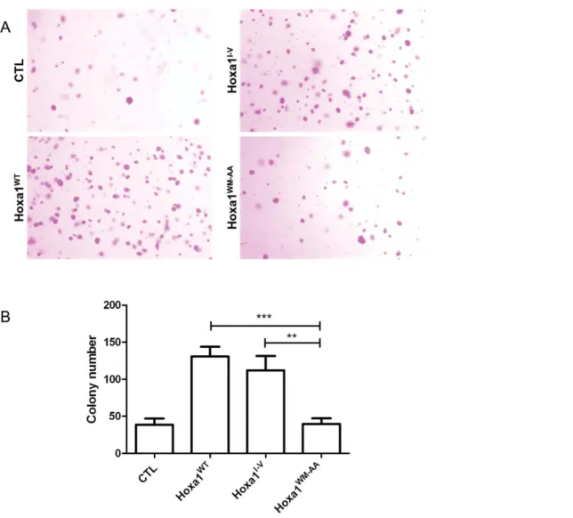
Tumor formation is associated with anchorage independent cell growth. This propensity of cells to grow with loose substrate attachment can be assayed in soft-agar medium. Cell suspensions are mixed in low percentage agar and left for growing over 17 days. Cells able to grow in an anchorage-independent manner will form colonies easily viewed after crystal violet staining. Cell clones were grown in soft agar and colonies were counted after 17 days of culture. As depicted on Figure 5, a low number of colonies were formed by the CTL cells. In contrast about three times more colonies grew from the Hoxa1WTand Hoxa1I-Vexpressing clones. Finally, the Hoxa1WM-AA clones produced a similar amount of colonies as the control clones, demonstrating that the mutant Hoxa1 protein has lost its ability to promote anchorage-independent cell growth (Figure 5).
Hoxa1WM-AAexpressing cells show contact inhibition Tumor cells loose the contact inhibition normally observed for epithelial cells in vivo or in vitro when cells reach confluence. The loss of contact inhibition induced by oncogenes is classically monitored by a foci formation assay. In this assay, cells are Figure 1. Transcriptional activity and relative expression of
Hoxa1 variants. (A) The Hoxa1 target reporter EphA2-r42B-luc is activated in MCF7 cells in the presence of expression vectors for Hoxa1WT, Hoxa1I-Vwhile not in the presence of Hoxa1WM-AAor of an empty (CTL) plasmid. In each experiment, the pML-EphA2-r42B-luc reporter plasmid was transfected in combination with expression vectors for both Prep1 and Pbx1a. Results were calculated by a luciferase/b-galactosidase ratio and represented as means 6 S.D. of triplicates. ***, p,0.001 (ANOVA2). (B) Detection of Hoxa1 variant proteins from whole cell lysates obtained from transiently transfected MCF7 cells reveal that Hoxa1WT, Hoxa1I-Vand Hoxa1WM-AAproteins are equally expressed and stable. No Hoxa1 protein could be detected from MCF7 cells or from cells transfected with an empty (CTL) expression vector. Detection of constitutively expressed b-actin protein was performed as control load.
transiently transfected to express oncoproteins and are left to grow for three weeks. Untransfected MCF7 cells displaying an epithelioı¨d phenotype are responsive to contact inhibition and show very few, if any, foci after three weeks of culture. The
transient transfection of Prep1a and Pbx1 expression vectors in control clones did not enhance foci formation (CTL, Figure 6). In contrast, transfecting Hoxa1WT or Hoxa1I-V together with the cofactors resulted in the appearance of numerous foci (Figure 5). Most significantly, when the hexapeptide mutant was cotrans-fected with the cofactors, a small amount of foci was observed, which was not distinguishable from the situation where only the cofactors were expressed. This assay therefore shows that the Hoxa1WM-AAprotein has lost the ability to relieve the cells from their contact inhibition. This again supports that the hexapeptide mutation suppresses the oncogenic potential of Hoxa1.
Discussion
While a continuously increasing number of studies report correlations between Hox genes misexpression and several types of cancers, only a few Hox genes have been identified to actually impact on cancer progression, as genuine oncogenes or tumor suppressors [11,13]. Hoxa1 has been reported to be abnormally expressed in breast carcinomas [8,25] and to act as a mammary oncogene [29]. Like many other Hox proteins, Hoxa1 can interact with the TALE homeoproteins Pbx. This interaction relies on a hexapeptidic motif of Hoxa1. It has indeed been demonstrated that substituting two amino acids (WM to AA) in this hexapeptide motif abrogated the formation of Hoxa1-Pbx1a complexes on cognate target DNA sequences [59,60]. Further, we have previously shown that disrupting the Hoxa1-Pbx interaction severely impaired its developmental activity. Indeed, by substitut-ing these two amino acids (WM to AA) critically involved in the docking to Pbx, we generated knockin mice which phenocopied the Hoxa1 knockout, suggesting that the Hoxa1-Pbx partnership is crucial to the Hoxa1 function. [55]
Here, we addressed the importance of the hexapeptide integrity for the oncogenic potential of Hoxa1. We demonstrate that the Hoxa1WM-AAhexapeptide mutant lost its ability to stimulate cell Figure 2. Characterization of MCF7 clones for the constitutive expression of Hoxa1 variants. (A) Expression of Hoxa1, Neomycin resistance (Neo), Pbx1 and b-actin genes was detected by RT-PCR. While MCF7 cells do not express Hoxa1, clones obtained the stable transfection of Hoxa1WT, Hoxa1I-Vand Hoxa1WM-AAcoding plasmids express the Hoxa1 variants at similar levels (b-actin used as reference). All cells express the endogenous Pbx1 gene. (B) The Hoxa1 and (C) PBX1B protein immunolocalisation reveals that both proteins localize into the cell nucleus. doi:10.1371/journal.pone.0025247.g002
Figure 3. Activation of a Hoxa1 target reporter in mammary carcinoma cell clones. The Hoxa1 target reporter EphA2-r42B-luc is activated in cell clones for Hoxa1WTand Hoxa1I-Vwhile not in Hoxa1 WM-AA
clones. In each experiment, the pML-EphA2-r42B-luc reporter plasmid was transfected in combination with expression vectors for both Prep1 and Pbx1a. The constitutively active pCMV-LacZ reporter plasmid was added as a transfection control. Results were calculated by a luciferase/b-galactosidase ratio, pooled for each type of clones and represented as means 6 S.D. of triplicates. *, p,0.05 and ***, p,0.001 (ANOVA 2).
proliferation, anchorage-independent cell growth and loss of contact inhibition. Thus, this hexapeptide motif is required to confer to Hoxa1 its oncogenic potential, supporting the view that this critically relies on the ability of Hoxa1 to interact with Pbx.
The involvement of Hox-Pbx interaction in cancer stimulation is supported by several studies aiming at evaluating the impact of HOX-PBX dimer disrupting molecules on cancer cell properties. These molecules were either synthetic peptides mimicking the hexapeptide motif from HOX proteins [61,62,63,64], or mimetic compounds obtained from molecular modelling and combinatorial libraries [65]. Such antagonist molecules have been shown to specifically block proliferation and promote apoptosis of melano-ma, ovarian, pancreatic and non-small-cell lung cancer cells in which members of the HOX family are deregulated [61,63]. Blocking the activity of HOX protein by interfering with their binding to PBX co-factor also reduced the growth of tumor cells in vivo [61,63]. The cell behavior modifications induced by these
inhibitors of the HOX-PBX interaction were further correlated to transcriptional changes indicative of a loss of malignancy [61,62,63]. In a similar approach, Fernandez et al. [66] showed that a dominant negative mutant of PBX reduced the oncogenic activity of HoxB7 and correlated well with increased apoptosis and decreased cell cycling. Finally, mutating the hexapeptide of HOXB4 has also shown to impair its ability to provoke cell transformation [67]. All these studies together with our data suggest that the interaction between Hox and Pbx proteins is a potential therapeutic target for distinct types of cancers.
Nevertheless, disrupting the Hox-Pbx interaction could not always result in a simple functional invalidation of the Hox activity. Indeed, a double mutation in the hexapeptide motif of the mouse Hoxb8 did not result in a loss-of-function of the protein as it is shown here for Hoxa1 and as we previously showed for the Hoxa1WM-AAknockin mice [55,68]. The knockin allele of Hoxb8 coding for a hexapeptide mutant protein indeed appeared as a neomorph. Thus, in contrast to what stands for Hoxa1, the hexapeptide-mediated interaction with Pbx would rather have a modulatory implication on the activity of Hoxb8. The use of hexapeptide mimetic peptides or of related molecules in a therapeutical perspective should then be considered on a case-by-case basis [68] and it would be worth addressing the functional importance of the hexapeptide for additional Hox proteins involved in cancer stimulation.
Although the integrity of the hexapeptide is required for the oncogenic activity of Hoxa1, this does not necessarily imply that the Hoxa1-Pbx interaction is involved in the Hoxa1-mediated oncogen-esis. We cannot formally exclude that the loss of oncogenic potential due to the hexapeptide mutation is independent of the loss of Pbx interaction. Indeed, the hexapeptide might be involved in other critical interactions as has been shown for other Hox proteins. For example, study of the hexapeptide motif of Antennapedia, a Hox protein from drosophila, has revealed that it is involved in an interaction with a TATA-binding associated factor linking Antenna-pedia to the transcripitonal machinery [69]. However, hexapeptide-mediated interactions with other proteins than Pbx have never been reported for Hoxa1, its paralogues or its invertebrate homologues.
Intriguingly, while Hoxa1 expression stimulated cell growth, expression of the Hoxa1WM-AAvariant resulted in a decrease in cell growth with respect to control cells in one of our assays. This suggests that beside the loss of transcription activity and Pbx interaction displayed by Hoxa1WM-AA, this variant could exert a dominant negative effect towards proteins involved in cell proliferation. It is highly expectable that Hoxa1 is involved in diverse protein-protein interactions other than with the sole TALE transcription factors. The WM-AA substitution would not invalidate all those interactions so that although being inactive in mediating Hoxa1-Pbx dimer formation on DNA, this mutant still interacting with other factors to be identified could impair the activity of some of those interactors thereby acting as a dominant negative.
The present study identifies the hexapeptide as a key determinant of Hoxa1 oncogenic properties. Considering the growing body of evidence that Hox proteins can be critical actors in several kinds of cancers, deciphering the modalities of their oncogenic or oncosuppressive activities will undoubtedly be relevant for the clinic and future therapeutic developments. Materials and Methods
Plasmid constructions
Expression vectors for Hoxa1 derivatives were obtained from the previously described pGIH309, pGIH327 and pGIH328 constructs [54]. Shortly, pGIH309 bears the wild-type Hoxa1 cDNA Figure 4. The expression of Hoxa1WM-AAin human mammary
carcinoma cells does not result in increased cell proliferation and growth. (A) WST-1 based proliferation assays were performed for MCF7-Hoxa1WT, MCF7-Hoxa1I-V, MCF7-Hoxa1WM-AA and MCF7-CTL clones. The proliferation index was determined for each clone as described in Materials and Methods. Results were pooled for each type of clones and represented as means 6 S.D. of triplicates. *, p,0.05 (ANOVA 2). (B) Cells for MCF7-Hoxa1WT, MCF7-Hoxa1I-V, MCF7-Hoxa1 WM-AAand MCF7-CTL clones were inoculated, kept in culture for 16 days and counted after day 4, 7, 9, 11, 14 and 16. Growth curves represent the mean of four independent experiments.
(Hoxa1WT) under the control of a CMV enhancer/promoter module. pGIH327 is similar to pGIH309 but harbours a mutant Hoxa1 cDNA in which an alternative splice site has been mutated (Hoxa1I-V) which in turn results in an I-to-V amino acid substitution in the Hoxa1 protein [58]. pGIH328 also contains a Hoxa1 cDNA sequence invalidated for the alternate splicing and is additionally modified to code for the WM-to-AA substitution in the Hoxa1 hexapeptide (Hoxa1WM-AA). To allow selecting stably transfected cells for these expression vectors, a Neomycin resistance marker has been added in pGIH309, pGIH327 and pGIH328 to give rise to pGIH364, pGIH367 and pGIH368, respectively. An empty vector only coding for Neomycin resistance has been obtained as control for all the experiments (pNeo). Details regarding the plasmid constructs are available upon request. Reporter plasmids EphA2-r4-Luc [70] and pCMV-LacZ [71], as well as expression vectors for Pbx1a [54] and Prep1 [72] have been described elsewhere.
Cell culture and transfections
The MCF7 cell line (ATCC #HTB-22) and transfected derivatives were maintained at 37uC in a humidified, 5% CO2
atmosphere in DMEM 4.5 g/L D-glucose supplemented with 10% heat-inactivated fetal bovine serum (FBS), 100 IU/ml penicillin and 100mg/ml streptomycin and 2 mM L-glutamine (Gibco). MCF7 cells were stably transfected with pNeo, pGI364, pGIH367 and pGIH368 plasmids, by use of the Gene Pulser Xcell System (Bio Rad). Transfectants were selected in 1 mg/ml G418 (Gibco). Transient co-transfections for luciferase reporter assays were carried out with the Transfectin reagent (BioRad). One day prior to transfection 80 000 cells per well were seeded in 24-well plates. Each transfection involved a total amount of 1.05mg of DNA, containing: 0.625mg of reporter plasmid (pML-EphA2-r42B-luc); 0.125mg of Hox expression vector; 0.125mg of Pbx1a expression vector; 0.125mg of Prep1 expression vector; and 0.05mg of internal standard reporter plasmid (pCMV-LacZ). In co-transfec-tions aimed at detecting foci formation, 200 000 cells were seeded in 36-mm Petri culture dishes. They have been transfected after 24 hours with 1mg of Hoxa1 or control expression vector and 1mg of each of the Pbx1a and Prep1 expression vectors with the Transfectin reagent (BioRad). As positive control, a plasmid coding for the oncogene hRAS, was used.
Figure 5. The expression of Hoxa1WM-AAin human mammary carcinoma cells does not result in increased anchorage independent cell growth. Cells were grown in soft agar and colonies were revealed by crystal violet staining (A) MCF7-Hoxa1WT and MCF7-Hoxa1I-Vcells produced a lot of colonies in soft agar while CTL and MCF7-Hoxa1WM-AAonly provide a modest colony growth. (B) For each culture, colonies were counted in three random microscopic fields at 16X magnification. Results were pooled for each type of clones and represented as means 6 S.D of triplicates. **, p,0.01; ***, p,0.001.
Western blotting
For detection of Hoxa1 proteins expression, transiently transfected cells were lysed with RIPA buffer (1 mM EDTA, 0.1% SDS, 1% Nonidet P40, 0.5% NaDeoxycholate, 50 mM TrisHCl pH7.5, 250 mM NaCl, protease inhibitors). Whole cell lysates were run on a SDS-PAGE, blotted on a nitrocellulose
membrane and revealed with an anti-Hoxa1 rabbit antibody (1/ 500; Sigma HPA004933), anti-rabbit bovine IgG-horseradish peroxidase (HRP)-conjugated antibody (1/3000; SantaCruz sc-2379). The protein load for western blotting was controlled by detecting b-actin with a HRP conjugated anti-b-actin antibody (1/ 3000 Sigma A3854).
Reverse transcriptase-PCR
Total RNA was extracted using Trizol Reagent (Invitrogen). RNA quantification was performed on a Nanodrop apparatus (Thermo Scientific). Onemg of RNA was then reverse transcribed into cDNA and amplified with the Expand Reverse Transcriptase (Roche) and Taq Polymerase (Westburg) respectively. Primers for RT-PCR were as follows: Hoxa1 (forward), 59-CCTTATGGCCCCTATGGA-39; Hoxa1 (reverse), 59-TTCTCAGATGATTCTTCCGTT-39; b-actin (forward), 59-GCTGGAAGGTGGACAGCGA-39; b-actin (reverse), 59-GGCATCGTGATGGACTCCG-39; NeoR (forward), 59-AAT-GAACTGCAGGACGAGGC-39; NeoR (reverse), 59- CAACGC-TATGTCCTGATAGC-39; Pbx1 (forward), 59-TCAGAGATG-GATGCGAGGGCGAAGAGACGC-39; Pbx1 (reverse), 59-TTTGGCAGCATAAATATTGGC-39. All RNA samples were treated with DNase I to avoid genomic DNA contamination. Immunostaining and fluorescence microscopy
Immunodetections of Hoxa1 and PBX1B were performed either on stably or on transiently transfected cells. In both cases, cells were seeded on glass cover-slips in 24-well plates. For stable clones, twenty-hours after seeding, cells were fixed in 4% formalin and blocked in 10% powder milk. Cells were incubated at 4uC with the anti-Hoxa1 rabbit antibody (1/50, Sigma HPA004933) or anti-Pbx1B (41.1) mouse antibody (1/50, Santacruz sc-101852) overnight. They were washed and incubated respectively with a fluorescein coupled anti-rabbit IgG antibody (1/100, GE Health-care N1034) or with an Alexa Fluor 555 coupled anti-mouse IgG antibody (1/1000, Cell Signaling 4409) for 1 h. Cover-slips were mounted in vectashield with DAPI medium (Vector Laboratories H1200) and viewed under Polyvar microscope (Reichert Jung). For transiently transfected cells, the same procedure was applied, except that cells were firstly transfected 24 hours after seeding and then processed for immunostaining 24 hours after transfection. Luciferase reporter assay
Cells were harvested 48 h after transfection for enzymatic assays. Lysis and enzymatic activity dosages were performed with the b-gal Reporter Gene Assay (Chemiluminescent) kit (Roche) and the Luciferase Reporter Gene Assay (High Sensitivity) kit (Roche), according to the instructions provided by the manufac-turer. For each transfection, the constitutively active pCMV-LacZ reporter was used as a control, so that the relative luciferase activity was calculated by a luciferase/b-galactosidase ratio. Cell proliferation and growth assays
The WST-1 assay is a colorimetric method based on the cleavage by mitochondrial dehydrogenase of the tetrazolium salt generating a detectable product, formazan. Two thousand cells for each MCF7 clone were seeded in 96-well plates in complete DMEM, with 2 wells devoid of cells as blank samples. Cells were allowed to seed overnight at 37uC in 5% CO2. Twenty-four hours after seeding, medium was changed with DMEM supplemented with 1% FBS. Two days after seeding, 10ml of WST-1 reagent was added to the cells medium for 4 hours. The absorbance for the formazan product (440 nm) and the background control (620 nm) were recorded every hour by a multiplate reader Figure 6. Hoxa1WT and Hoxa1I-V relieve MCF7 cells from
contact inhibition, while expressing Hoxa1WM-AA does not. MCF7 cells were transiently transfected for Hoxa1WT, Hoxa1I-V and Hoxa1WM-AA, together with Pbx1a and Prep1 cofactors, and grown for three weeks. Controls included cells transfected for the potent oncogene hRAS or cells transfected for Pbx1a and Prep1 only. Foci formation was observed for hRAS, Hoxa1WTand Hoxa1I-Vtransfected cells (arrowheads) while not for CTL and Hoxa1WM-AAcells.
(SpectraMax 190, Molecular Devices). For each time point, the difference between the measures at 440 nm and 620 nm was calculated and inserted on a graph as a function of time. The slope of each curve was calculated and represented the cell proliferation index.
For growth recording, 5.0 104cells of each MCF7 clone were seeded in 6-well plates in complete DMEM. Medium was changed every 2 days. After 4, 7, 9, 11, 14 and 16 days of culture, cells were counted. Values were reported on a graph representing the cell growth of MCF7 clones.
Anchorage-independent growth assay
Anchorage-independent growth was assayed in soft agar. Cells were plated into 24-well plates in growth medium (DMEM) containing 0.3% agarose, on top of a layer of 0.6% agarose gel (Sigma A9045). After 17 days, cells were stained with crystal violet for 1 h and colonies were counted under a binocular (Wild M3B – Van Hopplynus Instrument) in three random microscopic fields at 16X magnification.
Foci formation assay
MCF7 cells were seeded in 36-mm Petri dish and co-transfected as mentioned above. Once confluence was reached, medium was changed every 3 days. After 2 weeks, cultures were fixed with formalin, stained with 1% rhodamine B (Sigma R6626) and washed with PBS to bleach the non-focal monolayer. Foci were observed under a binocular (Leitz Wetzlar).
Acknowledgments
We thank Bernard Peers for providing the pCS2-Prep1 vector, Jin Chen and H. Earl Ruley for the Epha2-r4-luciferase reporter plasmid.
Author Contributions
Conceived and designed the experiments: RR. Performed the experiments: SD AT JL. Analyzed the data: RR SD AT JL. Contributed reagents/ materials/analysis tools: CL CG AN. Wrote the paper: SD. Contributed to manuscript redaction: RR.
References
1. Favier B, Dolle P (1997) Developmental functions of mammalian Hox genes. Mol Hum Reprod 3: 115–131.
2. Narita Y, Rijli FM (2009) Hox genes in neural patterning and circuit formation in the mouse hindbrain. Curr Top Dev Biol 88: 139–167.
3. Alexander T, Nolte C, Krumlauf R (2009) Hox genes and segmentation of the hindbrain and axial skeleton. Annu Rev Cell Dev Biol 25: 431–456. 4. Magli MC, Barba P, Celetti A, De Vita G, Cillo C, et al. (1991) Coordinate
regulation of HOX genes in human hematopoietic cells. Proc Natl Acad Sci U S A 88: 6348–6352.
5. Mallo M, Wellik DM, Deschamps J (2010) Hox genes and regional patterning of the vertebrate body plan. Dev Biol.
6. Hwang JH, Seok OS, Song HR, Jo JY, Lee JK (2009) HOXC10 as a potential marker for discriminating between amnion- and decidua-derived mesenchymal stem cells. Cloning Stem Cells 11: 269–279.
7. Svingen T, Tonissen KF (2003) Altered HOX gene expression in human skin and breast cancer cells. Cancer Biol Ther 2: 518–523.
8. Cantile M, Pettinato G, Procino A, Feliciello I, Cindolo L, et al. (2003) In vivo expression of the whole HOX gene network in human breast cancer. Eur J Cancer 39: 257–264.
9. Pfeifer GP, Rauch TA (2009) DNA methylation patterns in lung carcinomas. Semin Cancer Biol 19: 181–187.
10. Rice KL, Licht JD (2007) HOX deregulation in acute myeloid leukemia. J Clin Invest 117: 865–868.
11. Abate-Shen C (2002) Deregulated homeobox gene expression in cancer: cause or consequence? Nat Rev Cancer 2: 777–785.
12. Shah N, Sukumar S (2010) The Hox genes and their roles in oncogenesis. Nat Rev Cancer 10: 361–371.
13. Cillo C (2007) Deregulation of the Hox Gene Network and Cancer. in Hox Gene Expression Papageorgiou S ed. pp 121–133.
14. Chen H, Sukumar S (2003) Role of homeobox genes in normal mammary gland development and breast tumorigenesis. J Mammary Gland Biol Neoplasia 8: 159–175. 15. Friedmann Y, Daniel CA, Strickland P, Daniel CW (1994) Hox genes in normal
and neoplastic mouse mammary gland. Cancer Res 54: 5981–5985. 16. Chen F, Capecchi MR (1999) Paralogous mouse Hox genes, Hoxa9, Hoxb9,
and Hoxd9, function together to control development of the mammary gland in response to pregnancy. Proc Natl Acad Sci U S A 96: 541–546.
17. Lewis MT (2000) Homeobox genes in mammary gland development and neoplasia. Breast Cancer Res 2: 158–169.
18. Chu MC, Selam FB, Taylor HS (2004) HOXA10 regulates p53 expression and matrigel invasion in human breast cancer cells. Cancer Biol Ther 3: 568–572. 19. Raman V, Martensen SA, Reisman D, Evron E, Odenwald WF, et al. (2000)
Compromised HOXA5 function can limit p53 expression in human breast tumours. Nature 405: 974–978.
20. Carrio M, Arderiu G, Myers C, Boudreau NJ (2005) Homeobox D10 induces phenotypic reversion of breast tumor cells in a three-dimensional culture model. Cancer Res 65: 7177–7185.
21. Gilbert PM, Mouw JK, Unger MA, Lakins JN, Gbegnon MK, et al. (2010) HOXA9 regulates BRCA1 expression to modulate human breast tumor phenotype. J Clin Invest 120: 1535–1550.
22. Wu X, Chen H, Parker B, Rubin E, Zhu T, et al. (2006) HOXB7, a homeodomain protein, is overexpressed in breast cancer and confers epithelial-mesenchymal transition. Cancer Res 66: 9527–9534.
23. Murphy P, Hill RE (1991) Expression of the mouse labial-like homeobox-containing genes, Hox 2.9 and Hox 1.6, during segmentation of the hindbrain. Development 111: 61–74.
24. Barrow JR, Stadler HS, Capecchi MR (2000) Roles of Hoxa1 and Hoxa2 in patterning the early hindbrain of the mouse. Development 127: 933– 944.
25. Chariot A, Castronovo V (1996) Detection of HOXA1 expression in human breast cancer. Biochem Biophys Res Commun 222: 292–297.
26. Mukhina S, Mertani HC, Guo K, Lee KO, Gluckman PD, et al. (2004) Phenotypic conversion of human mammary carcinoma cells by autocrine human growth hormone. Proc Natl Acad Sci U S A 101: 15166–15171. 27. Waters MJ, Conway-Campbell BL (2004) The oncogenic potential of autocrine
human growth hormone in breast cancer. Proc Natl Acad Sci U S A 101: 14992–14993.
28. Mertani HC, Zhu T, Goh EL, Lee KO, Morel G, et al. (2001) Autocrine human growth hormone (hGH) regulation of human mammary carcinoma cell gene expression. Identification of CHOP as a mediator of hGH-stimulated human mammary carcinoma cell survival. J Biol Chem 276: 21464–21475. 29. Zhang X, Zhu T, Chen Y, Mertani HC, Lee KO, et al. (2003) Human growth
hormone-regulated HOXA1 is a human mammary epithelial oncogene. J Biol Chem 278: 7580–7590.
30. Mohankumar KM, Xu XQ, Zhu T, Kannan N, Miller LD, et al. (2007) HOXA1-stimulated oncogenicity is mediated by selective upregulation of components of the p44/42 MAP kinase pathway in human mammary carcinoma cells. Oncogene 26: 3998–4008.
31. Mohankumar KM, Perry JK, Kannan N, Kohno K, Gluckman PD, et al. (2008) Transcriptional activation of signal transducer and activator of transcription (STAT) 3 and STAT5B partially mediate homeobox A1-stimulated oncogenic transformation of the immortalized human mammary epithelial cell. Endocri-nology 149: 2219–2229.
32. Zelinski DP, Zantek ND, Stewart JC, Irizarry AR, Kinch MS (2001) EphA2 overexpression causes tumorigenesis of mammary epithelial cells. Cancer Res 61: 2301–2306.
33. Ogawa K, Pasqualini R, Lindberg RA, Kain R, Freeman AL, et al. (2000) The ephrin-A1 ligand and its receptor, EphA2, are expressed during tumor neovascularization. Oncogene 19: 6043–6052.
34. Graichen R, Liu D, Sun Y, Lee KO, Lobie PE (2002) Autocrine human growth hormone inhibits placental transforming growth factor-beta gene transcription to prevent apoptosis and allow cell cycle progression of human mammary carcinoma cells. J Biol Chem 277: 26662–26672.
35. Zhang X, Emerald BS, Mukhina S, Mohankumar KM, Kraemer A, et al. (2006) HOXA1 is required for E-cadherin-dependent anchorage-independent survival of human mammary carcinoma cells. J Biol Chem 281: 6471–6481. 36. Mann RS, Affolter M (1998) Hox proteins meet more partners. Curr Opin
Genet Dev 8: 423–429.
37. Foronda D, de Navas LF, Garaulet DL, Sanchez-Herrero E (2009) Function and specificity of Hox genes. Int J Dev Biol 53: 1404–1419.
38. Berthelsen J, Zappavigna V, Ferretti E, Mavilio F, Blasi F (1998) The novel homeoprotein Prep1 modulates Pbx-Hox protein cooperativity. Embo J 17: 1434–1445.
39. Mann RS, Morata G (2000) The developmental and molecular biology of genes that subdivide the body of Drosophila. Annu Rev Cell Dev Biol 16: 243–271. 40. Mann RS, Chan SK (1996) Extra specificity from extradenticle: the partnership
between HOX and PBX/EXD homeodomain proteins. Trends Genet 12: 258–262.
41. Neuteboom ST, Murre C (1997) Pbx raises the DNA binding specificity but not the selectivity of antennapedia Hox proteins. Mol Cell Biol 17: 4696–4706.
42. Chang CP, Shen WF, Rozenfeld S, Lawrence HJ, Largman C, et al. (1995) Pbx proteins display hexapeptide-dependent cooperative DNA binding with a subset of Hox proteins. Genes Dev 9: 663–674.
43. Pellerin I, Schnabel C, Catron KM, Abate C (1994) Hox proteins have different affinities for a consensus DNA site that correlate with the positions of their genes on the hox cluster. Mol Cell Biol 14: 4532–4545.
44. Laughon A (1991) DNA binding specificity of homeodomains. Biochemistry 30: 11357–11367.
45. Huang H, Rastegar M, Bodner C, Goh SL, Rambaldi I, et al. (2005) MEIS C termini harbor transcriptional activation domains that respond to cell signaling. J Biol Chem 280: 10119–10127.
46. Burglin TR (1998) The PBC domain contains a MEINOX domain: coevolution of Hox and TALE homeobox genes? Dev Genes Evol 208: 113–116. 47. Moens CB, Selleri L (2006) Hox cofactors in vertebrate development. Dev Biol
291: 193–206.
48. Mukherjee K, Burglin TR (2007) Comprehensive analysis of animal TALE homeobox genes: new conserved motifs and cases of accelerated evolution. J Mol Evol 65: 137–153.
49. Piper DE, Batchelor AH, Chang CP, Cleary ML, Wolberger C (1999) Structure of a HoxB1-Pbx1 heterodimer bound to DNA: role of the hexapeptide and a fourth homeodomain helix in complex formation. Cell 96: 587–597. 50. Knoepfler PS, Kamps MP (1995) The pentapeptide motif of Hox proteins is
required for cooperative DNA binding with Pbx1, physically contacts Pbx1, and enhances DNA binding by Pbx1. Mol Cell Biol 15: 5811–5819.
51. Chang CP, Brocchieri L, Shen WF, Largman C, Cleary ML (1996) Pbx modulation of Hox homeodomain amino-terminal arms establishes different DNA-binding specificities across the Hox locus. Mol Cell Biol 16: 1734–1745. 52. Shen WF, Chang CP, Rozenfeld S, Sauvageau G, Humphries RK, et al. (1996) Hox homeodomain proteins exhibit selective complex stabilities with Pbx and DNA. Nucleic Acids Res 24: 898–906.
53. Shen WF, Rozenfeld S, Lawrence HJ, Largman C (1997) The Abd-B-like Hox homeodomain proteins can be subdivided by the ability to form complexes with Pbx1a on a novel DNA target. J Biol Chem 272: 8198–8206.
54. Remacle S, Shaw-Jackson C, Matis C, Lampe X, Picard J, et al. (2002) Changing homeodomain residues 2 and 3 of Hoxa1 alters its activity in a cell-type and enhancer dependent manner. Nucleic Acids Res 30: 2663–2668. 55. Remacle S, Abbas L, De Backer O, Pacico N, Gavalas A, et al. (2004) Loss of
function but no gain of function caused by amino acid substitutions in the hexapeptide of Hoxa1 in vivo. Mol Cell Biol 24: 8567–8575.
56. LaRosa GJ, Gudas LJ (1988) Early retinoic acid-induced F9 teratocarcinoma stem cell gene ERA-1: alternate splicing creates transcripts for a homeobox-containing protein and one lacking the homeobox. Mol Cell Biol 8: 3906–3917. 57. Fernandez CC, Gudas LJ (2009) The truncated Hoxa1 protein interacts with
Hoxa1 and Pbx1 in stem cells. J Cell Biochem 106: 427–443.
58. Phelan ML, Rambaldi I, Featherstone MS (1995) Cooperative interactions between HOX and PBX proteins mediated by a conserved peptide motif. Mol Cell Biol 15: 3989–3997.
59. Phelan ML, Featherstone MS (1997) Distinct HOX N-terminal residues are responsible for DNA recognition specificity by HOX monomers and HOX.PBX heterodimers. J Biol Chem 272: 8635–8643.
60. Lampe X, Samad OA, Guiguen A, Matis C, Remacle S, et al. (2008) An ultraconserved Hox-Pbx responsive element resides in the coding sequence of Hoxa2 and is active in rhombomere 4. Nucleic Acids Res 36: 3214–3225. 61. Morgan R, Pirard PM, Shears L, Sohal J, Pettengell R, et al. (2007) Antagonism
of HOX/PBX dimer formation blocks the in vivo proliferation of melanoma. Cancer Res 67: 5806–5813.
62. Plowright L, Harrington KJ, Pandha HS, Morgan R (2009) HOX transcription factors are potential therapeutic targets in non-small-cell lung cancer (targeting HOX genes in lung cancer). Br J Cancer 100: 470–475.
63. Morgan R, Plowright L, Harrington KJ, Michael A, Pandha HS (2010) Targeting HOX and PBX transcription factors in ovarian cancer. BMC Cancer 10: 89.
64. Aulisa L, Forraz N, McGuckin C, Hartgerink JD (2009) Inhibition of cancer cell proliferation by designed peptide amphiphiles. Acta Biomater 5: 842–853. 65. Ji T, Lee M, Pruitt SC, Hangauer DG (2004) Privileged scaffolds for blocking
protein-protein interactions: 1,4-disubstituted naphthalene antagonists of transcription factor complex HOX-PBX/DNA. Bioorg Med Chem Lett 14: 3875–3879.
66. Fernandez LC, Errico MC, Bottero L, Penkov D, Resnati M, et al. (2008) Oncogenic HoxB7 requires TALE cofactors and is inactivated by a dominant-negative Pbx1 mutant in a cell-specific manner. Cancer Lett 266: 144–155. 67. Krosl J, Baban S, Krosl G, Rozenfeld S, Largman C, et al. (1998) Cellular
proliferation and transformation induced by HOXB4 and HOXB3 proteins involves cooperation with PBX1. Oncogene 16: 3403–3412.
68. Medina-Martinez O, Ramirez-Solis R (2003) In vivo mutagenesis of the Hoxb8 hexapeptide domain leads to dominant homeotic transformations that mimic the loss-of-function mutations in genes of the Hoxb cluster. Dev Biol 264: 77–90. 69. Prince F, Katsuyama T, Oshima Y, Plaza S, Resendez-Perez D, et al. (2008)
The YPWM motif links Antennapedia to the basal transcriptional machinery. Development 135: 1669–1679.
70. Chen J, Ruley HE (1998) An enhancer element in the EphA2 (Eck) gene sufficient for rhombomere-specific expression is activated by HOXA1 and HOXB1 homeobox proteins. J Biol Chem 273: 24670–24675.
71. Matis C, Chomez P, Picard J, Rezsohazy R (2001) Differential and opposed transcriptional effects of protein fusions containing the VP16 activation domain. FEBS Lett 499: 92–96.
72. Goudet G, Delhalle S, Biemar F, Martial JA, Peers B (1999) Functional and cooperative interactions between the homeodomain PDX1, Pbx, and Prep1 factors on the somatostatin promoter. J Biol Chem 274: 4067–4073.