Direction des bibliothèques
AVIS
Ce document a été numérisé par la Division de la gestion des documents et des archives de l’Université de Montréal.
L’auteur a autorisé l’Université de Montréal à reproduire et diffuser, en totalité ou en partie, par quelque moyen que ce soit et sur quelque support que ce soit, et exclusivement à des fins non lucratives d’enseignement et de recherche, des copies de ce mémoire ou de cette thèse.
L’auteur et les coauteurs le cas échéant conservent la propriété du droit d’auteur et des droits moraux qui protègent ce document. Ni la thèse ou le mémoire, ni des extraits substantiels de ce document, ne doivent être imprimés ou autrement reproduits sans l’autorisation de l’auteur.
Afin de se conformer à la Loi canadienne sur la protection des renseignements personnels, quelques formulaires secondaires, coordonnées ou signatures intégrées au texte ont pu être enlevés de ce document. Bien que cela ait pu affecter la pagination, il n’y a aucun contenu manquant.
NOTICE
This document was digitized by the Records Management & Archives Division of Université de Montréal.
The author of this thesis or dissertation has granted a nonexclusive license allowing Université de Montréal to reproduce and publish the document, in part or in whole, and in any format, solely for noncommercial educational and research purposes.
The author and co-authors if applicable retain copyright ownership and moral rights in this document. Neither the whole thesis or dissertation, nor substantial extracts from it, may be printed or otherwise reproduced without the author’s permission.
In compliance with the Canadian Privacy Act some supporting forms, contact information or signatures may have been removed from the document. While this may affect the document page count, it does not represent any loss of content from the document.
THE ROLE OF TRANSFORMING GROWTH FACTOR-BETA 1 IN
STEROIDOGENESIS, CELL PROLIFERATION, AND APOPTOSIS IN
CULTURED BOVINE GRANULOSA CELLS
par
XIAOFENG ZHENG
Département de biomédecine vétérinaire Faculté de médecine vétérinaire
Thèse présentée à la Faculté des études supérieures et postdoctorales ' en vue de l'obtention du grade Philosophiae Doctor (Ph. D.)
en sciences vétérinaires option reproduction
Août 2008
Université de Montréal
Faculté des études supérieures et postdoctorales
Cette thèse intitulée
THE ROLE OF TRANSFORMING GROWTH FACTOR-BETA 1 IN
STEROIDOGENESIS, CELL PROLIFERATION, AND APOPTOSIS IN
CULTURED BOVINE GRANULOSA CELLS
présentée par XIAOFENG ZHENG
a été-évaluée par un jury composé des personnes suivantes:
David W. Silversides, président-rapporteur Paul D. Carrière, directeur de recherche Yves Tremblay, codirecteur
Christine Théoret, membre du jury Éric Asselin, examinateur externe
RÉSUMÉ
L'infertilité bovine est une contrainte importante à la rentabilité de l'industrie laitière. La croissance folliculaire anormale, l' anoestrus et l' anovulation sont parmi les principales causes du déclin de la fertilité chez les vaches laitières hautes productrices. Afin de trouver des solutions à ces problèmes, une meilleure connaissance des mécanismes endocriniens qui régulent la croissance normale des follicules ovariens est nécessaire. La stimulation par les gonadotropines des follicules ovariens réceptifs est associée à une augmentation de la synthèse de l'oestradiol-17~ (E2) et de la progesterone (P4). L'E2 est un
marqueur clé de la croissance et de la sélection du follicule dominant, alors que la P 4 est
nécessaire au déclenchement de l'ovulation. Les cellules de la granulosajouent un rôle important pour déterminer la destinée des follicules en développement soit vers
l'ovulation, soit vers l'atrésie parce que ces cellules produisent de l'E2 et de la P4 et sont la
cible des signaux impliqués dans la prolifération cellulaire et l' apoptose.
Le «transforming growth fac.tor-~ 1» (TGF -~ 1) est un facteur important produit par les cellules folliculaires parce qu'il modifie la sécrétion de l'E2 et de la P4• Plusieurs études
effectuées sur des cellules provenant de différents tissus ont rapporté des effets biologiques contradictoires du TGF -~ 1. Par exemple, le TGF -~ 1 stimule la sécrétion de l'E2 et de la P 4
dans les cellules de la granulosa obtenues à partir de rates immatures traitées à l'E2 alors que le TGF-~1 inhibe la sécrétion de l'E2 et de la P4 dans les cellules de la granulosa
IV
d'action du TGF-~l sur les enzymes stéroïdogènes qui transforment les androgènes, l'oestrone (El) et le cholesterol en E2 et en P4 dans des cellules de granulosa bovines stimulées à la FSH et des cellules de granulosa quiescentes non-stimulées. Le deuxième objectif était de déterminer l'effet du TGF-~l sur la différentiation et la prolifération cellulaire et sur l'apoptose. Les cellules de granulosa bovines obtenues à partir de follicules de 2-5 mm ont été mises en culture dans un milieu sans serum en présence de FSH ou en absence de FSH (cellules quiescentes). Les cellules de granulosa quiescentes se sont lutéinisées spontanément en augmentant la secretion de P 4 et l'expression de l' ARNm des enzymes impliquées dans la synthèse de la P4 : StAR, CYPllA1, HSD3B et GSTA. L'ajout de FSH a ralenti le processus de lutéinisation en augmentant la synthèse de l'E2 et l'expression des enzymes CYP 19A1 et HSD17B1 et en inhibant l'expression de StAR. Dans les cellules de la granulosa stimulées par la FSH, le TGF-~l a inhibé l'augmentation d'E2 et de P4 en inhibant les enzymes stéroïdogènes correspondantes. TGF-~l a inhibé l'expre~sion du récepteur de FSH (FSHr) et a inhibé la stimulation par la FSH de l'expression de la
CYP 19A1 et de la HSD17B1 (mais pas de la HSD17B7) et a inhibé StAR, CYP llA1, HSD3B
et GSTA. TGF-~l a aussi inhibé la lutéinisation des cellules de granulosa quiescentes tout en préservant une certaine capacité oestrogénique. De plus, cette étude a démontré que l'activité réductionnelle de la HSD17B qui transforme l'El en E2 est très élevée dans les cellules de la granulosa bovine et que cette activité n'est pas affectée par la FSH ni par TGF -~ 1 et semble être corrélée avec la HSD 17 B7 plutôt que la HSD 17 B 1. Le traitement avec TGF -~ 1 a diminué de façon significative la proportion de cellules de granulosa dans la
phase proliférative du cycle cellulaire (S, G2 et M) et a augmenté la mortalité cellulaire par apoptose, tel que révélé par une augmentation de caspase-3 clivée. En résumé, ces résultats démontrent que le TGF-~l inhibe la lutéinisation et maintient les cellules de granulosa dans un état plus immature en s'opposant à l'action de la FSH. En agissant de la sorte, TGF-~l
pourrait jouer un rôle physiologique pour limiter la prolifération et la différentiation des cellules de granulosa dans les follicules antraux en croissance et pourrait être impliqué dans le processus de sélection du follicule dominant.
Mots-clés : TGF-~l, bovin, ovaire, follicule, cellule de la granulosa, steroïdogenèse, FSH, stéroïdes, cycle cellulaire, apoptose, caspase-3
VI
ABSTRACT
Infertility in dairy cattle restrains the economic success of this industry. Abnormal foUic1e growth, anoestrus and anovulation are among the principal causes of dec1ining fertility in high-yielding dairy cattle. To resolve these problems, more basic knowledge is required to understand the endocrine mechanisms that control normal growth of the ovarian foUic1e. Gonadotropin stimulation of responsive follic1es is associated with increased synthesis of estradiol-17~ (E2) and progesterone (P4). E2 is a key marker of growth and selection of the dominant follic1e, ~d P 4 synthesis is necessary for the induction of ovulation. Granulosa cells play an important role in determining the fate of the developing foUic1e towards ovulation or atresia because these cells produce E2 and P 4 and are the main target for cell proliferation and apoptosis signaIs.
Transforming growth factor-~l (TGF-~l) is an important factor produced by the foUic1e cells and is known to modify E2 and P 4 secretion, ovarian function and fertility. The physiological effects ofTGF-~l on granulosa cells is unc1ear.In rodent granulosa cells of E2-treated immature rats, TGF-~l stimulates E2 and P4 in vitro, but in cultured granulosa cells of domestic animaIs, TGF-~l inhibits E2 and P4 . The first objective ofthis study was to determine the mechanism of action ofTGF-~l on the steroidogenic enzymes that transform androgens, estrone (El) and cholesterol to E2 and P4 in FSH-stimulated and quiescent bovine granulosa cells. The second objective was to study the effect ofTGF-~l
on granulosa cell differentiation, proliferation and apoptosis. Bovine granulosacells were obtained from 2-5 mm foUic1es and were cultured in serum-free medium in the presence or
absence of FSH. Quiescent granulosa cells spontaneously Iuteinized by increasing P 4 secretion and the mRNAexpression ofP4-related enzymes: StAR, CYP11A1, HSD3B and
GSTA. The addition ofFSH slowed down this process by increasing E2 and E2-synthetic
enzymes CYP 19A1 and HSD17B1 and by inhibiting StAR mRNA. In FSH-stimulated granulosa cens, TGF -~ 1 inhibited the rise in E2 and P 4 secretion and inhibited the
expression of the corresponding steroidogenic enzymes. TGF~~l inhibited expression of FSH receptor (FSHr) mRNA and inhibited FSH~induced expression of CYP 19A1 and
HSD17B1 (but notHSD17B7) and StAR, CYP11A1, HSD3B and GSTA. In quiescent
granulosa cells, TGF-~l aIso inhibited luteinization of granulosa cells but preserved estrogenic capacity. Furthermore, this study shows that HSD 17B reducing activity that transforms El to E2 is very high in bovine granulosa cells, is unaffected by FSH and
TGF-~ 1 and appears to be correlated with HSD 17 B7 but not HSD 17 B 1. TGF -~ 1 treatment significantly decreased the proportion of granulosa cens in the proliferative phase of the cell cycle (S, G2 and M) and increased cell death by apoptosis as indicated by an increase of c1eaved caspase-3. Overall, these results demonstrate that TGF-~ 1 inhibits luteinization and keeps granulosa cens in a more immature state by opposing FSH action. By acting in this manner, TGF -~ 1 may have a physiological role to limit proliferation and differentiation of granulosa cells in growing antrai follicles and may be involved in the process of
selection of the dominant follic1e.
Keywords : TGF-~l, bovine, ovary' follicle, granulosa cell, steroidogenesis, FSH, steroids, cell cycle, apoptosis, caspase-3
V111
TABLE OF CONTENTS
INTRODUCTION ... 1
1. Overview of the bovine estrous cycle ... 1
1.1 Regulation of the ovarian cycle ... 1
1.2 Follicular development ... 2
1.2.1 Primary follicle and an'tral follicle development.. ... 3
1.2.2 Dominant follicle selection and ovulation ... 5
1.2.3 Follicular atresia ... 7
2. Steroidogenesis in the ovary ... 13
2.1.1 Classes of follicular steroids ... 15
2.1.1.1 Estrogen ... 16
2.1.1.2 Progestin ... 17
2.1.1.3 Androgen ... 17
2.1.2 Expression of steroidogenic enzymes and gonadotropin receptors during bovine follicular development ... 18
2.1.3 Steroidogenesis in the atretic follicle ... 21
2.2 Regulation of steroidogenesis in cultured bovine granulosa cells ... 23
3. Cell proliferation and apoptosis in granulosa cells ... 27
3.1 Cell cycle ... 27
3.1.1 Proliferation in granulosa cells ... 28
3.2 Apoptosis ... 29
3.2.1 Apoptosis in granulosa cells ... 30
3.2.2 Cell cycle and apoptosis ... 32
4. Characterization ofTGF-~ ... 36
4.1 Signalling cascade ofTGF-~ in the cell ... 37
4.2.1 Expression and regulation of TGF -~ 1, 2, 3 and TGF -~ type l, II receptors in the
ovary ... 40
4.2.2 Effects ofTGF-~ on function ofgranulosa cens ... 41
HYPOTHESIS AND OBJECTIVES ... 44
ARTICLE ONE ... 46
Title: Role of transforming growth factor-~ 1 in gene expression and activity of estradiol and progesterone-generating enzymes in FSH-stimulated bovine granulosa cens ... 46
ARTICLE TWO ... 92
Title: Transforming growth factor-~ 1 inhibits luteinization and promotes apoptosis in bovine granulosa cens ... 92
GENERAL DISCUSSION ... 134
1. TGF -~ 1 influence on steroidogenesis in cultured bovine granulosa cens ... 134
2. TGF-~ 1 influence on survival, death and morphology of granulosa cens ... 137
3. Role ofTGF-~1 during fonicle development and future directions ... 138
CONCLUSION ... 147
x
LIST OF TABLES
Table 1. In vivo expression of steroidogenic enzymes and gonadotropin receptors during bovine follicular development shown by in situ hybridization ... 26
LIST OF FIGURES
INTRODUCTION
Figure 1. Various stages of the ovarian cycle of the cow ... : ... 10
Figure 2. Honnone levels in blood during the estrous cycle of a cow ... Il Figure 3. A summary of the honnonal control of the ovarian cycle ... 12
Figure 4. The ~4 and ~5 pathways of gonadal steroid synthesis ... 24
Figure 5. The two-cell, two-gonadotropin theory offollicular steroidogenesis ... 25
Figure 6. A summary ofapoptotic and survival signalling pathways ... 34
Figure 7. TGF-~ family signalling ... 43
ARTICLE ONE Figure 1. Effect of FSH dose on secretion of E2 and P 4 and rnRNA expression of steroidogenic enzymes in bovine granulosa cells ... 76
Figure 2. Effect oftime in culture on secretion of E2 and P 4 and mRNA expression of steroidogenic enzymes in bovine granulosa cells ... 78
Figure 3. Effect ofTGF-~l on E2 and P4 secretion ... 80
Figure 4. Effect ofTGF-~l on conversion ofandrogens to E2 ... 82
Figure 5. Effect ofTGF-~l on HSD17B reducing activity and HSD3B dehydrogenase/isomerase activity ... 84
Figure 6. Effect ofTGF-~l on rnRNA expression ofsteroidogenic enzymes and FSHr .... 86
Figure 7. Effect ofFSH and TGF-~l on total cell protein and morphology ofgranulosa cells ... : ... 88
XlI
ARTICLETWO
Figure 1. Effect oftime in culture on secretion ofE2 and P4 and mRNA expression of steroidogenic enzymes in bovine granulosa cells ... 122 Figure 2. Effect ofTGF-~1 on E2 and P4 secretion from cultured quiescent bovine
granulosa cells ... 124 Figure 3. Effect ofTGF-~1 on conversion of androgens to E2 and 17~-HSD reducing
activity ... 126 Figure 4. Effect ofTGF-~1 on rnRNA expression of steroidogenic enzymes in quiescent
bovine granulosa cells ... 128 Figure 5. Effects ofTGF-~1 treatment on apoptosis and cell cycle phase of granulosa cells
in the presence and absence of FSH ... 130 Figure 6. Western blot analysis of cleaved caspase-3 in granulosa cells treated with TGF -~ 1 in the presence or absence ofFSH ... 132
GENERAL DISCUSSION
Figure 8. Factors influencing steroidogenesis in cultured bovine granulosa cells ... 141 Figure 9. Factors influencing survival and death of granulosa cells ... 143 Figure 10. Possible role ofTGF-~1 during follicle development.. ... 145
AA A4 ALK5 AMH
AR
BA bFGF bp BMP cAMP cdk cDNA CL CYP19A1 CYP17 CYPllA1 DHEA DHT DNA El E2 E2F -EGF FF Foxo3a FSHLIST OF ABBREVIA TIONS
antral atresia androstenedione
activin receptor-like kinase 5 anti-mullerian honnone
androgen receptor basal atresia
basic fibroblast growth factor base pair
bone morphogenetic protein cyclic andenosine monophosphate cyclin-dependent kinase
complementary DNA corpus luteum
cytochrome P450 aromatase
cytochrome P450 17 alpha-hydroxylase cytochrome P450 side chain cleavage dehydroepiandrosterone
dihydrotestosterone deoxyribonucleic acid estrone
17~-estradiol
DNA-binding transcription factors epidennal growth factor
follicular fluid
foxo transcription factor3a follicle-stimulating honnone
FSHr
GO
G1 G2 GAPDH GDF-9 GnRH GSTA hCG HSD3B HSD17B IGF IP3K kDa KGF KL LH LHr LIF M MAPK MEK rnRNAP
4P
s PCR PTEN PGF2afollicle-stimulating hormone receptor quiescence phase
first gap phase second gap phase
glyceraldehyde-3-phosphate dehydrogenase growth differentiation factor-9
gonadotropin-releasing hormone
a glutathione S-transferase
human chorionic gonadotropin .3~-hydroxysteroid dehydrogenase .
17~-hydroxysteroid dehydrogenase
insulin-like growth factor inositol trisphosphate 3 kinase kilodalton
keratinocyte growth factor kit ligand
luteinizing hormone
luteinizing hormone receptor leukaernia inhibitory factor
the mitotic phase
mitogen-activated protein kinase mitogen-activated protein kinase messenger RNA
progesterone pregnenolone
polymerase chain reaction
phosphatase and tensin homo log deleted on chromosome 10) prostaglandin F 2a
PKA PR RB RIA
RNA
RT S StAR T TGF-~TNFa
VEGF prote in kinase A progesterone receptorretinoblastoma tumor suppressor proteins radioimmunoassay
ribonuc1eic acid reverse transcriptase DNA synthesis phase
steroidogenic acute regulatory protein testosterone
transforming growth factor-~
tumor necrosis factor
a
XVI
DEDICATION
To my great parents and my parents-in-Iaw for their love, understanding and support.
ACKNOWLEDGEMENTS
Great thanks to my supervisor, Dr. Paul D. Carrière, for aH the opportunities to explore the field and the great amounts ofhelp given on this project as weH as his patience to make this thesis possible.
AIso, 1 have great pleasure to thank Dr. Bingtuan Wang, as close as a relative and a good friendto me, for his recommendation to study and work at CRRA.
My gratitude goes on to my co-supervisor, Dr. Yves Tremblay, for his impressive encouragement and support.
1 am also grateful to aH the professors and individuals at CRRA who have cared for me and helped me with great advice and technical support during the course of my Ph.D.
Consequently, they have made me more confident to explore the wonderful world of reproduction and motivate me to pursue a research career. Great heartfelt respect to Micheline Sicotte, Mira Dobias, Dr. Christopher A. Priee, Dr. Derek Boerboom, Dr. Jacques G.Lussier and Dr. Bruce D.Murphy and aH the people in CRRA.
And 1 would always remember the happy talks and the exchanges of ideas with Dr. Mingiu Cao and Dr.Yuanyi Li. 1 am so glad to make friends with them. Long live the friendship.
1 would also like to express my gratitude to aH the members of my thesis committee. 1 quite appreciate the investment of their valuable time and energy into this work.
1
INTRODUCTION
1. Overview of the bovine estrous cycle
The estrous cycle comprises the recurring physiologic changes that are induced by reproductive hormones in females. The estrous cycles start after puberty in sexually mature females. The four stages of the cycle (described bellow) succeed one another continuously until death and are interrupted only during pregancy and periods of anestrus. Typically the estrous cycle has been divided into four stages, 1. Proestrus: period of follicle development, occurring subsequent to luteal regression and ending at estrus; 2. Estrus: period of sexual receptivity; 3. Metestrus: period of initial development of the CL; 4. Diestrus: period of mature CL (Davidson 2007) (Figure 1). Based on the hormonal changes measured in blood (Figure 2), the estrous cycle has been further divided into a follicular phase (proestrus and estrus) which is characterized by the production ofE2, and a luteal phase (metestrus and
,
diestrus) which is characterized by the producti'on of P4 (Davidson 2007).
1.1 Regulation of the ovarial!. cycle
The ovarian cycle corresponds to repetitive changes in development of follicles, formation of the CL along with the secretion of hormones. Successful manipulation of the ovarian cycle for induction of estrus requires a good understanding of the physiological changes that occur throughout the estrous cycle. Generally, the hypothalamo-pituitary-gonadal axis regulates the production of reproductive hormones that control the ovarian cycle through a feedback communication system (Figure 3). A major product of the
hypothalamus is GnRH, which induces the secretion of FSH and LH by the anterior pituitary. GnRH, FSH and LH are released in a pulsatile way. FSH and LH regulate follicular development (Armstrong & Dorrington 1977). The theca cells of the bovine follicle respond to LH and synthesize ~ from cholesterol. The ~ passes by diffusion into the granulosa cell, and under the influence ofFSH, ~ is converted to E2 by CYP19Al and HSD17B (Fortune 1986; Hillier et al. 1994). When secretion ofE2 reaches a certain
threshold level, and P 4 from the regressing CL decreases, a positive feedback signalls generated in the hypothalamus and pituitary gland to increase the frequency of GnRH and LH pulses leading to generation of a GnRH surge and a LH surge which is responsible for ovulation induction. At the same time, estrous symptoms are triggered by the high
concentration ofE2 in blood. The LH surge is responsible for the luteinization of granulosa cells which consists in a switch from E2 to P 4 secretion. The increase in P 4 is necessary for ovulation induction and after ovulation, P 4 secretion by the CL is necessary to maintain pregnancy. During pregnancy, P4 sends a negative feedback signal to decrease GnRH for inhibition of ovulation. If the ovum is not fertilized, the endometrium of the non-pregnant bovine uterus will release PGF2a for regression of CL. Oxytocin produced in the CL is also known to play a role in luteolysis. When P 4 decreases in blood along with luteolysis, GnRH pulses start to increase again to promote a new ovarian cycle (Peters 1987).
1.2 Follicular development
Follicular development is a complex process involving communication among the oocyte, the granulosa cells, the thecal cells, and the inner cell environment of the ovary.
3
These communications occur through endocrine factors, the gonadotropic hormones, and ovarian cytokines which lead to expression of genes and proteins that determine the fate of the follicle (Fortune 1994; Hillier et al. 1994; Matzuk et al. 2002; Webb et al. 2007). The successful development of a follicle in single ovulating species includes activation of primordial follicles (Skinner 2005), growth of primary follicles to antral follicles (McNatty et al. 1999; Webb et al. 1999; Fortune 2003), dominant follicle selection (Fortune et al. 2001; Webb & Campbell 2007), and development of the preovulatory follicle toward ovulation (Richards et al. 2002). In cattle, the pattern of growth of antral foUicles greater than 4 mm is called a follicular wave. Typically two or three successive waves of follicular development are observed during the estrous cycle and a large dominant follicle of 10-13 mm will emerge from each wave (Sirois & Fortune 1988). The follicular waves are distinct to the extent that regression of the large dominant follicle usually begins before the onset of growth of a new follicular wave. The dominant follicle that reaches maximium size during the lute al phase usually undergoes atresia, whereas the dominant follicle present at the time ofluteal regression ovulates (Adams et al. 1992; Fortune 1994).
1.2.1 Primary follicle and antral follicle development
In the mammalian perinatal ovary, oocytes arrested in the diplotene stage of meiosis-I become surrounded by a single, squamous layer of somatic cells to form a finite population of non-growing primordial follicles which are progressively depleted during the reproductive life span (Peters 1969). Follicular development begins after the activation of primordial follicles. Although the mechanism triggering initiation of primordial follicle
growth remains largely unknown, studies suggest that activation of primordial follicles may be controlled by the balance between stimulatory and inhibitory factors. These stimulatory. factors include KL, LIF, BMP's, KGF and bFGF which promote the primordial to primary follicle transition (Skinner 2005). Fox03a and PTEN act as inhibitory factors because knock out ofFox03a and PTEN pro teins in mouse induced rapid activation of egg-containing primordial follicles (Castrillon et al. 2003; Reddy et al. 2008) .
. When the activation of primordial follicles occurs, primary follicles are fonned and
the ~urrounding single layer of somatic cells become cuboidal and proliferative and are
thereafter called granulosa cells (Van Voorhis 1999). Ovarian transplant studies in both cattle and sheep demonstrated that it takes approximately 4 months for primordial follicles to reach the size of a dominant antral follicle (Webb & Campbell 2007). It appears that primary and preantral follicles require more time to grow than antral follicles. A study in cattle precisely estimated the time required for follicle growth, which took 27 days to grow from 0.13 to 0.67 mm, 6.8 days to grow from 0.68 to 3.67 mm and 7.8 days to grow from 3.68 to 8.56 mm CLussier et al. 1987). The slowly growing phase of primary follicles to preantral follicles may be attributed to a lack of activation by FSH and sorne important factors at this period. In the FSHr knockout mouse, follicles continue to grow from the primary to the preantral follicle stage but folliculogenesis is blocked prior to antral follicle fonnation (Kumar et al. 1997). Hypophysectomy and GnRH-agonist-induced
5
less than 2 mm is FSH-independent (Webb et al. 2003). Additionally, recent studies have shown that T and VEGF stimulate the primary to secondary foUide transition in bovine foUides in vitro (Yang & Fortune 2007, 2008).
Primary foUides lack an independent blood supply but antral foUides receive blood from arterioles with an anastomotic network of capillaries just outside the basal lamina. This blood supply allows the follide to be exposed to factors circulating in the blood stream. In antral foUi des, stromal cells near the basal lamina of the follide become aligned parallel to each other and form a theca interna and a theca externa (Van V oorhis 1999). The cells of the theca layer play a critical role in steroidogenesis beyond the primary foUide stage. Once foUides enter the antral follicular growth.phase, the cal cells express LHr and granulosa cells express FSHr and these foUides respond to gonadotropins to increase secretion of steroid hormones. Eventually, antral follides enlarge both by the proliferation of granulosa cells and by an increase in the size of the oocyte (Hunter et al. 2004). During the antral foUide stage, the expression of steroidogenic enzymes in theca and granulosa cells are essential steps to ensure production of steroid hormones.
1.2.2 Dominant follicle selection and ovulation
In single ovulating species, like cattle, the process of dominant follide selection assures growth of one or occasionally two antral foUides but eliminates the remaining antral foUides by atresia (Fortune et al. 2001; Ginther et al. 2001) .. The mechanism of dominant foUide selection is not well understood, but it is believed that LH is important in
this process. The knockout of LHr in female mi ce showed follicular maturation only until the antral stage, but never to the preovulatory stage, and these mice never ovulate (Lei et al. 2001; Zhang et al. 2001). LH appears to replace the effect of FSH in granulosa cells when the antral follicle is selected to be the dominant follicle (Hunter et al. 2004; Mihm et al. 2006). Granulosa cells in the selected antral follicle acquire LHr to allow them to increase aromatization in response to LH (Ginther et al. 1996). Although there was an increase in E2 production by cultured bovine antral granlilosa cells following stimulation with LH
(Rouillier et al. 1996), LH do es not appear to stimulate CYP 19A l, and the role of LHr acquisition remains to be determined. Sorne studies also suggest a key role for changes in the intrafollicular IGF system in selection of the dominant follicle (Fortune et al. 2001; Mihm
&.
Austin 2002). In the dominant follicle of the follicular phase, enhanced expression of the proliferative gene CCND2( cyclin D2) and the anti-apoptotic gene GADD45B(growth arrest and DNA damage-inducible45~) in granulosa cells may support further growth of the dominant follicle, while enhanced expression ofCYPI9Al, INHA(inhibin, ~
A) or OSAP(ovary-specific acidic protein) may support the increased E synthesizing capacity characteristic of the dominant follicle, and enhanced expression of the proliferative genes, anti-apoptotic gene and steroidogenic genes in granulosa cells may support further growth of the dominant follicle towards ovulation (Ndiaye et al. 2005; Mihm et al. 2006; Mihm et al. 2008).
7
1.2.3 Follicular atresia
Discussion of follicular development is not complete without considering the process of atresia. In mammals of single ovulating species, it has been recognized that >99% of follicles present at birth become atretic with <1 % achieving ovulation (Tilly et al.
1991). Generally, atresia can occur at any stage offollicular development and is
accompanied by a series of changes in steroidogenesis and morphology. However careful analysis has revealed that atresia is not equaUy prevalent across aU stages. In human foUicles, the highest rates of atresia occur in early antral foUicles (Gouge on 1986). Similar findings have been revealed in cattle (Lussier et al. 1987).
During follicular atresia, the mechanism of cellioss is widely recognized as apoptotic ceU death (Tilly et al. 1991). Within the follicle, the granulosa ceU is the most susceptible ceU type undergoing apoptosis associated with atresia of the maturing follicle. However, pig, chicken and rat exhibit theca cell apoptosis which occurs much later than granulosa ceU apoptosis during the process of atresia (Tilly et al. 1991; Tilly et al. 1992; Palumbo & Yeh 1994; Foghi et al. 1998; Clark et al. 2004). The significance ofthis is not fully understood; however, it is believed that theca ceUs may perform an anti-apoptotic function for the adjacent granulosa cells (Johnson 2000). In support ofthis, bovine granulosa cells derived from small follicles were found to be less likely to undergo apoptosis in response to serum withdrawal when they were co-cultured with theca cells (Tajima et al. 2002).
Morphologically, atretic follicles are generally distinguished by oocyte involution, avascular theca and debris in the antrum. In addition, irregular shape of the follicle, increased intracellular spaces between granulosa cells, and nuclear pyknosis in the granulosa celllayer have been shown in the atretic follicle. Intemucleosomal DNA cleavage of granulosa cells is also a hallmark of apoptosis (Guthrie et al. 1995). Based on the morphology of follicular atresia, a study described two basic phenotypes of AA and BA. In AA follicles, the cell death begins at the antrum and progresses towards the basal lamina, however, in BA follicles, the direction of cell death is reversed. Macrophages breach the follicular basal lamina of BA but not AA foUicles (lrving-Rodgers et al. 2001).
In cattle, when follicle mature or become atretic, concentration of A4, P 4 and E2 in follicular fluid are usually related to a certain size or stage of follicular development (lreland & Roche 1983). In the early studies, it has been recognized that atretic large follicles have a higher concentration ofboth androgens and P4·than E2 in follicular fluid, whereas healthy dominant follicles have a lower concentration of either androgens or P 4 than E2 in their follicular fluid (lreland & Roche 1982). The different concentration of E2 in the healthy versus atretic follicle may be attributed to a high abundapt aromatase activity in granulosa cells of healthy follicles compared to atretic follicles (Tilly et al. 1992; Fortune et al. 2001). Recently, a transcription level study also showed that CYPIIAI and HSD3B expression was uninterrupted in granulosa cells of early atretic follicle supporting the
9
production of P 4, however a decrease of E2 secretion was due to a 10ss of the expression of
CYP19Al (Irving-Rodgers et al. 2003). Based on the different concentration ofsteroids observed in follicular fluid, early studies suggest that the ratio of concentration ofE2 to P4
in FF is a reliable method of distinguishing healthy from atretic antral follicles in the cow (Ireland & Roche 1983). However, measurement of the concentration ofE2 and P4 is not suited for determining atresia and health in small antral follicles due to insufficient amount offollicular fluid. (Irving-Rodgers et al. 2003).
Proestrus
+
Estrus
=
Follicular phase
Metestrus
+
Diestrus
=
Luteal 'phase
Hormone level ln blood
18
o
«
.
Il FoU.pnase;
FSH'
.~.
EstruslH
4
.'"
"
,
",
8
:';:> - - - - ..
~ ~~, , . ',
'i
12
16
20
2' Il ' ' .luteal.
phase' FoILphas~. P4- E2
,OVô~lJhif,orr'
.
,. ',)t. ,
11
Figure 2. Hormone levels in blood during the estrous cycle of a cow. (Modified from Peters 1987).
-1
t
Oestradiol positive feedback1
Inhibin negative feedback on-1/
FSH release1
- Oestrogens, FSH Oestrus Oestrus .... ""'-,..-_ _ _ _ _ .... behaviour canges LH-
...
-
,
1
1
Progesterone, negative feedback1
Progestational changesFigure 3. A summary of the hormonal control of the ovarian cycle (Adapted from Peters 1987).
13
2. Steroidogenesis in the ovary
AlI steroids are derived from cholesterol, which is a 27-carbon compound. It appears that circulating cholesterol is the most important precursor to ovarian follicular steroidogenesis. Cellular cholesterol inc1udes free cholesterol, cholesterol stored in the cell membrane, and cholesterol stored in cytoplasmic lipid droplets. Once cholesterol has entered the cell, it undergoes a series of enzymatic conversions to either 21-carbon progestins, 19-carbon androgens, or 18-carbon estrogens (Gore-Langton & Armstrong
1994). The production ofthese steroid hormones is regulated largely by the relative levels and the tissue-specific array of steroidogenic enzymes expressed at the cellular lev el. The first step in the steroidogenic pathway is the transportation of cholesterol into the
mitochondria from the outer mitochondrial membrane to the inner mitochondrial membrane. During the last few years two proteins were shown to be critical for this process: the mitochondrial translocator protein, previously known as peripheral-type benzodiazepine receptor, and the steroidogenic acute regulatory protein (StAR) (Stocco & Clark 1996). These two proteins functionally interact to facilitate cholesterol transport and may be part of a larger multimeric mitochondrial complex of proteins as sem bled to
facilitate the hormone-induced cholesterol transfer into mitochondria. This complex might inc1ude proteins such as the mitochondrial voltage-dependent anion channel, the
translocator protein-associated protein P AP7 which also functions as an A kinase anchor prote in that binds and brings into the complex the regulatory subunit la. of the cAMP-dependent protein kinase (Papadopoulos et al. 2007). In the mitochondria, the cholesterol
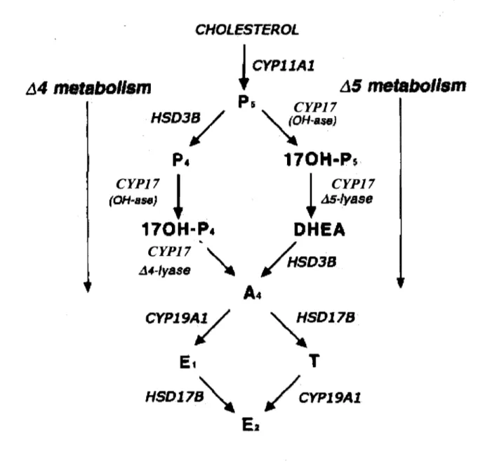
can be metabolized to Ps via CYPIIAI. Ps can be further metabolized by two different enzymes. The first is HSD3B, which converts pregnenolone to P 4. The HSD3B is located in the endoplasmic reticulum and is responsible for the fonnation of P 4. The second pathway of metabolism of P 5 is via CYP 17 which is located in the endoplasmic reticulum of thecal cens and is responsible for the conversion ofPs to dehydroepiandrosterone which is then converted to ~ by HSD3B. CYP17 can also convert P4 to~. These two alternative pathways for the metabolism of pregnenolone are called the ~4 and ~5 pathways (Gore-Langton & Annstrong 1994; Conley & Bird 1997) (Figure 4), which refers to the number of the first carbon having a double bond in either the first (~4) or second (~5) ring of the steroid molecule. The ~4 and ~5 pathways are observed in human, rat, bovine species, and the ~5 pathway is the preferred pathway in the human and bovine species (Zuber et al.
1986; Fortune 1986). Similar to the bovine species, follicular cens of the human ovary also appear to be unable to utilize the ~4 pathway by lack of expression of the enzyme
converting P4 to ~ (Fevold et al. 1989; Lin et al. 1991). However, the rat exhibits the ability to metabolize P4 directly to ~ by the ~4 pathway (Namiki et al. 1988; Fevold et al. 1989). Once ~ is fonned within the ovarian theca cens, it can either be converted to T via a HSD17B or be converted to an estrogen by CYP19Al in granulosa cells. Bovine
granulosa cens prefer to metabolize ~ to El by the enzyme CYPI9Al, and then the El is metabolized to E2 by HSD17B (Conley & Bird 1997). In the bovine species, HSD17Bl has
been identified (Sahmi et al. 2004). In the rodent, HSD17B type 1, 7 and 12 which are known to convert El to E2 have been identified in the ovary. HSD17B type 7 and 12 are
15
found in the endoplasmic reticulum and type 1 is cytoplasmic (Labrie et al. 1997; Luu-The et al. 2006). CYP19Al is a cytochrome P450 enzyme located in the endoplasmic reticulum. This enzyme is responsible for the conversion of ~ to El or the conversion of T to E2•
CYP 19 A 1 is a single enzyme encoded by a single gene (Fisher et al. 1998).
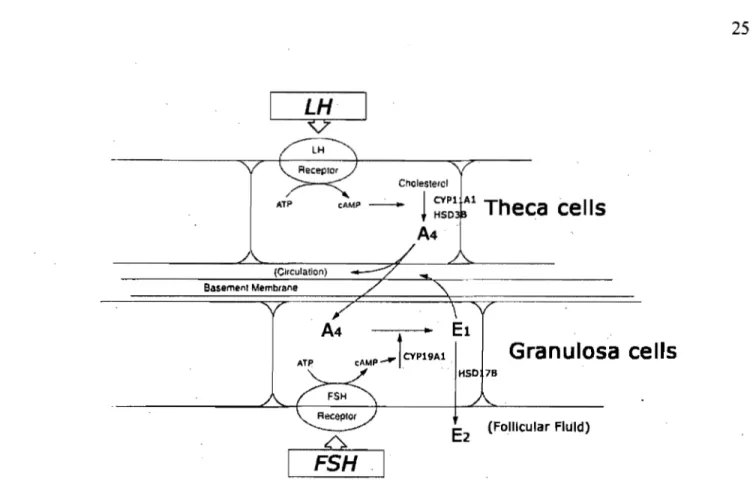
2.1 Steroidogenesis and the.two-cell and two-gonadotropin hypothesis
The follicular somatic cell population is composed of two major cell types, the theca and granulosa cell. These two types of cells express steroidogenic enzymes to pro duce steroid hormones at different stages of follicular development. The cell, two-gonadotropin hypothesis has been confirmed in the bovine species (Armstrong &
Dorrington 1977; Fortune 1986; Hillier et al. 1994; Adashi et al. 1995) (Figure 5). Under theeffects of LH, the early steps of steroidogenesis take place in the theca, such that cholesterol is converted into Ps and consequently to either P4, or the androgens ~ or T. These androgens pass by diffusion into the granulosa cell of the ovarian follicle, where, under the influence of FSH, they are converted into estrogens by CYP 19 A 1 and HSD l7B 1. Granulosa cells also have the capability to produce P 4 when the follicle reaches the large antral and preovulatory stages through expression of the P4-generating enzymes CYPllAI and HSD3B (Bao & Garverick 1998).
2.1.1 Classes of follicular steroids
Ovarian follicles secrete three classes of steroids: androgens, estrogens and progestins. Steroids have autocrine and paracrine effects, and directly influence the
,
reproductive system via specific steroid nuc1ear receptors. In recent decades, development of knockout mouse models has provided new evidence to understand the function of steroids in vivo.
2.1.1.1 Estrogen
The three major naturally occurring estrogens are E2, estrone and estriol. The active form of estrogen is E2. During follicular development before the pre-antral stage, FSHr are expressed in granulosa cells, and FSH stimulation is linked to the production of E2
(Erickson & Shimasaki 2001). Several functions of estrogen have been described in the follic1e. Estrogen stimulates 1) granulosa cell prolifération (Richards 1980), 2) the synthesis of granulosa cell insulin-like growth factor 1 (Hernandez et al. 1989), 3) the level of
estrogen receptor (Richards 1975),4) the number and size of intracellular gap junctions (Burghardt & Anderson 1981),5) the expression ofCYP19Al mRNA (Luo & Wiltbank 2006) and 6) induces LHr (Wang & Greenwald 1993). Conversely, estrogen attenuates granulosa cell apoptosis (Quirk et al. 2004) and inhibits androgen synthe sis in theca cells (Leung & Armstrong 1979). CYP19Al is a unique granulosa cell enzyme responsible for transformation of estrogen (Fisher et al. 1998). Estrogen affects the function of the ovary via granulosa cell estrogen receptor a and ~ (Sar & Welsch 1999; Sharma et al. 1999). Knockout of estrogen receptors and CYP 19 A 1 have shown that estrogen is essential fof folliculogenesis beyond the antral stage and is necessary to maintain the female phenotype of ovarian somatic cells (Drummond 2006).
17
2.1.1.2 Progestin
The active form ofprogestin is P4. The production ofP4 and its metabolites is one of the major biosynthetic activities of the ovary. Although P4 is secreted predominantly by the CL, P 4 is also secreted by theca cells and granulosa cells of developping follic1es. In the female reproductive tract, P 4 plays key roles in ovulation, implantation and the maintenance ofpregnancy (Graham & Clarke 1997). P4 performs these functions through two forms of PR, PR-A and PR-B (Spelsberg et al. 1972; Kastner et al. 1990). In the mouse, knockout of PR-A, blocked ovulation supporting the role OfP4 in ovulation induction. PR-B knockout were ovulatory and produced viable offspring (Mulac-Jericevic et al. 2000; Mulac-Jericevic
et al. 2003).
2.1.1.3 Androgen
Androgens, primarily ~ and T, are produced by theca cells. These are the immediate biosynthetic precursors of the estrogenic steroids. High ratios of androgens to estrogens have been reported in FF ofnonovulatory and atretic follic1es (McNatty et al. 1979; Carson et al. 1981). Moreover, a single injection ofDHT (non-aromatizable
androgen) to cyc1ing mice reduced the number of large follic1es by 15% and left the mi ce subfertile (Nandedkar & Munshi 1981). Androgens also inhibit FSH-stimulated LHr
expression by granulosa cells and enhance granulosa cell apoptosis (lia et al. 1985; Billig et al. 1993). These studies have been conducted in the rat and indicate that androgens impede follicular development, especially at the antral follic1e stage. However, in the early stages offolliculogenesis, androgens appear to promo te follicular growth (Walters et al. 2008). In
support of this, administration of androgens to female rhesus monkeys and pregnant ewes (prenatally treated fetuses) stimulates primordial follicle initiation and increases the number of growing follicle and overall follicie survival (Vendola et al. 1998; Steckler et al. 2005; Forsdike et al. 2007). In primate and gilts, androgen stimulates follicle growth and
increases FSHr rnRNA expression inprimary or preovulatory follicles (Weil et al. 1999;
Cardenas et al. 2002). Treatment of androgens in cultured bovine granulosa cells also increased FSHr mRNA expression (Luo & Wiltbank 2006). However, these studies do not account for the possibility of androgen conversion and hence indirect androgen actions since androgens stimulate CYPl9Al activity and E2 production (Hamel et al. 2005). Although, it is not clear if aIl the follicular effects observed can be attributed conclusively to direct androgen actions, the production of the AR conditional knockout model has
provided a fundamental advance in unraveling the roles of AR-mediated androgen action in female reproductive physiology (Hu et al. 2004b). Female conditional AR knockout mi ce have longer estrous cycles and reduced fertility. Their ovaries contain normal numbers of follicles, although large antral follicles appeared to have fewer granulosa cells and there were reduced n~bers of CL (Hu et al. 2004b).
2.1.2 Expression of steroidogenic enzymes and gonadotropin receptors during bovine follicular development
During follicular development, steroid hormone secretion and steroidogenic enzyme expression have been used as an index offollicular development. More recently, the bovine model has been used to study the relationship between steroidogenesis and follicular
development. In this review, bovine follicular development has been divided into six stages, primordial, primary, preantral, early antral, antral, and later antral follicle stages. The expression of steroidogenic enzymes and gonadotropin-receptors (FSHr and LHr) during the different stages of bovine follicle development is shown in Table.1.
19
The primordial follicle is formed during fetallife or soon after birth and is
composed of a primary oocyte surrounded by a squamous layer of pre-granulosa cells. There is no evidence of active steroidogenesis in primordial follicles. Entry of primordial follicles into the growth phase occurs throughout the reproductive life span and is
characterized by conversion of the flattened pre-granulosa cells surrounding the oocye into a single layer of cuboidal granulosa cells. This follicle is now termed a primary follicle. In the bovine species, expression ofFSHr mRNA was first localized in granulosa cells at the primary follicle stage (Xu et al. 1995a; Bao et al. 1997b), but there was no expression of steroidogenic enzymes. In the mouse, knockout of FSHr has been shown to block antral follicle formation without affecting growth of primordial, primary and preantral follicles (Kumar et al. 1997). This suggests that FSH stimulation may not be a limiting step on primary follicle development. However, sorne studies suggest that FSH may improve the proliferation of bovine granulosa cells by acting at the primary follicle stage (Garverick et al. 2002; Hunter et al. 2004; Webb et al. 2007).
Following formation oftheca cells around the granulosa cells in the preantral folli,ele, mRNA for LHr, CYPIIA1, CYPI7, and HSD3B are expressed in theca cells, and
expression generally increases with growth ofpreantral and early antral follicles (Xu et al. 1995a,b; Bao et al. 1997a, b). In granulosa cells at these follicular stages, there is still expression ofFSHr without expression ofCYP19Al. Moreover, granulosa cells do not express P4-generating enzymes, such as CYPIIAI and HSD3B. These results indicate that theca cells of follicles at preantral and early antral follicle stages are only able to synthesize androgens and progesterone, and granulosa cells are still steroid-inactive.
The follicular development to an early antralfollicle (0.4 to 4mm in diameter) is believed to be gonadotropin-independent because growth of antral follicles continues up to 4mm but not above when endogenous concentrations of gonadotropins are suppressed by hypothalamic stalk transection or by removal or inhibition of gonadotropin secretion by a GnRH agoni st (Awotwi et al. 1984; Campbell et al. 1995; Gong et al. 1996). The
observation ofFSHr and LHr knockout mouse models corroborates that development from primordial to early antral follicle stage is gonadotropin-independent (Kumar et al. 1997; Lei et al. 2001; Zhang et al. 2001).
During the antralfollicle stage (4 to 8 mm in diameter), theca cells keep expressing mRNA ofLHr, CYPIIA1, CYPI7, and HSD3B to promote the secretion ofandrogens and progesterone (Xu et al. 1995a, b; Bao et al. 1997a, b). At this stage, expression of
21
CYP19Al appears in granulosa cells (Xu et al. 1995b) which respond to FSH in vitro (Gutierrez et al. 1997a), suggesting that these follicles are able to convert androgens to E2.
Granulosa cells do not express mRNA ofLHr, CYPIIA1, HSD3B, which suggests that granulosa cells at the antral follicle stage are not yet capable ofproducing P4. In the antral and later antral follicular stages, the granulosa and theca cells express FSHr and LHr. The steroid honnone production is increased by gonadotropin stimulation (Campbell et al.
1995). These stages can be considered gonadotropin-dependent.
The later antral follicle stage is defined as growth of the dominant follicle to the preovulatory follicle stage. At this stage, rnRNA ofLHr, StAR, CYPIIA1, CYP17 and HSD3B are expressed in the theca cells. In granulosa cells, LHr, CYPIIAI and HSD3B mRNA expression are detected for the first time in addition to FSHr and CYP19Al. Furthennore, GSTA is expressed at the later antral follicle stage and codes for a protein with high HSD3B activity (Rabahi et al. 1999; Raffalli-Mathieu et al. 2007). These results indicate granulosa cells are able to convert androgen to E2, and also secrete P4. Ps
production by granulosa cells could be utilized by thecal cells to produce androgen as well (Fortune 1986).
2.1.3 Steroidogenesis in the atretic follicle
The depletion of primordial follicles occurs as the result of one of two processes: atresia or entry into the growth phase. Atresia occurs at any stage of follicular development (Lussier et al. 1987). In granulosa cells of preantral and early antral follicles, morphological
signs, such as pyknotic nuclei, are more useful to determine follicular atresia because granulosa cell steroidogenesis is not very active in these follicles. However, changes in steroidogenesis are earlier signs of atresia than changes in morphology in antral and later antral follicles (Xu et al. 1995a, b).
Several studies have shown that healthy antral follicles have higher concentrations of E2 than either P 4 or androgen, while atretic antral follicles have elevated concentrations of P 4 or androgens, resulting in a low ratio of the concentration of E2 to P 4 in follicular fluid
\
of atretic larger antral follicles (lreland & Roche 1982; McNatty et al. 1984; Spicer et al. 1987). In agreement with the decreased secretion of estradiol in atretic antral follicles, expression ofmRNA for CYP19Al and CYP19Al activity in granulosa cells is the most affected in the early atretic follicle, while expression ofmRNA for LHr, CYPIIA1, P450C17, HSD3B and StAR in theca cells and FSHr in granulosa cells is less sensitive to atresia, which preserves the secretion of progesterone or androgens in theca cells during the early atretic process (McNatty et al. 1984; Price et al. 1995; Bao et al. 1997a, b). In a few follicles at the advanced or late stages of atresia, the remaining few layers of granulosa cells express CYPIIA1, HSD3B, and StAR mRNA (Xu et al. 1995b, a; Bao et al. 1997b, a; Braw-TaI & Roth 2005), and these follicles have been confirmed as BA which me ans granulosa cell death can commence from the basal lamina and progresses toward the antrum (lrving-Rodgers et al. 2003). Sorne layers of granulosa cells in basal atretic follicles are morphologically healthy and spontaneously luteinize with CYPIIA1, HSD3B, and
23
StAR mRNA expression similar to that found in granulosa cells during luteinization caused by the preovulatory LH surge (Tilly et al. 1992; Irving-Rodgers et al. 2003).
2.2 Regulation of steroidogenesis in cultured bovine granulosa ceUs
1
In vivo, the complexities of hormonal interactions withJhe cytokines produced by
1
ovarian cells limit our investigations of steroidogenesis in follicle development. To
examine regulation of steroidogenesis, many studies focused on culturing granulosa cells in
vitro to mimic the changes of steroidogenesis during follicle development.
Granulosa cells in culture luteinize spontaneously, as demonstrated by the shi ft in the principal secretory product from E2 to P 4 (Luck et al. 1990; Meidan et al. 1990).
However, in b9vine granulosa cells harvested from small antral follicles and cultured in serum-free me?ium in the presence oflow doses ofFSH or IGF, E2 secretion increases gradually over 6 days of culture (Rouillier et al. 1996; Gutierrez et al. 1997a; Gutierrez et al. 1997b; Rouillier et al. 1998; Sahmi et al. 2004). The E2-secreting potential of cultured granulosa cells may be associated with the maturation or differentiation stage of the
follicle. Although granulosa cells from large antral follicles could secrete E2, these follicles rapidly 100 se the potential to produce estradiol with time of culture. Conversely, granulosa cells from small follicles consistently increase E2 secretion with time, which suggests that granulosa cells from these follicles are less mature and retain the potential to secrete E2 (Gutierrez et al. 1997a).
CHOLESTEROL
JCYPIIAl
~
A5mefabollsm
.14
matabOIIsm
Ps
CYP17
'{H-ase)
170H-P$·
CYP17
1
1
CYP17
(OH .. aH) ~
t
M-Iysse170H-P.
DHEA
CYP17",
/..
.
.d4-IysSB "II'
HSD38
A4
CYPl9~
~SDl7S
Et
T
HSDl7S""-
/
CYPl9Al
El
Figure 4. The â4 and â5 pathways of gonadal steroid synthesis. (Modified from Conley 1997)
LH
Cholesterol
ATP cAMP
~
!
~~l
AlTheca cells
(Circulation)
ATP
A4
--,---Il00110
El
cAMP _lCVP19Al
Granulosa cells
HSO 78
E2
(Follicular Fluld)FSH
25
Figure 5. The two-cell, two-gonadotropin theory offollicular steroidogenesis. (Modified from Adashi 1995)
Bovine Follicle Antral Follicle (4mm-8mm) Later Antral Follicle >8mm Enzymes & Gonadotropln FSHr(GC) FSHr (GC) LHr (TC) J. StAR 2. CYPIIAI 3. CYPI7 4.HS038 FSHr (Ge) LHr(TC) J. StAR 2. CYPIIAI 3. CYP17 4. HS038 FSHr (GC) LHr (TC) J. StAR 2. CYPllAl 3. CYP17 4. HS038 5. CYP19Al FSHr (GC) LHr(TC, GC) 1. StAR 2. CYPJJAI 3. CYP17 4. HSD38 5. CYP19Al
tbecal cells GCs: granulosa cells, FF: follicular nuid, A4: Androstenedione, P4: Progesterone, El: Estrone, E2: Sc Ta Cb Ta GCa FF 1 1
'
~4
oU Sc Ta nr ~P4 EI- E2~E2 oU Isf
S ..
oU-
T...a-~T Sc Tc."
L CIl oU (Ba. n QL 1997b) (Xu ~tI". 199511) (Xu ~/III. 1995b) (Ba. n QI. 1997b) rI QI. 1995b) (Xu et III. 1995-.) (8aQ dtll. 1998) nal.19958) (XU nQI. 1995b) (8ao ~I al. 1998) (XunQI.1995.) (Xuc>tal.199Sb) (BaD c>l al. 1998) (8ao c>/ al. 1997.)Table 1. In vivo expression of steroidogenic enzymes and gonadotropin receptors during bovine follicular development shown by in situ hybridization.
27
3. Cell proliferation and apoptosis in granulosa cells
In any developing tissue, cell proliferation must be controlled in such a manner that allows sorne cells to reach terminal differentiation while others are eliminated by
programmed cell death or apoptosis. Primordial follicles enter into the growing phase with two potential destinies: ovulation or atresia. Successful follicle development for ovulation requires granulosa cell differentiation accompanied by proliferation (van Wezel & Rodgers 1996; Robker & Richards 1998). Interruption of either proliferation or differentiation will cause exclusion of granulosa cells by apoptosis. Follicular development will depend on the intricate effects of growth factors, gonadotropins (LH and FSH), and steroid hormones on proliferation or apoptosis of granulosa cells (Asselin et al. 2000; Jiang J. Y. et al. 2003; Quirk et al. 2004).
3.1 Cell cycle
Although information on the mechanisms regulating cell cycle progression in granulosa cells is insufficient, much progress in understanding the regulation of the cell cycle has been made in other cell models, such as cancer cells (Sherr & Roberts 1999; Lundberg & Weinberg 1999). The cell cycle consists of four distinct phases: G 1, S, G2 and M. Activation of each phase is dependent on the proper progression and completion of the previous one. Cells that have temporarily or reversibly stopped dividing are considered to have entered a state of quiescence called GO phase. Mechanisms regulating the çell cycle are briefly described here based on previous studies (Sherr & Roberts 1999; Lundberg & Weinberg 1999; Quirk et al. 2004). Cell cycle progression is mediated by a protein family
(
of cdk that are sequentially activated by binding to specific cyclin proteins synthesized periodically during the cell cycle. The mitotic decision in eukaryotic cells is made at the G 1 phase of the cell cycle, when mitogens initiate the division process. During early- to mid-G 1 phase, progression requires the formation of complexes of cdk4 or cdk6 with cyclin D and cdk2 with cyclin E. The cdk4/6-cyclinD complex combined with the cdk2-cyclin E will initiate hyperphosphorylation of the RB, resulting in loss of its ability to inhibit cell cycle progression during late-G 1 phase. Hyperphosphorylation of RB permits liberation of the transcription factors of the E2F family that upregulate expression of the cyclins, kinases, and other proteins necessary for continuation through the S, G2 and M phases of the cell cycle. Formation of additional complexes between various cdks and their cyclin partners are also required for progression through S, G2 and M phases.
3.1.1 Proliferation in granulosa cells
Growth rates vary with the size of the follicle; in the bovine species, a follicle takes 27 days to grow from 0.13 to 0.67 mm, 6.8 days from 0.68 to 3.67 mm and 7.8 days from 3.68 to 8.56 mm (Lussier et al. 1987). Growth rate ofbo"ine follicles was estimated based on the mitotic index of granulosa cells (Lussier et al. 1987). In primordial follicles, the oocyte is surrounded by a single layer of nondividing granulosa cells arrested in GO phase of the cell cycle. Once primordial follicles leave this quiescent state, the granulosa cells , have entered into the cell cycle but proliferation is exceedingly slow (Hirshfield 1991).
Under the influence of gonadotropins, growth factors and steroids, granulosa cells of antral follicles enter into cell cycle progression which results in a rapid burst of proliferation and
29
the fonnation of large preovulatory follicles (Rao et al. 1978). The rapid phase of proliferation in granulosa cells was measured by a marked increase in the labelling of granulosa cells by tritiated thymidine (Rao et al. 1978), as well as by 5-bromodeoxyuridine (BrdU) incorporation or ca1culation of the mitotic index (Lussier et al. 1987; Gaytan et al. 1996). Treatment with IGF or estradiol in cultured bovine granulosa cells aiso induced an increase in GO/G 1 to S phase progression (Quirk et al. 2004; Hu et al. 2004a). When selected follicles reach the preovulatory stage, the LH surge tenninates follicular growth by causing granulosa cells to exit the cell cycle and remain in the GO stage (Rao et al. 1978; Stocco et al. 2007).
3.2 Apoptosis
Apoptosis is programmed cell death and provides a means for multicellular organisms to eliminate unwanted cells in response to developmental signaIs or toxic stimuli. The histological features of apoptosis in tissue include cytoplasmic vacuolization, chromatin condensation as well as the appearance of eosinophilic apoptotic bodies as round cytoplasmic masses or as masses of pyknotic chromatin sUITounded by a narrow rim of the cytoplasm. In culture, the features of apoptosis include cell shrinkage, surface convolutions, fonnation of protuberances with buddings and subsequent fonnation of the apoptotic bodies (Wyllie 1993; Hussein 2005). The process of apoptosis is controlled by a diverse range of cell signaIs, such as toxins, honnones, growth factors, nitric oxide or cytokines (Asselin et
al. 2000; Popov et al. 2002; Brune 2003; Jiang et al. 2003). It is weIl established that
evidence from studies of a number of cell types indicates several apoptotic signal pathways leading to cell death (Tilly et al. 2004). Two general apoptotic signal pathways have been identified. The intrinsic pathway is generated by internaI signaIs targeting mitochondria functionality, while the extrinsic pathway is directly triggered by the binding of death molecules (TNF and Fas) to cell surface receptors (Amsterdam et al. 2003; Hussein et al. 2003) (Figure 6). The intrinsic and extrinsic pathways activate the death factors in the ceU, such as Bid, cytC, Apaf-l to generate the death signaIs. At the end of any death signal transduction, apoptotic signal requires activation of the caspases to accomplish transduction of death signal in marnmalian cells (Tilly et al. 2004). Caspases inelude both 'initiator' caspases (8 and 9) and 'effector' caspases (3, 6 and 7). Once caspases 3, 6 and 7 activated, this executioner enzyme is irreversible, and results in the eleavage of numerous structural and functional proteins (such as PARP which is DNA repair protein) and the
internueleosomal eleavage of genomic DNA. (Matikainen et al. 2001; Johnson & Bridgham 2002; Tilly et al. 2004). Cell death pathways are opposed by anti-apoptotic proteins that protect mitochondrial membrane integrity (Bel-x, Bel-2), block caspase activity (inhibitor of apoptosis factors :IAPs) or prevent signaling via death receptors (the adaptor protein, FLIP). Expression ofthese anti-apoptotic proteins is regulated by gonadotropins (FSH and LH) and locally produced growth factors (IGF-I and EGF family ligands) via cell survival signaling pathways.
3.2.1 Apoptosis in granulosa ceUs
31
follicles, atresia is initiated mainly by the death of the oocyte, and percentage of atresia at these stages is lower than in antral foUicles (Lussier et al. 1987; Morita & Tilly 1999). It is established that degeneration of atretic antral follicles in mammalian ovaries can be
explained by apoptosis of granulosa cells and theca cells (Tilly 1996). Within the follicle, the most obvious cell type undergoing apoptosis in association with maturing foUicle atresia is the granulosa cell (Tilly et al. 1991; Tilly et al. 1992; Palumbo & Yeh 1994). The apoptosis of granulosa cells has been observed on the basis of the changes in morphology and also detected by the terminal deoxynucleotidyl transferase-mediated dUTP nick end-labeling (TUNEL) technique, DNA agarose gel electrophoresis with the formation of a DNA ladder, and flow cytometry that measures degraded DNA which appears as cells with hypodiploid DNA content and are represented in so-called "sub-G 1" peaks on DNA histograms. (Nicoletti et al. 1991; Wyllie 1993; Rouillier et al. 1998; Yang &
Rajamahendran 2000; Irving-Rodgers et al. 2001). Moreover, the signaIs, receptors and intracellular signaUing pathways leading to apoptosis within granulosa cells have been obtained from studies of genetic mouse and in vitro cultured cells. Although studies in terms of apoptosis .are still ongoing, it appears that multiple molecules are involved. These molecules include survival factors, such as gonadotropins, IGF-l, E2, interleukin-lB, EGF, bFGF, and TGF-a; and also include apoptotic factors, such as caspase, Fas antigens, reactive oxygen species, interleukin-6, androgens, bax, p53, and TNF-a (Asselin et al. 2000; Jiang et al. 2003; Quirk et al. 2004; Hussein 2005). Caspase-3 is a caspase common to several different apoptosis pathways (Matikainen et al. 2001; Johnson & Bridgham
2002). Caspase-3 is active1y involved in follicular atresia, and present in granulosa cells of atretic, but not healthy follicles (Boone & Tsang 1998; Nicholas et al. 2005). Furthermore, several intracellular signalling pathways have been linked directly to granulosa cell survival as well, including gonadotropin- and vasoactive intestinal peptide-induced cAMP
formation, MAPK-ErK and IP3K-AKt (Flaws et al. 1995; Johnson et al. 1999; Gebauer et al. 1999; Asselin et al. 2001; Johnson et al. 2001).
3.2.2 Cell cycle and apoptosis
The transition from preantral to antral follicles occurs after exposure of the
granulosa cells to FSH. Then, rapid proliferation and differentiation of the granulosa cells is initiated during antral foUicular development. At this period, the proportion of bovine follicles undergoing atresia increases dramatically with follicular diameter and the rate of atresia doubles between 2 and 8 mm (Lussièr et al. 1987). In the rat, the highest frequency of atresia occurs in antral follicles with relative1yrapid granulosa cell proliferation in comparison to smaller foUicles (Hirshfield & Midgley 1978). These observations provide evidence that granulosa cells that are dividing rapidly are most susceptible to apoptosis. In agreement with this finding, in vitro studies have demonstrated that cell survival is linked to maintenance or stimulation of G l-to-S phase transition whereas blocking this transition increases susceptibility to apoptosis (Quirk et al. 2004). In addition, the LH surge
terminates follicular growth by causing granulosa cells to exit the cell cycle and remain in
1
the GO stage (Rao et al. 1978; Hirshfield 1991). When the granulosa cell remains in the GO stage of the cell cycle, postmitotic differentiation happens and prevents granulosa cell
33
susceptibility to apoptosis, and a sign of differentiation of granulosa cells at this period is an increase in secretion ofP4 (Lundberg & Weinberg 1999; Peluso 2003). Based on the
expression pattern of the PR, it is predictable that P 4 inhibits apoptosis in cultured
granulosa cells isolated during the gonadotropin (LH) surge or periovulatory period (Peluso & Pappalardo 1998; Svensson et al. 2000).
Figure 6. A summary of apoptotic and survival signalling pathways. The absence of sufficient anti-apoptotic prote in expression (dotted green lines) is proposed to tip the balance in favor of activating pro-apoptotic pathways (solid red lines). Abbreviations: aCasp.-3, -7, -8, activated caspases; Apaf-l, Apoptosis Protease Activating Factor-l; Bid and tBid, intact or tnrncated BH3 Interacting Domain death agonist; cytC, cytochrome C; DD, death domain; DED, death effector domain; EGF, epidermal growth factor; F ADD, Fas-Associated Death Domain; FLIP, Flice-like inhibitory protein; FSH,
follicle-stimulating hormone; Inhibitor of Apoptosis Prote in, IAP; IGF-I, insulin-like growth factor 1; LH, luteinizing hormone; PARP, Poly-(ADP-ribose)-polymerase; PKAIcAMP, prote in kinase A/cyclic AMP signaling pathway; PKBI Akt, protein kinase BI Akt signaling
pathway. Each of the pro teins and cell signaling pathways depicted has been characterized in hen granulosa cells, in vitro. (adapted from
~ath Receptor
Ligands,
Extrinsic
Death
ReceptorsPathway
In
trins
ic
Cell Survival
FSH-R~R
EGF Farnily Ugands; IGF-IIl
P
a wa
th
y
PKA/cAMP." :
PKB/Akt.
(nutritional dcprivation : . :FLIP growth factor/gonadotropin .: .
JI
.
...
Bid~~
.
=--~
t
-8 tBid
-withdrallal, oxidativc strus) . ." .