HAL Id: hal-02502802
https://hal-mines-albi.archives-ouvertes.fr/hal-02502802
Submitted on 16 Oct 2020
HAL is a multi-disciplinary open access
archive for the deposit and dissemination of
sci-entific research documents, whether they are
pub-lished or not. The documents may come from
teaching and research institutions in France or
abroad, or from public or private research centers.
L’archive ouverte pluridisciplinaire HAL, est
destinée au dépôt et à la diffusion de documents
scientifiques de niveau recherche, publiés ou non,
émanant des établissements d’enseignement et de
recherche français ou étrangers, des laboratoires
publics ou privés.
A review of current treatments strategies based on
paromomycin for leishmaniasis
A.P.S. Matos, A.L. Viçosa, Maria-Inês Ré, E. Ricci-Júnior, C. Holandino
To cite this version:
A.P.S. Matos, A.L. Viçosa, Maria-Inês Ré, E. Ricci-Júnior, C. Holandino. A review of current
treat-ments strategies based on paromomycin for leishmaniasis. Journal of Drug Delivery Science and
Technology, Elsevier, 2020, 57, pp.101664. �10.1016/j.jddst.2020.101664�. �hal-02502802�
A review of current treatments strategies based on paromomycin for
leishmaniasis
A.P.S. Matos
a,b,c,∗∗, A.L. Viçosa
d, M.I. Ré
b, E. Ricci-Júnior
c, C. Holandino
aaLaboratório Multidisciplinar de Ciências Farmacêuticas, Departamento de Fármacos e Medicamentos, Faculdade de Farmácia, Universidade Federal Do Rio de Janeiro,
Cidade Universitária, Rio de Janeiro, RJ, Brazil
bToulouse University, CNRS, Rapsodee Research Center, IMT Mines Albi, Jarlard Campus, F-81013, Albi 09, France
cLaboratório de Desenvolvimento Galênico - LADEG, Departamento de Fármacos e Medicamentos, Faculdade de Farmácia, Universidade Federal Do Rio de Janeiro, Rio de
Janeiro, RJ, Brazil
dLaboratório de Farmacotécnica Experimental, Instituto de Tecnologia Em Fármacos – Farmanguinhos, Fundação Oswaldo Cruz, Rio de Janeiro, RJ, Brazil
Keywords: Leishmaniasis Paromomycin Current treatment Drug delivery systems
Advanced pharmaceutical formulations Combined treatment
A B S T R A C T
Leishmaniasis is a neglected disease caused by protozoan parasites of the Leishmania genus, which affects many people in several countries. This disease has three major clinical forms: cutaneous, mucocutaneous and visceral. The current treatments consist of an intravenous, intralesional or intramuscular administration of pentavalent antimonials, but other drugs can be used, among them, amphotericin B, pentamidine, paromomycin and mil-tefosine. However, these therapies have many side effects. Hence, there is an increase of studies searching for new formulations using different technologies and different routes of administration for leishmaniasis treatment. Paromomycin sulfate (PM) is an aminoglycoside antibiotic, belonging to class III of biopharmaceutical classi-fication system, used intravenously and topically with leishmanicidal activity. This review will provide a general overview of PM current leishmaniasis treatments and new PM formulations. Treatments using PM are available in ointments or creams for topical administration and PM solution for intramuscular administration. The topical treatment with PM presents low efficacy, probably related to low drug permeability across the skin. To improve PM permeability and efficacy, researchers are establishing micro and nanotechnologies. However, further stu-dies are still required to investigate more physicochemical properties and in vitro/in vivo parameters.
1. Introduction
Leishmaniasis is a parasitic infectious disease caused by approxi-mately 20 species of protozoan of the Leishmania genus and transmitted
by female phlebotomine sandflies [1–4]. This disease belongs to the
group of neglected diseases and is one of the major health problems in
the world [4,5], more specifically in 98 countries [6,7], with 12 million
sick people and 2 million new cases reported annually [5–9]. An
esti-mate of 26,000 to 65,000 deaths occur each year [10]. Furthermore,
cases of Leishmania and HIV (human immunodeficiency virus) co-in-fection are increasing and have been described in 35 countries
[3,5,7,11].
Leishmaniasis parasites have a digenetic life cycle with two mphological forms: promastigotes, form flagellated in the digestive or-gans of sand fly vector and amastigotes, form no flagellated in the
phagolysosome of mammalian host macrophages [3,4,7,12–15].
This disease is manifested in three major clinical forms: cutaneous,
mucocutaneous and visceral [3,4,8,11,12,16]. The severity and form of
clinical manifestations depend on the infecting parasite species, site of inoculum, the number of parasites inoculated and host immunity
re-sponse [17–19].
Cutaneous leishmaniasis (CL) is the most common type of this dis-ease through the appearance of skin lesions developing at the area of
sandfly bite [7,13,18], mostly ulcers and maybe leave life-long scars
and serious disability [10,15]. The CL lesions usually appear on the
face, neck, arms, and legs [7,8]. For cutaneous leishmaniasis, the
spe-cies of Leishmania most common are Leishmania major, Leishmania tro-pica, Leishmania mexicana, Leishmania braziliensis and Leishmania pana-mensis [2,4,13,18]. CL is endemic in more than 70 countries, in which 90% of the cases occur in seven countries (Afghanistan, Algeria, Brazil,
Pakistan, Peru, Saudi Arabia and Syria) [2–4,6].
Mucocutaneous leishmaniasis (MCL) is characterized by hemato-genous or lymphatic dissemination of parasites from cutaneous lesion
[2,7] and caused by Leishmania amazonensis, Leishmania braziliensis,
∗Corresponding author. Laboratório Multidisciplinar de Ciências Farmacêuticas, Departamento de Fármacos e Medicamentos, Faculdade de Farmácia,
Universidade Federal do Rio de Janeiro, Cidade Universitária, Rio de Janeiro, RJ, Brazil. E-mail address:anapaulasmatos@ufrj.br(A.P.S. Matos).
Leishmania panamensis, Leishmania guyanensis, Leishmania major, Leish-mania infantum and LeishLeish-mania tropica [2,4,8]. MCL is manifested by nasal inflammation followed by nasal cartilage infiltration and de-struction of nasal septum and can cause partial or total dede-struction of
nose, mouth and throat mucous membrane [2,7,10,13,15]. MCL
pre-sents more than 90% of cases in four countries: Bolivia, Brazil, Ethiopia and Peru. Nevertheless, there is no reported number of cases over the
year and by countries of this type of leishmaniasis [20].
Visceral leishmaniasis (VL), also known as kala-azar, is the most severe manifestation of leishmaniasis, in which the parasites infected
vital organs [3,16]. VL is manifested by hepatosplenomegaly,
pro-longed fever and pancytopenia [3,8] and, if untreated, can be fatal
[3,8,10,15,16]. Leishmania infantum and Leishmania donovani are the
Leishmania species responsible by VL [13,16]. Visceral leishmaniasis is
endemic in 65 countries and more than 90% of cases occur in five
countries (Bangladesh, Brazil, India, Nepal and Sudan) [3,4,6,7,16].
Table 1gathers active pharmaceutical ingredients (APIs) in current
use for the treatment of leishmaniasis.
The first-line drugs used to leishmaniasis treatment is an in-travenous, intralesional or intramuscular administration of pentavalent antimonials as meglumine antimoniate and sodium stibogluconate
(SSG) [7,9,14,15,19]. The mechanism of action of these compounds is
still not properly understood, but the drugs can inhibit glycolysis step of
metabolism and fatty acid oxidation of the parasite [4,7,10,21] and
pentavalent form is reduced to trivalent form [15]. The most frequent
side effects of these drugs are myalgia, arthralgia, anorexia and leu-kopenia. Furthermore, pentavalent antimonials can be cardiotoxic, nephrotoxic and hepatotoxic, which limited these drugs use in pregnant
and elderly people [21–24].
The second-choice treatment is an intravenous administration of amphotericin B, a polyenic antibiotic with high leishmanicidal activity. This drug binds to the ergosterol molecules present in the cytoplasmic membrane of parasites increasing membrane permeability and ion
in-flux [4,15,22,23,25]. There are four commercial formulations available
of this drug: amphotericin B deoxycholate, liposomal amphotericin B, cholesterol dispersion of amphotericin B and lipid complex of
ampho-tericin B [21–23]. Amphotericin B deoxycholate causes more side
ef-fects including fever, headache, nausea, vomiting, tremors and hypo-tension. Moreover, all amphotericin B formulations still present nephrotoxicity and cardiotoxicity and are restricted to the hospital environment. Liposomal and lipid-base formulations present lower toxicity but are more expensive than amphotericin B deoxycholate
[21–24].
Pentamidine is an aromatic diamidine, which has been marketed in the form of two salts: isethionate (di-b-hydroxyethane sulfonate) and mesylate (di-b-hydroxymethyl sulfonate) and are administered by
in-travenous or intramuscular routes [4,22,23]. The mechanism of action
may be related to a decrease of mitochondrial membrane potential by
drug accumulation in the mitochondria [4,22,23]. This drug can induce
different side effects such as hypoglycemia, hyperglycemia, ne-phrotoxicity and hepatotoxicity. Furthermore, pentamidine can cause
insulin-dependent diabetes [21–23].
Miltefosine is, originally, an anticancer drug (hexadecylpho-sphocholine), which shows interesting results in leishmaniasis treat-ment and has been considered an advancetreat-ment in the research of new treatment of this disease specially because can be administered orally
[4,24,25]. The miltefosine mechanism of action is based on drug
in-tracellular accumulation in parasites through transporters [4,22,23].
This drug shows several side effects such as vomiting, diarrhea, toxicity in the gastrointestinal tract. Furthermore, it presents a teratogenic
ef-fect and is prohibited for pregnant women [21–24]. As other drugs used
to treat leishmaniasis, miltefosine is already triggering resistance in
parasites [4,24]. Since 2017 is on the market one product containing
miltefosine (Milteforan®) for canine visceral leishmaniasis in Brazil
[26].
Sitamaquine is an aminoquinoline developed for visceral
Table 1 Current treatments of leishmaniasis (adapted from Bezerra de Menezes et al., 2015; Lindoso et al., 2012; Zulfiqar et al., 2017). Drugs Administration route Dosage Advantages Disadvantages Resistance Amphotericin B deoxycholate Intravenous 0.75–1 mg/kg/day (15 or 20 days daily or alternately) Primary resistance is unknown Need slow intravenous infusion, toxicity, unstable in high temperatures Laboratory strains Liposomal amphotericin B Intravenous 3–5 mg/kg single dose or 10–30 mg/kg total dose High effective and low toxicity Need slow intravenous infusion, high cost, unstable in high temperatures Not documented Miltefosine Oral 100–150 mg/day for 28 days Effective and safe Cost, poor patient compliance, cannot be used in pregnant patients Laboratory strains, some cases reported in India Paromomycin Intramuscular, intravenous or topic 15 mg/day for 21 days or 20 mg/kg for 17 days Low cost Efficacy varies between and within regions Laboratory strains Pentamidine Intramuscular 3 mg/kg/day every other day for 4 injections Short treatment Efficacy varies between Leishmania species Not documented Pentavalent antimonials Intramuscular, intravenous or intralesional 20 mg/kg/day for 28–30 days Easily availability and low cost Length treatment, painful injection and toxicity Common Sitamaquine a Oral 2 mg/kg/day for 21 days Effective Toxicity Not documented a.Sitamaquine is in phase II study for leishmaniasis treatment .
leishmaniasis treatment. Its mechanism of action affects the mobility, morphology and growth of the protozoans, through electrostatic in-teraction between polar ionic groups of the phospholipids present in the
parasite membrane and positive charge of the drug [4,27]. It is a new
medicine, which is currently in phase II clinical trials in India and
Kenya, presenting promising results for oral use [27]. The worst
pro-blem with this drug is the poor knowledge of the toxicity of its
meta-bolic [4].
Imidazoles (ketoconazole) and triazoles (fluconazole, itraconazole) are antifungal compounds with the same mechanism of action. These drugs act binding to the ergosterol molecules in the cytoplasmic membrane of parasites increasing membrane permeability. Triazoles are less toxic, interfere less with human synthesis of cholesterol and are
slower metabolized than imidazoles [17,22,23]. These compounds can
be administered orally and present lower toxic side effects than
pen-tavalent antimonials [17].
Paromomycin is an aminoglycoside antibiotic, discovered in the
1960s, with leishmanicidal activity [4,22,23], but more effective
against cutaneous leishmaniasis [4,28]. The mechanism of action of this
compound is not well known. Different routes can administer this drug (intramuscular, intravenous and topical). The most frequent side effects of this drug are ototoxicity, local pain (injectable paromomycin)
[4,22,23], erythema, vesicles and skin irritation [29].
Heat therapy or Thermotherapy has already been described in the
literature as an alternative treatment of cutaneous leishmaniasis [30].
There are several methods and several devices to provide localized heat therapy. One of these devices, ThermoMed® was approved by Food
Drug and Administration (FDA) for CL treatment [30]. In general,
Thermotherapy is applied locally for 30 s, heat of 50 °C, in one to four
weekly sessions and with local anesthesia [29,31]. There are some
studies that compared the efficacy of thermotherapy with meglumine antimoniate and showed efficacy similar to pentavalent antimonials
with safety results [29,32–34]. However, this therapy cannot be applied
in cutaneous lesions near mucosa or in lesions, which compromise
mucosa [34]. Furthermore, hyperpigmentation and secondary
infec-tions are side effects currently reported for this therapy [29].
There are also other local therapies for cutaneous leishmaniasis
treatment as cryotherapy, phototherapy and CO2 laser. Cryotherapy
consists of liquid nitrogen applied to the lesion for 15–20 s twice or
three times per session and repeated weekly until healing [31,35]. This
therapy acts destroying infected tissues and showed effectiveness
spe-cially combined with meglumine antimoniate [29,35]. The side effects
of this therapy include erythema, hypo- or hyperpigmentation
sec-ondary infections and burning [29].
CO2laser is a technique that used a source of continuous CO2laser
in lesions with pulse width between 0.5 and 5 s, power around
30–100 W and it is applied in one or a few sessions [29,36]. This
therapy promotes the thermolysis of infected tissues. Moreover, it
presents efficacy with few side effects such as hyperpigmentation, hy-pertrophic scarring and persistent redness. However, more studies are
required to evaluate the efficacy in different Leishmania species [29].
Photodynamic therapy (PDT) acts by a production of reactive oxygen species (ROS) using a photosensitizer molecule in the presence
of low-intensity visible light to promote cell death [29,37–39]. This
therapy is non-invasive, does not affect intact skin around the lesions
[37,40]. Porphyrins precursors, specially 5-aminolevulinic acid (ALA)
or methyl-aminolevulinic acid (MAL), and phthalocyanines are often used as photosensitizers with visible red light (633 nm) application
[29,38,40]. PDT requires special medical equipment and repeatable
applications [40]. The side effects of this therapy include pruritus, mild
burning sensation, redness and hypo or hyperpigmentation [29,40].
The current treatments using all mentioned APIs have several pro-blems, such as severe side effects, low patient adhesion, high cost and
parasite resistance [4,5,13,19–21], which have even motivated the
development of novel drugs to enhance leishmaniasis treatment [22,41]
or more effective and safer therapies [14].
Based on these problems, Paromomycin is the focus of this review because this API has been studied for topical administration, an inter-esting route for local treatment, especially for CL treatment. Furthermore, formulation strategies containing this API alone or in combined therapy have been proposed to address its poor efficacy for VL and CL treatments. Therefore, this review aims to provide a general overview of Paromomycin current leishmaniasis treatments (ointments, creams, injectable formulations) and new paromomycin formulations using diverse types of drug delivery systems (microparticles, solid lipid nanoparticles, liposomes among others).
2. Paromomycin sulfate (PM)
PM (Fig. 1) is an aminoglycoside antibiotic [3,4,11,14,42,43]
pro-duced by a fermentation of Streptomyces rimosus [11,42,43] with
ef-fective against a broad spectrum of bacteria and protozoa, including
leishmaniasis [11,25,42,43]. This drug is commercially available in its
salt form (sulfate) and has empirical formula C23H45N5O14 xH2SO4,
molecular weight (Mw) 615.6 g of drug base or Mw896.86 g for the salt
form [44–46]. PM is soluble in water and insoluble in organic solvents.
It presents high hygroscopic and chemical degradation in high
tem-peratures (> 200 °C) [47,48]. Furthermore, PM belongs to class III
(high solubility and low permeability) of the Biopharmaceutical
Clas-sification System (BCS) [47] and it has poor oral absorption [42,44].
The mechanism of PM is not completely understood, but inhibition
of translocation and recycling of ribosome subunits
[4,11,14,15,43,49–54] as well as modification in mitochondrial
mem-brane potential [4,14,15,41,49,51–53] are probably responsible for
protein synthesis inhibition and mitochondrial effects, respectively as
illustrated inFig. 2.
3. Current paromomycin treatments
The first report of PM formulation against leishmaniasis was the development of an ointment, which contained 15% of PM and 12% of
methylbenzethonium chloride for cutaneous leishmaniasis [55]. This
ointment used in a topical treatment twice a day for six or more days showed efficacy against mice and humans infected with L. major in
Israel [55,56]. Krause and Kroeger, in 1994, analyzed this same
oint-ment against L. panamensis and obtained an efficacy around 70% in
Ecuador [57]. In 1997, Ozgoztasi and Bavdar evaluated the
effective-ness of 15% of PM and 12% of methylbenzethonium chloride ointment in comparison with oral treatment with ketoconazole against L. major and observed a complete cure in 37.5% of patients treated with PM
ointment while ketoconazole presented no effect [58]. Arana and
co-workers, in 2001, performed a double-blind study with PM ointment (15% of PM and 12% of methylbenzethonium chloride) used twice a day for 20 days in Guatemala against L. mexicana and L. braziliensis. The Fig. 1. Chemical structure of PM
authors verified that PM ointment had more efficacy than placebo [59]. Asilian and Davami, in 2006, compared the efficacy of 15% of PM and 12% of methylbenzethonium chloride ointment with photodynamic therapy in patients infected by L. major in Iran and verified that PDT
was more effective than PM ointment [60]. Nowadays, it is available a
commercial formulation only in the Israel market as Leishcutan®. This formulation presents local toxicity, which was described as local irri-tancy associated with the use of methylbenzethonium chloride
[16,61,62].
Another study, developed in England, involved the development of an ointment with 15% PM and 10% urea. This formulation presented lesser toxicity than ointment with 15% PM and 12% of methylben-zethonium. However, further studies are required to confirm the
effi-cacy of this preparation (15% PM + 10% urea) [63]. In Honduras,
Neva and colleagues, in 1997, analyzed the efficacy of this same oint-ment against L. mexicana and L. infatum and detected the inefficacy of this formulation in lesions treatment, suggesting the PM, probably, had
no efficacy against these leishmania species [64]. In Iran, Asilian and
colleagues, in 2003, evaluated the clinical and parasitological effec-tiveness of the ointment with 15% PM and 10% urea against L. major and detected an efficacy increase proportional to the treatment time
[65]. Another group, in 2005, evaluated the same ointment (15% PM
and 10% urea), in Iran, and obtained no effect of this PM ointment in
cutaneous leishmaniasis treatment [66]. In 2004, Armijos and
co-workers compared the effectiveness of intramuscular meglumine anti-moniate with topical treatments containing PM (ointment with 15% of PM and 12% of methylbenzethonium chloride and ointment with 15% PM and 10% of urea) in Ecuador against New World Leishmania spe-cies. They observed that topical treatment was safe but presented lower efficacy than meglumine antimoniate and no differences between the two topical PM ointments were observed. However, they encouraged the use of PM formulations as viable alternatives for people without
access to antimonials treatment [67]. Faghihi and Tavakoli-kia, in
2003, compared, in Iran, the effectiveness of 15% PM and 10% of urea ointment with intralesional meglumine antimoniate against L. major and observed lower efficacy of PM formulation in comparison with
antimoniate [68]. In 2005, Shazad and colleagues compared two
dif-ferent treatments (15% PM and 10% of urea ointment and intralesional meglumine antimoniate) against L. major and verified no differences in the percentage of cure, suggesting PM ointment was safe and effective
as intralesional meglumine antimoniate [69].
In 1999, a cream containing 15% of PM and 0.5% gentamicin was
developed [62]. The effectiveness of this formulation was evaluated
against four Leishmania species (L. major, L. mexicana, L. amazonensis and L. panamensis) and compared with others PM formulations (PM alone, 15% of PM and 12% of methylbenzethonium chloride ointment and ointment with 15% PM and 10% of urea). The research group observed that 15% of PM and 0.5% of gentamicin had more efficacy than the other PM formulations, especially against New World
Leish-mania strains, and apparently low toxicity [62]. Another group, in 2009
and 2013, compared the effectiveness of 15% of PM and 0.5% of gen-tamicin cream and 15% of PM cream against L. major and verified no
differences of efficacy between these two creams [61,70]. One study
held in Peru and Panama, in 2013, evaluated the efficacy, safety and pharmacokinetics parameters of 15% of PM and 0.5% gentamicin cream and compared to 15% of PM cream. In both formulations, the
same amount of PM had systemic absorption [71]. In Brazil, since 2005,
one gel formulation containing 10% PM for topical use has been de-veloped in laboratory scale, with good results against mice infected
with L. amazonensis and infected hamsters with L. braziliensis [72].
From 2013 up to now, the scale-up of the production of this formulation is under development in partnership with a governmental brazilian pharmaceutical industry (Instituto de Tecnologia em Fármacos – Farmanguinhos/Fiocruz) and Drug for Neglected Diseases Institute
(DNDi) [73].
Soto and co-workers, in 1998, evaluated the combination of topical 15% of PM and 12% of methylbenzethonium chloride with injectable antimoniate and discovered that PM did not increase the effectiveness
of meglumine antimoniate against L. panamensis [74]. El-On and
co-workers, in 2007, analyzed a combined treatment with 15% of PM and 12% of methylbenzethonium chloride ointment with imiquimod against L. major. There was no difference between treatments with PM alone in infected mice BALB/c and with a combination of PM and imiquimod
[75].
In 2006, PM was approved for the treatment of visceral leishma-niasis in India. Sundar and colleagues, in 2007, evaluated the efficacy of intramuscular PM for VL in India and discovered that PM had the same
efficacy of amphotericin B administered intravenously [76]. In
Ban-gladesh, another study of intramuscular PM, in 2015, in phase IIIb of the human clinical trial, showed an efficacy around 94% considered an effective and safe treatment for visceral leishmaniasis in that country
[77]. Parenteral PM has been used for VL and one study in Kenya
showed 79% of patients cured by this formulation, while only 50% of patients were cured in another study in Colombia with a higher dose of PM for CL treatment. These studies suggested that injectable PM present
more effective results against VL than CL [78]. Musa and colleagues, in
2012, performed a multi-center phase III study in 4 East Africa coun-tries (Ethiopia, Kenya, Sudan and Uganda) to evaluate the efficacy and safety of PM intramuscular treatment and combined PM intramuscular and SSG intramuscular or intravenous treatment and the results were compared with SSG intramuscular or intravenous monotherapy. Monotherapy with SSG intramuscular or intravenous was more effec-tive than monotherapy with PM intramuscular after six months post-treatment. However, there was no difference in effectiveness between combined therapy with antimoniate therapy, suggesting that the com-bined treatment (PM and antimoniate) could be used for VL therapy in
East Africa [79].
In 2010, the World Health Organization (WHO) recommended a new visceral leishmaniasis treatment in East Africa. This treatment consists of a combined therapy containing a pentavalent antimonial
(SSG) and PM intramuscular [80]. One study held in South Sudan, in
2016, investigated and compared the effectiveness of SSG monotherapy and combined therapy (SSG + PM) for severe post-kala-azar dermal leishmaniasis. In this study, combined treatment was more effective than monotherapy, suggesting that combined treatment is an
inter-esting option for post-kala-azar dermal leishmaniasis treatment [81].
Kimutai and co-workers, in 2017, performed a pharmacovigilance study of a treatment using intravenous or intramuscular SSG combined with intramuscular PM in Eastern Africa for visceral leishmaniasis treatment. They monitored 3126 patients infected with L. donovani and treated with combined therapy. In this research, 95.1% of patients treated presented initial cure and, only 0.3% had non-response to the treatment. Thus, the authors suggest that WHO recommendation for SSG + PM combined therapy is safe, effective and adequate as first-line
VL treatment in East Africa [82].
There are some surveys under development using combined therapy. One study in phase III of the human clinical trial, in Bangladesh, evaluated the safety and efficacy of combined regimen for VL treatment. They compared the monotherapy (liposomal amphoter-icin B – Ambisome®) current treatment with combined therapy as Ambisome® with miltefosine (single dose of liposomal amphotericin B + seven days of miltefosine), Ambisome® with PM (single dose of liposomal amphotericin B + ten days of PM) and PM with miltefosine (ten days with both treatment). After six-month post-treatment, high cure rates were obtained for all therapies (98.1% for monotherapy, 99.4% for Ambisome® + PM, 94.4% for Ambisome® + miltefosine and 97.9% for PM + miltefosine). In accordance with these results, the authors proposed the use of a combined regimen as an option for VL
treatment in that region [83].
In 2017, Hendrickx and co-workers evaluated in vitro and in vivo responses of combined treatment (PM + miltefosine) against L.
infantum. The in vivo responses in hamster model showed more efficacy of the combined therapy than monotherapy Moreover, no resistance was detected when combined treatment was applied. Therefore, the
authors recommended this therapy as an option for VL treatment [84].
Another study published in 2017 investigated in vitro/in vivo effec-tiveness of combined therapy (chloroquine + amphotericin B, chlor-oquine + miltefosine and chlorchlor-oquine + PM) against two species of cutaneous leishmaniasis, L. major (Old World specie) and L. mexicana (New World specie). In vitro assays, involving combined treatment using chloroquine and amphotericin B promoted an increase of toxicity in host cells, while treatment using chloroquine + miltefosine pre-sented an enhancement of the antileishmanial activity of miltefosine against L. major and no changes in efficacy was observed against L. mexicana. Furthermore, chloroquine + PM showed an increase of an-tileishmanial activity against two Leishmania species. In vivo assays were carried out only with chloroquine + PM treatment. PM alone and chloroquine + PM promoted a reduction in lesion size of BALB/c in-fected with L. major, while no significant reduction was found in mice infected with L. mexicana. In relation to parasite load assay, they ob-tained a decrease in the number of parasites detected in mice skin, infected with L. major and treated with PM alone and chlor-oquine + PM. However, parasite burdens had similar results in all groups tested (placebo, PM alone, chloroquine alone and chlor-oquine + PM) in L. mexicana infected mice. The authors suggested that combined treatment (PM + chloroquine) provided limited effectiveness
compared to PM monotherapy [85]. Schwartz and co-workers, in 2018,
published a study comparing the efficacy of PM alone cream, human antibody alone (TNFα) cream and cream containing PM + anti-body (anti-TNFα) to control the local inflammatory response in BALB/c
mice infected with L. major [86]. The authors observed that the topical
application of antibody alone had no effect in the parasite load and the lesion size still increased. However, mice treated with PM alone and combination therapy (PM + anti-TNFα) showed a similar reduction in parasite load and reduction in lesion size. Combination therapy (PM + anti-TNFα) promoted a significant decrease in neutrophilic
in-filtrate, which mediated the downregulation of TNFα, interleukins (1β
and 17) and CCL3 leading to smaller lesions than PM alone treatment. Nevertheless, further studies are required to explore the administration
of antibodies and combination therapy (PM + anti-TNFα). Table 2
presents an overview of the current PM formulations for leishmaniasis treatment in the world tested in humans.
According toTable 2, PM intramuscular has the best results of
ef-ficacy, but this treatment was only practiced in two regions (India-Bangladesh and East Africa). PM ointment and PM cream presented variable efficacy, which can be related to Leishmania specie treated and Table 2
Current PM formulations for leishmaniasis treatment in the World.
Treatment Dosage Effectiveness Country Reference
Topical – Cutaneous leishmaniasis Parenteral – Visceral leishmaniasis
PM 15 mg/kg/day for 19 days 79% Kenya (Berman, 1997)
PM 22.5 mg/kg/day for 14 days 50% Colombia (Berman, 1997)
Intramuscular – Visceral leishmaniasis
PM 11 mg/kg/day for 21 days 95% India (Sundar et al., 2007)
PM 11 mg/kg/day for 21 days 94.2% Bangladesh (Jamil et al., 2015)
PM combined with parenteral or intramuscular SSG or PM monotherapy
15 mg/kg/day of PM + 20 mg/kg/day of SSG for 17 days or
20 mg/kg/day of PM for 21 days 91% for PM + SSG,84% for PM monotherapy East Africa (Musa et al., 2012)
PM combined with parenteral or
intramuscular SSG 15 mg/kg/day of PM + 20 mg/kg/day of SSG for 17 days 95.1% East Africa (Kimutai et al.,2017)
PM combined with parenteral Ambisome or PM combined with oral Miltefosine
5 mg/kg single dose of Ambisome + 15 mg/kg/day of PM for 10 days or 15 mg/kg/day of PM for 10 days +2.5 mg/kg/day of Miltefosine for 10 days
99.4% for PM + Ambisome,
97.9% for PM + Miltefosine Bangladesh (Rahman et al.,2017)
Intramuscular – Post-kala-azar Dermal leishmaniasis PM combined with intramuscular
Table 3 Clinical studies involving PM for leishmaniasis treatment (adapted from Clinical Trials, 2019). Title of study Treatment Type of leishmaniasis Country Status* An open label randomized study to access the safety and efficacy of short course paromomycin PM Visceral India Completed Paromomycin for individuals with uncomplicated cutaneous leishmaniasis PM Cutaneous United States Available An effectiveness study of paromomycin IM injection PM Visceral Bangladesh Completed Pharmacokinetics, Safety, and Efficacy Trial of Walter Reed (WR) 279,396 (Paromomycin + Gentamicin Topical Cream) and Paromomycin Topical Cream WR 279,396 (15% PM + 0.5% gentamicin) topical cream and 15% PM topical cream Cutaneous Panama Completed Safety, Efficacy, and Pharmacokinetics (PK) of Topical Paromomycin/Gentamicin Cream WR 279,396 (15% PM + 0.5% gentamicin) topical cream Cutaneous United States Terminated Safety, Efficacy and Pharmacokinetics (PK) Study of WR 279,396 Versus Paromomycin WR 279,396 (15% PM + 0.5% gentamicin) topical cream and 15% PM topical cream Cutaneous Peru Completed Safety and efficacy study of paromomycin PM and Amphotericin B Visceral India Completed Phase 3 Study of WR 279,396 and Paromomycin Alone WR 279,396 (15% PM + 0.5% gentamicin) topical cream and 15% PM topical cream Cutaneous Panama Completed Phase 3 Study to Evaluate WR 279,396 vs. Paromomycin Alone WR 279,396 (15% PM + 0.5% gentamicin) topical cream; 15% PM topical cream and cream without drug Cutaneous Tunisia Completed Expand Access/Assess Safety and Efficacy of Paromomycin intramuscular (IM) Injection PM Visceral India Completed Efficacy/Safety of Sodium Stibogluconate (SSG) Versus Paromomycin (PM) and SSG/PM Combination PM; SSG and combination PM + SSG Visceral Ethiopia and Kenya Completed Miltefosine/Paromomycin Phase III Trial PM; miltefosine; SSG Visceral Ethiopia and Kenya Recruiting Topical paromomycin PM; pentamidine isethionate; placebo Cutaneous Bolivia Completed Phase III, Study of Three Short Course Combo (Ambisome ®, Miltefosine, Paromomycin) Compared with AmBisome ® Liposomal amphotericin B; liposomal amphotericin B + miltefosine; liposomal amphotericin B + PM; miltefosine + PM Visceral Bangladesh Completed Cosmetic outcome of leishmaniasis scar after WR279,396 application WR 279,396 (15% PM + 0.5% gentamicin) topical cream Cutaneous leishmaniasis scar Tunisia Completed WR 279,396 WR 279,396 (15% PM+ 0.5% gentamicin) topical cream Cutaneous France Completed Efficacy of Topical Liposomal Form of Drugs Liposomal meglumine antimoniate (Glucantime); liposomal PM; placebo Cutaneous Iran Completed WR 279,396 open label treatment protocol WR 279,396 (15% PM + 0.5% gentamicin) topical cream Cutaneous Tunisia Terminated Combination therapy in India Amphotericin B deoxycholate; liposomal amphotericin B + miltefosine; liposomal amphotericin B + PM; miltefosine + PM Visceral India Completed Safety and Efficacy Study to Evaluate Different Combination Treatment Regimens Amphotericin B deoxycholate; Ambisome + miltefosine; Ambisome + PM; miltefosine + PM Visceral India Completed A Study to Evaluate the Efficacy and Tolerance of WR279,396 for Old World Leishmaniasis WR 279,396 (15% PM + 0.5% gentamicin) topical cream with Tegaderm dressing; WR 279,396 (15% PM + 0.5% gentamicin) topical cream with Gauze and Tape dressing Cutaneous Tunisia Completed Topical treatment with WR 279,396: A phase 2 study in the Old World WR 279,396 (15% PM + 0.5% gentamicin) topical cream and placebo Cutaneous France and Tunisia Completed Short Course regimens for treatment of Post-kala-azar dermal leishmaniasis (PKDL) PM, Ambisome and Miltefosine Post-kala-azar dermal Sudan Recruiting Topical treatment of recalcitrant ulcerative Old World Leishmaniasis with WR 279,396 WR 279,396 (15% PM + 0.5% gentamicin) topical cream Cutaneous United States Terminated Oral Miltefosine plus Paromomycin in American Cutaneous Leishmaniasis Miltefosine; PM; Miltefosine + PM Cutaneous Bolivia Not yet recruiting WR 279,396 – Walter Reed (WR) US Army Medical Research Patent for the development of a cream containing 15% PM and 0.5% gentamicin. * Legend Status: Not yet recruiting: The study has not started recruiting participants; Recruiting: The study is currently recruiting participants; Available: Expanded access is currently available for this intervention, and patients who are not participants in the clinical study may be able to gain access to the drug, biologic, or medical device being studied; Terminated: The study has stopped early and will not start again. Participants are no longer being examined or treated; Completed: The study has ended normally (that is, the participant's last visit has occurred).
the chosen period or frequency of treatment. Furthermore, PM + urea showed lower efficacy than other topical formulations (PM + methyl-benzethonium chloride and PM + gentamicin).
There is no evidence, which supports topical treatment containing
PM against New World Leishmania species [72], due to these
for-mulations present lesser efficacy than parenteral meglumine
anti-moniate [87]. This is probably the reason why PM formulations have
not yet been approved for leishmaniasis treatment in Brazil [88].
Moreover, PM current topical formulations present low permeability because this drug belongs to class III of BCS. Although these problems, there are some clinical studies under development or already achieved
using PM formulations.Table 3presented the clinical studies published
in Clinical Trials.gov site using keywords as “paromomycin” and
“leishmaniasis”.
In accordance withTable 3, it is possible to observe that Tunisia and
India are the countries with more clinical trials involving PM for-mulations. In addition, the PM formulation more evaluated is 15% PM + 0.5% gentamicin cream for topical cutaneous leishmaniasis treat-ment, which could mean an increase of researchers’ interest in devel-oping topical formulations for this disease treatment.
Furthermore, the parasite resistance of PM current formulations has been reported and it is related to the decrease of PM uptake by
Leishmania [48,89]. The parasite resistance, the few studies of PM
ef-ficacy in New World, the few numbers of clinical studies, the toxicity of formulations presented on the market and the poor permeability of this drug are motivating the development of new PM formulations for leishmaniasis treatment.
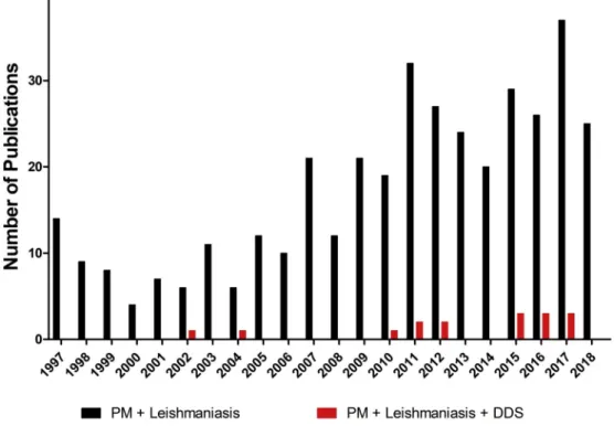
Fig. 3shows the number of publications between 1997 and 2018,
obtained in database “Web of Science”, using keywords as
“par-omomycin + Leishmaniasis” and “paromomycin +
Leishmaniasis + drug delivery systems (DDS)”. These data revealed that studies on PM drug delivery systems for leishmaniasis were not numerous before 2009. However, a growing interest in this topic has been observed from 2010 and more specifically within the last three years. This review will focus on the new formulations using DDS con-taining PM as monotherapy and in combined therapy developed in the last years.
4. Recent advancements in formulation strategies for leishmaniasis
The recent advancements in formulation strategies for leishmaniasis treatment using PM (microspheres, liposomes, solid lipid nanoparticles
- SLN, gels, polymeric films, emulsions) are presented inTable 4. They
will be discussed firstly for VL and then for CL leishmaniasis treatments. 4.1. Visceral leishmaniasis
Microspheres and liposomes are the two types of DDS proposed for the treatment of VL.
4.1.1. Microspheres
Microspheres can be defined as solid spherical particles ranging in size from 1 to 1000 μm. They can be produced from different materials as polymers (natural or synthetic), wax and proteins as carrier matrices
for drug delivery (Fig. 4) [90]. Microspheres can be produced by
dif-ferent processes such as spray drying, emulsification-solvent
evapora-tion, polymerization and coacervation among others [91,92].
Gen-erally, they have advantages compared to conventional dosage pharmaceutical formulations like lower toxicity, improved bioavail-ability, increased stability and extended drug delivery in a specific site
of action [93].
In 2011, Khan and Kumar [48] proposed a formulation based on
protein microspheres containing PM for visceral leishmaniasis treat-ment and parenteral use. This formulation aimed to overcome the limitations of most PM formulations (low efficacy, high doses required or local toxicity). The choice of albumin as a drug carrier is due to its biodegradability, nontoxicity and low cost. Moreover, albumin is ap-proved by FDA for clinical use. Spray drying was the process used to produce these PM-loaded albumin microspheres. It is a rapid, well es-tablished, reproducible and one-step process of converting a liquid formulation to a powder. The liquid formulation, sometimes a solution, an emulsion or a suspension, is sprayed through a nozzle into a chamber that simultaneously has hot gas being blown into it. As the liquid droplets are released through the nozzle and enter in contact with the
Fig. 3. Number of Publication of Paromomycin (PM) among 1997–2018 obtained from database ‘Web of Science’ using keywords as “paromomycin + Leishmaniasis” and “paromomycin + Leishmaniasis + drug delivery systems (DDS)”.
Table 4 Recent advancements in formulations strategies for PM. Formulation strategies Material Encapsulation method Type of leishmaniasis/Specie/ Use References Microspheres Albumin Spray drying Visceral/ L. donovani /Parenteral (Khan et al., 2013b, 2013a; Khan and Kumar, 2011) Liposomes Cholesterol, dicetyl phosphate, triglycerol monostearate, hexaglycerol distearate, decaethylene glycol mono n-hexadecyl ether or hexa glycol mono n-hexadecyl ether Fusion method Visceral/ L. donovani /Parenteral (Williams et al., 1998) Liposomes Phosphatidylcholine and stearylamine Conventional solvent evaporation method Visceral/ L. donovani /Parenteral (Banerjee et al., 2011) Liposomes Phosphatidylcholine, polyethyleneglycol, dimiristoyl phosphatidylcholine, dipalmitoyl phosphatidylcholine, dimiristoyl phosphatidylglycerol, diestaroyl phosphatidylethanolamine, dipalmitoyl phosphatidylglycerol, stearylamine Dehydration -rehydration method Visceral/ L. infantum /Parenteral (Gaspar et al., 2015) Liposomes Soybean phosphatidylcholine with or without cholesterol Solvent evaporation method and reverse-phase evaporation method Cutaneous/No leishmanicidal activity analyzed/Topical (Ferreira et al., 2004) Liposomes Soybean phosphatidylcholine, cholesterol, propyleneglycol, vitamin E, propylparaben, methylparaben Fusion method Cutaneous/ L. major /Topical (Jaafari et al., 2009) Liposomes Soybean phosphatidylcholine with or without cholesterol, propyleneglycol, vitamin E, propylparaben, methylparaben Reverse-phase evaporation method Cutaneous/ L. major /Topical (Carneiro et al., 2010) Liposomes Soybean phosphatidylcholine, cholesterol, sodium cholate, propylparaben, methylparaben, propyleneglycol, vitamin E Fusion method Cutaneous/ L. major /Topical (Bavarsad et al., 2012) Liposomes Cholesterol, phosphatidylcholine, phosphatidylglycerol, phosphatidylethanolamine, miltefosine Freeze-drying double emulsion method Cutaneous/ L. major /Topical (Momeni et al., 2013) Liposomes Soybean phosphatidylcholine, cholesterol, sodium cholate, propylparaben, methylparaben, propyleneglycol, vitamin E Fusion method Cutaneous/No leishmanicidal activity analyzed/Topical (Kalantari et al., 2014) Solid lipid nanoparticles Stearic acid or cetyl palmitate, Tween 80, Span 85, sodium sulfate Microemulsion method or solvent difusion method Cutaneous/No leishmanicidal activity analyzed/Topical (Ghadiri et al., 2012, 2011) Solid lipid nanoparticles Stearic acid, Tween 80 Modified high shear homogenization microemulsion method Cutaneous/ L. major and L. tropica /Topical (M. Heidari-Kharaji et al., 2016; Maryam Heidari-Kharaji et al., 2016; Kharaji et al., 2015) Hydrophilic gel Hydroxyethylcellulose Fusion method Cutaneous/ L. brazilensis and L. amazonensis /Topical (Gonçalves et al., 2005) Hydrophilic gel Hydroxyethylcellulose and methylbenzethonium chloride Fusion method Cutaneous/ L. braziliensis /Topical (Santos et al., 2008) Nanogel Poloxamer 407 Cold method Cutaneous/ L. major and L. infantum /Topical (Brugués et al., 2015) Hydrophilic gel Hydroxyethylcellulose, propyleneglycol, methylparaben Fusion method Cutaneous/ L. major and L. amazonensis /Topical (Mussi et al., 2007) Hydrophilic gel Hydroxyethylcellulose, propyleneglycol, methylparaben Fusion method Cutaneous/ L. major and L. amazonensis /Topical (Aguiar et al., 2010, 2009) Hydrophilic gel Hydroxyethylcellulose, propyleneglycol, methylparaben Fusion method Cutaneous/ L. braziliensis /Topical (de Morais-Teixeira et al., 2015) Hydrogel Chitosan, 2-hydroxipropyl β-cyclodextrin, β-glycerolphosphate, transcutol Mixture and homogenization Cutaneous/ L. major /Topical (Schwartz et al., 2014) Polymeric film Hydroxyethylcellulose, hydroxypropylmethylcellulose, gentamicin Fusion method Cutaneous/ L. major /Topical (Tolouei et al., 2011) Oil/Water emulsion Silicone based polymeric lipophilic surfactant, polyalkylene oxide block copolymer, paraffin oil Emulsification by homogenization Cutaneous/No leishmanicidal activity analyzed/Topical (Gomes et al., 2010)
hot gas, the solvent content of each droplet is removed by evaporation,
thus turning it from liquid to powder form [91,92,94]. The authors
prepared three different concentrations (2%, 5% and 8% w/v) of a total solid content with drug and albumin (1:9 w/w ratio) and analyzed the physicochemical properties and in vitro release of the microspheres. PM was not thermally degraded during the spray drying process. The PM-loaded albumin microspheres ranged between 2 μm and 4 μm in size,
being suitable for macrophage uptake [48].
In 2013, Khan and colleagues described two different studies
in-volving these PM-loaded albumin microspheres [51,95]. Firstly, they
evaluated the pharmacokinetic parameters of the microspheres ad-ministered intravenously in male rats, developed and validated a new bioanalytical method for quantifying PM until 40 ng/mL using a deri-vatization method (PM lacks strong chromophore). The PM-loaded al-bumin microspheres presented no nephrotoxicity when compared to
PM intramuscular injection [51]. Khan and co-workers then evaluated
the efficacy and stability of the PM-loaded albumin microspheres and observed a significant increased efficacy of this formulation compared to PM solution and a good stability in all temperature and humidity conditions tested, from which they considered that it could be a good
choice for VL PM treatment against L. donovani [95]. However, the
critical point of this formulation is the administration route (in-travenous), which is difficult for patient adhesion. Furthermore, more in vitro/in vivo studies are required including evaluate antileishmanial activity against L. infantum (New World Leishmaniasis specie). 4.1.2. Liposomes
Liposomes are bilayer phospholipid spherical vesicles (Fig. 4) that
are already present in the market [14,96,97]. They are great candidates
for the intracellular delivery system and antileishmanial drug delivery. Liposomal formulations generally present lower toxicity and higher efficacy than conventional formulations for leishmaniasis treatment
[96,97]. Furthermore, they are biocompatible, biodegradable and, as
drug delivery systems are able to increase drug efficacy and stability
[41].
Liposomes are classified as multilamellar vesicles (MLV) and uni-lamellar vesicles (large uniuni-lamellar vesicles – LUV or small uniuni-lamellar vesicles – SUV). They can be prepared by different methods as me-chanical dispersion (sonication, micro-emulsification, fusion, mem-brane extrusion among others), solvent dispersion (reverse-phase eva-poration, solvent injection) and detergent removal (detergent dialysis)
[98,99].
The arrival on the market of the first liposome formulation with amphotericin b for leishmaniasis treatment (amphotericin b liposomal – Ambisome®) motivated the increased number of researches on liposome formulations for other leishmanicidal drugs. Following this path, Williams and co-workers, in 1998, developed a liposome formulation
with PM to treat visceral leishmaniasis [100]. They used a different
molar ratio of surfactants, cholesterol and dicetyl phosphate melted and glucose solutions. The formulations with hexa or decaethylene glycol mono n-hexadecyl ether showed good stability and PM entrapment. Liposomes prepared with hexaethylene presented higher size (500 nm) and a maximum PM load of 20 μg/mL, whereas decaethylene glycol led to lower liposomes (200 nm) with higher drug content (between 20 μg/ mL– 40 μg/mL). In vitro and in vivo responses against L. donovani of PM liposomes were evaluated and compared to free PM. PM liposomes prepared with decaethylene glycol mono n-hexadecyl ether promoted higher parasite suppression in the liver. However, in the spleen and the bone marrow, PM liposomes presented suppression results similar to
the control group [100].
Other group working with liposomes [49] have studied cationic
phosphatidylcholine with stearylamine liposomes containing PM for intravenous single-dose treatment. They evaluated the in vitro and in vivo behavior of this formulation. The liposomes were prepared by the Fig. 4. Different types of drug delivery systems.
conventional solvent evaporation method with 10% of entrapment of efficacy of PM. In vitro analyses against L. donovani revealed a higher efficacy of PM liposomes, needing a lower dose than free PM to give the same effect. Moreover, mice treated with PM liposomes presented a higher reduction of parasite burden, with no in vivo toxicity. The au-thors also evaluated the ability to induce protective immunity of this formulation. The PM liposomes promoted an immunomodulatory effect
on CD4+and CD8+T cells for gamma interferon production and
down-regulated interleukin-10. This effect could represent longer protection
and higher efficacy than Ambisome® [49].
Another study in 2015 analyzed the efficacy of PM associated with liposomes, for parenteral administration, in infected murine models
with L. infantum [101]. Six formulations of PM liposomes were
devel-oped varying the lipid composition by dehydration-rehydration method (lipids dissolved in chloroform, dried and hydrated with PM aqueous solution). It was observed that the inclusion of polyethylene glycol (PEG) in lipid composition or stearylamine reduced 30% of the en-trapment efficiency in comparison with formulations prepared without PEG and stearylamine. This finding was attributed to the increase of zeta potential, which affects drug/lipid interactions. In the stability study, they analyzed four stability conditions (PM liposomes in buffer suspension at 4 °C, PM liposomes in inactivated fetal bovine serum at 37 °C, lyophilized and lyophilized with cryoprotector trehalose) and discovered that the best one was the PM liposome lyophilized with trehalose. They obtained PM liposomes with size lower than 120 nm. In vitro studies were developed with three formulations. One formulation, with negative charge surface, presented more than 90% internalization levels, which indicates that negative charge and rigidity of the liposome membrane promoted the cells uptake. In the biodistribution study, the PM liposomes showed preferential targeting in the liver, lung and spleen, while free PM presented a fast elimination from the blood-stream. Furthermore, the PM liposomes showed efficacy against L. in-fantum and no renal toxicity, suggesting that these formulations are an
interesting option for visceral leishmaniasis treatment [101].
4.2. Cutaneous leishmaniasis
Liposomes, SLN, and hydrogels are the three types of DDS more investigated for the treatment of CL.
4.2.1. Liposomes
Liposomes (Fig. 4), as described before, are bilayer phospholipid
vesicles and one of the drug delivery systems also studied for CL treatment with PM. Ferreira and co-workers, in 2004, evaluated the skin permeation of two types of PM liposomes for topical
administra-tion [102]. They prepared large multilamellar vesicles (MLV) by solvent
evaporation method (conventional method) and large unilamellar ve-sicles (LUV) by reverse-phase evaporation method (method with high aqueous space and able to encapsulate aqueous material) using only soybean phosphatidylcholine or a mixture of soybean phosphati-dylcholine and cholesterol as a lipid MLV presented a low PM capsulation of around 7.5% compared to LUV. Moreover, PM en-capsulation in LUV depended on the lipid composition, varying from 27.2% when prepared from a mixture of lipids (soybean phosphati-dylcholine and cholesterol) to 41.9%, when prepared only with soybean phosphatidylcholine. Thus, the skin permeation study was carried out only with PM-LUV. In the permeation test, PM-LUV presented low permeation across intact skin (around 1.5% permeated). Although the permeation of PM was higher across stripped skin than across intact skin, PM-LUV permeated lower than blank-LUV or PM solution after 10 h. The explanation was that PM liposomes, when used in stripped skin, promoted a drug-controlled release. More studies are required to investigate PM skin permeation, but this formulation could be an
al-ternative for leishmaniasis treatment [102].
Other researchers also developed PM liposomes for topical use. In
2009, Jaafari and co-workers [103] prepared two formulations of PM
liposomes by fusion method, varying the percentage of the drug (10% and 15%). The fusion method is a simple, solvent-free process, leading to an ideal viscosity for direct application in the skin. The two PM li-posomes presented comparable particle sizes (less than 500 nm) and encapsulation efficiencies (around 60%). They evaluated the in vitro and in vivo efficacy of the PM liposomes. In vitro studies against L. major showed that PM liposomes were three to four times more effective than PM solution. Furthermore, in vivo studies with BALB/c mice infected with L. major demonstrated that mice treated with liposomal formula-tion presented a significant reducformula-tion of lesion size after one week of treatment. After 12 weeks, PM liposomes showed a reduction in para-site burden, while blank liposomes had an increase of parapara-site burden. No significant differences were detected between the formulations prepared with different PM loads (10 or 15%). Based on these results, liposomes could be an interesting carrier for PM, however, further
biodistribution and immunomodulatory studies are required [103].
Another study with PM liposomes was conducted in 2010 using the
reverse-phase evaporation method [104]. The authors prepared two
LUV (PM with soybean phosphatidylcholine and PM with a mixture of soybean phosphatidylcholine and cholesterol) and investigated skin permeation in BALB/c mice infected with L. major. The surface charges of liposomes were modified after loading PM (drug promoted a de-crease of negative charges), probably due to an interaction between PM and the external monolayer of liposomes. Permeation test through in-tact skin of mice showed low levels of permeated drug (1.9% of PM solution and 4.8% of PM liposomes prepared with soybean phosphati-dylcholine and cholesterol), which increased with PM liposomes pre-pared with soybean phosphatidylcholine (7.2%). This formulation also
presented the highest level of permeation (14.6 μg/cm2) across stripped
skin. The lower values of permeation observed with PM liposomes prepared with cholesterol can be explained by the cholesterol presence that increases the rigidity of vesicles decreasing skin permeation of the drug. In in vivo assay, the treatment held with PM liposome gel was significantly better than treatment with free PM gel. This study also verified that liposomes promoted controlled drug release and increased
the topical delivery properties of this drug [104].
In 2012, Bavarsad and co-workers prepared, by fusion method
fol-lowing a previous study [103], a new class of liposomes called
trans-fersome [105]. Transfersomes are elastic vesicles formed by bilayer
phospholipid with edge activator, which promotes bilayers deform-ability. This deformability allows drug permeation across intact skin
when applied in non-occlusive conditions [105]. These authors
eval-uated the in vitro and in vivo effects of this formulation. Eighteen for-mulations were prepared, varying the percentage of lipids and the percentage of ethanol using factorial design. All the formulations pre-sented more than 50% of drug entrapment, but formulations containing 2% of sodium cholate were most stable. Nine PM transfersomes (for-mulations with 6% of sodium cholate and/or with 10% of ethanol) were excluded from the study due to their instability. In vitro studies against L. major showed that transfersomes formulations were more effective than PM cream or solution. Skin permeation investigated with four transfersomes (those with best in vitro properties and more stable ve-sicles) showed that these formulations retained more than 60% PM, while PM cream retained only 13%. PM transfersomes reduced lesion sizes and promoted lower parasite burden in vivo studies, without complete cure 12 weeks post-infection. No differences were observed between the four PM transfersomes in these studies. In conclusion, PM transfersomes produced with 2% of sodium cholate, with or without ethanol, could be an alternative option for cutaneous leishmaniasis
treatment [105].
In 2013, Momeni and co-workers [97] prepared liposomes by a
method based on the freeze-drying of double emulsions, which is re-ported to be a method with high encapsulation efficiency for hydro-philic or hydrophobic drugs and being able to produce sterile small unilamellar liposomes. Liposomes containing only PM were not gener-ated by this method, because phospholipid and PM produced
agglomerated particles, which could require further studies to control agglomeration. The authors then decided to associate another API (miltefosine) to PM and generate a combined therapy. PM-miltefosine liposomes were prepared with cholesterol, resulting in an encapsulation efficiency lower than 40%, considered too low for in vivo assay. In addition, there was not PM liposome produced by the same method for
comparison between mono and combined therapy [97].
Kalantari and co-workers, in 2014, investigated the possible ne-phrotoxicity and hepatotoxicity of PM liposomes using male Wistar rats
model [106]. They prepared PM liposomes by fusion method following
one previous study [103]. The toxicity of formulation was evaluated in
three periods of treatment (10, 20 and 30 days). No differences in the liver and kidney weight index were detected between the group re-ceiving PM liposomes topically twice a day and the control group. Also, the histopathological characteristics after the treatment provided that PM liposomes could be toxicity if used in long-term treatment. In fact, after 30 days, the liver showed reversible swelling cells, whereas the kidney presented a mild renal tubular necrosis. These changes were not observed after 10 or 20 days of treatment with PM liposomes, sug-gesting that PM liposomes could be used as a short treatment without
promoting nephrotoxicity and hepatotoxicity [106].
4.2.2. Solid lipid nanoparticles (SLN)
SLN (Fig. 4) are a class of nanoparticles, which have a lipid matrix
core stabilized by surfactants with a size lower than 1000 nm, solid at room temperature. SLN show some advantages as low toxicity, improve drugs solubility and bioavailability, large surface area and controlled
drug release [107]. Furthermore, these nanoparticles can be used as a
carrier for hydrophilic or hydrophobic drugs [96,107] and have been
studied as delivery system for hydrophilic and hydrophobic drugs
combined, especially for cancer treatment [108]. This system can be
suitable for several routes, such as pulmonary, ocular, intranasal,
sub-cutaneous, dermal, rectal intravenous and oral [96,107–109]. The use
of physiological lipids like triglycerides and cholesterol promotes more compatibility and safety of SLN formulations. Moreover, SLN have potential use in epidermal application with controlled release, lower
skin irritation and active protection [53,96]. SLN can be produced by
various methods, among them, emulsification solvent evaporation, high-pressure homogenization, hot homogenization, solvent diffusion,
hot microemulsion dilution [110,111].
Concerning PM formulations, a work about PM loaded-SLN was
published in 2011 [109]. SLN were prepared by two different methods:
microemulsion (aqueous solution with PM dispersed in melted lipids under agitation and immediately dispersed in cold water) and solvent diffusion (aqueous phase with PM was heated, added in organic phase containing solvent and lipids, homogenized and dispersed in cold water). The authors applied an experimental design to evaluate the estimated effects of three physicochemical properties (entrapment ef-ficiency, particle size and polydispersity index) of these systems. The best PM-SLN system was prepared by microemulsion with 39% of PM entrapment efficiency. It provided a prolonged release profile over 24 h
with a 64% PM release. Its thermal behavior was characterized [109].
In 2012, the same group of authors extended the study of PM loaded-SLN, improving the formulation using a statistical experimental design and two parameters (particle size and entrapment efficiency)
[28]. Some characteristics were predicted as the amount of drug,
per-centage of surfactant, ratio lipid/drug, method to prepare the SLN and lipid composition. After prediction, the PM-SLN formulation (90 mg of PM, 0.75% of surfactant, stearic acid as lipid and a ratio of 1:4 drug/ lipid) was prepared by microemulsion and the product obtained pre-sented the following characteristics: 42% of PM entrapment efficiency,
prolonged release during 24 h with 64% PM release [28].
In 2015, Kharaji and co-workers [53] evaluated the in vitro efficacy
of PM-SLN against L. tropica and L. major based on the optimal
for-mulation described in 2012 [28]. Four formulations of PM-SLN were
prepared by a modified high shear homogenization microemulsion
technique varying the percentage of PM-SLN (oil phase) in the micro-emulsion and the particle size. Moreover, in vitro cytotoxicity against human monocyte (THP1) and promastigotes and amastigotes of L. major and L. tropica response were evaluated. The results showed that free drug and blank SLN presented non-toxic effect, while all formula-tions PM-SLN showed cytotoxicity in THP1 cells. However, PM-SLN obtained with higher particle size (PM-SLN 15% 980 nm and PM-SLN 15% 1500 nm) were very toxicity against monocyte and excluded from the other assays. The two formulations with lower particle size (PM-SLN 15% 120 nm and PM-(PM-SLN 12.5% 240 nm) showed cytotoxicity only when used in high concentrations (> 6000 μg/mL), suggesting that PM-SLN toxicity is size-dependent. In the case of effective against pro-mastigotes and apro-mastigotes of L. tropica and L. major, the blank solid lipid nanoparticle presented no effect and the two formulations PM-SLN showed more efficacy than pure drug. When comparing in vitro results obtained of PM-SLN formulations, PM-SLN 15% 120 nm exhibited more efficacy and lower toxicity than PM-SLN 12.5% 240 nm. The fluores-cence microscopy confirms that PM-SLN formulations had lower in-fection levels than free drug and Blank-SLN. Therefore, PM-SLN pro-moted an increase in the effectiveness of PM and could be an interesting
alternative for leishmaniasis treatment [53].
The same group of authors (Kharaji et al.), in 2016, analyzed in vivo
efficacy of PM-SLN, developed in their previous study [53], against
BALB/c mice model infected with L. tropica and L. major [112,113]. The
first study was conducted to evaluate in vivo response against Leish-mania tropica using the most effective PM-SLN formulation (PM-SLN
15% 120 nm) [113]. In that work, they observed that PM-SLN has no
toxicity after 1 week administered in healthy mice. Moreover, BALB/c mice were infected with L. tropica and treated with two different ad-ministrations of PM-SLN (intramuscular and intralesional). Spleens and lymph nodes were removed and submitted to parasite burden, parasite load quantification and cytokine measurement. PM-SLN administered intramuscular and intralesional presented lower levels of parasite burden and parasite load when compared to parasite levels in blank-SLN, free PM and mice without treatment. Furthermore, amphotericin B, PM-SLN intramuscular and intralesional showed higher responses of IFN-γ secretion and nitric oxide production, suggesting that PM-SLN is effective and improve PM efficacy against leishmaniasis. These results support that solid lipid nanoparticle is a new and promising alternative
for cutaneous leishmaniasis treatment [113].
A subsequent study of the same group evaluated in vivo response against L. major using the PM-SLN formulation with the best results in vitro [112]. The lymph nodes of BALB/c mice infected with L. major were removed and submitted to parasite burden and cytokine mea-surement. Results showed that mice treated by the intramuscular route with PM-SLN formulations had lower levels of parasite burden than mice treated with amphotericin B, suggesting that PM-SLN and am-photericin B work similarly in parasite inhibition. Additionally, PM-SLN and amphotericin B presented higher levels of cytokine productions (IFN-γ and interleukin 4) and higher nitric oxide levels when compared to mice groups without treatment, with blank-SLN or free PM
treat-ment. These results reinforced the previous findings [106] that PM-SLN
can improve PM efficacy in leishmaniasis treatment [112].
4.2.3. Gels
Hydrophilic gels (Fig. 4), also known as hydrogels, are cross-linked
polymer networks dispersed in water medium produced by one or more
monomers reaction [114,115]. They can present some interesting
properties as stimuli sensitivity (physical or chemical) and aqueous swelling, and due to these properties, they have been studied for several applications. Hydrogels can be classified on several ways according to the synthesis route (homopolymers, copolymers or multipolymers), physical structure (amorphous, crystalline, semi-crystalline or hydro-colloids), ionic charge (anionic, cationic, amphiphilic or neutral), size (macrogels, microgels or nanogels) and bonds nature (chemical or physical). They can be produced by physical stimuli (temperature,