ÉCOLE DE TECHNOLOGIE SUPÉRIEURE UNIVERSITÉ DU QUÉBEC
THESIS PRESENTED TO
ÉCOLE DE TECHNOLOGIE SUPÉRIEURE
IN PARTIAL FULFILLMENT OF REQUIREMENTS FOR THE DEGREE OF
DOCTOR IN ENGINEERING Ph.D.
BY David LABBÉ
DEVELOPMENT AND PREVALIDATION OF A MEASUREMENT TOOL FOR THE PIVOT SHIFT PHENOMENON OF THE KNEE
MONTRÉAL, JANUARY 11TH 2010 © David Labbé, 2010
JURY PRESENTATION
THIS THESIS HAS BEEN EVALUATED BY THE FOLLOWING BOARD OF EXAMINERS
Ms Nicola Hagemeister, Thesis director
Département du génie de la production automatisée à l’École de technologie supérieure
M. Jacques A. de Guise, Thesis co-director
Département du génie de la production automatisée à l’École de technologie supérieure
M. Jean Arteau, President of the jury
Département de génie mécanique à l’École de technologie supérieure
M. Rachid Aissaoui, Member of the jury
Département du génie de la production automatisée à l’École de technologie supérieure
M. Michael J. Dunbar, External examinator
Department of orthopaedic surgery at Dalhousie University
THIS THESIS HAS BEEN PRESENTED AND DEFENDED BEFORE A BOARD OF EXAMINERS AND PUBLIC
DECEMBER 14TH 2009
ACKNOWLEDGEMENTS
First and foremost, I would like to thank my thesis directors: Nicola Hagemeister and Jacques de Guise. These past few years, I have learned a tremendous amount from both of you in the field of biomedical research. This, however, pales in comparison to what you have taught me about professionalism. You have shown me that one can be an excellent professor while still being respectful and kind to everyone around. Nicola, your guidance and our many discussions about the implications of my project and of Ph.D. studies in general were my guiding light when I was overcome by doubt. You are a great teacher and a better person. Jacques, you are an example of professionalism and integrity. Your enthusiasm is contagious to everyone who works with or for you. I hope I can pass on to my future students the lessons I have learned from both of you.
I would also like to thank the members of the jury who have accepted to take the time to read and evaluate my thesis. I would particularly like to express my appreciation to Dr. Dunbar who is traveling from Halifax to attend my thesis defense.
This project could not have gotten off the ground without the precious collaboration of numerous orthopaedic surgeons, namely Drs. Guy Grimard, David Baillargeon, Patrick Lavigne, Véronique Godbout, Julio Fernandes, Pierre Ranger, Vincent Massé, Peter Glavas and David Blanchette. I am extremely grateful for the time you took out of your hectic schedules to participate in this project. I am very impressed by the work you do and I applaud your dedication to research.
Without the numerous subjects who accepted to be evaluated for this study, there would have been no data to analyze. Although research involving human subjects certainly comes with its challenges, it also offers many rewards. Time and time again I was surprised by people’s willingness to participate, many times despite a long commute, for no reason other than for the sake of science or to lend a helping hand.
Since I started working at the Laboratoire de recherche en imagerie et orthopédie (LIO), I have worked with too many people to thank each one personally. When I arrived at the LIO with no background in orthopaedics or biomechanics, I felt a little intimidated but quickly found that there was always someone willing to help out. I have learned a great deal from so many of you. I must give special thanks to Gerald Parent for his technical support throughout this project and to Brigitte Dubreuil whose organizational skills and enthusiasm make everything so much easier at the LIO. On a more personal level, I have made many friends that made it so much more fun to come to work every morning. More specifically, Alex and Guillaume have become good friends. Your friendship as we took on the challenge of doctoral studies has been much appreciated.
I would have never been in a position to pursue Ph.D. studies had it not been for the support of my parents and family. Your help and encouragement throughout my studies are what made it possible for me to find something I really love doing and to dedicate so much time and energy to it. I am eternally grateful for everything you have done.
Finally, I would like to thank my closest friends, whom I could always count on to not talk to me about my project. Thank you Carl, Remy, Hugo, Mathieu, Francis, Magalie and Jonathan for your support and most of all, for the fun times shared in your company. I would be remiss if I didn’t thank Marie-Christine for her companionship throughout this entire project; your support has meant a lot to me. Last but not least, I would like to thank Elise for her encouragement during the long hours put into the writing of this thesis and for her support and reassurance in the hours leading up to my thesis defense. I look forward to returning the favor for years to come.
DEVELOPMENT AND PREVALIDATION OF A MEASUREMENT TOOL FOR THE PIVOT SHIFT PHENOMENON OF THE KNEE
David LABBÉ ABSTRACT
Rupture of the anterior cruciate ligament (ACL) is one of the sports injuries with the highest incidence. The ACL is essential to knee joint stability; it’s function is to limit anterior subluxation and internal rotation of the tibia relative to the femur. When a rupture occurs, the knee joint presents increased laxity and a rotational instability. During daily activities, this instability often results in a feeling that the knee is slipping, or giving way.
Traditional clinical tests measure the increase in knee laxity. Such measures are useful for diagnosing a rupture but they give no indication of the impact of the rupture on knee joint function. The pivot shift test is a clinical test that reproduces and grades the rotational instability following ACL-rupture. It is the only test that has been shown to correlate to subjective criteria of knee joint function such as patient satisfaction, return to physical activity and episodes of giving way. The pivot shift test is graded based on the clinician’s interpretation of a subjective scale, rendering it poorly reliable, especially in the hands of a less experienced clinician. There is currently no objective method for grading the pivot shift. The objective of this thesis was therefore to develop such an objective method to attribute the grade based on recorded knee joint kinematics.
To do so, we first developed a system to attach electromagnetic sensors to the lower limb and record the knee joint kinematics of subjects during the pivot shift test. Two separate data acquisition protocols ensued. In the first, three orthopaedic surgeons each performed the test on twelve different subjects and subjectively graded the pivot shift they produced. The aim of this phase was to develop a method capable of diminishing the variability introduced in the recordings by the clinicians’ gestures. In the second phase, an additional 53 subjects were evaluated by one of eight different surgeons. The subjective grades they attributed were used as a gold standard for data analysis. Principal component analysis (PCA) was used to determine which features of the kinematics explain most of the variability between recordings. Using these features, a support vector machine (SVM) based classifier was developed to automatically attribute the pivot shift grade.
The results showed that the variability between clinicians could be diminished by an average of 20% using the velocity of flexion applied by the evaluating clinician. This yielded significant differences between the pivot shift grades for many kinematic parameters. The ACP analysis showed that translation is the most important component of the pivot shift and that its velocity and acceleration are more important than its actual amplitude. Using the most important features, the SVM-based classifier obtained substantial agreement with the clinicians, with 95% of the recordings being classified within one grade of the clinician’s attribution.
This thesis has shown that the most important kinematic features of the pivot shift are not those that have traditionally been the focus of quantitative studies. It has also demonstrated the feasibility of objectively attributing the pivot shift grade based on a recording of its kinematics.
Keywords: anterior cruciate ligament, pivot shift, knee joint instability, knee joint kinematics, grade classification
DÉVELOPPEMENT ET PRÉ-VALIDATION D’UN OUTIL DE MESURE DU PHÉNOMÈNE DE PIVOT SHIFT DANS L’ARTICULATION DU GENOU
David LABBÉ RÉSUMÉ
La rupture du ligament croisé antérieur (LCA) est une des blessures sportives les plus fréquentes. Le LCA est essentiel pour la stabilité du genou; ses fonctions principales sont de limiter la translation antérieure et la rotation interne du tibia par rapport au fémur. Lorsqu’une rupture survient, le genou montre une laxité accrue et une instabilité rotatoire. Durant les activités quotidiennes, cette instabilité résulte souvent en un sentiment de glissement où les patients ont l’impression que leur genou cède sous leur poids.
Les tests cliniques traditionnels mesurent la laxité du genou. Cette laxité est utile pour diagnostiquer une rupture mais ne donne aucune information relative à l’état fonctionnel du genou. Le test du pivot shift est un test clinique qui reproduit l’instabilité rotatoire et lui attribue un grade. Il s’agit du seul test qui corrèle avec des critères subjectifs de l’état fonctionnel du genou tels que la satisfaction du patient, son retour à l’activité physique et le nombre de fois que son genou cède. Ce test est gradé selon l’interprétation du clinicien et suivant une échelle subjective, ce qui le rend peu fidèle, particulièrement pour des cliniciens moins expérimentés. Actuellement, il n’existe aucune méthode objective d’attribuer le grade du pivot shift. L’objectif de cette thèse était donc de développement une telle méthode subjective pour attribuer le grade à partir d’un enregistrement de la cinématique du genou. Pour ce faire, nous avons développé un système pour fixer des capteurs électromagnétiques au membre inférieur et enregistrer la cinématique du genou des sujets pendant le test du pivot
shift. Deux protocoles d’acquisition de données s’en sont suivis. Dans le premier, trois
orthopédistes ont chacun exécuté le test du pivot shift sur douze sujets et ont subjectivement gradé le pivot shift produite. L’objectif de cette phase était de développer une méthode capable de diminuer la variabilité introduite dans les enregistrements par le geste des cliniciens. Dans la seconde phase, 53 sujets additionnels ont été évalués par un de huit orthopédistes différents. Les grades attribués par les orthopédistes ont servis de gold
standard pour l’analyse subséquente. L’analyse par composantes principales (ACP) a été
utilisée pour déterminer quelles caractéristiques cinématiques expliquent la plus grande partie de la variabilité entre les enregistrements. À partir de ces caractéristiques, un classificateur de type machine à vecteurs de support (MVS) a été développé pour automatiquement attribuer le grade du pivot shift.
Les résultats ont montré que la variabilité entre les enregistrements des différents cliniciens peut être diminué de 20%, en moyenne, en utilisant la vitesse de flexion appliquée par chaque clinicien. Cette diminution permet des différences significatives entre les différents grades de pivot shift pour plusieurs caractéristiques cinématiques. L’analyse par ACP a montré que la translation est la composante la plus importante du pivot shift et que son accélération et sa vitesse sont plus importantes que son amplitude. En utilisant les
caractéristiques les plus importantes, le classificateur MVS a obtenu une concordance substantielle avec les cliniciens. Quatre-vingt quinze pourcent des enregistrements ont été classifiés à un garde ou moins de celui attribué par le clinicien.
Cette thèse a démontré que les caractéristiques les plus importantes du pivot shift ne sont pas ceux qui ont traditionnellement fait l’objet d’études quantitatives. Elle a aussi démontré la faisabilité d’attribuer objectivement le grade du pivot shift à partir d’un enregistrement de la cinématique du genou.
Mot clés: ligament croisé antérieur, pivot shift, instabilité du genou, cinématique de l’articulation du genou, classification du grade
TABLE OF CONTENTS
Page
INTRODUCTION ...1
CHAPTER 1 THEORETICAL FRAMEWORK ...3
1.1 Introduction ...3
1.2 Anatomy of the tibiofemoral joint ...3
1.2.1 The cruciate ligaments ... 5
1.3 Kinematics of the asymptomatic knee ...8
1.4 The ACL-deficient knee ...9
1.4.1 Static tests ... 11
1.4.2 Dynamic tests ... 14
1.5 Increased laxity versus knee joint stability ...16
CHAPTER 2 PROBLEM STATEMENT AND OBJECTIVES ...19
2.1 Problem statement ...19
2.2 Hypotheses ...20
2.3 Objectives ...21
CHAPTER 3 LITTERATURE REVIEW ...22
3.1 Introduction ...22
3.2 The pivot shift phenomenon ...22
3.2.1 Grading the pivot shift ... 22
3.2.2 Variations of the pivot shift test ... 24
3.2.3 Reliability of the pivot shift test ... 25
3.3 Instrumented knee joint laxity evaluations ...28
3.3.1 Anteroposterior laxity ... 28
3.3.2 Rotational laxity ... 32
3.4 Skin movement artifacts ...34
3.4.1 Skin mounted marker optimization ... 35
3.4.2 Percutaneous pins ... 36
3.4.3 External attachment systems ... 37
3.5.1 Early methods ... 43
3.5.2 In vivo analysis of the pivot shift kinematics ... 44
3.6 Conclusion ...56
CHAPTER 4 METHODOLOGICAL CHOICES AND SITUATION OF THE ARTICLES WITHIN THE THESIS ...58
4.1 Summarized description of the experimentations ...58
4.1.1 Inter-observer variability ... 58
4.1.2 Characterization of the pivot shift ... 59
4.1.3 Attachment system ... 61
4.1.4 Instrumentation ... 63
4.1.5 Definition of the coordinate system ... 64
4.2 Computation of translations and rotations ...68
4.3 Classification method...70
4.4 Situation of the articles within the scope of the thesis ...71
CHAPTER 5 ARTICLE I: ACCOUNTING FOR VELOCITY OF THE PIVOT SHIFT TEST MANOEUVRE DECREASES KINEMATIC VARIABILITY ...73
5.1 Introduction ...76
5.2 Materials and methods ...77
5.2.1 Experimental protocol ... 79
5.2.2 Data analysis ... 81
5.3 Results ...82
5.4 Discussion ...87
5.5 Acknowledgements ...91
CHAPTER 6 ARTICLE II: FEATURE SELECTION USING A PRINCIPLE COMPONENT ANALYSIS OF THE KINEMATICS OF THE PIVOT SHIFT PHENOMENON IN THE KNEE ...92
6.1 Introduction ...95
6.2 Methods...96
6.2.1 Experimental protocol ... 96
6.2.2 Data analysis ... 97
6.2.3 Principal component analysis ... 98
XI
6.4 Discussion ...102
6.5 Aknowledgements...105
CHAPTER 7 ARTICLE III: A CLASSIFICATION METHOD FOR AN AUTOMATIC AND OBJECTIVE ATTRIBUTION OF THE PIVOT SHIFT GRADE ...106
7.1 Introduction ...109
7.2 Materials and methods ...110
7.2.1 Population ... 110 7.2.2 Experimental protocol ... 112 7.2.3 Data classification ... 113 7.3 Results ...115 7.4 Discussion ...117 7.5 Aknowledgements...121
CHAPTER 8 SUPPLEMENTARY RESULTS ...122
CHAPTER 9 DISCUSSION ...124
9.1 General aspects ...124
9.1.1 Definition of the pivot shift... 125
9.1.2 Progress accomplished toward quantitative measure ... 126
9.1.3 Difficulty in quantifying the pivot shift ... 126
9.2 Limitations and specific aspects ...127
9.2.1 Validation of the method ... 127
9.2.2 Use of multiple recordings from a single subject ... 129
9.3 Fuzzy gold standard ...130
CHAPTER 10 CONCLUSION AND OUTLOOK ...132
APPENDIX I NUMBER OF PIVOT SHIFT EVALUATIONS PER CLINICIAN ...135
APPENDIX II PARTICULARITIES OF CLINICIAN TECHNIQUE ...136
APPENDIX III SUBJECT CHARACTERISTICS ...137
APPENDIX IV TYPICAL KNEE JOINT KINEMATICS DURING THE PIVOT SHIFT TEST ...140
APPENDIX V KNEE JOINT KINEMATICS OF THE PIVOT SHIFT RECORDINGS WHERE THERE IS A DISAGREEMENT OF MORE THAN ONE
GRADE BETWEEN THE CLINICIAN AND CLASSIFIER ...144 APPENDIX VI KINEMATICS OF THE PIVOT SHIFT THAT WERE USED BY THE
SVM-BASED ALGORITHM ...149 BIBLIOGRAPHY ...159
LIST OF TABLES
Page
Table 1.1 The mean sensitivity and specificity values of three clinical tests for
diagnosis of ACL rupture, from a meta-analysis ...17
Table 3.1 Maximum anterior translation and internal rotation reached by 10 different examiners during the pivot shit test on a cadaveric knee ...26
Table 3.2 Summary of the in vivo studies that quantified the pivot shift ...55
Table 3.3 Summary of the in vivo studies that quantified the pivot shift (contd) ...56
Table 4.1 Utilization of pivot shift recordings for each article ...60
Table 5.1 Number of knees evaluated for each grade ...81
Table 5.2 Grades attributed by each clinician, for each subject ...82
Table 5.3 Spearman’s rank coefficients and P values for different kinematic parameters with regards to the clinician-attributed pivot shift grade ...83
Table 5.4 Comparison of statistical analysis of kinematic data with regards to attributed grade for normalized and non-normalized data ...86
Table 5.5 Spearman’s rank correlation between kinematic parameters and the clinical grades attributed by an orthopedic surgeon ...87
Table 6.1 Number of knees evaluated for each grade ...96
Table 6.2 The list of kinematic features included in the principal component analysis ...98
Table 6.3 Loading factors of the pivot shift parameters ...102
Table 7.1 Grade distribution of the pivot shift recordings ...112
Table 7.2 Attribution of the pivot shift grade by clinicians and a SVM-based classifier ...116
Table 7.3 Classification of the pivot shift recordings by clinicians and a SVM-based classifier, into groups formed by grades 0, 1 and 2,3 ...117
Table 8.1 The CMC coefficients of four attachment systems for the recording of knee joint kinematics during gait ...123
LIST OF FIGURES
Page
Figure 1.1 The bony structures of the knee joint. ...4
Figure 1.2 The ligaments and menisci of the knee joint. ...5
Figure 1.3 The anteromedial (AM) and posterolateral (PL) bundles of the ACL. ...6
Figure 1.4 The anatomical axes of the knee and the rotations about these axes. ...8
Figure 1.5 The most widespread clinical tests for ACL deficiency, divided by type. ....10
Figure 1.6 Illustration of the tibia subluxed and after its sudden reduction, called the pivot shift. ...11
Figure 1.7 Illustration of a clinician performing the Lachman test. ...12
Figure 1.8 An illustration of the anterior drawer test. ...13
Figure 1.9 Illustration of a clinician performing the pivot shift test. ...14
Figure 1.10 Illustration of a clinician performing the jerk test, a subluxation test. ...15
Figure 3.1 A clinician performing the pivot shift test with the tibia in the three positions suggested by Jacob et al.: internal rotation, neutral and external rotation. ...23
Figure 3.2 The KT1000 installed on a patient’s lower limb. ...29
Figure 3.3 The Rolimeter arthrometer on a patient’s lower limb. ...31
Figure 3.4 The GNRB arthrometer...32
Figure 3.5 A device proposed by Musahl et al. to measure tibial rotation. ...33
Figure 3.6 (A) The position of the subject in the open MRI system; (B) The pressure applied by the clinician to produce anterior displacement and internal rotation of the tibia. ...34
Figure 3.7 Illustration of a percutaneous pin with reflective markers attached. ...36
Figure 3.8 Different variations of the Cleveland Clinic and Helen Hayes methods, compared by Manal et al. ...38
XV
Figure 3.10 The exoskeleton installed on the lower limb of a subject. ...40 Figure 3.11 The FTD and Helen Hayes plaques installed on a lower limb. ...41 Figure 3.12 (A) Attachment system proposed by Marin et al. (B) Attachment
system proposed by Goujon et al. ...42 Figure 3.13 Fixation of the motion-capture sensors to the bone in the study by
Bull et al. ...45 Figure 3.14 Translation of the center of the tibia relative to the femur during the
pivot shift test before and after ACL reconstruction ...46 Figure 3.15 Experimental setup of the Lane et al. study. ...47 Figure 3.16 The angle of p created by the arc of motion of the pivot shift reduction
and the arc of motion of the reference flexion, in the sagittal plane. ...48 Figure 3.17 The experimental setup for the studies of Kubo et al. and Hoshino et al. ....49 Figure 3.18 Correlation between the clinical grade of pivot shift and the measured
maximum posterolateral velocity. ...50 Figure 3.19 Attachment system made of custom molded thermoplastic plates. ...51 Figure 3.20 The femoral clamp and stabilizing bar developed by Amis et al. ...52 Figure 3.21 Simultaneous measurement of the translation and rotation components
of the motion of the femur using bone- and skin-mounted attachment
systems during a pivot shift test. ...53 Figure 3.22 (A) Graph of anterior laxity versus knee flexion (B) Graph of internal
rotation laxity versus knee flexion. ...53 Figure 3.23 Area of anteroposterior translation of knee joint center during the pivot
shift test. ...54 Figure 4.1 The femoral component of the attachment system we developed for
data acquisition. ...62 Figure 4.2 The tibial component of the attachment system we developed for
data acquisition. ...63 Figure 4.3 Illustration of the movement of hip circumduction, performed to
Figure 4.4 Illustration of the movement of flexion/extension, performed to define the mean axis of flexion. ...66 Figure 4.5 Illustration of the movement of slight flexion/maximum extension,
performed to define the neutral position. ...67 Figure 4.6 The coordinate systems of the tibia and femur as defined by the
modified FP method. Adapted from Primal Pictures Ltd ...68 Figure 4.7 Representation of the matrices containing the position and orientation
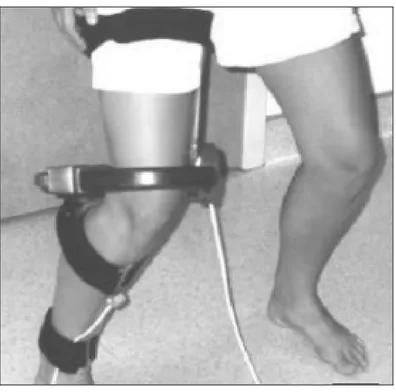
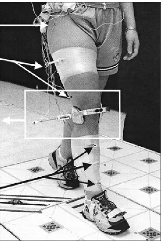
of the femoral and tibial sensors relative to the electromagnetic source. ...69 Figure 5.1: The attachment system used to fix motion capture devices to the
lower limb. A) the femoral component, B) the tibial component. ...78 Figure 5.2 A clinician performing the pivot shift test on a subject. ...79 Figure 5.3 Mean and standard deviation values for length of acceleration vector
according to the attributed grade. ...84 Figure 5.4 The kinematics of two grade 3 pivot shifts, produced by two different
clinicians, on a single subject. Black: Flexion angle (°); red: posterior translation (mm); green: external tibial rotation angle (°); blue: velocity of linear translation (mm/sec); magenta: acceleration of linear translation (mm/sec2). Acceleration values have been multiplied by a factor of 30 for visual representation. ...85 Figure 6.1 The scree plot of the 15 principal components (PCs). ...100 Figure 6.2 Cumulative percentage of total variance explained by the principal
components. ...101 Figure 7.1 Representation of the distribution of the pivot shift recordings obtained
from 56 subjects and 8 orthopaedic surgeons. ...111 Figure 7.2 A clinician performing the pivot shift test on a subject.Erreur ! Signet non défini. Figure 7.3 Classification of the pivot shit recordings according to their clinical
LIST OF ABBREVIATIONS AND ACRONYMS 2D Two-dimensional
3D Three-dimensional
AC Alternating current
ACL Anterior cruciate ligament ACLD Anterior cruciate ligament deficient AM Anteromedial
ANOVA Analysis of variance DOF Degrees of freedom
EBM Evidence-based medicine PCA Principal component analysis PCL Posterior cruciate ligament
PL Posterolateral
LCL Lateral collateral ligament MCL Medial collateral ligament MRI Magnetic resonance imaging RMS Root mean square
LIST OF SYMBOLS AND UNITS mm millimeter
mm/sec millimeter per second
mm/sec2 millimeter per second squared N Newton
INTRODUCTION
Evidence-based medicine (EBM) is described as “the conscientious, explicit and judicious use of current best evidence in making decisions about the care of individual patients”1. Different elements of evidence-based medicine (EBM) have been applied for many years but its major concepts were only popularized by Cochrane in 19722 and the term “evidence-based medicine” first appeared in medical literature in 19923. Since that time, EBM has gained in popularity and its benefits have gained widespread acceptance.
The use of EBM implies the capacity to evaluate a subject’s status or outcome using scientific methods. In EBM, evaluations made by so-called “medical experts” are the least valid form of evidence. To scientifically evaluate a subject’s status or outcome, one must have access to objective and reliable measures that can be compared to those found in the literature or to measures taken prior to treatment. Such a measure is not readily available for the evaluation of knee joint function following rupture of the anterior cruciate ligament (ACL).
Rupture of the ACL is amongst the most common musculoskeletal injuries with an incidence of over 100 000 each year in the United States4. Assuming a similar incidence in Canada, that’s over 10 000 such injuries annually. The impact of an ACL rupture varies greatly from one patient to the next and while objective measurements of knee laxity can be used for diagnosing a rupture, only a more complex and dynamic test called the pivot shift test can assess the functional impact of the injury on the affected knee5. This test is applied in an unconstrained and therefore variable manner and the clinician performing the test evaluates its result subjectively5.
In this thesis we conducted two sets of experimentations with the general objective of developing a more objective means of evaluating the pivot shift test. To do so, we first took into account the complex kinematics of the knee joint and the pathomechanics of the ACL-deficient knee. We also considered the clinical tests used to evaluate these pathomechanics,
with a focus on the pivot shift test (Chapter 1). In light of this, we established the problem statement and the specific objectives of this thesis (Chapter 2). Next, we conducted a review of current literature about the pivot shift test and the various factors that impact its results. We explored the methods, results and shortcomings of previous studies that were aimed at objectifying the pivot shift test (Chapter 3). In Chapter 4, we present the methodological choices we made in conducting our study and go on to situate the three articles within the context of the larger study and its objectives.
The first of these articles presents a method for reducing the inter-observer variability of the pivot shift maneuver and identifies kinematic parameters related to the pivot shift grade (Chapter 5). The second article presents a novel method for identifying the kinematic parameters that explain most of the variability between pivot shift recordings (Chapter 6). The third and final article presents a classification method that objectively attributes the pivot shift grade. It does so based on the recorded knee joint kinematics, using the methods described in the first two articles (Chapter 7). A general discussion of the study is presented in Chapter 9, followed by the conclusion and outlook (Chapter 10).
This study advances the understanding of the kinematics of the pivot shift and takes it one step further by providing a method for objective grade attribution based on these kinematics. Moreover, it represents a big step towards the development of a quantitative measure of the pivot shift phenomenon.
CHAPTER 1
THEORETICAL FRAMEWORK 1.1 Introduction
The knee is the largest and most complex joint of the human body. It is composed of two separate articulations: that of the tibia with the femur (the tibiofemoral joint) and that of the patella with the femur (the patellofemoral joint). In this study, we focus on the tibiofemoral joint, which is primarily a hinge joint, but its hinge movements are combined with gliding and sliding, making it a triaxial joint6.
This chapter focuses on the anatomy and complex kinematics of the asymptomatic tibiofemoral joint as well as the impact of an ACL rupture on tibiofemoral joint kinematics and function. The different types of clinical tests used to evaluate this impact are presented and the relevance of each is discussed.
1.2 Anatomy of the tibiofemoral joint
The knee is a synovial joint, which means that a fibrous capsule, called a synovial capsule, surrounds it. The cavity formed by this capsule contains synovial fluid that lubricates the articulating surfaces. The capsule of the knee is unique in that it does not completely surround the joint. Rather, it covers the lateral and posterior faces of the knee and is deficient on the medial and anterior faces, where the patellar ligament and medial patellar retinaculum fill most of the gap. The capsule is supplemented and strengthened by the five extracapsular ligaments (patellar ligament, fibular collateral ligament, tibial collateral ligament, oblique popliteal ligament and arcuate popliteal ligament)6.
Figure 1.1 The bony structures of the knee joint. Adapted from Visible Body
The three main bones of the knee joint are: the femur, the tibia and the patella (Figure 1.1). The femoral condyles rest atop the tibial plateau, forming the tibiofemoral joint. The lateral and medial menisci lie on the tibial plateau and serve to disperse the weight of the body and reduce friction between the tibia and the femur. These menisci are crescent-shaped fibrocartilaginous structures and are fused to the tibia along their outer circumference but are detached from it on their inner edge. They have a concave geometry that espouses the shape of the femoral condyles.
The stability of the knee joint is the result of its surrounding soft tissue structures (Figure 1.2): the synovial capsule, the extraarticular ligaments, the intraarticular ligaments and several tendons of muscles that act on the knee joint. The main tendons that act as stabilizers of the knee are the tendons of the quadriceps muscles and the tendon of the semitendinosis. The aforementioned five extracapsular ligaments are tense when the knee is in extension and they play a role in limiting hyperextension. The medial and lateral collateral ligaments also resist valgus and varus stress, respectively6.
5
The intracapsular ligaments of the knee are called the cruciate ligaments because they cross each other in the intercondylar fossa of the femur. Because this work is aimed at measuring the pivot shift, which is a symptom of ACL rupture, the anatomy and function of the cruciate ligaments are presented in greater detail.
Figure 1.2 The ligaments and menisci of the knee joint. Adapted from Netter’s Atlas of Anatomy
1.2.1 The cruciate ligaments
The cruciate ligaments are named according to their insertion site on the tibia. Thus, the posterior cruciate ligament (PCL) inserts posteriorly to the ACL on the tibia, 1-1.5 cm inferior to the posterior rim of the tibia, in the PCL fovea. On the femoral side, it inserts to the anterolateral border of the medial femoral condyle. The average PCL is 38 mm long but because its femoral fixation is extracapsular, its intracapsular length is shorter than that of the
ACL and it has a less oblique orientation. Its average width is 13 mm and like the ACL, it is composed of two main bundles: anterolateral (AL) and posteromedial (PM). The AL bundle is tense during in flexion and the PL bundle is tense in extension7.
The PCL’s primary function is to limit posterior translation of the tibia relative to the femur. Its secondary functions are to resist varus and valgus forces as well as hyperextension. A PCL rupture leads to an increased posterior translation of up to 20 mm. The PCL’s resistance to tension is generally considered to be above 2000 N, slightly higher than that of the ACL. Hyperextensions and posterior impacts to the proximal tibia are the most frequent injury mechanisms7.
Figure 1.3 The anteromedial (AM) and posterolateral (PL) bundles of the ACL.
Taken from Fu8
The ACL inserts to the tibia posteromedially to the anterior horn of the lateral meniscus. Its posterior limit is approximately 2 mm anterior to the tibial fixation of the PCL. On the
7
femoral side, it fixes to the posteromedial border to the lateral condyle. The average length of an ACL is 38 mm with an average width of 10 mm. When tension is applied to it, it stretches and lengthens by 1-2 mm. The ACL is composed of interlaced collagen fibers contained within a synovial membrane. The tibial nerve innervates it and its blood supply comes primarily from the middle genicular artery7. The ACL is known to have a proprioceptive function9, 10 and approximately 1% of its volume is comprised of nerve structures11.
As is the case for the PCL, the ACL is composed of two distinct bundles. These bundles, named for their fixation points, distinguish themselves by the orientation of their collagen fibers, which run parallel to their axes. The posterolateral bundle is tense in full extension and slight hyperextension and the anteromedial bundle is tense in flexion (Figure 1.3).
The ACL’s primary function is to limit anterior translation of the tibia relative to the femur. Its secondary functions are to limit internal tibial rotation, varus and valgus stresses and hyperextension. On average, the ACL can resist a tension of close to 2000 N and has a rigidity of 242 N/mm. During passive knee extension, the ACL is only tensed between 170 and 180 degrees. Active knee extension between 50 and 110 degrees does not tense the ACL7. However, additional contraction of the quadriceps and hamstring muscles does affect the tensile loading of the ACL within this range. It has been demonstrated in vitro that a quadriceps force of 400N increases the tensile force of the ACL up to 60 degrees of flexion. At higher angles, the same force relieves the load. Hamstring activation reduces the load on the ACL throughout the entire range of flexion of the knee12.
The ACL is most frequently torn without contact, usually during a rapid deceleration combined with a pivot. Other injury mechanisms include hyperextension and excessive internal tibial rotation13.
1.3 Kinematics of the asymptomatic knee
The asymptomatic tibiofemoral joint has a much larger range of motion in flexion/extension than in the other axes but it is nonetheless not strictly a hinge type joint. In fact, the tibiofemoral joint allows some degree of rotation in all 3 anatomical axes (Figure 1.4). The gliding of the femur on the tibia also produces translations in the 3 anatomical axes.
Figure 1.4 The anatomical axes of the knee and the rotations about these axes.
Adapted from Trilha et al.14
On average, the knee can flex over a range of approximately 140-160 degrees. This arc of flexion can be divided into three subarcs: (1) the arc of terminal extension, also called
screw-home, which ranges from maximum extension to about 10° of flexion; (2) the arc of active
function which ranges from about 10 to about 120°; and (3) the arc of passive flexion which ranges from about 120° to maximum flexion 15.
As the knee extends from 10-20° of flexion towards the limit of passive extension (-5 to 5), the medial femoral condyle glides on the tibia’s medial condyle, which produces an external tibial rotation. This mechanism is due to a combination of bony geometry and of the restraint
9
applied by the ACL. When the knee is in full extension, no significant tibial rotation is possible without concomitant flexion15.
In the arc of active function, the medial femoral condyle rotates to produce a combination of flexion and longitudinal (internal/external) rotation but barely any translation (1.5 mm at most). The lateral condyle rolls but also slides anteroposteriorly (15 mm during flexion). This allows longitudinal rotation around an axis that passes through the medial condyle and flexion about an axis that passes through both condyles. As result of this asymmetrical sliding, between 10° and 120° of flexion, the tibia rotates internally about 30°15.
At 90° of flexion, the tibia is free to rotate about 30° longitudinally without concomitant flexion. Conversely, the knee can flex from 20 to 90° without concomitant tibial rotation. Throughout its entire arc of flexion, very little abduction/adduction occurs in the weight-bearing knee. In the non weight-weight-bearing knee, a range of 15 degrees is normally attainable15.
In arc of passive flexion, both condyles roll back onto the posterior horns of the menisci and lose contact with the tibia itself. The knee can thus be said to be subluxed. This range of flexion, between 120 and 140 to 160° is only possible with externally applied forces15.
1.4 The ACL-deficient knee
As previously stated, the primary function of the ACL is to limit anterior translation of the tibia and its secondary functions include limiting internal tibial rotation. Therefore, the ACL-deficient knee displays increased anterior translation and tibial rotation when subjected to external forces and moments16. A medial translation of the tibia and increased abduction rotation has also been linked to ACL-deficiency, especially in low flexion angles17. In weight bearing conditions, the tibial plateau is persistently subluxed anteriorly between 0 and 90° of flexion18.
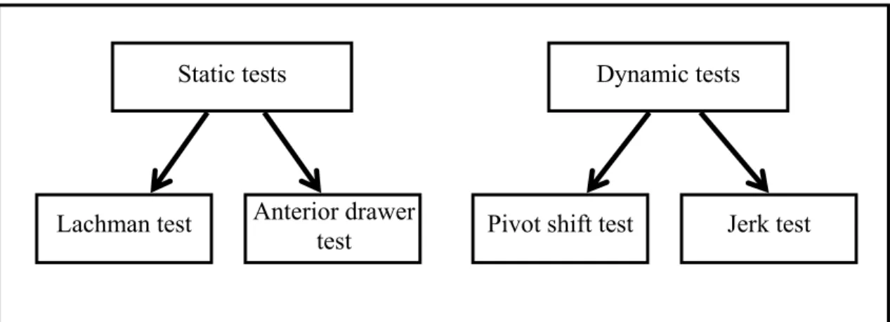
Clinical evaluation of a knee suspected of being ACL-deficient involves two categories of manual tests that are used to verify the restraints applied by the ACL and thus the ligament’s integrity: static tests and dynamic tests (Figure 1.5).
The objective of the static tests is to isolate a specific ligament, the ACL in this instance, by applying a force that is almost parallel to its collagen fibers. The clinician aims to test the restraint due to the ACL while limiting solicitation of surrounding soft tissues. The two most popular static tests for testing the ACL’s integrity are the Lachman test19 and the anterior drawer test20.
Figure 1.5 The most widespread clinical tests for ACL deficiency, divided by type. As for the dynamic tests, they aim to reproduce a more complex rotational and translational instability, called the pivot shift21. The pivot shift was first described as a slipping knee because patients complained of slipping and a feeling of insecurity. Other patients described the feeling as the knee giving way or crumpling up. They described a feeling of complete insecurity when the knee moved into a position of flexion.
Anatomically, the pivot shift is characterized by an anterior subluxation of the lateral tibial plateau relative to the femoral condyle as the knee approaches extension and the spontaneous reduction of subluxation during flexion22. Figure 1.6 shows the subluxed and reduced
Static tests Dynamic tests
Lachman test Anterior drawer
11
positions of the tibial plateau. The subluxation has a rotational as well as a translational component so that the subluxation is greater on the lateral side of the tibial plateau than on the medial side. Both the pivot shift test23 and the jerk test24 are used to reproduce this symptom in a clinical setting.
Figure 1.6 Illustration of the tibia subluxed and after its sudden reduction, called the pivot shift.
Taken from Bull et al.21
1.4.1 Static tests
The so-called static clinical tests are used to evaluate the amount of AP laxity in the tibiofemoral joint. The Lachman test is performed with the patient in supination and his knee flexed approximately 20°. The clinician stabilizes the femur with one of his hands and applies an anterior force to the tibia with his other hand (Figure 1.7).
Figure 1.7 Illustration of a clinician performing the Lachman test.
Taken from Ebell et al.25
The clinician evaluates the amount of anterior translation produced in order to verify the integrity of the ACL. As originally described by Torg et al. 19, an evaluation is considered positive when there is noticeable anterior translation with a soft or mushy endpoint. Nowadays, the Lachman test is usually graded using the amplitude of translation produced, but different authors have used different grading systems. The International Knee Documentation Committee (IKDC) separates knee laxity into 4 grades: grade A, normal (0-2 mm); grade B, nearly normal (3-5 mm); grade C, abnormal (6-10 mm); and grade D, severely abnormal (> 10 mm). The clinician can approximate the amount of translation or use an arthrometer such as the KT-1000, as will be discussed in the literature review. Many clinicians also compare the amount of translation produced on the examined knee to that produced on the controlateral knee and look for significant difference as an indicator of ACL deficiency.
The anterior drawer test is also performed with the patient in supination but for this test the examined knee is flexed between 60 and 90 degrees with the foot resting flat on the examining table. The clinician places both hands around the tibia, near the knee and jerks the tibia anteriorly (Figure 1.8). As is the case with the Lachman test, high anterior translation is
13
an indicator of rupture and the difference in translation between both knees has been shown to be more telling than the actual translation26. The grading systems used for the anterior drawer test are generally identical to those used to grade the Lachman test.
Figure 1.8 An illustration of the anterior drawer test.
Taken from Ebell et al.25
Although both static tests presented here aim to evaluate AP laxity, their sensitivity in diagnosing ACL rupture is very different. Many studies have compared the sensitivity (percentage of true positives) and specificity (percentage of true negatives) of both these tests 26-30. The consensus has been that both tests have high specificity but only the Lachman test has high sensitivity. In fact, the Lachman test has been shown to be a sufficiently sensitive diagnostic means to warrant surgery without confirmation using MRI29, 31. The anterior drawer test, on the other hand, has been shown to be unreliable28 and of little use in diagnosing ACL ruptures26. A recent meta-analysis of more than 10 comparative studies puts the average sensitivity of the Lachman test at 86% versus 62% for the anterior drawer test32.
1.4.2 Dynamic tests
As their name implies, dynamic tests are performed in a dynamic manner while applying forces and moments to the joint. They allow for an evaluation of more complex bone movements that cannot be evaluated using static tests. However, they are performed in a much less constrained manner, which implies that they are more variable and the produced movements are harder to quantify.
Figure 1.9 Illustration of a clinician performing the pivot shift test.
Taken from Ebell et al.25
Many dynamic clinical tests have been proposed. They can be separated into two main categories: reduction tests and subluxation tests21. In the reduction tests, the leg is initially in full extension and is slowly flexed under a valgus moment (the knee is held in place with one hand placed against its lateral side while the other hand applies an abductor force to the lower leg).
During flexion, a sudden reduction of the anteriorly subluxed lateral tibial plateau (the pivot shift) is observed in the ACL-deficient knee. This reduction is graded as 0 (none), 1 (glide), 2 (clunk) or 3 (gross clunk) by the evaluating clinician. The first documented reduction test was described by Galway et al. in 197223 and consists of conducting the aforementioned test
15
with the patient supine (Figure 1.9). MacIntosh (a coauthor of Galway’s work) later described his version of the pivot shift test as being similar to Galway’s but with the foot of the examined limb internally rotated21. This test (known as the pivot shift test), which is widely used by clinicians, has thus been attributed to both authors. Other reduction tests, such as that of Slocum et al.33 have been proposed but their use is not widespread.
Figure 1.10 Illustration of a clinician performing the jerk test, a subluxation test.
Taken from Losee et al.34
The subluxation tests are essentially the opposite of reduction tests. In subluxation tests, the knee is initially flexed and is extended under a valgus moment and an internal rotation of the tibia as seen in Figure 1.10. During extension, the lateral tibial plateau suddenly subluxates anteriorly. The first and most widely used variation of the subluxation test is called the jerk test24. In the jerk test, the knee is initially flexed to 90° with the hip flexed to 45° and the tibia internally rotated (Figure 1.10). During extension the subluxation of the lateral femorotibial articulation is maximal around 30° and then as extension continues, a spontaneous relocation occurs, which is known as a jerk. The authors claim that this jerk test is more sensitive than reduction tests24. However, this claim is not backed by any data in their publication.
Nonetheless, the pivot shift test (which is a reduction test) remains the most widely used clinical test for observing the rotational and translational instability of the knee. A combination of both tests is sometimes used, as one is the opposite of the other. In that sense, if the knee is placed under valgus moment and the tibia is placed under internal rotation while the knee is flexed and extended, both tests are being combined and the instability should be observed during both flexion and extension. The pivot shift test is discussed in further detail in Chapter 3.
1.5 Increased laxity versus knee joint stability
As stated, the sensitivity of the Lachman test in diagnosing ACL ruptures has been shown to be very high26, 28, 29, 32. Several studies have compared the sensitivity of the pivot shift test to that of the Lachman test. There is a consensus amongst these studies that the Lachman test is more sensitive but there is considerable disagreement as to the sensitivity of the pivot shift test. It has been reported to be as low as 18%35 and as high as 90%28. In their recent meta-analysis, Benjaminse et al.36 found sensitivity to be in the 30-40% range without anesthesia and in the 85-90% range with anesthesia (the effect of anesthesia on the pivot shift test is discussed in Chapter 3).
Its specificity, on the other hand, has consistently been reported as being very high and superior to that of the other tests. Another recent meta-analysis, this one by Scholten et al. 32, confirms the finding that the Lachman test is the most sensitive and the pivot shift test, the most specific (Table 1.1). Benjaminse et al. 36 reported similar results. In other words, a positive pivot shift test indicates an ACL rupture and a negative Lachman test rules it out.
17
Table 1.1 The mean sensitivity and specificity values of three clinical tests for diagnosis of ACL rupture, from a meta-analysis
Adapted from Scholten et al.32
Mean sensitivity Mean specificity
Lachman test 86% 91%
Anterior drawer test 62% 88%
Pivot shift test 35% 98%
Although it is obviously relevant to diagnose an ACL rupture, it is often the level of knee joint function that is of most interest and that clinicians aim to restore. The evaluation of knee joint function is generally done using any number of subjective variables and validated functional scales, such as the Lysholm scale37. Several studies have demonstrated that no significant relationship exists between anterior laxity (instrumented or not) and subjective measures of functional outcome38-42.
A recent study involving 202 subjects has shown that the pivot shift does have significant associations with satisfaction, partial giving way, full giving way, difficulty cutting, difficulty twisting, activity limitation, overall knee function, sports participation and Lysholm score38. Subjects with higher-grade pivot shift tests had less satisfaction, more limitations and lower knee function. The authors conclude that their findings support the functional importance of the pivot shift phenomenon and the clinical relevance of the pivot shift test.
The presence of a post-operative pivot shift has recently been shown to correlate with poor patient subjective evaluations and poor knee function scores43; patients with a post-operative pivot shift were 14.4 times more likely to have an unsatisfactory outcome than patients without a pivot shift.
Such studies underline the importance of the pivot shift and of its grade, in contrast to the Lachman test. Both tests are important parts of a clinical evaluation but serve different purposes. The pivot shift test’s specificity makes it a valuable complement to the Lachman test in establishing a diagnosis but more importantly, the pivot shift test is the only test which can be used to assess the level of knee joint function and predict long term outcome. As such, many studies have now concluded that the objective of reconstructive surgery should be to eliminate the presence of a pivot shift and not to simply diminish AP laxity43-46.
19
CHAPTER 2
PROBLEM STATEMENT AND OBJECTIVES 2.1 Problem statement
As mentioned in the introduction, the main problem with current clinical evaluations following ACL rupture is that only the pivot shift test, a highly subjective test, can assess knee joint function. The clinician attributes the grade of the pivot shift relying on his interpretation and experience5, 21. Moreover, the nature of the grading scale renders the grade poorly repeatable5. Indeed, it has been shown that different clinicians frequently attribute different grades to a same patient47.
No objective method for evaluating the pivot shift test currently exists, despite several attempts in the literature48-53. In the absence of an objective pivot shift measurement tool, it is difficult for less experienced clinicians to attribute a grade with a sufficiently high level of confidence for it to be used in determining the course of treatment. Moreover, a subjective measure is inadequate for following the progression of a patient’s status or for comparing pre- and post-operative evaluations. This is an obstacle not only to evaluating the success of individual surgeries but, more importantly, to comparing the outcomes of different surgical procedures. Similarly, it is difficult to compare the cohorts of different studies in terms of the pivot shift grade. As a consequence of the lack of an objective measure, the anteroposterior laxity, as evaluated by Lachman’s test using the KT-1000, is generally used for comparing outcomes or subjects. As established in Chapter 1, AP laxity has no significant correlation to knee joint function.
Many obstacles stand in the way of the development of a pivot shift measurement tool and one of the most important is the ability to record the kinematics of the tibia and femur with high precision. The artifacts caused by skin to bone displacement around the knee are important enough in relation to the bone movements to mask critical information54, 55. It has been demonstrated that surface mounted markers near the knee move up to 17 mm rms
relative to the underlying bone during passive knee flexion54. A study by Amis et al.48 has recently shown that the sudden reduction of the tibial plateau that defines the pivot shift was clearly recorded by a motion capture device fixed to the bones using intra-cortical screws. However, a non-invasive attachment system that was simultaneously recording knee joint kinematics recorded only a very small percentage of this translation. While it may be foreseeable to use intra-cortical screws for some per-operative evaluations of the pivot shift, this solution is obviously not one that can be transferred to a clinical setting. Thus, recording knee joint kinematics in a non-invasive manner and with sufficient precision remains a major concern in developing a measurement tool.
Skin displacement artifacts are a problem that pertains to the recording of the pivot shift but the nature of the pivot shift itself introduces additional challenges. The interpretation of what constitutes a given grade is not the only aspect that differs between clinicians. The pivot shift test is an unconstrained test, meaning that the clinician’s gesture is not guided and there is no measure of applied forces. As such, it is inevitable that the applied forces and moments and the resulting kinematics will differ somewhat between clinicians. It has been shown that the position of the limb during the test as well as the amount of force applied directly impact the produced pivot shift kinematics56, 57. Thus, not only are clinicians interpreting differently from their colleagues, they are also producing different bone displacements. A study by Noyes et al.47 showed that the anterior subluxation of the medial tibial plateau induced by 11 clinicians performing the pivot shift test on a cadaveric knee varied from 6 mm to 16.9 mm.
2.2 Hypotheses
Our main hypothesis for this thesis is that the kinematics of the tibia and of the femur can be recorded during the pivot shift test and that they can be used to attribute the grade in a more objective manner. This hypothesis relies on four specific hypotheses:
• We hypothesize that significant differences exist between the kinematics of different grades of the pivot shift.
21
• We hypothesize that tibiofemoral joint kinematics can be recorded with sufficient reliability to allow for these differences to be revealed.
• We hypothesize that inter-clinician variability can be reduced using particularities of each clinician’s gesture such that the aforementioned kinematic differences are still observed when the evaluations are performed by many different clinicians.
• We hypothesize that variability between recordings of a same grade can be reduced sufficiently such that a classification algorithm can distinguish the different grades with high accuracy.
2.3 Objectives
The main objective of this study is to develop an objective method of attributing the pivot shift grade. Our specific objectives in doing so are:
• To develop a tool capable of recording the knee joint kinematics of the pivot shift phenomenon.
• To diminish the variability introduced by the clinician in order to increase the kinematic differences between each pivot shift grade.
• To characterize the kinematics of the pivot shift and identify which aspects of these kinematics are important in grading the pivot shift.
• To develop a classifier capable of attributing the pivot shift grade in a manner similar to that of an experienced clinician based solely on a kinematic recording of the pivot shift test.
CHAPTER 3
LITTERATURE REVIEW 3.1 Introduction
This chapter will present an overview of research that has been conducted about the pivot shift test: its grading, the different variations of technique that have been proposed and the reliability of the test. Different knee evaluation devices developed to quantify the static tests will be presented and their strengths and weaknesses discussed. The impact of skin movement artifacts and different possible solutions will then be presented, as this is a major obstacle when passing from the quantification of a static test to that of a more dynamic test such as the pivot shift test. This will be followed by a detailed look at the methodology and results of previous studies that have attempted to measure and quantify the pivot shift.
This literature review will set the table for the methodological choices and show how this thesis is positioned with regards to previous work.
3.2 The pivot shift phenomenon
3.2.1 Grading the pivot shift
When performing the pivot shift test, the clinician must not only determine whether or not the pivot shift is present but also grade it, if it is present. The grade indicates the severity of the instability. For grading to be possible and relevant, the criteria must allow examiners to clearly distinguish between different grades. This grading serves three purposes. First of all, it is claimed that the test is subtle enough for examiners to assess different injuries and discriminate between them based on the grade. The severity of the pivot shift is also critical in determining which type of treatment is appropriate21, 58. Finally, grading is useful in a research perspective as it allows for comparisons between limbs with similar degrees of pivot shift.
23
According to the grading system that was originally described by Galway et al.23, the pivot shift is graded as a 0 (absent or negative), 1+ (slight slip), 2+ (moderate slip) or 3+ (momentary locking). Bach et al.59 suggested a minor change to this grading, suggesting a grade of 0.5 be added for “trace”. This proposed change was never widely adopted.
Jakob et al.57 proposed a grading system that is less subjective, yet it doesn’t require a measuring device. They suggest that performing the test using internal, neutral and external tibial rotation and noting in which positions a pivot shift is elicited can indicate the grade. Figure 3.1 shows the three positions in which the test is performed. This grading system is in fact less subjective because the examiner does not have to quantify the displacement. It has however been refuted by other authors59 and its use has been very limited.
Figure 3.1 A clinician performing the pivot shift test with the tibia in the three positions suggested by Jacob et al.: internal rotation,
neutral and external rotation. Adapted from Jacob et al.57
Despite these proposed adaptations, the standard grading remains similar to that initially described by Galway. The grading scale of the IKDC is now the one that is used in a majority of studies. Its grades are described as: 0 (absent), 1 (glide), 2 (clunk) and 3 (gross).
3.2.2 Variations of the pivot shift test
Anesthesia
The variation that has been shown to influence results of the examination the most is anesthesia. Various researchers have shown the pivot shift test consistently results in higher grades when performed under anesthesia. Anesthesia makes the surrounding muscles limp, which eliminates their effect on knee kinematics. This allows the effect of the ligaments to be isolated because without anesthesia, the muscles surrounding the knee often offer resistance to guard against pain.
Donaldson et al.27 found that the pivot shift test was much more sensitive when performed under anesthesia. In their study, which was previously presented, they examined 101 knees with acute ACL injuries. Only 35% of ACL deficient knees had a positive pivot shift test without anesthesia versus 98% under anesthesia. Similarly, Harilainen 60 found a positive pivot shift in only 13 of 350 knees with acute ACL rupture versus 87 when under anesthesia. Norwood et al.61 also found similar results with 36 knees evaluated with the jerk test. It is worth noting, however, that the aforementioned studies involved knees with acute injuries and that the pivot shift test is presumably more suited for evaluation of chronic injuries, when pain and swelling have subsided, allowing for less resistance to the test. On the other hand, studies have shown that the pivot shift is more sensitive under anesthesia, whether the injury is acute or chronic62, 63.
Lower limb position
Another factor that has been investigated by many authors is the effect of the positioning of the lower limb on the results of the pivot shift test. Here, there is no consensus and there are actually many authors who have published very different results. The variant which was
25
most often studied and which is most disputed is the rotation (internal, neutral or external) of the tibia. Other authors have investigated the effect of hip abduction and flexion.
The rotation of the tibia during the pivot shift test has been debated over the years and there is still no clear consensus. A majority of authors recommend applying internal rotation22, 24, 64 but other recommended external rotation34, 59, 65 or at least avoidance of internal rotation47, 66. Daniel et al.67 found that the pivot shift grade was not affected by tibial rotation although they did not focus on tibia rotation but rather on comparing measurement devices.
Most studies that specifically compared different tibial rotations found that the pivot shift test gives best results when the tibia is in external rotation. Two such studies also investigated the effect of the hip joint position. The original pivot shift test, as described by Galway et al.22, was performed with the hip in a neutral position and foot and tibia in internal rotation. In two different studies, Bach et al.59 and Petermann et al.56 both examined the effect of different combinations of tibial rotations and hip joint positions.
These studies show that the position that best allows for the pivot shift to be observed is with the hip in 30° abduction and the tibia in 15° external rotation. For all positions of the hip including the traditional neutral position, results are best with the tibia in external rotation. These results suggest that the traditional pivot shift test should be modified; however tests must be done to verify if these suggested improvements also apply when patients are not under general anesthesia.
3.2.3 Reliability of the pivot shift test
As discussed in chapter 2, the pivot shift test is a subjective test, meaning that there is no way to precisely measure the intensity of the pivot shift. Each examiner must rely upon his experience and judgment in determining if the pivot shift is observable and if so, to what degree. It has been well documented that different examiners elicit the pivot shift in different ways and for some patients, the pivot shift may be present under one clinician’s examination
but not under another’s21, 47. However, not many studies have investigated the reproducibility of the test. For any test to be considered a valid examination, it is important for it to be as reproducible as possible between clinicians but also between examinations of the same clinician on different days. Furthermore, the grade of the pivot shift is used to decide which type of treatment should be pursued and in research, to compare various patients. Thus, the grading must also be reproducible for it to have clinical relevance or scientific value.
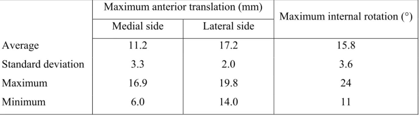
Noyes et al.47 used a single cadaveric lower limb and 10 experienced knee surgeons in order to analyze the knee motions induced when the pivot shift test is performed. The objective of this group’s research was not specifically to determine the differences in grading between each examiner although their results do allow for such comparisons. For the study, the cadaveric knee was intact except for the ACL, which was cut. The femoral head was replaced with a ball and socket joint and the entire limb was mounted on an examining table. Each examiner was told to perform the pivot shift test as they normally would in their practice. Tibiofemoral motion was measured with a six degree-of-freedom instrumented spatial linkage. Table 3.1 presents the full results for the 10 examiners.
Table 3.1 Maximum anterior translation and internal rotation reached by 10 different examiners during the pivot shit test on a cadaveric knee
Adapted from Noyes et al.47
Maximum anterior translation (mm)
Maximum internal rotation (°) Medial side Lateral side
Average 11.2 17.2 15.8
Standard deviation 3.3 2.0 3.6
Maximum 16.9 19.8 24
27
Much variability can be observed in Table 3.1 but a few values are of particular interest. First, the maximum anterior translation induced for the medial tibial plateau varied from 6 to 16.9 mm between examiners. Those examiners who produced the most internal tibial rotation induced the least anterior tibial translation. Therefore, the amount of internal rotation that is applied has a direct impact on the pivot shift and it is recommended to limit the internal tibial torque applied during the test. The amount of anterior subluxation of the lateral tibial plateau also varied, from 14 to 19.8 mm. This variability is especially important in the grading of the pivot shift phenomenon; therefore it is possible that one examiner could have graded it a 2 while another graded it a 3.
While this test has many limitations, most of them related to the fact that a cadaveric limb was used, it does show that different clinicians may obtain very different results when performing the pivot shift test on a same patient. The authors believe this underlines the need for a clinical device able to measure anterior and posterior subluxations of the medial and lateral plateaus under controlled loading conditions to accurately grade the pivot shift and make the test more reproducible.
In a more recent study, Bull et al.50 found that there was very large variation, between knees, in the pivot shift kinematics, despite all evaluations being performed by a single experienced orthopaedic surgeon. External tibial rotation was 13 ± 8° combined with a posterior tibial translation of 12 ± 8 mm.
No studies were found that compared results for a single examiner performing the test on a same patient on different days. While there is reason to believe that under anesthesia the knee will react in the same manner day after day, a same clinician may not repeat the test with the same loads each time. When the patient is not under anesthesia, there is also the added possibility that the leg muscles might affect the pivot shift differently on different days. The muscles may be more or less fatigued or tense on different days, thus restricting
the kinematics in different ways. Such an investigation would be interesting because if this is the case, it would add to the concern with regards to the reproducibility of the test.
3.3 Instrumented knee joint laxity evaluations
Efforts are currently under way to develop quantitative measurement tools for dynamic tests such as the pivot shift test. The static tests, such as Lachman’s test, are much less complex and a number of measurement instruments have been commercially available for several years. Because AP displacement is a component of the pivot shift, this section will present these instruments, which were developed to quantify AP laxity. It will also present instrument for measuring the more recently described rotational laxity.
3.3.1 Anteroposterior laxity
The KT1000
The KT1000, developed to measure the position of the tibia relative to the femur in the sagittal plane, has been commercially available since 198268. Since that time, many studies have assessed its accuracy and reliability and countless others have used the KT1000 as an objective measure of AP laxity to compare outcomes of different procedures for ACL-reconstructive surgery.
The KT1000, shown in Figure 3.2, is comprised of a support for both thighs and both feet to insure that the lower limbs are flexed to approximately 25° and have a similar tibial rotation69. The lateral face of each foot rests against the foot support, resulting in an external rotation of 15-25°. The arthrometer is fixed to the shank using Velcro straps. Two sensors are integrated to the KT1000: one is placed against the patella and the second, over the tibial tuberosity. The tibia is free to move is the AP direction when a load is applied.
29
Figure 3.2The KT1000 installed on a patient’s lower limb.
Taken from Sernert et al.70
Using the handle situated over the tibial tuberosity, the clinician applies an anterior or posterior load to the tibial. Two distinct audible signals indicate when loads of 67 N and 89 N are attained. These are the two loads that are used for the standardized evaluation. A gauge indicates the amplitude of the displacement that is produced between the two sensors as a result of the applied load. The reference position (zero displacement) is set by applying successive loads of 89 N and then releasing until a stable return-to-normal position is obtained.
Daniel et al. 71 were the first to publish results using the KT1000. Their results were obtained from 338 healthy subjects and 89 symptomatic subjects. They obtained average anterior displacement of 5.7 mm for healthy subjects versus 13 mm for ACL deficient subjects. Moreover, 92% of healthy subjects had a difference of less than 2 mm between both knees compared with ninety-six percent of symptomatic subjects. Kowalk et al.72 tested the KT1000’s accuracy in vitro and found it to be good (0.13 mm). They concluded that its accuracy showed great potential for clinical application. Rijke et al.73 showed that the tool