HAL Id: pastel-00004044
https://pastel.archives-ouvertes.fr/pastel-00004044
Submitted on 18 Jul 2008HAL is a multi-disciplinary open access archive for the deposit and dissemination of sci-entific research documents, whether they are pub-lished or not. The documents may come from teaching and research institutions in France or abroad, or from public or private research centers.
L’archive ouverte pluridisciplinaire HAL, est destinée au dépôt et à la diffusion de documents scientifiques de niveau recherche, publiés ou non, émanant des établissements d’enseignement et de recherche français ou étrangers, des laboratoires publics ou privés.
Contributions and Applications to Knowledge Based
Segmentation
Maxime Taron
To cite this version:
Maxime Taron. Registration & Modeling of Shapes with Uncertainties: Contributions and Appli-cations to Knowledge Based Segmentation. Mathematics [math]. Ecole des Ponts ParisTech, 2007. English. �pastel-00004044�
& %
TH `ESE
pr´esent´ee pour l’obtention du titre de
DOCTEUR DE L’ ´ECOLE NATIONALE
DES PONTS ET CHAUSS ´EES
Sp´ecialit´e : Informatique
par
Maxime TARON
Recalage & Mod´elisation de Formes avec Incertitudes
Contributions et Applications `a la Segmentation
avec a priori Statistique
Registration & Modeling of Shapes with Uncertainties:
Contributions and Applications to Knowledge Based Segmentation
Soutenue le 15 novembre 2007 devant le jury compos´e de :
Pr´esident du jury
J´erome Garot
CHU Henri Mondor - Paris XII
Rapporteurs
Michael J. Black
Brown University
Isabelle Bloch
ENST
Alison Noble
University of Oxford
Examinateurs
Herv´e Delingette
INRIA
Renaud Keriven
Ecole des Ponts
Directeurs de th`ese Nikos Paragios
Ecole Centrale de Paris
Marie-Pierre Jolly Siemens Corporate Research
Introduction (Version Franc¸aise) . . . . 15
Contexte et Motivations . . . 16
Contributions . . . 19
1. Introduction . . . . 23
1.1 Context and Motivations . . . 25
1.2 Contributions . . . 27
1.3 Thesis Plan . . . 28
2. Shape Registration . . . . 31
2.1 Introduction . . . 32
2.2 Shape Definition and its Relation to Deformation . . . 35
2.2.1 Shape Representation with Moments . . . 37
2.2.2 Shape Representations with Discrete Geometric Models . . . 38
2.2.3 Shape Representations with Continuous Geometric Models . . . 40
2.2.4 Implicit Representations . . . 42
Distance Transform . . . 43
2.3.1 Global Deformations . . . 46
2.3.2 Local Deformations . . . 48
Free Form Deformations . . . 50
Thin Plate Splines . . . 54
Extracting Similarity transform from Thin Plate Spline . . . 57
2.4 Optimization . . . 58
2.4.1 Optimization Strategies . . . 61
Optimization algorithms . . . 62
2.4.2 Narrow Bands and Distance Reinitialization . . . 63
2.4.3 Similarity Measures . . . 65
2.4.4 Landmark Registration . . . 66
2.4.5 Implementation Remarks . . . 66
2.4.6 Experimental results . . . 67
2.5 Estimation of Registration Uncertainties . . . 69
2.5.1 State of the Art . . . 70
2.5.2 Continuous Formulation: Towards Hessian Matrix . . . 73
2.5.3 Discrete Formulation: Uncertainty propagation . . . 75
2.5.4 Relations between the Hessian approach and Uncertainty propagation . . . 76
2.5.5 Scaling Uncertainty . . . 79
2.5.6 Experimental Results . . . 80
2.5.7 Testing Sequence . . . 81
3. Modeling Shape Variations . . . . 85
3.1 Introduction . . . 86
3.2 State of the Art in Statistical Interpretation of Samples . . . 89
3.2.1 Bayesian Learning . . . 90
3.2.2 Gaussian Assumption . . . 91
3.2.3 Gaussian Mixture Models . . . 93
3.2.4 Non Parametric Density Estimation . . . 97
3.3 Statistical Modeling of Shape Variations . . . 100
3.3.1 Initial Reference Shape . . . 101
3.3.2 Pose Subtraction and Local Registration . . . 102
3.3.3 Average Reference Shape Model . . . 103
3.4 Dimensionality Reduction with Linear Methods . . . 105
3.4.1 Uncertainty-Driven Principal Component Analysis with Uncertainty. . . . 105
3.4.2 Uncertainty-Driven Independent Component Analysis. . . 106
3.5 Variable Bandwidth Non-Parametric Approximation of Deformations . . . 110
3.6 Dimensionality Reduction and Non-Parametric Interpretation of Samples . . . 114
3.7 Conclusion . . . 115
4. Knowledge-Based Segmentation . . . 117
4.1 Introduction . . . 118
4.2 State of the Art in Segmentation . . . 121
4.2.1 Model Free Approaches . . . 121
Manifold-Driven Object Segmentation . . . 126
Manifold-Enhanced Object Segmentation . . . 128
4.3 Manifold-Enhanced Knowledge-Based Segmentation . . . 132
4.3.1 Image-Based Term . . . 133
4.3.2 Shape-Based Term . . . 136
4.3.3 Segmentation with Uncertainties . . . 138
4.3.4 Left Ventricle Segmentation from CT-scans . . . 140
4.3.5 Segmentation of the Corpus Callosum . . . 141
4.4 Conclusion . . . 144
5. Conclusions & Future Directions . . . 147
5.1 Introduction . . . 148
5.2 Contributions . . . 149
5.3 Future Directions . . . 150
Conclusion (Version Franc¸aise) . . . 153
Synth`ese des travaux . . . 154
Perspectives . . . 155
A. Derivation of the Segmentation Image Term . . . 157
B. Discrete Computation of Distance on Anisotropic Grids . . . 159
B.1 Numerical Gradient. . . 159
B.2 Fast Marching . . . 160
B.4 Signed Distance Initialization . . . 165
C. Uncertainty Driven, Kernel Based Shape Modeling for Digits Recognition . . . 167
2.1 From left to right, Delaunay Remeshing [144], Simplex representation [120], Mass Spring model [145]. . . 39 2.2 Examples of Continuous Models representation. From Left to Right, B-spline
curve fitting [76], Fourier surface Representation [171], m-reps [143] . . . 40 2.3 Implicit representation: (a-top) superquadrics [8] (a-bottom) hyperquadrics [35],
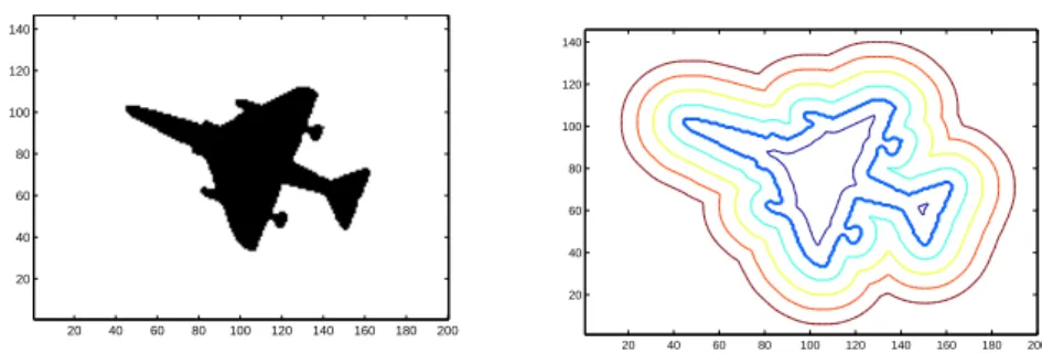
(b) topology change on level-set representation, (c) phase field toppology change [148]. . . 43 2.4 Displaying zero and non-zero iso-contour of the distance transform extracted from
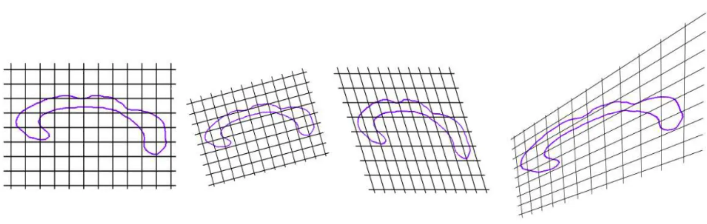
a sample shape of plane. . . 45 2.5 Classic global transformations. From left to right, identity, rotation+scaling, affine,
perspective transformation . . . 48 2.6 Example of Free Form Deformations. (middle) deformations using a 6×10. (right)
grid refinement with the same transformation and a 11 × 20 grid. . . 53 2.7 Example of TPS Transformation of the right ventricle. From left to right: initial
model with 4 control points; 4-points 3D TPS transform (equivalent to an affine transformation); Refinement of the transformation with 25 control points; TPS transform with 25 control points . . . 57 2.8 TPS transformation with 25 control points (Right) and its best affine approximation
(middle) and best similarity transform approximation (right) . . . 58 2.9 Full Registration process on Corpus Callosum with FFD. Registration using
sim-ilarity transform (top right), registration using affine transform (top left), registra-tion using 6 × 4 FFD lattice, registraregistra-tion using 12 × 7 FFD lattice . . . 68
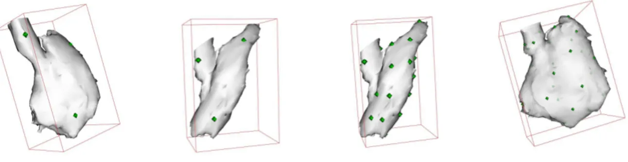
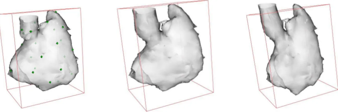
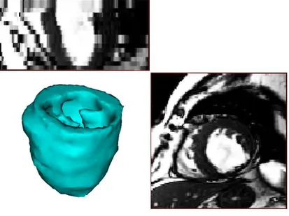
2.10 Representation of the manually segmented left ventricle from MRI data . . . 69 2.11 Full Registration process on cardiac left ventricle. (a) Initial pose. (b) After
au-tomatic initialization process. (c) Similarity transform. (d) 4 control points TPS transformation. (e) 60 control points transform. (f) 90 control points TPS-transform. . . 70 2.12 Projection of the covariance matrix Σ−1
Θ on the grid control points. Ellipses are elongated in the direction of the contour, and larger on distance control points. Ellipses are not represented at control points where uncertainty is too large. . . 80 2.13 Projection of the covariance matrix ΣΘ on the TPS control points. (a, b) Left
ventricle epicardium, (c) Left ventricle endocarium (same pose as (b)). . . 81 2.14 Registration and uncertainty computation on a manually segmented ventricle from
MRI data. Colormap shows the registration error (in mm.), (a) full view, (b) clipped view. (c) Uncertainty ellipses. . . 81 2.15 (a) An example of the registration output: from the reference model to a shape
of the training set (Θi). (b) This registration output with an additional random
perturbation (Θi,perturb). (c) Registration of the reference model on the perturbed
sample (transformation Θi,reg)+ uncertainty representation (Σi,reg). . . 82
2.16 This table shows that computing distances in the vector space of the transforma-tion may only be relevant if uncertainty informatransforma-tion is also used. With the use of uncertainty, the distance between the perturbed sample and its registration is al-ways smaller than the distance between the perturbed sample and the same sample without perturbation. . . 83 2.17 Various samples of registration . . . 84
3.1 Successive steps of the reference shape model building. (a) Mesh extracted from a automatically segmented ventricle with marching cubes, (b) Smoothed and deci-mated mesh, (c) Manual manipulation of the model, (d) the reference shape model with the set of control points describing the deformation . . . 102 3.2 A synthetic example of Independent Component analysis in the 2 dimensional
case. ICA will retrieve the linear function that will transform the distribution into a domain where the two components are statistically independent. . . 108
3.3 Density plot of independent components s = W.Θ, independence and Gaussian mixture estimation of components appears relevant. . . 109 3.4 The sampling of deformation candidates according to the uncertainty evaluation,
as a prior to Independent Component Analysis on the training set. . . 110
4.1 Image Based segmentation of the cardiac left ventricle in CT-images, constraining the transformation to a similarity transform and considering uniquely the endo-cardium (interface between blood pool and muscle). . . 136 4.2 Uncertainty computed on the segmentation result of cardiac left ventricle.
Repre-sentation of its projection on the control points as a set of ellipsoids. . . 139 4.3 The histograms of the greylevels of different areas of the myocardium. . . 140 4.4 Results of the segmentation process. Left: Segmentation of the myocardium
dis-playing the distance to the groundtruth along with a colormap considering four different sample cases outside of the learning database. All results are displayed for a TPS deformation using the ICA shape model with 90 control points. Middle column: The intersection of the segmentation with the data, papillary muscles are correctly segmented. Right: Distribution of errors displayed as histograms. All numerical values are expressed in millimeters. . . 142 4.5 Histograms of the corpus callosum and the background area. The of a use gaussian
mixture to model the corpus callosum and background intensity distribution in MR is appropriate (this figure should be seen in color). . . 143
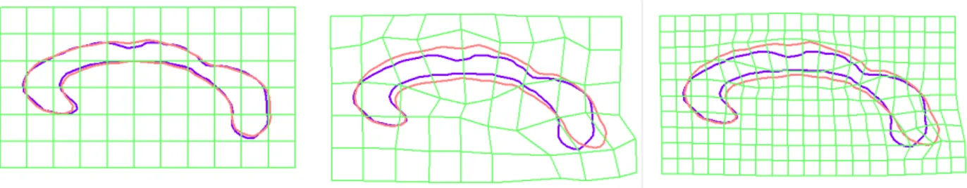
4.6 Segmentation with uncertainties estimates of the corpus callosum; (a) Automatic rough positioning of the model, (b) segmentation through affine transformation of the model (c) segmentation using the local deformation of the FFD grid and uncertainties estimates on the registration/segmentation process (this figure should be seen in color). . . 144
4.7 Additional segmentation results with uncertainty measures. . . . 145
B.1 The 3 types of points in Fast Marching algorithm . . . 161 B.2 The circular list of [211], containing N buckets with size dx. The active buckets
B.3 The rational balls in 3D for a (5 × 5 × 5). Left : the chamfer mask induce a norm on Z3, Left : the mask do not induce a norm on Z3 ( from [67] ) . . . 163 B.4 Decomposition of a Chamfer mask, forward and backward on a 3 × 3 mask . . . . 164 B.5 Sequential computation of the chamfer distance . . . 164 B.6 Initialisation of distance from binary images . . . 165 B.7 Initialisation of distance from an oriented contour . . . 166
C.1 (left) Distribution of the digits 3 and 4 in the space of likelihoods of belonging to
the classes ‘3’ and ‘4’. (middle) Distribution of the digits 4 and 9 in the space of likelihoods of belonging to the classes ‘4’ and ‘9’. (right) Distribution of the digits 3 and 9 in the space of likelihoods of belonging to the classes ‘3’ and ‘9’. . . 168
This Thesis work was performed mostly in Ecole Nationale des Ponts et Chauss´ees, and then Ecole Centrale de Paris. During these years, I was hosted by 3 research laboratories: CERTIS ( ” Centre d’Etudes et de Recherches sur les Techniques de l’Informations et des Syst`emes ” in Ecole de Ponts) , MAS (Applied Maths and Systems lab in Ecole Centrale) and the Imaging and Visualization department of Siemens Corporate Research who sponsored most of this research work. Also a PhD thesis is personal work, none of this would have been possible without the help of some people who counted most in the past 4 years. My greatest thanks go to Pr. Nikos Paragios, my PhD director, who guided me through the research work and helped me in keeping consistent motivation and faith which is the first requirement to go forward in research. Also I would like to thank my co-director in Siemens Corporate Research, Marie-Pierre Jolly who welcomed me in Princeton every summer. I greatly appreciated her constant support and advice on any matters.
Next, I would also like to express my deep thanks to Alison Noble, Isabelle Bloch and Mickael Black for having accepted to review my manuscript despite the important amount of work this required. Many thanks also to J´erˆome Garot, Herv´e Delingette and Renaud Keriven for their participation in my thesis committee.
Beside contributions and working matters, I also received a great amount of support from all my colleagues and friends. I would like to especially mention two of my office mates, Cedric Allene and Noura Azzabou. Also many other colleagues from Ecole des Ponts have counted in my first years of Phd, starting with Renaud Keriven, Geoffray Adde and Olivier Juan who have been a precious relief on technical issues. Jean Yves Audibert gave me crucial hints on the statistical part while Charlotte Ghys brought a very different application to the method presented in this work with the treatment of facial expressions. Also, I would like to acknowledge especially Patrick Etyngier for being my best reference frame in the last month of my Thesis, Radhou`ene Neji for reminding us of the irony of our lives; Stephane Soppera and Emeric Sarron for their friendship and gentle pressure while approaching the end of this thesis.
La plupart des organismes vivants sont munis de centre de perception visuelle de plus ou moins grande complexit´e leur permettant d’interagir avec leur environnement imm´ediat. Le but princi-pal de la vision par ordinateur consisterait donc `a extraire et comprendre le contenu d’images afin de reproduire artificiellement cette capacit´e d’interaction `a l’aide de robots ou de bras motoris´es. Cependant l’interaction physique n’est pas essentielle, un but annexe tend `a utiliser ces techniques afin d’am´eliorer la qualit´e des liens homme/ordinateurs avec des applications li´ees au traitement des images, des flux vid´eo (surveillance, suivi d’objets et structures. . . ) ou encore l’aide au diag-nostic dans le milieu m´edical. Cette probl´ematique d’interpr´etation des images fait donc appel `a plusieurs domaines de recherche tels que la reconnaissance de structures, intelligence artificielle, vision par ordinateur, traitement des images, apprentissage, etc. Plus r´ecemment l’analyse des images m´edicales est apparue comme un axe de recherche important faisant appel `a plusieurs des branches pr´ec´edemment ´evoqu´ees. Depuis quarante ans les travaux de recherche sur ces sujets sont nombreux et touchent des domaines scientifiques aussi vaste que les math´ematiques appliqu´ees, l’informatique, la m´ecanique, la physiologie, biologie et l’anatomie.
Supposant que le syst`eme visuel de l’ˆetre humain est le plus performant (en termes d’acquisition et de traitement), de nombreux travaux ont ´et´e effectu´es afin d’en comprendre son fonctionnement et de pouvoir le reproduire en partie. Pourtant l’´etat de l’art sur la compr´ehension du syst`eme humain d´ecoulant des th´eories de David Marr, reste insuffisant pour pouvoir ˆetre appliqu´e di-rectement et l’essentiel des recherches applicatives de la vision par ordinateur vont principalement se focaliser sur diff´erentes techniques issues des math´ematiques appliqu´ees. De telles approches n´ecessitent la s´election d’un mod`ele (repr´esentation param´etrique du probl`eme) ´etablissant un lien avec les observations (images, donn´ees m´edicales, sorties de syst`emes st´er´eoscopiques ou multi vues, etc.) ainsi que l’utilisation de techniques efficaces pour estimer les param`etres du mod`ele. Il existe donc une infinit´e d’approches possibles pour r´esoudre un probl`eme de perception visuelle. Par ailleurs ces probl`emes sont tr`es souvent mal pos´es car les d´ependances existant entre observa-tions et mod`ele sont non lin´eaires, faiblement contraintes et donc difficiles `a optimiser.
Par ailleurs, le choix de l’approche utilis´ee pour r´esoudre un probl`eme li´e `a la perception vi-suelle est largement justifi´e par le domaine d’application. Par exemple, le positionnement automa-tique et la localisation d’obstacles seront consid´er´es en roboautoma-tique, l’estimation des d´eplacements et le suivi d’objets sont utilis´es pour traiter les flux vid´eos et les techniques de reconstruction ou d’estimation de la position d’objets tridimensionnels sont ´etudi´es lorsque des syst`emes st´er´eoscopique ou monoculaire sont utilis´es. Enfin, des mod`eles statistiques seront souvent utilis´es en traitement des images m´edicales.
Durant les deux derni`eres d´ecennies, nous avons pu observer d’importants progr`es dans les techniques permettant d’examiner les tissus biologiques de fac¸on non invasive. Les derni`eres g´en´erations de scanners fournissent des informations anatomiques et physiologiques ainsi qu’un faisceau de donn´ees li´ees `a certaines pathologies pouvant ˆetre exploit´ees pour un diagnostic pr´ecoce et suivies de mesures th´erapeutiques adapt´ees. La g´en´eralisation des techniques d’imageries m´edicales, l’am´elioration de la qualit´e des images et l’introduction de nouvelles modalit´es ont contribu´e `a pro-duire une large quantit´e de donn´ees devant ˆetre analys´ees. Ceci a rendu `a la fois indispensable et r´ealisable les techniques automatiques de pr´etraitement et autres outils d’´evaluation assist´es par ordinateur sur les images.
L’imagerie m´edicale fait r´ef´erence `a un ensemble de modalit´es d´ecrivant l’´etat des tissus et des os, ainsi que diverse informations physiologiques. Les diff´erentes techniques d’acquisitions parmi lesquelles comptent les Rayons X, Scanner, Imagerie `a r´esonance magn´etique, ´echographie, im-agerie `a tenseur de diffusion, tomographie `a ´emissions de positrons etc. pr´esentent des propri´et´es compl´ementaires. L’exploitation des informations issues de ces modalit´es constitue un d´efi mod-erne pour comprendre l’anatomie, les structures biologiques et les effets des pathologies sur ce qui est visualisable de leur fonctionnement (au travers des d´eplacements, diffusions de mol´ecules marqu´ees, flux sanguin, activit´e neuronale etc.) Finalement, l’aide au diagnostic m´edical peut ˆetre r´esum´e par le traitement de ces signaux multidimensionnels structur´es permettant de comprendre l’´etat des organes ´etudi´es. Une telle probl´ematique est clairement pluridisciplinaire et fait appel `a des domaines scientifiques allant de la biologie aux math´ematiques.
Contexte et Motivations
La mod´elisation de structures anatomiques complexes se d´ecompose g´en´eralement en trois ´etapes: (i) d´eterminer un ensemble de mesures anatomiques et pathologiques obtenues grˆace au traitement et l’exploitation des diff´erentes modalit´es d’imagerie m´edicales. (ii) proposer un mod`ele param´etrique en accord avec les indices anatomiques capable de d´ecrire l’ensemble des variations
de l’organe consid´er´e; (iii) les param`etres de ce mod`ele sont estim´es afin de reproduire le comporte-ment d’une nouvelle observation sur laquelle les mˆemes indices anatomiques et/ou pathologiques sont pr´esents. On peut alors comparer cette nouvelle observation avec celle d’un cas de r´ef´erence, sain, afin d’obtenir des indications aidant au diagnostic. Ce proc´ed´e est g´en´eralement accom-pagn´e par l’´etude d’un mod`ele statistique d´ecrivant les variations de l’organe, construit `a partir d’individus sains et malades afin de le comparer plus efficacement `a une nouvelle observation.
Les techniques de repr´esentation de formes et l’´etude de leurs variations pour des objets appar-tenant `a la mˆeme classe ou a des classes diff´erentes sont des probl`emes universels aux applications multiples. Les principales applications sont la segmentation avec a priori statistique de formes (l’extraction de structures particuli`eres sur des images), le ’tracking’ (suivi d’objets, l’obtention des positions et d´eformations successives d’une structure dans une s´equence d’images), la recon-naissance d’objets (classification permettant de d´ecider si un certain type d’objet est pr´esent dans une image), etc. L’extraction des informations caract´erisant l’´etat d’un organe reste un objectif ambitieux. Il s’agit d’abord d’interpr´eter les donn´ees en provenance des signaux source, et d’en fusionner les informations. Ces informations peuvent prendre la forme de signaux discrets tels que des images, volumes 3D ou 4D, sur lesquels les signaux pr´esentent un certain degr´e de corr´elation `a diff´erentes ´echelles. Retrouver le contenu s´emantique d’une image consiste alors en la descrip-tion de ces mesures `a l’aide d’un mod`ele math´ematique simple dont on estimera les param`etres. Si un tel proc´ed´e peut sembler simpliste, il faut en mesurer la complexit´e au regard des organes `a mod´eliser, pour lesquels la s´election d’un mod`ele math´ematique r´esulte d’un compromis en-tre complexit´e et faisabilit´e. D’une part, on cherchera `a cr´eer un mod`ele capable d’exprimer les diff´erents ´etats de l’organe ´etudi´e; et d’autre part, on d´eterminera les valeurs des param`etres de ce mod`ele uniquement `a partir de nouvelles mesures pouvant s’av´erer incompl`etes. Consid´erons un exemple simple tir´e du cycle cardiaque. Le coeur est un muscle poss´edant deux ventricules. Le ventricule gauche, le plus gros et le plus important agit comme une pompe et envoie le sang enrichi en oxyg`ene vers tous les tissus et organes. Le cycle cardiaque pr´esente deux ´etats essentiels: La fin de la diastole, correspondant `a l’instant o`u les ventricules sont le plus dilat´es, et la fin de la systole, l’instant o`u le coeur est le plus contract´e. La diff´erence entre les deux volumes ventriculaires `a ces deux instants est un indice physiologique important sur le fonctionnement cardiaque. Dans cette optique, l’extraction de donn´ees fait r´ef´erence `a l’obtention du volume de la cavit´e du ventricule gauche donc `a sa segmentation.
D`es lors que ces informations ont ´et´e extraites des images, l’´etape suivante r´esidera dans la s´election du mod`ele en vue de la s´eparation entre les cas normaux et les cas affect´es par diverses pathologies. Ceci est g´en´eralement effectu´e via un mod`ele statistique d´ecrivant les diff´erentes populations. Cette id´ee relativement simple suppose qu’un ensemble de mesures ont ´et´e extraites
sur diff´erents individus et ´etudie les diff´erences existant entre ces mesures. L’objectif de la phase de mod´elisation est de retrouver des comportements analogues entre des individus, qui pourront ensuite ˆetre utilis´es comme autant de r´ef´erences dans l’´etude de nouveaux cas. Ceci reste une vue simpliste de la tˆache `a effectuer ; dans la pratique des mod`eles plus complexes provenant de la th´eorie de l’estimation o`u des statistiques seront utilis´es. Par ailleurs, l’ensemble des observations est contraint par le mod`ele anatomique choisi. Si on consid`ere encore une fois l’exemple du cœur et de la fraction d’´ejection, on utilisera le fait que cette quantit´e pour un cœur sain est d’environ 60 D`es lors que ces mod`eles ont ´et´e d´etermin´es et que leurs statistiques ont ´et´e estim´ees/apprises `a partir des observations, le diagnostic est le r´esultat de la comparaison entre les mesures ef-fectu´ees sur un nouveau cas et les comportements th´eoriques appris `a partir de l’ensemble des cas de r´ef´erence. Cette ´etape n´ecessite encore une fois l’extraction d’informations `a partir d’image m´edicale et a pour objectif d’informer des risques potentiels encourus par le patient. Il faut n´eanmoins ˆetre conscient que le d´eveloppement de telles technologies n’a pas pour but de rem-placer ou marginaliser le rˆole du m´edecin : le traitement des images m´edicales, pour l’aide au diagnostic, doit se limiter `a fournir au m´edecin un panel d’outils appropri´es lui permettant de faire un diagnostic plus pr´ecis, prendre des d´ecisions plus rapidement tout en minimisant le risque pour le patient.
Parmi l’ensemble des ´etapes pr´esent´ees pr´ec´edemment pour l’aide au diagnostic, la plus com-plexe est la construction du mod`ele math´ematique de l’organe ´etudi´e. Cette ´etape a d’ailleurs un impact important sur les autres composantes du processus. Tout d’abord ce mod`ele est utilis´e pour aider `a l’extraction de l’organe `a partir des images biom´edicales ; ensuite, ce mod`ele constitue la base des comparaisons entre les sujets sains et malades. Les m´ethodes pr´esent´ees dans l’´etat de l’art, s´eparent g´en´eralement les ´etapes de mod´elisation d’un organe et l’´etape d’extraction de cet organe dans les images. En d’autres termes, ces m´ethodes consid`erent que l’on dispose d’un ensemble d’apprentissages et d’un mod`ele statistique dont les param`etres sont alors estim´es. Par ailleurs, le recalage constitue aussi un ´el´ement d´eterminant (le recalage est l’´etape durant laquelle tous les ´el´ements de l’ensemble d’apprentissages sont transform´es pour pouvoir ˆetre repr´esent´es dans le mˆeme r´ef´erentiel). Le recalage pouvant aussi ˆetre l’objet d’erreurs importantes, il donne lieu `a des mod`eles de formes erron´es pour lesquels les erreurs d’alignement se sont propag´ees. La s´election d’un mod`ele pour les d´eformations est aussi d´eterminante, il s’agit de cr´eer un mod`ele suffisamment simple pour pouvoir en estimer les param`etres de fac¸on robuste, et suffisamment complexe pour repr´esenter l’ensemble des d´eformations possibles, qu’ils correspondent `a des cas normaux (sains) ou anormaux (ayant subi diverses affections). Enfin ce mod`ele statistique sera con-sid´er´e pour aider au processus de segmentation, dans les cas o`u les informations contenues dans les images m´edicales ne sont pas suffisantes pour r´ealiser une segmentation pr´ecise. L’utilisation
d’un tel mod`ele dans le processus de segmentation est la pierre angulaire de l’aide au diagnostic m´edical.
Contributions
Cette th`ese a permis de traiter les probl`emes essentiels de la vision par ordinateur que constitue le recalage des surfaces (global et local), la mod´elisation de formes, et la segmentation avec a priori de forme. Dans cette optique nous avons consid´er´e une technique de repr´esentation de la forme bas´ee sur le calcul de cartes de distances. Cette technique b´en´eficie de nombreux avantages tant g´eom´etriques que math´ematiques et sera utilis´e tout au long de la chaˆıne de raisonnement.
Le recalage de formes ou de surfaces, est un probl`eme central de la vision par ordinateur, objet de nombreuses ´etudes et souvent utilis´e dans le traitement des images m´edicales. Ce probl`eme se r´esume `a d´eterminer une d´eformation particuli`ere permettant d’´etablir l’ensemble des correspon-dances entre deux formes ou deux ensembles de caract´eristiques extraites sur des images. Ces deux objets ´etant respectivement nomm´es la ’source’ (pouvant ˆetre d´eform´ee) et la ’cible’ sur laquelle la source est d´eform´ee, Dans le cadre de notre ´etude, les ensembles consid´er´es seront des formes repr´esent´es `a l’aide de cartes de distance. La contribution la plus importante de nos travaux r´eside dans l’introduction et l’utilisation des incertitudes dans le processus de recalage. Ainsi, l’action d’aligner deux surfaces n’est plus consid´er´e comme un probl`eme `a solution unique, mais comme ayant une infinit´e de solutions dont la distribution pourrait ˆetre repr´esent´ee par une densit´e de prob-abilit´e multimodale, dans un espace de grande dimension. Une telle approche consid´er´ee au niveau local, dans un voisinage de la solution de recalage obtenue, fournit une information quantitative sur la qualit´e du r´esultat. En d’autres termes, si on consid`ere une surface source align´ee sur une surface cible de fac¸on globale, le recalage local permet d’´etablir un ensemble de correspondances entre les surfaces, et les incertitudes d’introduire un ensemble de matrices de covariance indiquant le degr´e de confiance pouvant ˆetre donn´e `a ces correspondances. Le probl`eme de recalage a ´et´e formul´e de fac¸on tr`es g´en´erique, afin de le r´ealiser nous avons consid´er´e dans un premier temps une classe de d´eformations libres (FFD, ” Free Form Deformations ” ou d´eformations de formes li-bres) permettant de repr´esenter une d´eformation de l’espace ambiant ind´ependamment de la forme ou surface consid´er´ee. Dans un second temps les d´eformations de plaques minces ont ´et´e con-sid´er´ees (TPS, ” Thin Plate Spline ”, ou d´eformations des plaques minces), elles permettent une d´eformation des surfaces et induit une d´eformation de tout l’espace, la particularit´e r´eside dans la position des poign´ees contrˆolant la d´eformation (ou points de contrˆole), plac´ees `a la surface de l’organe ce qui en fait une transformation d´ependant sp´ecifiquement de l’organe, mais ayant une plus faible dimension que les d´eformations libres pour une pr´ecision ´equivalente. Ces
transfor-mations permettent de repr´esenter indiff´eremment des transfortransfor-mations locales ou globales, nous proposons donc d’en s´eparer la composante rigide indispensable dans la phase de mod´elisation.
D`es lors que le probl`eme du recalage a ´et´e r´egl´e, il s’agit de mod´eliser les variations pr´esentes dans un ensemble d’apprentissages. Concr`etement, il faut d´eterminer une densit´e de probabilit´e pouvant repr´esenter ces variations. Notre seconde principale contribution r´eside dans l’approche utilis´ee pour propager les incertitudes ´evalu´ees pendant le recalage dans la phase de mod´elisation. Nous avons d´evelopp´e deux approches ind´ependantes. La premi`ere g´en`ere un mod`ele de faible dimension utilisant les techniques d´evelopp´ees pour l’analyse en composante ind´ependante (ACI) et adapt´e `a la mod´elisation des d´eformations. Cette approche est utilis´ee et h´erite de l’information provenant des incertitudes. Pour cela, l’ensemble d’apprentissages est utilis´e comme une base pour g´en´erer de nombreux nouveaux ´echantillons, de sorte que les exemples avec une forte variance seront plus largement dispers´es, donnant un plus faible degr´e de confiance `a ces variations. Le calcul de l’analyse en composante ind´ependante avec les incertitudes est un mod`ele efficace si les variations des ´echantillons pr´esentent des variations ind´ependantes pouvant s’exprimer comme des mixtures de Gaussiennes. Pourtant une telle contrainte n’est pas syst´ematiquement r´ealis´ee sur des cas pratiques ; afin de pallier ce d´efaut, nous avons propos´e une approche non param´etrique utilisant des noyaux avec une covariance variable afin de mod´eliser les variations des ´echantillons. L’id´ee essentielle consid´erera que les ´echantillons les plus repr´esentatifs seront associ´es `a des variables al´eatoires Gaussienne dans l’espace des d´eformations, centr´ees sur la d´eformation et donc la matrice de covariance d´epend de l’incertitude locale de l’´echantillon consid´er´e.
Ayant ainsi construit deux mod`eles statistiques pour ces d´eformations, ils seront utilis´es pour effectuer une segmentation des organes sur des images m´edicales avec un a priori de forme. Pour cela, nous combinons une repr´esentation implicite des formes (avec des ensembles de niveaux ou level sets), avec des d´eformations param´etriques bas´ees sur un ensemble de points de contrˆole. Les points de contrˆole pourront ˆetre localis´es sur une grille r´eguli`ere (d´eformation de formes libres, FFD) ou sur une surface (d´eformation de plaques minces, TPS). Le mod`ele de moindre com-plexit´e utilise conjointement les TPS et l’analyse en composante ind´ependante pour la segmen-tation du ventricule gauche en imagerie scanner tridimensionnelle. Pour mod´eliser des organes qui pr´esentent des variations plus importantes ne montrant pas de structure et de r´egularit´e, nous avons utilis´e l’approche non param´etrique `a noyaux de taille variable associ´ee aux d´eformations de formes libres (FFD). Le corps calleux a ´et´e utilis´e comme exemple test pour cette approche, la segmentation ´etant consid´er´ee sur des vues sagittales en imagerie `a r´esonance magn´etique. Dans les deux cas consid´er´es, le r´esultat final de la segmentation a une interpr´etation qualitative et quantitative. Ainsi, le recalage tout comme la segmentation sont vues comme des probl`emes d’estimation, les incertitudes apportant des informations relatives aux donn´ees disponibles pour
effectuer cette tˆache (support visuel des images en segmentation et bruit inh´erent aux formes et surfaces pour le recalage). Enfin, ces mesures d’incertitudes contribuent `a la segmentation, dans une approche ind´ependante du choix des param`etres et permettant de retrouver d’importantes vari-ations de formes.
La th`ese suit ce sch´ema et est d´ecoup´ee en trois grands chapitres. D’abord le recalage, intro-duisant la technique de repr´esentation des formes, les diff´erentes transformations, les techniques d’optimisation et d’´evaluation des r´esultats. Ce chapitre introduit donc les mesures d’incertitudes en proposant deux approches compl´ementaires. Le second chapitre touche `a la mod´elisation des formes, introduisant les mod`eles param´etriques et non param´etriques et l’usage des incertitudes dans ces mod`eles. Le troisi`eme chapitre d´ecrit l’utilisation de ces m´ethodes pour la segmenta-tion et introduit les incertitudes dans le sch´ema d’´evolusegmenta-tion du mod`ele d´eformable ainsi introduit. Le dernier chapitre clos la th`ese en introduisant une s´erie de directions `a venir que peut prendre ces travaux. Enfin les appendices successivement le calcul des d´eriv´ees utilis´ees dans la phase de segmentation puis une application important du mod`ele statistique pour une application non m´edicale.
Introduction
Visual perception is a central component for most biological organisms. Despite an important de-gree of variation between natural organisms, even the most primitive element has some abilities to sense the environment through visual sensors. Understanding and extracting content from im-ages and reproducing this ability in artificial environments like intelligent robots, robotic arms, computer aided interpretation of images and video, human computer interaction, computer aided diagnosis, etc. is the primary goal of computer vision. Statistical pattern recognition, artificial intelligence robotics, computer vision as well as image processing and more recently medical im-age analysis are research domains which related visual perception to imim-age understanding. These domains have gained significant attention from the scientific community in the past four decades and now are established and well represented research areas in applied mathematics, computer and engineering sciences, physiology, biology and neurology. One can observe an interdisciplinary effort from scientists with diverse scientific backgrounds towards visual perception.
Under the certitude that the human vision system (acquisition as well as processing) is the most efficient visual perception system, recent efforts in the above mentioned vision related areas aim to reproduce human visual system. However, the current state of understanding of the human brain is far from being reproducible. Therefore, the available knowledge is not sufficient to replicate it, through the aid of sensors and computers complex biological vision systems. The main stream of research over the past decades in the area of computer vision lies mostly in the use of applied mathematics and engineering. In such a context, research scientists have abandoned David Marr’s theories and introduced parametric mathematical inference problem for specific tasks of visual perception, where the set of optimal parameters corresponds to the answer of visual perception. This process requires the selection of a model (parametric representation of the problem), the es-tablishment of a connection between the model and the observations (images, medical data, video,
stereo data, etc.) and the use of efficient strategies to recover the model parameters. One should point out that such strategies have an infinite number of possible solutions since one can formalize the same perception tasks with numerous mathematical models, use various means of introduc-ing the model to data dependencies as well as different techniques exist for the optimization of these dependencies. Furthermore, most of these approaches are ill-posed due to the non-linearity of the dependencies between parameters and observations, as well as due to the lack of sufficient constraints to guide the estimation problem.
Therefore, the definition of a universal solution to the visual perception problem is rather chal-lenging and strongly depends on the task to be accomplished. The task definition is also strongly related to the application area. For example, self-localization and obstacle avoidance are prob-lems often considered in robotics, motion estimation and tracking are studied in video processing, stereo reconstruction and 3D inference are investigated in computer vision, statistical modeling of samples and segmentation at looked at in medical image analysis.
In the recent years, we have observed a revolution on how human and biological tissues can be imaged in non-invasive ways. The latest generation of medical hardware provides anatomical, physiological and pathological data which can be used to perform early diagnosis, follow up and evaluation of therapeutic strategies. Constant improvements on the image quality as well as the introduction of new image modalities have generated an enormous amount of data to be analized. In such a context, the use of computer aided-techniques has emerged as an efficient pre-screening procedure with applications to diagnosis and post-treatment evaluation.
Medical images refer to a set of modalities describing the status of human tissues, bones and physiological information. This may refer to simple or very complex measurements and can be acquired in a number of ways. X-rays, Magnetic Resonance, Computer Tomography, Positron Emission Tomography, Diffusion Tensor Imaging are examples of acquisitions with varying and complementary properties. The exploitation of such an information space is a great challenge of our days and consists of understanding the anatomical structure of biological systems and in par-ticular the effect of pathologies on their complex mechanisms of operation. The task of computer aided diagnosis can be reformulated as follows: processing these n-dimensional signals in order to understand the current state of an organ of interest. Such in-depth modeling and understanding of complex biological systems is an interdisciplinary effort which involves researchers with different scientific backgrounds including physiology, biology, neurobiology, mathematics and engineering.
1.1 Context and Motivations
Modeling complex anatomical structures often consists of three steps; (i) A set of measures, of anatomical and pathological indices are recovered through the processing, understanding and ex-ploitation of medical image modalities, (ii) A (parametric) mathematical model that is consistent with the anatomy is proposed, it is capable of describing the operation (i.e. the statistical variation) of the organ or structure under consideration, and (iii) estimating the parameters of the model so that it can reproduce the behavior observed through the use of anatomical and pathological indices. Then one may compare this behavior with a reference, healthy behavior, to make a soft diagno-sis. Such a task is achieved through a statistical comparison between the model built to describe healthy individuals and the measurements obtained for the subject under consideration.
Shape representation and modeling of its variations inter and intra-class is considered to be a universal problem with applications to knowledge-based segmentation (extraction from images of a particular structure), tracking (recovering successive positions and deformations of a structure in a number of consecutive images), recognition (classification decision for the presence or not of a class of objects in an observed image), etc. Extraction of information regarding the state of an organ from biomedical images is a challenging task. One has to deal first with content interpretation from sparse local signals and then with the fusion problem. Images, volumes, 4D volumes etc. correspond to sampled continuous functions where measurements are only correlated at a local scale, therefore recovering content often consists of describing the measurements using a simple mathematical model and then estimating the parameters of this model using the measurements. While such a process seems very trivial, given the complexity of biological systems the selection of a model which is a compromise between complexity and tractability is not straightforward. On one hand, one would like a model that is capable of explaining the state of the organ; on the other hand, one should be able to determine the parameters of this model from sparse signals. Let us consider a ”simple” example: the heart cycle and in particular the cycle of the left ventricle which pumps oxygenated blood to all human tissues (including the most distant one). The cardiac cycle contains two important phases alternating contraction and dilatation. End-systole and end-diastole are the instants where the ventricle has its lowest and its highest volume. The relative volumetric difference between these instants (called ejection fraction) is a reliable indicator about the heart function. In such a context the task of content extraction refers to the recovery of the volume of the left ventricle.
Once information has been determined from images, the next step consists of determining ap-propriate means to model this information and being able to separate the diseased from the normal
cases. This is often done through statistical modeling of populations. The idea is fairly simple: assuming a set of measurements from different individuals, study how different these measure-ments are. The aim of modeling is to recover a common pattern of behavior for the observations between subjects which then can be used as a gold standard to compare with new cases. This is a rather simplistic representation of the task. In practice complex mathematical models from estimation theory as well as statistical inference are considered to construct these models. The observations are constrained from known anatomical models. In order to demonstrate this task, let us again consider the heart operation. It is known and will be confirmed that the ejection fraction should be about 60%, which means that the blood leaving the heart in every cardiac cycle is about 60% of the end-diastolic left ventricle volume. Eventually this is a rather simplistic example since the measurements refer to a single dimension, while in the most general case one can imagine measurements of high dimension with many indices to be statistically modeled.
Once models have been determined and have been estimated from observations, diagnosis refers to the task of comparing the measurement of the new subject with the expected theoreti-cal behavior of the model. This task involves again content extraction from biomeditheoreti-cal images and aims to produce an indicator of whether the subject under consideration has a potential risk. One should have in mind that this technology does not aim to replace or marginalize the role of physicians. The task of computer aided diagnosis consists in providing more appropriate means of content interpretation in medical images, which will allow faster and more accurate diagnosis, while producing new means for treatment and therapy evaluation.
Efficient mathematical organ representation is among the most challenging problems of the above mentioned processing chain because it has a large impact on all other components of the process. In particular it can be used as an aid to improve organ extraction from images, and also is the base of comparison between healthy and unhealthy subjects. State of the art methods often dis-associate the problem of organ modeling with organ extraction. In other words, given a set of training examples and a choice of the statistical model, they infer the model parameters. Registra-tion itself (bringing all examples of the training set in the same parameter space) is challenging and often erroneous, resulting in models where alignment errors have been propagated. Furthermore, the selection of a mathematical model to account for the variation of samples is also critical. First, one would like a simple mathematical model where robust parameter estimation is feasible. Fur-thermore, a need exists for a model capable of accommodating the variation of all samples while being able to cope with abnormal samples. Last, but not least these models should be considered efficiently to aid the segmentation process if the data support is not available and provide efficient diagnosis.
1.2 Contributions
In this thesis we aim to address the challenges that are surface registration (global and local), mod-eling shape variations (parametric and non parametric models) and knowledge-based segmenta-tion. To this end, we consider a state-of-the art shape/surface representation (distance transforms) with numerous desirable geometrical and mathematical properties which is then used along the proposed chain.
Shape/surface registration is a well studied problem in computer vision, computational geome-try and medical image analysis. The definition of the problem consists of deriving a transformation, which given a source and a target feature space, establishes correspondences between them. In the context of our research, feature spaces correspond to 2D curves and 3D shapes, represented using distance transforms. The main contribution of our work consists of the introduction of uncertain-ties in the registration process. Therefore, the transformation aligning the two surfaces becomes a multi-modal high-dimensional density function with both quantitative and qualitative expression of the process. In other words, given a source and a target representation as well as an initial global alignment between them, we are able to determine the set of correspondences and also associate the result with covariance matrices which indicate the amount of confidence for the obtained re-sult. In order to address generic surface registration this concept is customized for the case of free form deformations (FFD) which is a shape/surface free (domain-defined) representation of dense displacements. Next, we propose a similar estimation of a multivariate deformation density for the case of thin plate splines representations (TPS). This is an organ specific, low-dimensional transformation with control-points being defined on the surface. In order to account for the in-tegrated nature of this transformation (the global and the local are not separable), we propose a rigid-invariant form.
Once the registration problem has been addressed, the next task consists of modeling the vari-ations of the training set, or determining a continuous probabilistic representation of the observed density. The main contribution of our work consists of presenting two alternatives on propagat-ing registration uncertainties to the statistical model describpropagat-ing shape variations. The first con-sists of generating a reduced model that uses independent component analysis (ICA) to model the computed class of transformations. The traditional independent component analysis approach is improved and inherits the registration uncertainties through the augmentation of the sample set according to the observed uncertainties. Therefore, samples with high variance are dispersed and produce statistical shape models which are less confident in these areas. The uncertainty-driven ICA is an efficient computational statistical model if samples can be expressed as a mixture of
Gaussians. However, there are cases where imposing such a parametric constraint is not natural and the observed shape variation cannot be quantified using these models. In order to overcome this limitation, we propose a variable-bandwidth non-parametric approximation of the samples. The central idea behind such an approach is to consider the most representative examples in the training set and associate them with continuous Gaussian densities centered at the deformation with a covariance matrix that depends on the local uncertainty of the considered sample.
With these statistical models in hand, the next task to be addressed is knowledge-based segmen-tation and shape-based classification. Both statistical models are used to impose prior knowledge on the segmentation. We efficiently combine implicit parameter-free representations (level sets), with parametric control-point based deformations either defined on a regular grid (FFD) or con-sidered on a surface grid (TPS). The model with the least complexity associates thin plate splines deformations and independent component analysis and is used to segment the left ventricle in cardiac computer tomography and magnetic resonance images. For organs with larger shape vari-ations we propose the variable bandwidth non-parametric (VBD) approach associated with free form deformations (FFD). The Corpus Callosum is used as a test a case for shape-driven segmen-tation. In both cases, the final segmentation result has a qualitative and a quantitative interpresegmen-tation. Segmentation like registration is viewed as a statistical estimation problem where uncertainties can be determined which encode both the amount of support from the prior model as well as the local image support. Last, but not least in order to improve classification, both the deformation and the uncertainty are considered in such a statistical model which is relatively parameter-free and can capture important variations of the training examples.
The remainder of this document is organized in three main chapters, a registration one, a sta-tistical modeling one, and a segmentation one. Conclusions are part of that last section while non-related medical applications are presented in the appendix along with derivations of cost func-tions which were presented in the main document.
1.3 Thesis Plan
Chapter 2 is dedicated to shape registration which involves three aspects, Shape representations, nature and type of transformations and optimization strategies. We first review the most widely used shape representations and motivate our selection to use distance functions. Once the represen-tation has been established, we discuss/review the nature of deformations in particular focusing on dense parametric registration models. Two models are presented in details: free form deformations and thin plate spline deformations. Then, we briefly review the state-of-the art point-based
regis-tration methods (Iterated Closed Point, Shape Context, and Robust-Point-Matching) and introduce our approach on the space of implicit functions focusing on the explicit estimation of uncertainties which is the main contribution of this chapter for both deformations models being considered.
In the next chapter we first present the state of the art in modeling shape variations which of-ten requires the selection of the parametric model and the estimation of its parameters from the data. In particular we briefly review simple models like principal component analysis, mixture of Gaussians and kernel principal component analysis among others. Then we present in details the independent component analysis approach and then non-parametric density approximations with fixed and variable bandwidths. These models are then modified and enriched with the uncertainty-driven registration results presented earlier leading to more efficient representations of densities which can better capture the samples variation and can perform more efficient samples discrimina-tion/classification.
The last main chapter of the thesis is dedicated to knowledge-based image segmentation. First, we review model-free and model-based image segmentation as well as their applications to com-puter vision. In particular we focus on level-set based methods, as well as active shape models which are the most closely related with our approach. Then we introduce two novel prior models, one based on uncertainty-driven independent component analysis and one on variable metric ker-nels. For both models we consider a state-of-the-art image term which can be adjusted according to the application setting and explicitly determine segmentation uncertainties. The proposed meth-ods outperform complex approaches based on level sets as well as the ones with limited statistical capture (Gaussian assumptions) like active shape and appearance models. We demonstrate the per-formance of these methods using two applications, namely the segmentation of the left ventricle in cardiac images and the segmentation of the corpus callosum in brain images.
Conclusions and discussions are part of the last section of the document presenting first the main shortcoming and limitations of the approaches and then potential future perspectives. The document also contains an appendix where some of the contributions of the thesis are put in ev-idence for other application domains like statistical character recognition, facial animations and image morphing.
To conclude, this thesis evolves around shape, representation, registration, modeling, and image-based shape inference with constraints and well as primarily their applications in medical image analysis. It has produced (up to now), one major journal publication [185], one book chapter [136] two major conference publications [182, 181], two major workshop papers [184, 183] and three US patents (pending).
Shape Registration
Abstract – Shape registration consists of recovering a transformation that establishes certain correspondence between
two structures (curves/surfaces/etc.). The problem is often ill posed because (i) the structures of interest can be repre-sented in various ways, (ii) the set of allowable transformations is infinite, and (iii) the similarity between registered structures can be defined using various metrics. These 3 aspects of the registration problem are often treated as a whole, because the definition of shape takes multiple aspects in the state of the art and may carry deformations. This chapter will propose a definition of shape that meets the requirements of most of the descriptions and representa-tions that will be reviewed. Then we will show the advantage of using an implicit shape representation with a distance transform as an implicit deformable template for registration. The chapter will then present the class of parametric deformations used in the registration process. Finally we will introduce the uncertainty on the registration results as a way of expressing the variability on the retrieved deformation field.
2.1 Introduction
The problem of shape registration can be simply defined as follows: given two instances of the same object, find a transformation that will align them according to some similarity measure. It has been a problem widely studied over the past decades with many related applications. The above-mentioned definition involves three terms, shape instance, shape transformation and shape similarity/disimilarity. Shape model or shape parameterization is often the term used to describe the mathematical model behind the physical object. Such a model aims to capture certain math-ematical properties and often provides a continuous reconstruction. Transformation is the class of plausible geometric deformations which can be applied to the source shape towards improving the alignment of its transformed variant and the target shape. Last, but not least similarity is a mathematical term which defines measures of comparison between the transformed source shape and the target one.
The problem of efficient shape representations arises in numerous scientific domains like com-putational geometry, computer vision, graphics and animations, pattern recognition, comcom-putational and molecular biology, etc. The main aim is to determine an efficient mathematical model with nice computational properties which can be easily extracted and interpreted from a computer. These two desirable properties are often conflicting. Existing representations of shapes can be roughly clas-sified in three categories which are representations of increasing complexity: (i) moments-based, (ii) discrete models, and (iii) continuous models [121]. Moment-based representations focus on the observed local structure of the shape [101]. In such a context one seeks for moments (preferably invariants) which can be either extracted from the object silhouette or from the object area. These moments encode the local characteristics of the shape and do not always retain the global structure. Therefore they are efficient for shape matching, recognition, etc. but have little success when con-sidered for either registration or segmentation due to the fact that the inverse projection between these moments and the original shape is usually not trivial [215]. Discrete approaches exploit shape representations through a limited number of control points and some interpolation strategies, to-wards a complete geometric representation. One can then extract continuous geometric variables from this representation. The use of dense sampling leads to models of finer and finer precision that decreases the importance of the triangulation process, a widely considered technique to link control points. Subdivision surfaces are another example where the same concept is used. The main limi-tation of these represenlimi-tations lies in the fact that for the case of a finite number of control points, an important cross-dependency exists between the reconstructed shape and the sampling rule used to determine the positions of these points [206]. Furthermore, determining the geometric/physical properties of the shape either at a global or a local scale is far from trivial. Continuous models
are a geometric alternative to discrete models where one can recover a continuous/differentiable shape form through the use of interpolation functions. One can differentiate explicit from implicit representations in this category. In the former case, shapes are represented using either a num-ber of control points and a continuous interpolation function or through a linear composition of a set of orthogonal features [37]. These methods are a reasonable compromise between complexity and geometric efficiency, but they suffer from their inability to describe forms with multiple non-connected components. Implicit representations are a useful set of alternatives, where a shape is represented using the zero-level set of a continuous function. These representations inherit some nice invariance properties, are geometrically efficient but computationally expensive [16].
The transformation that relates two instances is in general unrelated to the parameterization that was considered to represent the source and the target shape. In general, one can decompose prior art in global registration and dense alignment [167, 197]. Global registration assumes that all shape elements (point clouds, control points, triangulated surfaces) are transformed with the same parametric model, which in most of the cases is customized to them by being a function of their position. Translation or more complex models like rigid, similarity, affine or homographic are examples of global models [88]. The estimation of the transformation parameters is often a well-posed problem due to its over-constrained nature. Each shape element will provide a constraint while the same unknown transformation parameters are to be determined from all constraints. On the other hand these models fail to capture local deformations and they can be very imprecise if large local changes are observed between the two shape instances. Local deformation models are the most appropriate tools for local alignment. These models assume that either points move individually (extreme case), or they undergo a rich deformation according to a predefined mathe-matical model [191, 172]. In the former case the estimation of the individual deformations is rather ill-posed and therefore additional regularization constraints are to be considered for the recovery of a meaningful solution. Continuous deformation models are an alternative to point-based dense registration. With spline-based deformation, the transformation is expressed as a combination of a limited number of basis functions. In order to account for local consistency, these models also inherit some elasticity properties leading to continuous deformation models [58]. Examples of these models are free form deformations, thin plate splines, finite elements, etc. These models offer a compromise between global and dense registration with the estimation of their parameters being a tractable computational problem. However, one should note that their performance heavily depends on whether or not the selected continuous model is capable of expressing the observed deformation. In particular the use of elasticity constraints which is required towards recovering a geometrically meaningful solution could have an important impact on the quality of the obtained result [209].
The definition of a similarity/dissimilarity metric is strongly related to both the shape param-eterization as well as the nature of the transformation. The Euclidean distance is the most natural metric when considering clouds of points independently of the nature of the transformation [208]. More advanced geometric features including normal, curvature or other higher order moments be-tween shapes can be used when a continuous representation is assumed independently from the nature of the transformation [5]. These methods often decompose the estimation process in two stages, first correspondences between shapes is retrieved and then the transformation that aligns them is estimated [217]. The correspondences are often recovered on an expanded feature space (local curvature, etc.) while the alignment is done using geometric distances. The iterated closest point, the robust point matching and the dual-bootstrap iterated closest point are examples of such methods [13, 175]. Implicit representations of shapes offer natural ways to define similarities di-rectly on this space [109]. Then the need of recovering correspondences is implicitly addressed. In such a context, well known metrics from the space of images have been adopted, like the sum of squared differences criterion, the normalized cross correlation, the mutual information, etc. Furthermore, numerous techniques can be used for optimizing these cost functions, like gradient descent, relation, combinatorial optimization, etc. Most of these methods are quite sensitive to the initial conditions and have no guarantee of convergence to the optimal solution if the deformation model is too dense (two many parameters to be determined).
The review of existing work in the area of shape registration shows that the representation problem is quite well addressed, which is also the case for the deformation models. On the other hand, defining appropriate metric functions that measures the similarities between shapes is still an open problem. The choice of a proper metric is related to every aspect of image registration, that are representation, registration model and similarity. However, it seems that the choice of im-plicit representations can provide enough freedom on exploring various metrics as well as various deformations models. In terms of optimization, all methods presented in the literature are to a certain degree sensitive to noise. Moreover when solving mathematical inference and imposing geometric consistency even the optimal solution to the problem cannot guarantee that the obtained correspondences are the right anatomical ones. One can overcome this limitation through means of providing quantitative and qualitative registration results. In such a context we view registra-tion as a statistical estimaregistra-tion problem when one aims to recover the mean soluregistra-tion and the local uncertainty.
The remainder of this chapter is organized as follows. In section 2 we review shape representa-tions, while in section 3 we discuss prior art on global and local registration. The last introductory section of this chapter describes the state of the art in optimization techniques. Then, we introduce our general registration approach that involves a global and a local component focusing on the
in-troduction of uncertainties. This model is customized to deal with organ-specific problems through the use of a TPS deformation model, as well as generic deformation models through an FFD.
2.2 Shape Definition and its Relation to Deformation
Contours, surfaces, boundaries and their mathematical descriptors often called shape are the key to our world as they are the most important visual features for the identification of most objects. In the scientific literature, various definitions of the term shape exist. All of them agree to limit the shape to the geometry, subtract any color or illumination information and therefore differentiate it from the ’appearance’. A simple definition of the space of surfaces and volumes considers a measurable subset of R2 or R3. However this is too generic, and not efficient from a computational perspective, therefore more specific subsets should be considered in computer vision. The work of Delfour and Zolesio [54] and Charpiat [27] add some constraints to this basic definition to create the set of ’shape of interest’. These definitions are implicitly used in most of the state of the art, but require an explicit and somehow restrictive form to prove the equivalence between common distance functions defined in this manifold.
Let S represent a measurable subset of R2, contained in a ’hold all’ open bounded domain also called the ’image domain’ Ω. It is possible from this point to define the set of smooth shapes C0 (resp. C1, C2). For any x on the boundary of S (∂S) there exist a neighborhood N and a C0 (resp.
C1, C2) scalar function (f ) such that ∂S ∩ N can be represented as the epigraph of f :
∂Ω ∩ N = {x ∈ Ω|f (x) <= 0}.
This definition implies that the boundary of a C1 or C2 cannot have multiple points and is repre-sented by a simple regular curve (or surface in the 3D case). Restricting the shape of interest to the set of smooth shapes is usually considered in most of the literature as it is compatible with the discrete computer world. However considering smooth shapes in the continuous space Rn is
not sufficient as one can design sequences of shapes with constant area and perimeter diverging to infinity. This kind of paradox was formally removed by introducing a constraint on the maximum curvature of the boundary ∂S. This constraint may be released on a finite set of points to allow shapes with sharp angles. From this formal definition, one can extract two particular subsets full
shapes and boundary shapes, which are a practical way to classify the descriptors:
• The full shapes F are defined as the subset of S that satisfy that the closure of their interior
• The boundary shape B is the subset of S such that the shape is equal to its boundary (B = {Ω ⊂ D : ∂Ω = Ω}).
A slightly different approach to the concept of shape relates it directly with its object counter-part which can be presented under several poses and therefore present certain variability. Kendall in 1977 [94], and Dryden and Mardia in 1988 [112] proposed a simple shape representation that consisted of points sets and defined the resulting shape as whatever remains once a simple groups of transformations such as similarity (translation, rotation and scaling) is factored out. Therefore a shape can be defined as the equivalence classes of object views according to such group of transfor-mations. However there is at least three reasons that make the task of identifying shape according to the former definition challenging: (i) 2D images are the representations of objects from the real 3D world, so the 2D problem would take advantage of considering the projective transform rather than similarity. Furthermore, occlusions and degenerative views (different 3D views producing the same 2D projections) make the shape definition non-unique.. (ii) Two shapes perturbed due to noise should present minor local differences and have to be identified as identical. This introduces robustness in the representation and the importance of it also depends on the application. (iii) Re-covering the geometric properties of the shape (not only the representation) is critical since such properties are the core features to registration. This definition of shape plus deformation find its foundation in [193], where the process of identifying similar shapes using deformable grids was introduced. In such a way one can say that the concept of deformable template was born 90 years ago. This idea was then formalised in the 80’s introducing the group action defining the variability within object class in the General Pattern Theory as introduced by Grenander in [75].
It is clear that many tasks in computer vision have to do with the comparison of shapes, having two different shapes, one should be able to say whether these come from the same object. Therefore we denote two categories of shape representations which are used in computer vision:
• Moments also known as descriptors transform a shape into a set of features or 1D functions.
These features are often designed to carry invariant properties with respect to some group of spatial transformation and therefore offer some robustness in identification and shape comparison tasks.
• Geometric Models present a set of features or usually scalars or functional that are used to
reconstruct a unique shape in a continuous manner. For identification purpose, these types of representation are closely related to shape registration. Geometric models are twofold:
Discrete Geometric Models: A shape is represented with a finite set of scalars, a contin-uous shape may be reconstructed given a interpolation framework between the scalar
![Fig. 2.1: From left to right, Delaunay Remeshing [144], Simplex representation [120], Mass Spring model [145].](https://thumb-eu.123doks.com/thumbv2/123doknet/2696673.62921/40.892.122.804.502.671/right-delaunay-remeshing-simplex-representation-mass-spring-model.webp)
![Fig. 2.2: Examples of Continuous Models representation. From Left to Right, B-spline curve fitting [76], Fourier surface Representation [171], m-reps [143]](https://thumb-eu.123doks.com/thumbv2/123doknet/2696673.62921/41.892.204.720.482.649/examples-continuous-models-representation-fitting-fourier-surface-representation.webp)
![Fig. 2.3: Implicit representation: (a-top) superquadrics [8] (a-bottom) hyperquadrics [35], (b) topology change on level-set representation, (c) phase field toppology change [148].](https://thumb-eu.123doks.com/thumbv2/123doknet/2696673.62921/44.892.126.813.362.593/implicit-representation-superquadrics-hyperquadrics-topology-representation-toppology-change.webp)