Alterations of the epigenome in brain cancer: loss of 5-hydroxymethylcytosine.
Par
Carolyne Mary Lowry
Programmes de Science des radiations et imagerie biomédicale
Mémoire présenté à la Faculté de médecine et des sciences de la santé en vue de l’obtention du grade de maitre ès sciences (M. Sc.)
en Sciences des radiations et imagerie biomédicale
Sherbrooke, Québec, Canada Octobre, 2016
Membres du jury d’évaluation
J. Richard Wagner, Programme de science des radiations et imagerie biomédicale Benoît Paquette, Programme de science des radiations et imagerie biomédicale
Raymund Wellinger, Département de Microbiologie et Infectologie © Carolyne Mary Lowry, 2017
[Dédicace] I would like to dedicate this manuscript to all brain cancer patients and to the 130 surgery patients that have given us a cause to fight for.
R
ÉSUMÉAltérations de l’épigénome dans le cancer du cerveau: perte de 5-hydroxyméthylcytosine.
Par
Carolyne Mary Lowry
Programmes de Sciences des radiations et imagerie biomédicale
Mémoire présenté à la Faculté de médecine et des sciences de la santé en vue de l’obtention du diplôme de maitre ès sciences (M.Sc.) en Sciences des radiations et imagerie biomédicale, Faculté de médecine et des sciences de la santé, Université de Sherbrooke,
Sherbrooke, Québec, Canada, J1H 5N4
5Mtéthylcytosine (5mC) est une marque épigénétique qui peut être oxydée en 5-hydroxyméthylcytosine (5hmC) par ten-eleven translocation (TET) oxygénases. Ceci amène à l’étape initiale de la déméthylation de 5mC. Le niveau de 5hmC est plus élevé dans le cerveau par rapport aux autres organes. Par contre, ce niveau a une réduction marquante au cours du développement d’une tumeur cérébrale, notamment le glioblastome multiforme (GBM). Toutefois, il n’y a aucun mécanisme connu pour expliquer cette anomalie. Les objectifs de ce projet étaient de (1) discerner l’implication de la voie de déméthylation obtenu par médiation de TET et (2) d’avoir une compréhension plus profonde de la constitution épigénétique des tumeurs cérébraux. (1) Des cellules U87 ont été incubées avec 5mC, 5hmC, 5-fomylcytosine (5fC) et une co-incubation de 5hmC avec 3,4,5,6-tétrahydro-2’-déoxyuridine (dTHU). Les échantillons ont été récoltés à 0, 24, 48, 96 heures. (2) 130 tumeurs cérébraux (GBM = 79; grade II/III = 51) étaient obtenus directement de chirurgie et mise en suspension dans un tampon d’extraction d’ADN le jour même. Les échantillons d’U87 et de tissu tumoral ont subi les protocoles d’extraction et de digestion d’ADN. Le pourcentage par cytosine (%/C) était calculé par la quantification de 5mC, 5hmC, 5fC, 5-hydroxyméthyluracile (5hmU) et 5-formyluracile (5fU) en utilisant LC-MS/MS. (1) Les incubations d’U87 ont démontrées la possibilité augmenter les niveaux génomique de 5hmC, mais aussi une légère augmentation des niveaux de 5mC. Les niveaux de 5hmC ont accru de 1.9 fois après 96hrs. Par contre, aucune variation n’a été observée dans les niveaux de 5fC. Les incubations de 5hmC et 5fC ont été accompagnées par une élévation des niveaux de 5hmU et 5fU respectivement. L’addition de dTHU avec 5hmC avait diminué l’incorporation de 5hmU par 65%. (2) Dans les tumeurs cérébraux, les niveaux moyens de 5mC, 5hmC et 5fC étaient de 4.0, 0.15 et 0.021%/C respectivement. Les quantités de 5hmU et 5fU étaient grandement plus faible dans le GBMs que dans les tumeurs de bas grades. La présence de 5hmU et 5fU dans les tumeurs et leur augmentation durant l’incubation des U87 indiquent une activité de désamination, qui peut, en particulier, entraver les niveaux de 5hmC. En outre, malgré l’incubation avec 5mC, les niveaux de 5hmC et 5fC n’ont pas augmentés suggérant un dysfonctionnement de TET. L’activité de TET est maintenue dans GBM, mais altérée dans les tumeurs de bas grades à cause de mutation isocitrate dehydrogenase-1 (IDH1). Par conséquent, la forte activité de désamination et la déficience en TET peuvent conduire à une réduction épigénétique.
S
UMMARYAlterations of the epigenome in brain cancer: loss of 5-hydroxymethylcytosine. By
Carolyne Mary Lowry
Science des radiations et imagerie biomédicale Program
Thesis presented at the Faculty of medicine and health sciences for the obtention of Master’s degree diploma maitre ès sciences (M.Sc.) in Sciences des radiations et imagerie
biomédicale, Faculty of medicine and health sciences, Université de Sherbrooke, Sherbrooke, Québec, Canada, J1H 5N4
Methylcytosine is an epigenetic mark, which can be oxidized to 5-hydroxymethylcytosine (5hmC) in DNA by ten-eleven translocation (TET) oxygenases. It is an initial step in the demethylation of 5mC. Levels of 5hmC is relatively high in the brain compared to other organs, but these levels are known to be significantly reduced during the development of a brain tumor, especially in glioblastoma multiforme (GBM). However, no known mechanisms may fully explain this abnormality. The objectives of my project were to (1) understand the implications of the demethylation pathway mediated by TET, and (2) gain a deeper insight in the epigenetic make-up of brain tumors. (1) U87 cells were incubated with 5mC, 5hmC, 5-formylcytosine (5fC) or co-incubated of 5hmC with 3,4,5,6-tetrahydro-2’-deoxyuridine (dTHU) over a timeline of 0, 24, 48 and 96 hours. (2) 130 brain tumors (GBM= 79; grade II/III= 51) were obtained directly from surgery and immediately suspended in DNA extraction buffer. Both cell samples and tumor tissues underwent DNA extraction and DNA digestion protocols. The percent per cytosine (%/C) was obtained by quantification of 5mC, 5hmC, 5fC, 5-hydroxymethyluracil (5hmU) and 5formyluracil (5fU) using LC-MS/MS. (1) Cellular incubations showed that it is possible to increase levels of 5hmC in DNA, but also a slight increase in 5mC levels throughout the experiment. 5HmC levels dramatically increased by 1.9-fold after 96h. On the other hand, no increase was observed in 5fC levels. Both 5hmC and 5fC incubations were accompanied by high increases in 5hmU and 5fU levels respectively. The addition of dTHU to the 5hmC incubation decreased 5hmU incorporation by 65%. (2) The average levels of 5mC, 5hmC and 5fC, in brain tumors, were 4.0, 0.15 and 0.021 %/C respectively. 5HmU and 5fU levels were present at comparable levels of 5hmC and 5fC. Levels of 5hmC, 5hmU and 5fU were significantly lower in the DNA of GBM specimens. There was a strong correlation between 5mC with 5hmC and 5fC in GBM, but this was absent in low grade tumors. The presence of 5hmU and 5fU in brain tumor and the increase in their levels during cell incubations indicate a deamination activity in these cancerous cells, which may impinge on the cellular levels of 5hmC, in particular. Furthermore, upon the incubations with 5hmC, downstream levels of 5fC did not increase suggesting a TET malfunction. TET activity is maintained in GBMs, but impaired in low grade tumors due to isocitrate dehydrogenase-1 (IDH1) mutations. Therefore, in brain tumors, a strong deamination activity and TET impairment may lead to epigenetic reduction of 5hmC.
T
ABLE OF CONTENTRésumé ... ii
Summary ... iii
Table of content ... iv
List of figures ... vi
List of tables ... vii
List of abbreviation ... viii
Introduction ... 1 Problematic ... 11 Objectives ... 11 Hypotheses ... 11 Article 1 ... 13 Abstract ... 16 Introduction ... 17
Methods and Materials ... 19
Cell Culture and Treatment ... 19
Toxicity ... 19 DNA Extraction ... 19 DNA Digestion ... 20 LC-MS/MS analysis ... 21 Statistics ... 21 Results ... 22 Discussion ... 28 Acknowledgements ... 30 References ... 31
Secondary Project (unpublished) ... 36
Materials and Methods ... 36
Tumor Specimens ... 36
Sample Preparation ... 36
Statistics ... 37
Comparison of methyl cytosine related modifications in tumor specimens ... 38
Correlations between modifications ... 40
Discussion ... 42 DNA Hypo/Hypermentylation ... 42 5hmC in brain tumors ... 44 Implications of TET ... 46 Deamination activity ... 48 Conclusion... 51 Acknowledgements ... 52 List of references ... 53
L
IST OF FIGURES IntroductionFigure 1 Cytosine Methylation ... 4
Figure 2 Conversion of 5mC to 5hmC...……….5
Figure 3 TET Oxidative Process……….………6
Figure 4 Base Excision Repair Mechanism ………..……….8
Article 1 Figure 1 Structure of DNA methylation and demethylation intermediates. ... 22
Figure 2 Toxicity 5mC, 5hmC, 5fC, 5hmU and 5fU nucleosides toward U87 cells in culture....……….………...23
Figure 3 Treatment of cells with 5mC ………..………...…24
Figure 4 Treatment of cells with 5hmC ………..………..………...25
Figure 5 Treatment of cells with 5fC ………..……….…26
Figure 6 Treatment of cells with 5hmC in the presence of dTHU ………..……….…27
Secondary Project (Unpublished) Figure 5 LC-MS/MS analysis of 5methylcytosine oxidation products in DNA………..….37
Figure 6 Levels of 5mC in brain tumors. ... 38
Figure 7 Levels of 5hmC and 5fC in brain tumors....………...39
Figure 8 Levels of 5hmU and 5fU in brain tumors………...…40
Figure 9 Correlation between 5mC with 5hmC and 5fC………..41
Figure 10 DNA methylation with the aid of S-adenosylmethionine……..……...…46
Figure 11 Demethylation pathway promoted in GBMs...………..……...47
L
IST OF TABLESL
IST OF ABBREVIATION 5caC 5fC 5fU 5hmC 5hmU 5mC 8-oxo-dG AID α-KG APOBEC BER °C C CDA CNS D DNMTs dTHU G GBM hMTH1 IDH MGMT ROS SAH SAM T 5-carboxylcytosine 5-formylcytosine 5-formyluracil 5-hydroxymethylcytosine 5-hydroxymethyluracil 5-methylcytosine 8-oxo-7,8-dihydro-2’-deoxyguanosine (8-oxo-dG) Activation-induced deaminase α-KetoglutarateApolipoprotein B mRNA-editing enzyme complex Base excision repair
Celsius Cytosine
Cytidine deaminase Central nervous system Deuterium
DNA methyltransferases
3,4,5,6-tetrahydro-2’-deoxyuridine Guanine
Glioblastoma (Glioblastoma Multiforme) Human Mut T homologue 1
Isocitrate dehydrogenase-1
O6-methylguanine-DNA-methyltransferase
Reactive oxygen species S-adenosylhomocysteine S-adenosylmethionine Thymine
TET TDG WHO
Ten-eleven translocation enzyme family Thymidine DNA glycosylase
I
NTRODUCTIONCancer is a known occurrence within the human population. It has drawn much attention in research, and has led to multiple efforts to both better understand it, as well as to find a solution to ease, if not cure, the suffering of those affected. Within the human population, cancer may be caused by various factors, such as endogenous factors, for example reactive by-products of our metabolism, as well as exogenous factors, for instance toxic chemicals and UV radiation (Irigaray et al. 2007).
Whatever the reason may be, cancer as a whole is considered to account for 40% of unnatural cause of death, thus ranking it as the number one cause. In 2015 alone, 196,900 new cases of cancer where diagnosed in Canada (Canadian Cancer Society’s Advisory Committee on Cancer Statistics 2015). The varieties of cancers that may befall an individual are innumerable. There are some that have grabbed much attention throughout much of today’s media. Of these are breast, prostate, lung and colorectal cancers, which, despite the multiple attempts to cure them, account for 51% of all cancers diagnosed (Canadian Cancer Society’s Advisory Committee on Cancer Statistics 2015).
Despite the “fame” of these cancers, there are others just as important, and yet not as well known. One of them being the appearance of a cancerous mass in the brain, or a brain tumor. There was an estimated 3000 new cases of brain cancer within Canada in 2015 (Canadian Cancer Society’s Advisory Committee on Cancer Statistics 2015). In general, brain cancer is known to be a leading cause of cancer related deaths (Soffietti et al. 2002; Van Den Bent 2003). Stats Canada ranks it as the 9th highest mortality rate related to cancer
in general (Canadian Cancer Society’s Advisory Committee on Cancer Statistics 2015). A tumor within the brain or, in general the central nervous system (CNS), may cause various symptoms in patients. The list of symptoms is long, since they vary greatly depending on the location of the tumor. Some of these symptoms may be prolonged headaches, seizures and impairment of the motor and sensory functions (Gril et al. 2010). Thus, their impact on the individual is quite dramatic.
The most common brain tumors are those that grow rapidly and are metastatic in origin. Known as brain metastases, or secondary brain cancer, they develop in individuals
already diagnosed with cancer. The fraction of cancer patients who develop a brain metastases have been considered to be 20-40% of cancer patients (Arnold & Patchell 2001; Soffietti et al. 2002). 40-50% of brain metastases stem from primary lung cancers, whereas another 20-30% arise from breast cancers (Patel & Mehta 2007; Gril et al. 2010). However, these fractions may rise due to advanced neuroimaging techniques. The number of diagnosed brain metastases outweighs the number of primary brain tumors by a 10:1 ratio (Gril et al. 2010; Fortin 2012).
Alternatively, a primary brain cancer stems from brain tissue and develops as a cancer without any requirement of another pre-existing cancer. There are at least a hundred and twenty different specific types of tumors that may develop in the brain, such as anaplastic oligodendroglioma and gliosarcoma (Brain Tumour Foundation of Canada. 2015). However, even though there is a wide range of brain tumors, the most prominent one is the glioblastoma (GBM). Also known as glioblastoma multiforme, it can arise in two different ways; primary GBM known as de novo GBM, or secondary GBM. De novo GBM is considered to be the most common and aggressive primary brain tumor (Mashimo et al. 2014; Crespo et al. 2015). It constitutes about 95 percent of all GBM cases or about 23 percent of all diagnosed primary brain tumors (Ohgaki & Kleihues 2007; Najafi et al. 2012; Sturm et al. 2014; Zadran et al. 2014). They have been classified by the World Health Organization (WHO) as grade IV astrocytomas (Louis & Ohgaki 2007; Crespo et al. 2015). The expected survival duration for patients suffering from ‘primary’ GBM rarely exceeds five years, where the average survival rate is one year with a median of 16 months (Krex et al. 2007; Mashimo et al. 2014; Zadran et al. 2014). As for ‘secondary’ GBM, it arises from lower-grade tumors, which would consist of WHO’s grade II and grade III, which are astrocytomas and anaplastic astrocytomas respectively, and only represent the remaining 5% of GBM cases (Ohgaki & Kleihues 2007; Sturm et al. 2014). Therefore, ‘primary’ GBMs are considered to be most prominent within GBM studies.
Various studies have focused on GBMs with a variety of outcomes. One focus of study pertains to their epigenetic constitution. Epigenetics is a heritable change, which may consist of an addition of a chemical group on DNA that can affect gene expression without directly changing the DNA sequence (Hartl 2011; Hattori & Ushijima 2014). MicroRNA and histone alkylation can also carry epigenetic effects on gene expression (Pfeifer, Gerd.
P.Hahn 2014). Furthermore, DNA methylation is an epigenetic change, which can affect gene expression. DNA methylation occurs predominantly on cytosines (C) that contain a specific chemical bond known as a CpG dinucleotide (Marx 2012). A large number of CpG dinucleotides are in clusters known as CpG islands, where many of these islands a grouped in gene control regions, particularly regulatory and promotor regions (Sidiropoulos et al. 2005).
The methylation of DNA is an enzymatic process that is performed by DNA methyltransferases (DNMTs), which catalyzes the transfer of the methyl chemical group onto the fifth position of C (Figure 1) (Robertson 2001; Grassi 2003; Kim et al. 2012). There are three DNMT enzymes that can perform this reaction: DNMT1, DNMT3A and DNMT3B. DNMT1 functions to maintain DNA methylation patterns. It has the capability to methylate DNA during DNA replication to ensure the same methylation pattern on the daughter strand as found on the original DNA strand (Goffin 2002). DNMT1 is found to be in higher abundance than DNMT3A and DNMT3B, but the latter are no less important. Both DNMT3A and DNMT3B have the ability to methylate DNA de novo, because of their affinity for hemi-methylated or non-methylated DNA (Goffin 2002; Kim et al. 2012). It is important to note that hemi-methylated DNA is when only one of the DNA strands in the double helix is methylated and the other is not. In this state, the single methylation still carries the same DNA activity as unmethylated DNA (Deobagkar et al. 1990). Furthermore, hemi-methylated DNA can lead to de novo methylation as mentioned previously, but also in certain circumstances, it can maintain DNA methylation by recruiting DNMT1 (Nishiyama et al. 2013).
Figure 1. Cytosine Methylation
Cytosine methylation consists of the addition of a methyl chemical group on the fifth carbon of the cytosine ring by either DNMT1, DNMT3A or DNMT3B, thus creating 5mC.
Irrespective of which DNMT implicated in the addition of a methyl group to cytosine, it has been found to create a stable chemical modification known as 5-methylcytosine (5mC) (Bird 2001) (Figure 1). The creation of this fifth DNA base, now allows a transcriptional repression capacity within eukaryotes without affecting the genetic code (Klose & Bird 2006). Alternatively, histone alkylation may also carry an epigenetic effect on gene expression (Feil & Fraga 2012). In the case of 5mC, it has the capability to silence gene expression, thus giving it important roles in genome stability, differentiation and development (Baylin & Jones 2011; Nabel, Manning, et al. 2012; Robertson et al. 2014). Therefore, 5mC is a prominent subject in epigenetic research.
However, since 2009, a new epigenetic mark has attained much attention. 5-Hydroxymethylcytosine (5hmC) may have been mentioned as early as 1972, but it was in 2009, with the simultaneous discovery of high amounts of 5mC in Purkinje neurons and characterization of the ten-eleven translocation (TET) enzyme family, that 5hmC was officially accepted as a new epigenetic mark, thus becoming the sixth DNA base (Penn et al. 1972; Kriaucionis & Heintz 2009; Tahiliani et al. 2009). TET is a part of the family of α-ketoglutarate (α-KG) and Fe(II)-dependent dioxygenases (Tahiliani et al. 2009; Ito et al. 2010). The α-KG, required for TET activity, is created by isocitrate dehydrogenase (IDH1 & IDH2) proteins through oxidation and decarboxylation of isocitrate (Xu et al. 2011). 5mC has been shown to be converted to 5hmC by oxidation mediated by the TET enzymes (Tahiliani et al. 2009) (Figure 2). The specific roles of 5hmC have been greatly debated
amongst scientists. The widely accepted main role of 5hmC is as an important intermediate in the DNA demethylation pathway (Tahiliani et al. 2009; Ito et al. 2011).
Figure 2. Conversion of 5mC to 5hmC
The primary step in the oxidation pathway performed by TET enzymes consist of the oxidation of the methyl group on 5mC creating 5hmC.
The function of the DNA demethylation pathway is to remove the methyl group from the cytosine ring of 5mC, thus restoring cytosine. This process allows the reactivation of genes that were formerly supressed with the presence of 5mC. There are two versions of demethylation activity that can take place; passive and active demethylation. Passive demethylation depends on the down regulation of the DNMT1 enzyme activity. With its activity reduced, the maintenance of the methylation pattern is no longer kept up during DNA replication. Therefore, the levels of DNA methylation gradually decrease with every replication through passive dilution (Lu et al. 2015). Furthermore, passive demethylation may occur with 5hmC. In these circumstances, 5hmC is not recognized by DNMT1, which would prevent the maintenance of DNA methylation (Stroud et al. 2011; Ito et al. 2010). Therefore, passive demethylation may lead to a reduction in 5mC and 5hmC.
On the other hand, active demethylation requires the presence of various enzymes. A key player in the active demethylation pathway is the TET enzyme family. To initiate the demethylation process, as mentioned previously, TET would oxidize 5mC to 5hmC (Tahiliani et al. 2009; Koh et al. 2011). However, the TET oxidation process is a sequential procedure that does not stop at the initial formation of 5hmC. TET can further oxidize 5hmC creating 5-formylcytosine (5fC) finishing with its conversion to 5-carboxylcytosine (5caC) (Gu et al. 2011; Inoue et al. 2011; Ito et al. 2011; Faggio et al. 2011) (Figure 3).
Figure 3. TET Oxidative Process
The oxidation pathway performed by TET enzymes consist of the oxidation of the methyl group on 5mC creating 5hmC. When further oxidized, 5hmC converts to 5fC to then finally become 5caC.
However, the extent of the TET enzyme’s implication comes to an end with 5caC. TET does not have the capabilities to bring the demethylation pathway to a full closure by reinstating cytosine. Therefore, there are other enzymes that are recruited to fulfill this final task. These enzymes belong to the family of base excision repair enzymes, whose well known function is to repair oxidative and alkylation damage to DNA. Base excision repair (BER) involves the initial cleavage of the N-glycosidic and phosphodiester bonds, followed by removal of the abasic site, nucleotide addition by DNA polymerase and ligation. Thereby, 5hmC, 5fC and 5caC, and related modifications, may be removed from the genome and replaced with non-modified cytosine during base excision repair. With 5hmC, it is also possible to form 5-hydroxymethyluracil (5hmU) as an intermediate between 5hmC and cytosine through a deamination process. Generally, 5hmU is known as an oxidative product of thymine (T) created by reactive oxygen species (ROS) (Mouret et al. 1991). 5HmU opposite A in double stranded DNA is removed by BER enzyme known as single-strand selective monofunctional uracil DNA glycosylase 1 (SMUG1) (Abdel-Fatah et al. 2013).
The exogenous and endogenous factors, which tend to promote the development of cancer, are known to create oxidative stress in cells (Irigaray et al. 2007). Oxidative stress, in a general point of view, generates a constant amount of ROS as a result of normal mitochondrial electron transport that maintains cellular ATP levels. Under certain circumstances, there is an increase in ROS levels due to partial decoupling of electron transport or to activate NADPH oxidase and other cellular process during inflammation (Barciszewska et al. 2014). With high levels of ROS present, normal mediators to stabilize
and remove these oxidative agents become overwhelmed. Therefore, it may lead to the creation of various DNA damage, where some of them may approach the levels of epigenetic marks, such as 5hmC and 5fC. In general, the impact on DNA is directed in particular to guanine (G), leading to the creation of 8-oxo-7,8-dihydroguanine (8-oxo-G) (Mikkelsen et al. 2009; Volle et al. 2011). 8-oxo-G damage in DNA can challenge DNA repair processes and lead to mutations (G to T transversion) if the damage is not promptly repaired by the appropriate BER enzymes. The level of 8-oxo-G has been associated with an increase in the formation amyloid deposits within the brain, thus possibly accelerating the progression of Alzheimer’s disease (Davies 2000; Cencioni et al. 2013). Furthermore, oxidative stress tends to affect DNA methylation patterns, which may lead to other health problems such as increased chances of heart failure (North & Sinclari 2012).
The accumulation of 5hmU in cellular DNA may be due to either 5hmC deamination or thymine oxidation. Interestingly, 5hmU is also formed by the enzymatic oxidation of thymine in DNA by TET enzymes (Pfaffeneder et al. 2014; Robertson et al. 2014). Albeit, this reaction is much slower than the one with 5mC. Thus, the primary activity of TET would be directed on 5mC, but the dioxygenase activity of TET is not exclusive to cytosine derivatives.
As a potential pathway in the removal of 5hmC from DNA, 5hmC may undergo enzymatic deamination by activation-induced deaminase (AID) or apolipoprotein B mRNA-editing enzyme complex (APOBEC) (Nabel, Jia, et al. 2012; Burns et al. 2013; Robertson et al. 2014). The deamination performed by either enzyme leads to the problematic formation of a mismatch now present within the DNA similar to a T:G basepair. Therefore, the base excision repair (BER) pathway comes into play. One of the BER enzymes that would be used is thymidine DNA glycosylase (TDG), which can replace 5hmU with a non-modified cytosine (Morgan et al. 2004; Cortellino et al. 2011; Maiti & Drohat 2011; Nabel, Jia, et al. 2012) (Figure 4). The implications of TDG can, also, extend to 5fC and 5caC. It should be noted that TDG does not act on 5hmC:G basepairs, but only on subsequently oxidized forms (5fC or 5CaC) and the deaminated form, 5hmU. The excision of these modifications does not require an intermediate molecule. Indeed, they are preferentially excised by the TDG enzyme, since the enzyme has a higher affinity for these 5hmU:G basepairs than the T:G basepairs (Maiti & Drohat 2011) (Figure 4). Therefore,
with the addition of AID/APOBEC and BER/TDG, the deamination pathway, initiated by TET, would come to a full closure. Thus, DNA demethylation is a dynamic pathway.
Figure 4. Base Excision Repair Mechanism
Base excision repair (BER) pathway is used in the demethylation pathway to reinstate cytosine within DNA. An enzyme used for this mechanism is TDG. It act directly on 5fC and 5caC. However, 5hmC must be deaminated by to 5hmU before BER can function.
5HmC plays a primary role within the demethylation pathway. It is believed that 5hmC is an epigenetic mark because it plays a role in gene regulation similar to its predecessor 5mC. 5HmC has been found to be highly concentrated within enhancer and gene bodies although they are also prominent in promotor regions (Stroud et al. 2011). This may be related to the removal of 5mC that supresses gene expression within these areas. Therefore, enzymatic oxidation of 5mC can activate genes that were previously silenced by
high levels of 5hmC in these regions. However, many more studies will be necessary to understand the full impact of 5hmC in gene regulation (Colquitt et al. 2013; Shen & Zhang 2013). One study has demonstrated that 5hmC does have a gene regulation capacity, but opposite to that of 5mC. It was shown that, when there is a reduction of 5hmC, gene expression is reduced, and when there is an increase in 5hmC levels, expression increases (Jin, Wu, et al. 2011; Colquitt et al. 2013). Colquitt and co-workers have demonstrated this hypothesis using methylated immunoprecipitation to assess 5hmC/5mC ratios, and correlated these with the immunoprecipitation of Olfacotry marker protein expressed in horizontal basal cells, Neurogenin1 expressed in globose basal cells and Intercellular Adhesion Molecule 1 expressed in mature olfactory sensory neurons, where each cell types represented a step of the three stages in neurodevelopment respectively (Colquitt et al. 2013). Hence, 5hmC is a key intermediate in DNA demethylation, but if present it can also enhance gene expression. However, there is still uncertainty of the molecule’s possible alternate roles that it may possess.
Genomic 5hmC is found in cells of all tissues of the human body. However, its levels differ greatly from one tissue to another. For example, the spleen has levels of 5hmC ranging from 0.03-0.06% of C, whereas the heart has levels of 0.15-0.17% of C (Globisch et al. 2010). However, it is well established that 5hmC levels are highest within the CNS, in particular in the brain, which has levels ranging from 0.4-0.7% of C (Kriaucionis & Heintz 2009; Globisch et al. 2010; Li & Liu 2011). Within the brain itself, levels also vary amongst the different regions. Several studies have found that the levels of 5hmC are most abundant in the cortex (Feng et al. 2010; Globisch et al. 2010; Szwagierczak et al. 2010; Jin, Wu, et al. 2011; Li & Liu 2011). Nevertheless, the specific functional purpose explaining the high levels of 5hmC in the CNS is relatively unknown.
Oddly enough, the levels of 5hmC found in various tissues becomes greatly altered during the development of a tumor. The occurrence of hypohydroxymethylation, a reduction in hydroxymethylation levels (5hmC), is a key feature of cancer. Several studies have demonstrated this tendency in cancer (Kudo et al. 2012; Gambichler et al. 2013). For example, levels of 5hmC present in colon cancer tissue are 4-fold lower in comparison with normal tissue (Li & Liu 2011). Similar out-comes can be said for the development of brain tumors. In extreme cases, the levels of 5hmC are reduced by as much as 30-fold within a
brain tumor (Jin et al. 2011). One research group compared GBM cells to cells of normal brain tissue obtained from a brain bank. The results were staggering. Kraus and his associates estimated the percentage of 5hmC in cells that stain positive to antibodies toward 5hmC was 73.5%, 45.2% and 50.1% in healthy cortex, cerebral white matter and cerebellar cortex, respectively. Using an antibody approach to detect 5hmC, they found only 2.23% of 5hmC positive cells in GBM tissue (Kraus, Kolck, et al. 2015). These results magnify the dramatic reduction of 5hmC within GBMs. Furthermore, various studies have demonstrated that not only is 5hmC reduced in cancer, but that these levels may also further decrease as the cancer progresses to a more aggressive form (Kudo et al. 2012; Orr et al. 2012; Takai et al. 2014).
Problematic
The downregulation of TET enzymes in cancer leading to a reduction in 5hmC is a phenomenon observed in various malignancies (Yang et al. 2013; Gambichler et al. 2013; C. Liu et al. 2013). Hence, it may be a partial cause to the progression of certain cancers. Also, some mutations in isocitrate dehydrogenase-1 (IDH1) leading to a decline in TET enzyme activity, would inadvertently lower 5hmC levels as well (Waha et al. 2012). However, the true mechanisms explaining a significant reduction of 5hmC during the development and growth of GBMs, in particular, are still unclear.
Objectives
Objective #1 (Article, main project)
The primary goal of this research was to modulate the levels of 5mC and related modified bases (5hmC and 5fC) within cell culture that was incubated with these modified nucleosides. Using LC-MS/MS analysis the ratio of 5mC, 5hmC and 5fC per 100 cytosine bases allows a comparison of normal cellular levels of these modified nucleosides versus the levels in the cells incubated with these cytosine modifications. The final outcome would allow an insight in the demethylation pathway mediated by TET, which connects 5mC, 5hmC and 5fC together. Thus, any unusual enzymatic activity may be noted.
Objective #2 (Secondary Project)
Within brain tumors, the aberrant levels of 5hmC have been noted often. However, a deeper analysis of not only 5mC, 5hmC, and 5fC, but also the related deaminated derivatives (5hmU and 5fU) could lead to a more profound understanding of the epigenome of brain cancer. LC-MS/MS analysis of these modified nucleoside levels within low grade astrocytomas (grade II and III) and GBM (grade IV) tumors would demonstrate any variations in their levels and comparisons between the two tumor types would deepen the understanding of how GBMs epigenetically differ from its fellow brain tumors.
Hypotheses
Hypothesis #1
During the cellular incubation experiments, it is expected that 5mC, 5hmC and 5fC would be readily incorporated into the genomic DNA. Hence, the levels of these modifications
would increase. Therefore, with their initial levels higher than normal, it was anticipated that the other molecules further down the demethylation pathway would increase as well. Hypothesis #2
Whilst comparing the levels of 5mC and its potential intermediates within low grade astrocytomas (grade II and III) and GBM (grade IV) tumors, we expected to find the signature low levels of 5hmC as it has been reported in various studies. However, we may expect the levels of the alternate 5mC modifications to be abnormal, for example, levels of 5fC may be higher or those of 5hmU due to aberrant enzymatic activity in the demethylation pathway.
A
RTICLE1
Modulation of genomic 5-hydroxymethylcytosine and its demethylation derivatives upon incubation in cell culture with related modified nucleosides.
Authors: Carolyne Mary Lowry, Laurent-Olivier Roy, David Fortin & J. Richard Wagner
Status of the article: Submitted to PLOS ONE on July 27th 2016
Prologue: For the cellular experiments, I performed the 5hmC, 5fC and 5hmC + dTHU. Also, I performed the DNA extraction, digestion and the subsequent LC-MS/MS analysis. Furthermore, I wrote the initial draft of the manuscript. Laurent-Olivier Roy taught me the necessary cell culture technique for cellular incubation with the modified nucleosides. In addition, he has performed the toxicity assay and the 5mC cell incubation. Dr. David Fortin allowed me access to his level 2 laboratory for the cell studies along with kindly providing all required materials, i.e. cell media, and the U87 cell line. Finally, Prof. J. Richard Wagner taught me the use of the LC-MS/MS, and being my supervisor, he also helped me in the revision of the manuscript prior to submission.
Résumé : 5-Hydroxyméthylcytosine (5hmC) est une marque épigénétique nouvellement caractérisé. Celle-ci est le résultat de l’oxydation enzymatique de la 5-méthylcytosine (5mC) dans l’ADN par la famille des enzymes ten-eleven translocation (TET). Cet évènement peut expliquer une régulation diversifiée de certains gènes. 5HmC est présent dans tous les tissus de mammifères, mais ses niveaux sont plus élevés dans le cerveau. Dans le développement et la croissance de nombreux cancers humain, particulièrement le cancer du cerveau de niveau maligne, les niveaux de 5hmC sont largement réduits par des mécanismes encore inconnus. Dans l’ADN des cellules U87 en culture, les niveaux de 5hmC, ainsi que plusieurs modifications correspondantes (5mC, formylcytosine (5fC), 5-hydroxyméthyluracile (5hmU) et 5-formylcytosine (5fU)), sont suivi de près par LC-MS/MS lors d’une incubation avec 5mC, 5hmC et 5fC nucléosides purifiés. Nos résultats démontrent qu’il est possible d’augmenter le niveau génomique de 5hmC dans l’ADN cellulaire lors d’une incubation avec le nucléoside 5hmC, mais l’augmentation est faible
(environ 1-2 fois). Étrangement, l’incubation avec 5hmC a également conduit à une réduction du niveau d’origine génomique de 5fC. Cependant, durant l’incubation avec 5hmC ou 5fC, il y a une incorporation massive des dérivés d’uracile (5hmU et 5fU). Ce qui est intéressant, ceux-ci sont maintenus à des niveaux élevés après 3 jours d’incubation. Toutefois, lorsque les cellules ont été co-incubées avec un inhibiteur de l’activité de cytosine désaminase, 3,4,5,6-tétrahydro-2’-déoxyuridine, l’incorporation initiale de 5hmU dans l’ADN cellulaire a été réduite de 65%. Ces résultats indiquent que la désamination de 5hmC bloque son incorporation dans l’ADN cellulaire tout en conduisant à l’accumulation du dérivé 5hmU correspondant. Cette voie peut expliquer la présence de niveau élevé de 5hmU dans l’ADN cellulaire dans certaines conditions.
Modulation of 5-hydroxymethylcytosine and its demethylation
intermediates in cellular DNA by incubation with related modified
nucleosides
Carolyne Mary Lowry1, Laurent-Olivier Roy2, David Fortin3, J. Richard Wagner1*
1Départment de médecine nucléaire et radiobiologie, Faculté de médecine et sciences de la
santé, Université de Sherbrooke, Sherbrooke, Québec, Canada
2Département de pharmacologie, Faculté de médecine et sciences de la santé, Université de
Sherbrooke, Sherbrooke, Québec, Canada
3Service de neurochirurgie, Département de chirurgie. , Faculté de médecine et sciences de
la santé, Université de Sherbrooke, Sherbrooke, Québec, Canada
Key words: DNA damage, DNA repair, ten eleven translocation, formylcytosine, 5-hydroxyuracil, 5-formyluracil, tetrahydrouridine
Abstract
5-Hydroxymethylcytosine (5hmC) is a newly characterized epigenetic mark arising from the enzymatic oxidation of 5-methylcytosine (5mC) in DNA by the ten-eleven translocation (TET) enzyme family. This event can explain differential regulation of certain genes. 5HmC is present in all mammalian tissues, but its levels are the highest within the brain. In the development and growth of many human cancers, particularly malignant brain cancer, the levels of 5hmC are greatly reduced due to as yet to unidentified mechanisms. Here, the levels of 5hmC as well as several related modifications (5mC, 5-formylcytosine (5fC), 5-hydroxymethyluracil (5hmU) and 5-formyluracil (5fU)) in the DNA of U87 cells in culture was monitored by LC-MS/MS upon incubation with purified 5mC, 5hmC and 5fC nucleosides. Our results demonstrate that it is possible to increase the genomic level of 5hmC in cellular DNA upon incubation with 5hmC nucleoside albeit the increase is modest (about 1-2 fold). Interestingly, incubation with 5hmC also led to a reduction in the level of genomic 5fC. Most strikingly, however, incubation with either 5hmC or 5fC led to massive incorporation of the corresponding uracil derivatives of 5hmC and 5fC, i.e., 5hmU and 5fU, respectively. The steady state level of 5hmU and 5fU remained elevated after 3 days incubation. The initial incorporation of 5hmU into cellular DNA was reduced by 65% but not entirely when cells were co-incubated with an inhibitor of cytosine deaminase activity, 3,4,5,6-tetrahydro-2’-deoxyuridine. These results indicate that the deamination of 5hmC blocks its incorporation into cellular DNA while leading to the accumulation of the corresponding 5hmU derivative into DNA. This pathway may explain the presence of high levels of 5hmU in cellular DNA under certain conditions.
Introduction
The methylation of DNA by DNA methyltransferases (DNMTs) converts cytosine to 5-methycytosine (5mC), predominantly within CpG dinucleotides leading to 60-90% methylation of these sites in mammals (1,2). Cytosine methylation is a well-known epigenetic mark that regulates the expression of genes during development and maintains genetic stability thereafter (3,4). Although the presence of 5-hydroxymethylcytosine (5hmC) was reported in the 1970’s (5,6), this modification was only recognized as a novel epigenetic mark related to 5mC in 2009 (7,8). 5HmC arises from the enzymatic oxidation of 5mC by ten-eleven translocation (TET 1-3), a family of α–ketoglutarate (α-KG) and Fe(II)-dependent dioxygenases (8–10). The oxidation of 5mC to 5hmC is the initial step in the demethylation pathway mediated by TET, which ultimately converts 5mC back to cytosine. In this way, TET can directly alter gene regulation by reversing the effects of species and tissue specific gene methylation. In comparison to 5mC, 5hmC also accumulates to relatively high levels in the genome, displays a different distribution in the genome, and possesses a unique set of DNA and protein binding partners (11,12). A large proportion of 5hmC in the brain lies in gene-bodies (13–15) with substantial accumulation of 5hmC at non-CpG sites (16). Indeed, 5hmC appears to promote expression or act as a transition state between active and non-actively transcribed genes (15,17–19).
The mechanism of active demethylation from 5hmC to cytosine is not clear. TET enzymes have the ability to perform iterative oxidation of 5mC, thereby generating 5hmC as well as further oxidation products: 5-formylcytosine (5fC) and 5-carboxycytosine (5caC) (10). The latter two modifications are substrates for thymine DNA glycosylase (TDG), which has been implicated in the demethylation of 5mC (10,20–23). An animal knockout of TDG is also lethal at early stages of development (24,25). Recently, TDG, TET and DNA containing 5mC was shown to form a tight complex and thereby transform 5mC to cytosine in association with base excision repair (26). Alternative mechanisms of demethylation have been proposed. Uracil-DNA glycosylase UNG2 is implicated in the removal of 5caC, specifically activating the transcription of a methylation-silenced reporter gene (27). Finally, several groups have proposed the deamination of 5hmC to 5hmU as an initial step in demethylation by activation induced deaminase and apolipoprotein B mRNA-editing enzyme complex family of cytidine deaminases (AID/APOBEC) (28,29). The
co-expression of both TET1 and AID/APOBEC promoted the demethylation of 5hmC in cultured cells and adult mouse brain (21). Following deamination, 5hmU:G mispairs can be repaired by single-strand-specific mono-functional uracil DNA glycosylase 1 (SMUG), methyl binding domain protein 2 (MBD2) and TDG (30). Deamination was shown to amplify the removal of 5mC in densely methylated CpG loci by long patch BER and non-canonical mismatch repair processes (31). A functional role of AID deaminases in 5mC demethylation though has been questioned because of the lack of adverse effects of AID-deficiency during development in mice and humans (32,33) and the absence of 5hmU at potential sites of 5hmU-G mismatches in genomic DNA (34,35).
The relatively high level of 5hmC in the brain compared to other tissues points to an important function of this epigenetic mark in the central nervous system possibly as a means to enhance brain plasticity (36–38). Numerous studies have reported changes in the global level and distribution of 5hmC in the genome with the onset and pathology of neurological diseases (34). During the development of malignant brain tumors, the levels of 5hmC have been reported to fall dramatically as much as 30-fold in extreme cases (37). Furthermore, changes in the levels of 5mC and 5hmC have been correlated with the development and growth of glioblastoma, the staging of tumor progression, and patient survival (39). The underlying mechanisms for the reduction of 5hmC levels in brain cancer remain largely unknown. In an attempt to better understand the mechanism of 5mC demethylation, we have incubated immortalized human glioblastoma cells in culture with 5mC, 5hmC and 5fC as modified nucleosides. Our results indicate that the modified nucleosides are taken up into cells but that they are not directly incorporated into cellular DNA; rather they undergo deamination to the corresponding uracil derivatives that are efficiently incorporated into DNA.
Methods and Materials
Cell Culture and Treatment
U-87 MG cells (human glioblastoma, ATCC® - HTB-14) were cultured in the
presence of 5% CO2 at 37°C with Eagle’s Minimum Essential Medium (EMEM)
supplemented with 10% Fetal Bovine Serum (FBS), 1mM of both sodium pyruvate and non-essential amino acids, and penicillin and streptomycin (100UI/mL - 1× penicillin/streptomycin). Incubations with 5mC, 5hmC, or 5fC were carried out at a concentration of 36µM. Cells were collected usually after three time points: 0h, 24h 48h and 96h. The concentration of 3,4,5,6-tetrahydro-2’-deoxyuridine (dTHU) used in co-incubation experiments was 100-400µM. Cells were detached using 0.05% trypsin-EDTA, centrifuged and washed with phosphate buffered saline (PBS).
Toxicity
The toxicity of modified nucleosides was examined for U87 cells by the addition of 5mC, 5hmC, and 5fC, 5hmU and 5fU (0-200µM). The compounds were dissolved in water and filtered through a 0.22 micron filter to sterilize the solution before addition to cells in culture medium. After treatment, the surviving fraction was determined using the 3-(4,5-dimethylthiazol-2-yl)-2,5-diphenyltetrazolium bromide (MTT) viability/cytotoxicity assay. Briefly, 7500 cells/100µL/well were seeded in 96-well plates and allowed to adhere overnight. The culture medium was removed, cells were washed with PBS, and phenol-red free culture medium containing various concentrations of compounds was added. Ten µL/well of MTT reagent (5mg/mL) was added and the plate was incubated at 37ºC for 4h with light stirring. Then, 65µL of medium/well was withdrawn followed by the addition of 50µL/well of Triton X-100: isopropanol (1:10 ratio). The plate was returned at 37ºC for 4h with moderate/high stirring. Cell survival was estimated by the absorption at 570nm with a wavelength correction at 690nm using a PowerWave×340 and the KC junior software (Bio-Tek instruments).
DNA Extraction
Cell pellets containing ~1 million cells were suspended in extraction buffer (10mM Tris-Cl, 5mM EDTA-Na2 and 0.10mM desferrioxamine, pH 8.0) and sodium dodecyl
freeze-thaw cycles with liquid nitrogen. RNA was digested in the cell lysis mixture by the addition of 20µL of a freshly prepared solution RNase solution (Sigma R6513; 1mg/mL in 10mM Tris-Cl, 1mM EDTA-Na2, 0.1mM desferrioxamine, pH 8.0). Subsequently, protein
was broken down by the addition of a 15µL solution of a freshly prepared solution of Qiagen Protease (30AU) No. 19157; 1mg/mL concentration dissolved in Tris-Cl buffer, pH 7.5), which was incubated for 2h in a water bath at 37°C with vortexing every 20 min. Finally, DNA was precipitated from the mixture by the addition of 600µL of a saturated solution of NaI solution (7.6M NaI, 40mM Tris-Cl, 20mM EDTA-Na2 and 0.1mM
desferrioxamine), followed by the addition of 1mL of 2-propanol. The Eppendorf tube was inverted by hand until the aqueous and isopropanol phases merged together. To optimize the precipitation of DNA, the sample was placed in the freezer overnight at -10ºC. DNA was precipitated by centrifugation for 30 min at 4°C, and the supernatant was removed and discarded. The DNA pellet was washed twice, first with a solution 50% 2-propanol in water, and then with a solution of 70% ethanol in water. Each wash was followed by centrifugation at 10,000g for 10 min. The DNA pellet was dried by turning them upside down on a paper towel in ambient air for 30 min.
DNA Digestion
DNA pellets were dissolved in water containing desferrioxamine (0.1mM) and 5-hydroxycytosine (10µM) to prevent DNA artificial oxidation. Labeled internal standards was added from a standard mixture (5µL) containing 5mC (30μM), 5hmC (0.2μM), 5fC (0.2μM), 5hmU (0.5μM), 5fU (0.5μM) and 8-oxo-7,8-dihydro-2’-deoxyguanosine (8oxoG; 0.2μM). The digestion of DNA to its component nucleosides was accomplished in two steps. First, we added 5µL of 50mM sodium acetate buffer (pH 5.0) and 5µL of a stock solution of P1 nuclease (Sigma N8630; 1U/µL concentration in 50mM acetate buffer, pH 5.0). The mixture was incubated at 50˚C for 30 min. In a second step, we added 10μL of Tris-Cl buffer (250mM) and 5µL of snake venom phosphodiesterease (Sigma P3134; 0.002 U/µL concentration in 50mM Tris-Cl, pH 8.0) plus 5μL of alkaline phosphatase (calf intestine, Roche Diagnostics, 10713023001; 1U/µL in buffer provided by the manufacturer). The samples were incubated at 37°C for 60 min. Finally, 10μL of 0.22M phosphoric acid and 40µL of chloroform were added. The sample was briefly vortexed and
centrifuged for 10 min. The aqueous phase containing enzymatically hydrolyzed DNA was removed and stored at -20ºC before analysis.
LC-MS/MS analysis
The analysis of modified nucleosides was carried out using a HPLC system (Shimadzu) connected to a tandem MS instrument (API-3000, AB-Sciex). The chromatographic separation was achieved with an AcclaimTM PolarAdvantage II C18
(2.1mm x 250mm, 3µm) column maintained at 30°C. The modified nucleosides eluted from a gradient of ammonium formate buffer (5mM, pH 5.0) and 90% acetonitrile that started at 10% and increased to 30% acetonitrile within 15 min at a constant flow rate of 160μLmin-1. Twenty µL injection volumes from a total volume of 90µL for each sample
were employed. The MS parameters were optimized for the detection of each modified nucleosides as reported previously (40). Data collection and peak integration were performed using Analyst 6.2 software (AB-Sciex). The concentration of modified nucleosides was calculated from the ratio of integration areas obtained from the natural and corresponding isotopically labeled internal standard. The levels of the 5hmC, 5mC, 5fC and 5hmU per 100 non-modified cytosine residues were calculated based on the concentration of modified nucleoside (MS analyses) and the concentration of non-modified nucleosides (C, T, G, A) monitored by UV absorption at 260nm.
Statistics
Statistics were performed using the student t-test. The parameters used were a 1 tail, type 2 t-test within excel 2013 program.
Results
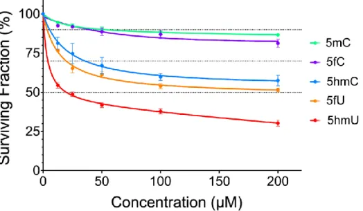
To assess the incorporation of modified nucleosides into cellular DNA, U87 cells were treated in culture medium with various nucleosides in combination with LC-MS/MS to measure the amount of 5mC, 5hmC and 5fC modifications in DNA (structures are shown in Fig 1). In addition, 5-hydoxymethyluracil (5hmU) and 5-formyluracil (5fU) were monitored to access the possibility that cytosine derivatives undergo deamination. Treatment of cells with either 5mC or 5fC showed minimal toxicity even at relatively high concentrations (200µM; <20% cell death; Fig 2). In contrast, treatment with 5hmC led to 40%-50% cell death at 200µM. We chose to incubate cells with a concentration of 5mC, 5hmC and 5fC of 36µM corresponding to no higher than 35% cell death for all three derivatives of deoxycytidine. The toxicity of the modified derivatives of 2’-deoxyuridine, 5hmU and 5fU, was also examined; they showed greater toxicity than the corresponding 2’-deoxycytidine derivatives.
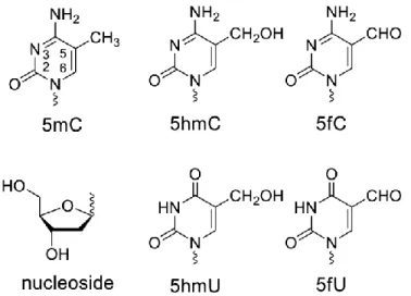
Fig. 1. Structure of DNA methylation and demethylation intermediates. Cells were incubated with the nucleoside (structure attached to a 2-deoxyribose moiety) and the modifications were assessed in cellular DNA as the nucleoside by LC-MS/MS.
Fig. 2. Toxicity 5mC, 5hmC, 5fC, 5hmU and 5fU nucleosides toward U87 cells in culture.
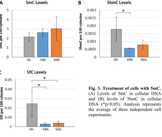
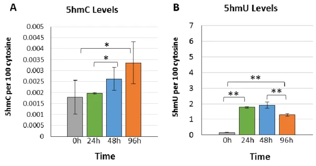
The percentage of endogenous 5mC to cytosine in the DNA of U87 cells was on average about 2%. Upon treatment with 36µM 5mC, there was no significant change in the level of 5mC in cellular DNA (Fig 3A). Upon incubation with 5mC nucleoside, the levels of 5hmC significantly decreased by 2.9-fold (p<0.05) to the levels observed in the control group (Fig 3B). Similar effects of 5mC incubation were also observed with the levels of 5fC, where there levels diminished by 8.7-fold and remained 6.5-fold lower than at 0h (Figure 3C). Thus, both 5hmC and 5fC decreased in cellular DNA upon incubation of U87 cells with 5mC nucleoside.
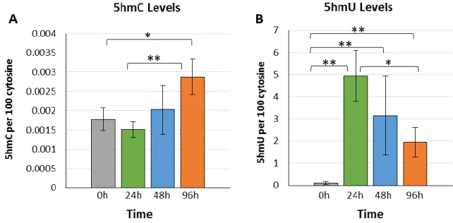
Next, we performed incubations with 5hmC nucleoside. The results showed a progressive increase of 5hmC in DNA giving a 1.9-fold increase after 96h incubation compared to the levels at initial and 24h incubation periods (Fig 4A). Interestingly, the levels of 5fC in DNA decreased at 48h (0.0065%) and remained low at longer times (0.014%) compared to the initial levels of 0.024% of 5fC per cytosine in DNA. A surprising level of incorporation was observed for the deamination product of 5hmC: 5hmU (Fig 4B). The amount of 5hmU in DNA increased from a basal level of 0.10% to 4.94% 5hmU per cytosine (49-fold; p<0.01) within the first 24h of incubation. After 24h, the levels of 5hmU in cellular DNA decreased to about half, indicating removal of this modification as a function of incubation (Fig 4B). The levels of 5hmU in cellular DNA remained high at about 2% 5hmU per cytosine even after 96h.
Fig. 3. Treatment of cells with 5mC. (A) Levels of 5mC in cellular DNA and (B) levels of 5hmC in cellular DNA (*p<0.05). Analysis represents the average of three independent cell experiments.
Fig. 4. Treatment of cells with 5hmC. (A) Levels of 5hmC in cellular DNA and (B) levels of 5hmU in cellular DNA. Error bars indicate the standard deviation from three independent cells experiments (*p<0.05; **p<0.01).
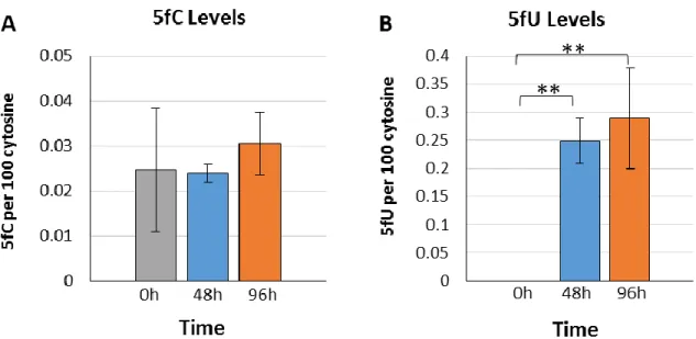
Following incubation studies with 5mC and 5hmC, we turned our attention to 5fC nucleoside. For this modification, there was no variation in the percentages of either 5mC or 5hmC incorporation into cellular DNA. Likewise, the levels of 5fC did not increase during incubation (Fig 5A). However, the levels of its deaminated derivative, 5-formyluracil (5fU) increased from nearly undetectable levels (~0%) to notably high levels of 0.25% within the first 48h of incubation (Fig 5B). The levels of incorporation of 5hmU into cellular DNA were about 20-fold higher than those observed for 5fU after exposure to the same concentration of 5hmC and 5fC, respectively (comparing Fig 4B and 5B).
Fig. 5. Treatment of cells with 5fC. (A) Levels of 5fC in cellular DNA and (B) levels of 5fU in cellular DNA. Error bars indicate standard deviation from three independent cells experiments (*p<0.05; **p<0.01).
Because 5hmU is generated from the deamination of 5hmC, we examined the effect of a well-known inhibitor of cytidine deamination activity, 3,4,5,6-tetrahydro-2’-deoxyuridine (dTHU) (39–42). The inhibition of cytidine deaminase activity in U87 cells reached a plateau at concentrations of dTHU above 200µM. When cells were incubated with both 5hmC (36µM) and dTHU (200µM), the levels of 5hmC in cellular DNA increased from about 2 to 3.3% of 5hmC per cytosine (Fig 6A). This effect was very similar to our previous results using only 5hmC nucleoside (Fig 4A). The addition of both 5hmC and dTHU also led to a substantial increase of 5hmU in cellular DNA within the same time frame (Fig 6B). A comparison of the effect of 5hmC with and without dTHU indicates that inhibition of cytidine deaminase activity by dTHU reduced the initial incorporation of 5hmU by about 65% at the 24h time point although the reduction was smaller at later time points.
Fig. 6. Treatment of cells with 5hmC in the presence of dTHU. (A) Levels of 5hmC in cellular DNA and (B) levels of 5hmU in cellular DNA. Error bars indicate standard deviation from three independent cells experiments (*p<0.05).
Discussion
Numerous nucleoside derivatives are genotoxic because they resemble natural nucleic acids in structure and become incorporated into cellular DNA. For example, 5-aza-2’-deoxycytidine and related nucleosides are taken up into cellular DNA, thereby inhibiting DNA methyltransferase activity, decreasing methylation and altering epigenetic gene regulation (43). Various halogenated derivatives of 2’-deoxycytidine and thymidine have been proposed as antiviral agents, anticancer drugs, as well as radiosensitizers due in part to their ability to accumulate in cellular DNA and interfere with transcription, replication and DNA repair (44–46). Nucleosides in cell culture can enter cells through nucleoside transporters. Once inside, the nucleosides can undergo phosphorylation to monophosphate and further to the di- and tri-phosphate forms by nucleic acid kinases for their subsequent incorporation into cellular DNA.
In view of the role of 5mC in epigenetics, random incorporation of this nucleoside into the genome would likely be devastating. For this reason, the incorporation of 5mC into cellular DNA of mammalian cells does not occur to any significant extent either artificially, upon exposure to high concentrations of the nucleoside, or naturally, during the salvage of nucleotides arising from DNA degradation. Thus, there was no incorporation 5mC in the DNA of U87 cells upon incubation with 5mC nucleoside (Fig 3A). Two pathways appear to be responsible for blocking the uptake of 5mC into cellular DNA: 1) the inefficient phosphorylation of 5mC particularly from mono to diphosphate derivatives by nucleoside monophosphate kinases; 2) the presence of a robust cytosine deaminase activity (CDAs) that efficiently induces the deamination of 5mC to thymidine at the level of the nucleoside and mononucleotide (47). Interestingly, incubation of cells with 5hmC nucleoside resulted in an increase in the level of 5hmC in cellular DNA by about 2-fold (Fig 4A and Fig 6A). This effect may be explained by either an inductive effect of 5mC nucleoside on TET1/3 activity that in turn converts 5mC to 5hmC or an inhibiting effect of 5mC nucleoside on the removal of 5hmC from DNA. Alternatively, the effect of 5mC may be due to an increase in the concentration of intracellular thymidine, as a result of the deamination of 5mC to thymidine by CDAs (48).
When cells were incubated in cell culture with 5hmC nucleoside, there was an increase in genomic 5hmC by about 1.5-fold. These results indicate that the blockade toward the incorporation of 5mC is somewhat alleviated for 5hmC compared to 5mC. It should be noted though that the triphosphate of 5hmC will be inserted essentially opposite any G during DNA polymerization, and thus, this method of insertion is not site-specifically directed toward CpG dinucleotides. From our analysis, the level of several other modifications related to 5hmC was also determined by LC-MS/MS. In sharp contrast to 5hmC, the amount of 5hmU, the deamination product of 5hmC, ascended to extraordinarily high values from 0.1% of cytosine in control cells to 5% of cytosine in treated cells cytosine (50-fold). Indeed, the levels of 5hmU were higher than that of endogenous 5mC. To understand the origin of 5hmU in DNA, we investigated the involvement of cytosine deaminase activities (CDAs) by treating cells with 5hmC in the presence of a well-known inhibitor of CDAs (dTHU). The addition of dTHU leads to the inhibition of both predominant types of CDA that are directed toward either nucleoside or monophosphate nucleotide substrates containing a cytosine moiety. Whereas dTHU inhibits CDAs at the nucleoside step, the phosphorylation of dTHU inside cells leads to phosphorylated dTHU that inhibits the deamination of nucleotides. Our results demonstrated a substantial decrease in the final level of 5hmU in cellular DNA treated with 5hmC in the presence of dTHU (Fig 6). Thus, we conclude that the massive accumulation of 5hmU in cells treated with 5hmC arises from deamination of 5hmC to 5hmU at either the nucleoside or nucleotide level followed by subsequent phosphorylation and incorporation into cellular DNA.
Several years ago, Boorstein and co-workers studied the incorporation of 5hmU into cellular DNA upon incubation of cells in culture with the nucleoside (49). They reported relatively high toxicity of 5hmU nucleoside when it was administered in cell culture to hamster V79 cells at concentrations between 0.1-1µM. The toxicity of 5fC in cell culture has also been reported at low concentrations for 5fU (50). In our experiments with 5hmC, the majority of U78 cells survived a treatment of as high as 36µM of 5hmC, indicating the 5hmC is much less toxic than 5hmU when given in cell culture. Although the incorporation of 5hmU into V79 cells was limited by toxicity, the level of 5hmU obtained by incubation with 5hmU (8 per 100 thymine) was comparable to that obtained by incubation with 5hmC
(5 per 100 cytosine) after exposure to the same concentration of nucleoside for 24h (51). Thus, exposure of cells to either 5hmC or 5hmU leads to the incorporation of 5hmU into cellular DNA with approximately the same efficiency. This is very interesting because 5hmU is much more toxic than 5hmC even though similar levels of incorporation of 5hmU are obtained for a given concentration. A possible explanation may include difference in the cell lines with respect DNA repair and ability to cope with high levels of DNA damage or alternative mechanisms of toxicity for 5hmU that are bypassed upon incubation with 5hmC. Recent studies suggest that 5hmU is also an epigenetic mark (52). The levels of 5hmU are notably higher in the DNA of mouse embryonic stem cells and human spermatocytes compared to somatic cells (33,53). The levels of 5hmU fluctuate with the expression of TET, they are more closely correlated with the levels of 5hmC than with those of 8oxoG, an indicator of oxidative DNA damage, and sequences containing 5hmU specifically bind chromatin remodeling proteins and transcription factors (33). These studies suggest that TET enzymes can also oxidize T to 5hmU in the genome in addition to its main role as a mediator of 5mC oxidation to 5hmC. In the present work, we show that 5hmU nucleoside is efficiently incorporated into cellular DNA when cells are incubated in culture with 5hmC nucleoside. Thus, it is reasonable to propose an alternative scenario to explain the presence of 5hmU in cellular DNA, which involves the turnover of cellular DNA containing 5hmC during either cell division or DNA repair followed by deamination of 5hmC to 5hmU and incorporation of the latter modification into DNA as an acceptable precursor within the nucleotide pool.
Acknowledgements
Cell culture supplies were a kind gift from Wisent Bioproducts. The authors thank the Natural Science and Engineering Research Council (NSERC) of Canada for financial support.
References
1. Bird A. MOLECULAR BIOLOGY: Methylation Talk Between Histones and DNA. Science (80- ) [Internet]. 2001 Dec 7;294(5549):2113–5. Available from: http://www.ncbi.nlm.nih.gov/pubmed/11739943
2. Szyf M. DNA methylation and demethylation probed by small molecules. Biochim Biophys Acta - Gene Regul Mech [Internet]. Elsevier B.V.; 2010;1799(10–12):750– 9. Available from: http://dx.doi.org/10.1016/j.bbagrm.2010.09.002
3. Baylin SB, Jones PA. A decade of exploring the cancer epigenome — biological and translational implications. Nat Rev Cancer [Internet]. Nature Publishing Group; 2011 Sep 23;11(10):726–34. Available from: http://dx.doi.org/10.1038/nrc3130
4. Ficz G, Hore TA, Santos F, Lee HJ, Dean W, Arand J, et al. FGF signaling inhibition in ESCs drives rapid genome-wide demethylation to the epigenetic ground state of pluripotency. Cell Stem Cell. 2013;13(3):351–9.
5. Penn NW, Suwalski R, O’Riley C, Bojanowski K, Yura R. The presence of 5-hydroxymethylcytosine in animal deoxyribonucleic acid. Biochem J. 1972;126(4):781–90.
6. Penn NW. Modification of brain deoxyribonucleic acid base content with maturation in normal and malnourished rats. Biochem J. 1976;155(3):709–12.
7. Kriaucionis S, Heintz N. The nuclear DNA base 5-hydroxymethylcytosine is present in Purkinje neurons and the brain. Science [Internet]. 2009;324(5929):929–30. Available from: http://www.sciencemag.org/content/324/5929/929.long
8. Tahiliani M, Koh KP, Shen Y, Pastor WA, Bandukwala H, Brudno Y, et al. Conversion of 5-methylcytosine to 5-hydroxymethylcytosine in mammalian DNA by MLL partner TET1. Science [Internet]. 2009;324(5929):930–5. Available from: http://www.pubmedcentral.nih.gov/articlerender.fcgi?artid=2715015&tool=pmcentre z&rendertype=abstract
9. Anderson DL. Hotspots, basalts, and the evolution of the mantle. Science [Internet]. 1981 Jul 3;213(4503):82–9. Available from: http://arxiv.org/abs/1308.5367
10. Ito S, Shen L, Dai Q, Wu SC, Collins LB, Swenberg JA, et al. Tet proteins can convert 5-methylcytosine to 5-formylcytosine and 5-carboxylcytosine. Science [Internet]. 2011 Sep 2;333(6047):1300–3. Available from: http://www.pubmedcentral.nih.gov/articlerender.fcgi?artid=3495246&tool=pmcentre z&rendertype=abstract
11. Shen L, Zhang Y. 5-Hydroxymethylcytosine: Generation, fate, and genomic distribution. Curr Opin Cell Biol [Internet]. Elsevier Ltd; 2013;25(3):289–96. Available from: http://dx.doi.org/10.1016/j.ceb.2013.02.017
12. Spruijt CG, Gnerlich F, Smits AH, Pfaffeneder T, Jansen PWTC, Bauer C, et al. Dynamic readers for 5-(Hydroxy)methylcytosine and its oxidized derivatives. Cell. 2013;152(5).
13. Jin S-G, Wu X, Li AX, Pfeifer GP. Genomic mapping of 5-hydroxymethylcytosine in the human brain. Nucleic Acids Res [Internet]. 2011 Jul 1;39(12):5015–24. Available from: http://nar.oxfordjournals.org/lookup/doi/10.1093/nar/gkr120
14. Stroud H, Feng S, Morey Kinney S, Pradhan S, Jacobsen SE. 5-Hydroxymethylcytosine is associated with enhancers and gene bodies in human embryonic stem cells. Genome Biol [Internet]. 2011;12(6):R54. Available from: http://www.pubmedcentral.nih.gov/articlerender.fcgi?artid=3218842&tool=pmcentre
z&rendertype=abstract
15. Wang T, Pan Q, Lin L, Szulwach KE, Song CX, He C, et al. Genome-wide DNA hydroxymethylation changes are associated with neurodevelopmental genes in the developing human cerebellum. Hum Mol Genet. 2012;21(26):5500–10.
16. Lister R, Mukamel E a, Nery JR, Urich M, Puddifoot C a, Nicholas D, et al. Global Epigenomic Reconfi guration During Mammalian Brain Development. Science. 2013;341(mC):629–43.
17. Colquitt BM, Allen WE, Barnea G, Lomvardas S. Alteration of genic 5-hydroxymethylcytosine patterning in olfactory neurons correlates with changes in gene expression and cell identity. Proc Natl Acad Sci U S A [Internet]. 2013 Sep
3;110(36):14682–7. Available from:
www.pnas.org/cgi/doi/10.1073/pnas.1302759110
18. Pfeifer GP, Kadam S, Jin S-G. 5-Hydroxymethylcytosine and Its Potential Roles in Development and Cancer. Epigenetics Chromatin. 2013;6(1):10.
19. Wen L, Li X, Yan L, Tan Y, Li R, Zhao Y, et al. Whole-genome analysis of 5-hydroxymethylcytosine and 5-methylcytosine at base resolution in the human brain. Genome Biol [Internet]. 2014;15(3):R49. Available from: http://www.pubmedcentral.nih.gov/articlerender.fcgi?artid=4053808&tool=pmcentre z&rendertype=abstract
20. Cortellino S, Xu J, Sannai M, Moore R, Caretti E, Cigliano A, et al. Thymine DNA Glycosylase Is Essential for Active DNA Demethylation by Linked Deamination-Base Excision Repair. Cell [Internet]. 2011 Jul;146(1):67–79. Available from: http://linkinghub.elsevier.com/retrieve/pii/S0092867411006623
21. Guo JU, Su Y, Zhong C, Ming G, Song H. Hydroxylation of 5-Methylcytosine by TET1 Promotes Active DNA Demethylation in the Adult Brain. Cell [Internet]. 2011
Apr;145(3):423–34. Available from:
http://linkinghub.elsevier.com/retrieve/pii/S0092867411002996
22. Kohli RM, Zhang Y. TET enzymes, TDG and the dynamics of DNA demethylation. Nature [Internet]. 2013;502(7472):472–9. Available from: http://www.nature.com/doifinder/10.1038/nature12750\nhttp://www.ncbi.nlm.nih.go v/pubmed/24153300\nhttp://www.pubmedcentral.nih.gov/articlerender.fcgi?artid=P MC4046508
23. Shen L, Wu H, Diep D, Yamaguchi S, D’Alessio AC, Fung HL, et al. Genome-wide analysis reveals TET- and TDG-dependent 5-methylcytosine oxidation dynamics. Cell [Internet]. Elsevier Inc.; 2013;153(3):692–706. Available from: http://dx.doi.org/10.1016/j.cell.2013.04.002
24. Cortázar D, Kunz C, Selfridge J, Lettieri T, Saito Y, MacDougall E, et al. Embryonic lethal phenotype reveals a function of TDG in maintaining epigenetic stability. Nature [Internet]. 2011 Feb 17;470(7334):419–23. Available from: http://dx.doi.org/10.1038/nature09672
25. Saito Y, Ono T, Takeda N, Nohmi T, Seki M, Enomoto T, et al. Embryonic lethality in mice lacking mismatch-specific thymine DNA glycosylase is partially prevented by DOPS, a precursor of noradrenaline. Tohoku J Exp Med [Internet].
2012;226(1):75–83. Available from:
http://joi.jlc.jst.go.jp/JST.JSTAGE/tjem/226.75?from=CrossRef\nhttp://www.ncbi.nl m.nih.gov/pubmed/22200605