HAL Id: pastel-00001725
https://pastel.archives-ouvertes.fr/pastel-00001725
Submitted on 28 Feb 2008
HAL is a multi-disciplinary open access
archive for the deposit and dissemination of sci-entific research documents, whether they are pub-lished or not. The documents may come from teaching and research institutions in France or abroad, or from public or private research centers.
L’archive ouverte pluridisciplinaire HAL, est destinée au dépôt et à la diffusion de documents scientifiques de niveau recherche, publiés ou non, émanant des établissements d’enseignement et de recherche français ou étrangers, des laboratoires publics ou privés.
Mouse models of peanut allergy Contribution of oral and
nasal sensitization to allergic reactions to peanut and
cross-reactivity with food and environmental antigens
Romy Fischer
To cite this version:
Romy Fischer. Mouse models of peanut allergy Contribution of oral and nasal sensitization to allergic reactions to peanut and cross-reactivity with food and environmental antigens. Life Sciences [q-bio]. INAPG (AgroParisTech), 2005. English. �NNT : 2005INAP0011�. �pastel-00001725�
INSTITUT NATIONAL AGRONOMIQUE PARIS-GRIGNON UNIVERSITY OF ALABAMA AT BIRMINGHAM
THÈSE
pour obtenir le grade de
Docteur de l’Institut National Agronomique Paris-Grignon Discipline : Immunologie
présentée et soutenue par
Romy Fischer
Ingénieur AgronomeSpécialisation : Nutrition Humaine
Le 8 novembre 2005
Mouse models of peanut allergy
Contribution of oral and nasal sensitization to allergic reactions to
peanut and cross-reactivity with food and environmental
antigens
Directeurs de thèse Pr. Associé Prosper Boyaka
Pr. Daniel Tomé
JURY
Pr. Daniel Tomé Président
Pr. Gabriel Peltre Rapporteur
Pr. Jean Fioramonti Rapporteur
Pr. Associé Prosper Boyaka Examinateur
Dr. Anne-Marie Davila-Gay Examinateur
Remerciements
Cette thèse a été réalisée en co-tutelle entre l’Immunobiology Vaccine Center de l’Université de Birmingham en Alabama, dirigé par les Professeurs Kohtaro Fujihashi et Jerry Mc Ghee et le laboratoire INRA de Physiologie de la Nutrition et du Comportement Alimentaire de l’Institut National Agronomique Paris-Grignon, dirigé par le Professeur Daniel Tomé.
Je veux remercier ici mon directeur de thèse, le Docteur Prosper Boyaka. Il m’a accueilli avec une gentillesse qui m’a particulièrement touchée et il m’a aidé dans les démarches difficiles que sont l’installation dans un pays étranger et l’adaptation à un nouveau mode de vie. Le soutien et la confiance qu’il m’a accordés ont été réels dès notre rencontre et n’ont jamais failli au cours des 4 ans que j’ai passé aux Etats-Unis. Il m’a aussi consacré un nombre incalculable d’heures, alors que les journées lui sont déjà bien trop courtes. En plus de sa grande gentillesse et sa disponibilité, je rends ici hommage à son savoir scientifique et lui suis reconnaissante de m’en avoir transmis une partie. J’espère que notre collaboration sera
fructueuse encore longtemps.
J’ai eu la chance d’avoir un autre directeur de thèse, Daniel Tomé, qui m’a suivie tout au long de ma thèse puis accueillie dans son laboratoire. Je lui suis reconnaissante de me fournir un poste de travail pour les années à venir.
TABLE OF CONTENT
ABSTRACT
FIGURE LIST AND ABBREVIATIONS INTRODUCTION
1. Food allergies 1
1.1. Definition and incidence of food allergies 1
1.2. Classes of food allergies 3
1.2.1. Class 1 or true food allergy 3
1.2.1.1. Main allergens and clinical symptoms 3 1.2.1.2. Cross-reactivity between food allergens 5 1.2.2. Class 2 or pollen-associated food allergy 5
1.3. Cellular and molecular mechanisms of food allergy 7
1.3.1. Th1 and Th2 cell subsets and responses 7
1.3.2. Allergic reaction to a primary allergen 9 1.3.2.1. Basic mechanisms of allergy: the Th2 hypothesis 9 1.3.2.2. Food allergy as a polyphenotypic disease 10 1.3.2.2.1. Data supporting the Th2 hypothesis 10 1.3.2.2.2. Role of Th1 cells in food allergy 12 1.3.2.2.3. Regulation of the immune response to dietary antigens 13
1.4. Cross-reactive allergic reaction 15
1.4.1. Antibody cross-reactivity 15
1.4.1.1. Molecular basis: structural identity between allergens 15
1.4.1.2. Cross-reactive protein epitopes 15
1.4.1.3. Cross-reactive carbohydrate epitopes 16
1.4.2. Induction of cross-reactive allergic symptoms by B and mast cell triggering 16
1.4.2.1. B cell triggering by a cross-reactive allergen 16 1.4.2.2. Mast cell triggering by a cross-reactive allergen 18
1.4.3. Basis for discrepancies between in vitro and clinical tests 18
1.4.3.2. Low-affinity antibodies 19
1.4.3.3. IgE assays and detection of cross-reactive antigens 19
2. Interplay between the GI and respiratory tracts in food allergy 22
2.1. Asthma induced by food ingestion or food inhalation 22
2.2. GI and respiratory tracts as sites of sensitization and reactivity to food allergens 23 2.2.1. NALT and PP’s: morphologic and functional similarities 23 2.2.2. Distinct features of NALT and Peyer’s patches 24
2.3. Mechanism of food-induced asthma 26
2.3.1. IgE-mediated asthma 26
2.3.2. Non-IgE-mediated asthma 28
3. Food allergy in the legume family 29
3.1. Peanut and lupine as primary allergens 29
3.1.1. Primary allergy to peanut 29
3.1.2. Primary allergy to lupine 30
3.1.2.1. Lupine in human consumption 30
3.1.2.2. Lupine allergens 30
3.1.2.3. Case reports of allergic reactions to lupine 31
3.2. Cross-reactivity between peanut and other legumes 32 3.2.1. Cross-reactivity in the legume family is clinically irrelevant in most cases 32 3.2.2. Some particular legumes may favor cross-reactive reactions 33
3.3. Peanut allergy and asthma 34
3.3.1. Aerosolized peanut as an allergic reaction-trigger 34
3.3.2. Aerosolized peanut as a sensitizing agent 35
4. Animal models of food allergy 36
4.1. Models to investigate mechanisms of food allergies 37
4.1.1. Studies in mice 37
4.1.1.1. Advantages and limitations of the mouse model of food allergy 37 4.1.1.2. Dissecting mechanisms: systemic sensitization / OVA + alum 38
4.1.1.3. Mimicking human food allergy: oral sensitization / protein extract + CT 38
4.1.2. Studies in nonrodent animals 39
4.1.2.1. The atopic dog model 39
4.1.2.2. The swine model 40
4.2. Models to predict the allergenicity of novel proteins 40 4.2.1. Advantages of animal models compare to in vitro studies 40 4.2.2. Studies in the Brown Norway rat: oral sensitization / no adjuvant 41 4.2.3. Studies in BALB/c: intraperitoneal sensitization / no adjuvant 41
4.3. Conclusion 42
MATERIAL AND METHODS 43
Peanut and legume extracts 44
Mice 45
Mucosal immunizations 45
Nasal challenge with peanut and unrelated proteins 46
Plasma antibody responses 46
Airway hyper-reactivity 48
Histology and determination of lung inflammation scores 50
Flow cytometry 51
Purification of CD4+ T cells 52
Quantification of cytokine and chemokine mRNA by real-time PCR 52
Bronchoalveolar lavage and cytospin 53
Cytokine ELISA 53
Statistics 54
RESULTS 55
1. Proteins in peanut and legume extracts 57
2. Reactivity of anti-peanut Abs against legumes 57
3. Role of mucosal routes of sensitization in peanut-specific Ab responses 61
The nasal route of immunization 63
4. Influence of the mucosal route of sensitization on airway responses to secondary exposure to peanut 70
Both nasally and orally sensitized mice experienced AHR after nasal peanut challenge 70 Nasal and oral sensitization promoted distinct lung responses to secondary exposure to peanut 74
Phenotype of lung mononuclear cells after nasal peanut challenge 77
5. Distinct Th cell and inflammatory cytokine responses in mice sensitized by the oral or nasal routes 78
Nasal peanut challenge induces higher Th2-type cytokine responses in orally sensitized mice 78 Role of Th1 and Th2 cytokines in the Ab response to peanut sensitization 80 Antibody responses in C57BL/6 WT and IL-4 KO mice 80 Antibody responses in BALB/c WT and IL-4/IL-13 KO mice 82 Antibody responses in C57BL/6 WT, IL-12p40 KO and IFN-γ KO mice 84 Role of Th1 and Th2 cytokines in lung inflammatory responses to peanut challenge 86 Lung inflammatory responses in IL-4 KO and IL-4/IL-13 double KO mice 86 Lung inflammatory responses in IL-12 KO and IFN-g KO mice 88
6. Biological significance of immune responses induced by oral vs nasal sensitization 90
In vitro reactivity of Abs from mice nasally or orally sensitized to peanut with legumes or unrelated antigens
90 Differential lung responses to nasal challenge with legumes or unrelated antigens 92 Recruitment of lung MAC-1+ cells after nasal challenge with legumes or unrelated antigens 92
DISCUSSION AND CONCLUSION……….………..……..96-112
REFERENCES………..………...…..115-135
RESUME
Il n’est pas rare que les personnes souffrant d’une allergie alimentaire présentent des troubles gastriques ou respiratoires au contact de protéines végétales de la même famille botanique que celle responsable de l’allergie alimentaire ou appartenant à une famille botanique différente. Les réponses de type Th2 et les IgE sont reconnus depuis longtemps pour leur rôle dans ces réactions allergiques, mais des publications récentes soulignent que des interactions cellulaires et moléculaires plus nombreuses et plus complexes sont impliquées. L’objectif de ce travail a été d’étudier l’influence du site initial de sensibilisation aux protéines de l’arachide sur les réponses adverses induites par exposition respiratoire à ces mêmes protéines ou à des protéines liées ou non à la famille des légumineuses. Pour ce faire, nous avons d’abord analysé la réponse sérique en anticorps après sensibilisation aux protéines de l’arachide par voie orale ou nasale en présence de toxine cholérique. Nous avons ensuite examiné le recrutement de cellules dans les poumons ainsi que la production de cytokines induit par la ré-exposition aux protéines d’arachide. Nos résultats ont montré que les taux d’IgE étaient élevés et ceux d’IgG spécifiques étaient faibles après sensibilisation par voie orale et en comparaison à ceux induits par la voie nasale. Les deux groupes de souris ont présenté une hyper-réactivité respiratoire après ré-exposition aux protéines de l’arachide. Cependant, les poumons des souris sensibilisées par voie orale étaient caractérisés par une éosinophilie forte et la présence de taux élevés de cytokines Th2, alors que ceux du groupe nasal étaient caractérisés par l’absence d’éosinophiles, un fort recrutement de neutrophiles et de macrophages ainsi que par la prédominance de cytokines inflammatoires. Enfin, l’exposition aux protéines d’autres légumineuses ou d’antigènes environnementaux a induit une réaction pulmonaire inflammatoire chez les souris sensibilisées par voie nasale mais pas chez celles sensibilisées par voie orale. Ces résultats suggèrent que la voie de sensibilisation a une influence cruciale sur la nature de la réaction immunitaire induite et nos deux modèles de souris constituent des outils intéressants pour étudier les réactions adverses dans la
population générale, sensibilisée aux protéines alimentaires au niveau du tractus gastro-intestinal ou chez certains professionnels de l’industrie agro-alimentaire, sensibilisés au niveau respiratoire.
ABSTRACT
Food-allergic patients can develop adverse reactions to members of the same and even unrelated botanical families. IgE and Th2-type responses play an important role in allergic reactions although it is now clear that these reactions involve other cellular and molecular interactions. We investigated the role played by the mucosal sites of initial priming to peanut on subsequent immune responses to peanut and reactivity to airway antigen challenge. For this purpose, we analyzed antibody responses to peanut immunization, as well as cytokine and airway responses to nasal antigen challenge in mice orally or nasally sensitized to peanut in the presence of cholera toxin. Oral peanut sensitization induced higher levels of IgE but lower IgG responses than nasal immunization. Both mice sensitized orally and nasally to peanut experienced airway hyper-reactivity upon nasal peanut challenge. However, orally sensitized mice exhibited higher levels of lung eosinophilia and IL-4 in response to peanut challenge. In contrast, higher levels of lung MAC-1+ cells and inflammatory cytokines were seen in nasally sensitized mice. Finally, nasal but not oral sensitization promoted lung inflammatory responses to unrelated antigens. These findings suggest that the initial route of sensitization influence the responses of peanut allergic individuals to airborne antigens and allergens.
FIGURES INTRODUCTION
Table 1 Allergens of class 1 food allergy ... 2
Table 2 Allergens of class 2 food allergy ... 4
Figure 1 Subsets of CD4+ T cells ... 6
Figure 2 Molecular and cellular mechanisms of class 1 food allergy (part 1)... 8
Figure 3 Molecular and cellular mechanisms of class 1 food allergy (part 2)... 11
Figure 4 Molecular basis of Ab cross-reactivity ... 17
Figure 5 Mast cell triggering by a cross-reactive protein... 17
Figure 6 Asthma and gastro-intestinal symptoms induced by food ingestion or inhalation... 21
Figure 7 Factors contributing to food allergy-induced asthma... 25
Figure 8 Mechanisms of eosinophil-mediated and eosinophil-independent asthma ... 27
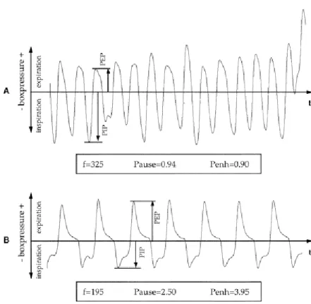
Figure 9 Barometric plethysmograph : parameters measured... 47
Figure 10 Barometric plethysmograph : changes in box pressure waveform ... 49
RESULTS Figure 11 SDS-PAGE of legume extracts... 56
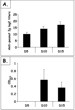
Figure 12 Peanut-specific Ab responses after IP sensitization... 58
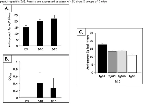
Figure 13 Peanut-specific Ab responses after IP sensitization with a high dose of PPE... 59
Figure 14 IP sensitized mice Ab reactivity with other legumes ... 59
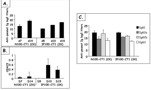
Figure 15 Kinetics and levels of Ab responses after IP and nasal sensitization ... 62
Figure 16 Influence of nasal doses of PPE on Ab responses... 62
Figure 17 Influence of nasal doses of CT on Ab responses ... 64
Figure 18 Kinetics and levels of IgG responses after nasal and oral sensitization ... 65
Figure 19 Levels of total and peanut-specific IgE after nasal and oral sensitization... 65
Figure 20 Influence of oral doses of PPE on Ab responses... 67
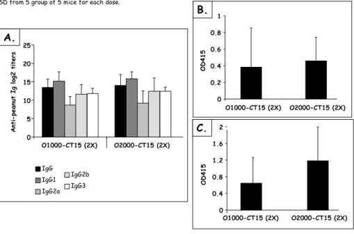
Figure 21 Influence of oral doses of CT on Ab responses ... 67
Figure 22 Influence of the antigen on Ab responses after nasal and oral sensitization ... 68
Figure 23 Airway hyper-reactivity after nasal challenge of orally or nasally sensitized mice ... 69
Figure 24 Cell recruitment in lung and BALF of mice sensitized with peanut ... 71
Figure 25 Cell recruitment in lung and BALF after nasal peanut challenge ... 72
Figure 26 Effect of the nasal peanut challenge on the phenotype of lung cells ... 73
Figure 27 Influence of the route of sensitization on the phenotype of lung cells ... 75
Figure 28 Cytokine responses in lungs and BALF of mice sensitized orally or nasally ... 76
Figure 29 Ab responses to peanut sensitization in IL-4 KO mice... 79
Figure 30 Ab responses to peanut sensitization in IL-4/IL-13 KO mice... 81
Figure 31 Ab responses to peanut sensitization in IL-12 KO and IFN-γ KO mice ... 83
Figure 32 Lung inflammatory responses in IL-4 KO and IL-4/IL-13 KO mice... 85
Figure 33 Lung cell phenotype after nasal challenge of IL-4/IL-13 KO mice. ... 87
Figure 34 Lung inflammatory responses to peanut in IL-12 KO and IFN-γ KO mice... 87
Figure 35 Orally or nasally sensitized mice Ab reactivity with other legumes ... 89
Figure 36 Lung inflammatory responses to nasal challenge with legume proteins... 91
Figure 37 Lung inflammatory scores after nasal challenge with legume proteins ... 91
Figure 38 Lung inflammatory responses to nasal challenge with unrelated proteins... 93
Figure 39 B and T cells in the lungs after challenge with legumes or unrelated antigens... 93
Figure 40 MAC-1+ cells in the lungs after challenge with legumes or unrelated antigens... 95
DISCUSSION Figure 41 Homing of effector cells induced in the NALT or GALT ... 99
Figure 42 Th1 and Th2 cytokines differentially affect Ab responses in orally and nasally sensitized mice... 103
Figure 43 Cellular and molecular events underlying the oral sensitization / nasal challenge model ... 107
ABBREVIATIONS Ab Antibody
AHR Airway hyper-reactivity
APC Antigen-presenting cell
BALF Bronchoalveolar lavage fluids BSA Bovine albumine serum
CDD Cross-reactive carbohydrate determinants
CP Crossing point
CT Cholera toxin
DBPCFC Double-blind placebo-controlled food challenge Der f Dermatophagoides farinae
ELISA Enzyme linked immunosorbent assay
FITC Fluorescein isothiocyanate
GALT Gut-associated lymphoid tissues GI tract Gastrointestinal tract
HRP Horseradish peroxydase
IFN Interferon IL Interleukine IP Intraperitoneal KO Knock-out M cells Microfold cells
MALT Mucosal-associated lymphoid tissues NALT Nasal-associated lymphoid tissues OAS Oral allergy syndrome
OVA Ovalbumine
PBS Phosphate-buffered saline
PE Phycoerythrin
Penh Enhanced pause
PPE Peanut protein extract
PP Peyer’s patch
RAST Radioallergosorbent assays
Th T helper
TNF Tumor necrosis factor WT Wild-type
1
1. Food allergies
Mucosal surfaces broadly interface with the environment making them the initial sites of interactions between cells of the immune system and pathogens or environmental antigens. The immune system must discriminate between pathogenic and innocuous stimuli and mount the most appropriate responses. Allergy is a disease resulting from inappropriate immune responses to otherwise harmless antigens. Symptoms of food allergy generally occur upon subsequent contact with a food product in individuals previously sensitized to it. They can also develop in the context of cross-reaction upon contact with an allergen different than the primary offending food product.
After defining a food allergy and closely characterize the two classes involved in this disease, we will detail mechanisms of both primary and cross-reactive reactions.
1.1. Definition and incidence of food allergies
Food allergies are immune-mediated adverse reactions to food products. These reactions are distinct from the other types of adverse reactions to foods, related to toxins, pharmacologically active chemicals, enzymatic deficiencies or psychological reactions. Food allergies include IgE-dependent but also IgE-independent reactions such as dietary protein enterocolitis, dietary protein prostitis and celiac disease. Recent studies estimate that IgE-mediated food allergies affect 6 % of children under 3 years of age and 4 % of the general population 1, a prevalence much higher than evaluated in the past 2. For example, the prevalence of peanut allergy has doubled among American children from 1997 to 2002 3.
high high ARA h2 Peanut lectin Peanut high low high intermediate intermediate β-conglycinin β β-conglycinin α SKTI** Soy lectin Gly m1 Soybean high intermediate intermediate β-lactoglobulin Casein BSA Milk Generalized Skin Gastrointestinal Respiratory Systemic high high intermediate low Ovalbumin Phosvitin Ovomucoid Conalbumin Egg Symptoms Resistance to digestion True food allergens *
* Sensitizing and symptom eliciting allergens
**Soybean Kunitz trypsin inhibitor
Table 1: Allergens of class 1 food allergy from Astwood JD et al. Nat Biotechnol. 1996
3
1.2. Classes of food allergies
It is well known that food allergies develop in genetically predisposed individuals 4, 5. Epidemiologic studies also suggest a major role of environmental factors for the development of allergic reactions 6-8. In this regard, the "hygiene hypothesis" is based on the assumption that the high rate of infectious diseases in developing nations could counter the development of allergic disorders 9. More important for this study, the immaturity of the gastrointestinal tract and subsequent increased internal permeability or breakdown of oral tolerance are believed to be involved in sensitization to foods in infancy 10. The resulting type of food allergy is named class 1 food allergy. Another type of IgE-mediated food allergy or class 2 food allergy is mainly seen in adults. This type of reaction unlikely results from impaired gastrointestinal functions and is believed to develop as a consequence of sensitization to inhalant allergens 11.
1.2.1. Class 1 or true food allergy
1.2.1.1. Main allergens and clinical symptoms
In class 1 food allergy, the sensitization process occurs in the gastrointestinal tract. The most important allergens involved in class I food allergy 1 include milk, egg, seafood, nuts and legumes, which are particularly resistant to gastric digestion 12, 13 (Table 1). These
allergies tend to be outgrown later in childhood, peanut being an exception 14, 15.
Symptoms of class 1 food allergy may affect different parts of the body in addition to the site of contact with the offending allergens. Gastrointestinal tract symptoms include
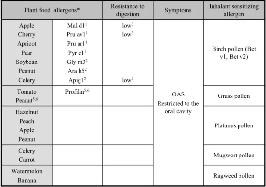
Ragweed pollen Watermelon Banana Mugwort pollen Celery Carrot Platanus pollen Hazelnut Peach Apple Peanut Grass pollen Profilin5,6 Tomato Peanut5,6
Birch pollen (Bet v1, Bet v2) OAS Restricted to the oral cavity low3 low3 rien rien rien rien low4 Mal d11 Pru av11 Pru ar11 Pyr c11 Gly m32 Ara h52 Apig12 Apple Cherry Apricot Pear Soybean Peanut Celery Inhalant sensitizing allergen Symptoms Resistance to digestion Plant food allergens*
Table 2: Allergens of class 2 food allergy
1Ebner C et al. J Allergy Clin Immunol. 1995
2Vieths S et al. Ann N Y Acad Sci. 2002
3Vieths S et al. Allergy. 1998
4Jankiewicz A et al. Int Arch Allergy Immunol. 1996
5Petersen A et al. J Allergy Clin Immunol. 1996
6de Martino M et al. Allergy. 1988
7Enrique E et al. Allergy. 2002
5 abdominal pain, vomiting and diarrhea 16. Allergic manifestations in the respiratory tract include rhinitis and asthma 17. Skin symptoms include urticaria, angio-oedema and dermatitis
18. Symptoms may be more severe when they affect the cardiovascular system. In fact, food
allergy remains a leading cause of anaphylaxis treated in emergency departments 19-21.
1.2.1.2. Cross-reactivity between food allergens
Antibody cross-reactivity has been described for many years between members of the same botanical family (i.e, legumes, tree nuts, cereals, avian food products, fish or shellfish) or between members of taxonomically unrelated families (legumes and nuts, shellfish and nuts). However, these antibody cross-reactions lead to different clinical outcomes. For example, antibody cross-reactivity in the legume family or in the cereal family is clinically irrelevant in most cases 22, 23, whereas the one among crustaceans 24 or fish 25 was reported to provoke clear clinical symptoms.
1.2.2. Class 2 or pollen-associated food allergy
Most fruit and vegetable allergens are labile to digestion (Table 2). These allergens may not effectively sensitize the organism after ingestion. However, they could contribute to food allergy via interaction with antibodies induced by primary sensitization with related inhalant allergens. Indeed, the immunologic basis for class 2 allergy is IgE cross-reactivity between common epitopes in pollen and fruit or vegetable allergens 26, 27.
Most fruit and vegetable food allergens only induce mild symptoms, restricted to the oral cavity, including itching in the mouth and throat and local edema 28. These symptoms are
Th1
Th2 IgG1, IgEMast cells
Eosinophils IgG2a Macrophages Neutrophils IL-4 IL-10 IFN-γ inhibition IL-4 IL-5 IL-13 stimulation Naïve Th Antigen IL-12 IL-4 IFN-γ
7 characteristic of the oral allergy syndrome (OAS) 29. The pollen-associated food allergies best described are the parabirch 30, 31 and ragweed-banana-melon 32 syndromes.
1.3. Cellular and molecular mechanisms of food allergy
The identification in 1986 of two distinct subsets of CD4+ T helper cells in mouse 33 greatly contributed to improve our understanding of mechanisms underlying allergic responses.
1.3.1. Th1 and Th2 cell subsets and responses
Th1 CD4+ T cells produce a profile of cytokines, including IFN-γ, that promotes IgG2a production by B cells 33. On the other hand, Th2 cells produce a profile of cytokines, including IL-4, IL-5 and IL-13, that promotes IgG1 and IgE production by B cells (Figure 1). Th1 and Th2 CD4+ T cells also influence the innate response. Indeed, chemokines that attract monocytes and neutrophils to inflammatory sites include RANTES and MIP-1β, respectively, secreted by Th1 cells. Further, secretion of IFN-γ by Th1 cells is a potent activator of macrophages. On the other hand, chemokines that attract eosinophils to inflammatory sites include CCL11 (eotaxin), secreted by Th2 cells. Secretion of IL-5 by Th2 cells induces the activation of eosinophils.
Th1 and Th2 cells have been shown to derive from a common precursor cell, or Th0. The cytokine environment surrounding the antigen-primed Th cell determines the subset that develops. IL-12 induces the development of Th1 cells, whereas IL-4 induces the development of Th2 cells. Th1 or Th2 cytokines also exert a cross-regulatory or inhibitory effect: IL-4
1. Sensitization phase 2. Reactivity phase IMMEDIATE REACTION APC Food protein IL-4 IL-13 Mucosa: GI tract Naïve Th B Th2 Th1 IL-4 CD40L CD40 TCR MHC- II CD4 TCR BCR
LATE PHASE REACTION
recruitment, activation IL-5, IL-9, GM-CSF,CCL11 Secondary exposure to food protein Ba,Mc Eo Th2 IL-4, IL-9 IL-10 leukotrienes cytokines
9 strikingly diminishes priming for Th1 cells whereas IFN-γ strikingly diminishes priming for Th2 cells.
Th1 and Th2 CD4+ T cells were later identified in humans 34, 35, although the Th1/Th2 dichotomy is less clear-cut in human as it is in mice models.
1.3.2. Allergic reaction to a primary allergen
The pathogenesis of allergy has been traditionally characterized by excessive Th2 CD4+ T cell cytokines 36 and a central role has been given to IgE Abs responses 37. However, it is now clear that broader cellular and molecular interactions occur during both the sensitization phase, when the organism develops antibodies specific to food antigens and the effector phase when the allergic reaction occurs after a secondary contact with the allergen.
1.3.2.1. Basic mechanisms of allergy: the Th2 hypothesis
The sensitization phase of the allergic reaction is initiated by the sampling of antigens by antigen presenting cells (DCs and macrophages) and presentation to B and T cells (Figure 2). Antigens that give rise to allergic reactions are thought to preferentially elicit naïve Th0 cells to differentiate into Th2-type cells 38. The signals provided by IL-4 39, 40 and IL-13 41 and the interaction between CD40 on B cells and its ligand on T cells 42 stimulate immunoglobulin class switching of IgM-bearing B cells and promote secretion of antigen-specific IgE. Secreted IgE bound to high affinity surface receptors (FcεRI) on mast cells and basophils, which are normal component of the GI tract.
The allergic reaction is triggered by a secondary exposure to the sensitizing allergen. The interaction of allergen with two or more IgE molecules cross-links FcεRI receptors on mast cells and triggers their degranulation 43. This results in the release of preformed mast-cell inflammatory mediators, such as histamine and tryptase and in the synthesis and release of newly generated lipid mediators, such as leukotriene and prostaglandin 44. These agents
rapidly mediate the symptoms of the immediate reaction.
Mast cells release cytokines that contribute to the recruitment of T cells and eosinophils. Furthermore, T cell secretion of IL-4 is not only responsible for the IgE isotype switching but also for the rolling on and adhesion to endothelial cells of circulating eosinophils, which are attracted into the gastrointestinal wall by both IL-5 and CCL11 45-47. Mediator release by eosinophils, basophils, and possibly other cell types is responsible for the late phase reaction.
1.3.2.2. Food allergy as a polyphenotypic disease 1.3.2.2.1. Data supporting the Th2 hypothesis
The “Th2 hypothesis in allergy” 48, as detailed above, is supported by studies with peripheral blood mononuclear cells showing that the food-specific T cell response in patients with food allergy is Th2-skewed when compared to that of food-tolerant individuals 49-51. It is also supported by genetic studies. Indeed, gene linkage and structure analysis, genome-wide screening and positional cloning have linked several genes to the potential for high IgE responses and IgE-dependent allergic reactions. One group of candidate genes is located in the 5q23-35 region in chromosome 5 and codes for IL-4, IL-5, IL-9 and IL-13, which increase IgE levels (IL-4 and IL-13) and favor eosinophil recruitment and survival (IL-5) and allergic manifestations.
APC Food protein GI tract Th0 Th1 IL-12, IL-18, IFN-a, IFN-γ Macrophages Inflammatory medoators
Figure 3 : Molecular and cellular mechanisms of class 1 food allergy (part 2)
from Bohle B et al. Mol Nutr Food Res. 2004
Three phases in food allergy 1. Sensitization phase
2. IgE-mediated effector phase Immediate reaction
Late-phase reaction after 6-8 hours 3. T cell-mediated effector phase after 48-72 hours
1.3.2.2.2. Role of Th1 cells in food allergy
As allergic symptoms are more readily inducible in the lung than in the GI tract, the role of Th1-type cells in the allergic process has been mostly investigated in animal models of asthma. Th1-type responses were initially believed to only counter Th2-type diseases, based upon the reciprocal regulatory effect of IFN-γ and IL-4. There is now evidence that Th1 cells could enhance rather than inhibit Th2 cell recruitment in asthma 52, 53. In a similar way, it is possible that food-specific Th1 cells mediate the appearance of food allergy symptoms. In this regard, patients suffering from atopic dermatitis developed enhanced skin symptoms upon bovine casein oral challenge. The majority of food-specific T cells isolated from the peripheral blood of these individuals were of the Th1 phenotype. Investigators showed that the initial phase of the skin disease was predominantly associated with Th2 cells, and was followed by a second phase mostly associated with Th1 cells and IgE-independent skin lesions 54. Other investigators proposed a role for food-specific Th1 cells in IgE-independent inflammatory reactions 55 (Figure 3) and suggest that food allergic reactions consist of three phases. During the initial sensitization phase, Th0 cells would differentiate into Th2 cells under the influence of IL-4. However, the presence of IL-12, IL-18, IFN-γ and IFN-α may induce the differentiation of Th1 cells as well. The second phase is the traditionally described IgE-mediated effector phase, which can be further divided into an immediate and a late phase that occurs after 6 to 8 hours. Th1 cells and macrophages are suggested to maintain an inflammatory response during the third phase, after 48-72 hours.
13
1.3.2.2.3. Regulation of the immune response to dietary antigens
The Th1/Th2 response to dietary antigens may be regulated by several factors, including Th cells involved in oral tolerance, and both pathogenic and non-pathogenic microorganisms. Another environmental factor that influences the immune response is the antigen.
The Th2 phenotype may be counterbalanced by a subset of CD4+ T cells suppressing antigen-specific immune responses, designated as T regulatory cells 56. These cells produce IL-10 and they are believed to be involved in oral tolerance, or the induction of non-responsiveness to orally administered antigens, including food 57. Other studies link TGF-β-producing Th3 cells to oral tolerance 58.
As mentioned before, the cytokine environment plays a major role in the differentiation of allergen-primed Th0 cells into Th1 or Th2 cells. Thus, microorganisms that alter the cytokine balance may regulate allergic responses. For example, probiotics (lactobacilli or bifidobacteria) have been shown to inhibit antigen-induced IgE secretion trough induction of IL-12 secretion by macrophages 59. Stimulation of the innate immune system by infectious agents, such as those that activate toll-like receptors (TLRs), may drive the immune system toward a Th1 phenotype and away it from the Th2 phenotype associated with food allergy. For example, Bashir et al 60 showed that oral sensitization with peanut proteins and the adjuvant cholera toxin (CT) induced peanut-specific IgE and anaphylactic symptoms in mouse strains lacking a functional TLR4 but not in their WT counterparts. When the composition of bacterial flora was reduced by antibiotic administration, TLR4 WT mice were as susceptible to the induction of peanut allergy as TLR4 mutant mice. Additional evidence for the role of TRL signaling in modulating allergic diseases is provided by
mechanistic studies in animal models of asthma 61-63 that demonstrated the protective effect of CpG DNA of bacterial or viral origin. Surprisingly in regard to the hygiene hypothesis, epidemiology studies showed that helminth infections, which induce strong Th2 and IgE responses 64 have a protective effect against allergy 65-67. A recent model of peanut allergy in the presence of the adjuvant CT showed that IgE levels and anaphylactic symptoms were greatly diminished in helminth infected mice. The allergic symptoms were similar in peanut allergic mice and in helminth infected mice that were given neutralizing Abs to IL-10 68. Thus, one hypothesis is that chronic parasitic infections induce a regulatory network that significantly reduces the risk of allergy 69.
The nature and amount of allergens represent the prime factors that regulate the allergic reaction. This is consistent with the fact that dietary avoidance remains the only effective way to prevent most food allergies. Most allergens are foreign proteins or glycoproteins with a molecular mass usually ranging between 5000 and 70 000. Many attempts were made to characterize such antigens but until now, there has been no indication for common structural or biological features to all allergens. Studies of in vitro stimulation of CD4+ T cells isolated from allergic donors showed the effect of different doses of allergen on Th1/Th2 differentiation. For example, Secrist et al. 70 found that CD4+ T cells from allergic donors produced high levels of IL-4 when stimulated with low concentrations of allergens but produced low levels of IL-4 when stimulated with high concentrations. This study also found that the quality and magnitude of response to the allergens was influenced by the nature of antigen presenting cells (i.e., B cells or macrophages). In this regard, the role of the innate immunity, and in particular APCs, in shaping the differentiation pathway of a naïve Th0 cell is still matter of debate. This question is of special interest since several food and environmental allergens (i.e., Der f) or other proteins associated with them could display properties such as enzymatic activities that could trigger innate responses.
15
1.4. Cross-reactive allergic reaction
Caracterizing a cross-reactive allergic reaction includes investigating the cross-reactivity of IgE antibodies but also its clinical relevance. As explained below, both phenomena are distinct.
1.4.1. Antibody cross-reactivity
1.4.1.1. Molecular basis: structural identity between allergens
Two allergens are cross-reactive if there is one IgE antibody that binds to both these proteins. Thus, structural characteristics of proteins are major determinants of cross-reactivity. The IgE cross-reactions are because of shared features at the level of primary and tertiary structures of proteins. Cross-reactivity is rare below 50 % sequence identity and usually requires more than 70 % amino acid identity 71.
1.4.1.2. Cross-reactive protein epitopes
Molecular biology has allowed the identification of many cross-reactive allergens and the increasing availability of purified recombinant proteins will help characterize them further at the molecular level 72. It turned out that most cross-reactive allergens could be grouped into a small number of protein families, fulfilling important biological functions and therefore highly conserved in their sequence and structure. They include profilin and pathogenesis-related proteins that are ubiquitous cross-reactive plant allergens involved in most pollen-associated food allergies 73-75, seed storage proteins (albumins and globulins),
responsible for cross-reactivity in the legume family 76 and tropomyosin proteins of crustaceans, house dust mites, cockroaches, and mollusks 77.
1.4.1.3. Cross-reactive carbohydrate epitopes
Antibody cross-reactivity, however, extends beyond the protein level. IgE antibodies from sera of allergic patients can be directed to carbohydrate determinants 78. Due to the high cross-reactivity of such IgE antibodies, these epitopes were called cross-reactive carbohydrate determinants (CDD) 79. In fact, they were shown to be extremely cross-reactive not only between plant-derived glycoproteins (IgE cross-reaction between carbohydrate pollen allergens and tomato, 80 peanut 81 or zucchini 82 has been demonstrated) but also to glycoproteins from invertebrate animals (house mite dust or fish allergens). This high degree of cross-reactivity was explained by the conserved structure of N-glycans from plants and invertebrate animals 83.
1.4.2. Induction of cross-reactive allergic symptoms by B and mast cell triggering
1.4.2.1. B cell triggering by a cross-reactive allergen
The immunologic phenomenon of cross-reactivity may induce allergic symptoms when the secondary (or cross-reactive) allergen is able to trigger B cells and mast cells. The cross-reactive allergen has to cross-link two monoclonal surface-bound antibodies in order to trigger a B cell. However, it is very unlikely that an allergen displays cross-reactive repeated epitopes. The phenomenon of B cell cross-reactive priming, hypothesized by Aalberse et al.84 describes how the involvement of a linking partner allows B cell triggering by a
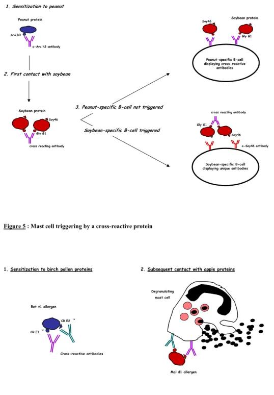
cross-1. Sensitization to peanut
2. First contact with soybean
Peanut protein Soybean protein a-Ara h3 antibody Ara h3 Soy46 Gly G1 cross reacting antibody
Peanut-specific B-cell displaying cross-reactive
antibodies
3. Peanut-specific B-cell not triggered
cross reacting antibody
a-Soy46 antibody
Soybean-specific B-cell displaying unique antibodies
Soybean-specific B-cell triggered
Soybean protein
Gly G1 Soy46
Gly G1
Soy46
Figure 4 : Molecular basis of Ab cross-reactivity
1. Sensitization to birch pollen proteins 2. Subsequent contact with apple proteins
Bet v1 allergen Cross-reactive antibodies Degranulating mast cell CR E2 * CR E1*
CR E1, CR E2: Two cross-reactive epitopes on the allergens Bet v1 from birch proteins and Mal d1 from apple proteins (J Chromatogr B Biomed Sci Appl 2001 May 25; 756 (1-2): 307-13)
*
Mal d1 allergen
Figure 5 : Mast cell triggering by a cross-reactive protein
reactive protein. Figure 4 illustrates the antibody repertoire broadening to unique (non cross-reactive) soybean allergens (the 46 kd fraction - Soy46 - evidenced by Eigenmann et al. 85 for example) in peanut-sensitized patients. In this theoretical situation, sensitization to peanut induces antibodies specific to Ara h3, a peanut allergen that has been shown share IgE epitopes with soybean glycinin G1 86. Upon subsequent contact with soybean proteins, these
antibodies might form immune complexes with glycinin G1 and present them to a naïve B cell with antibodies on it surface to the unique epitope Soy46. This may trigger the B cell to produce antibodies that are specific to soybean.
1.4.2.2. Mast cell triggering by a cross-reactive allergen
Unlike B cell antibodies, the antibodies on a mast cell are polyclonal. Thus, cross-linking of receptor bound IgE may be done be any cross-reactive molecule presenting two or more conserved epitopes (Figure 5).
1.4.3. Basis for discrepancies between in vitro and clinical tests
However, as mentioned before, in vitro cross-reactivity diagnostic tests frequently result in false-positive outcomes, which do not reflect clinical sensitivity patterns. This has especially been reported for plant foods like cereals or legumes 22, 23. Various reasons might explain these discrepancies, including monovalent CCDs, low affinity antibodies and problems inherent to the IgE assays.
19
1.4.3.1. Monovalent CCDs
Anti-carbohydrate IgE antibodies may play a role in the observed differences between
in vitro and clinical tests. In fact, monoglycosylated proteins, such as Arah1 from peanut,
may explain that clinical symptoms might not appear in patients whose IgE response is restricted to CCDs 87. Indeed, monovalent allergens are unable to induce histamine release by cross-linking IgE bound to the receptors of mast cells and basophils. However, a polyclonal IgE response to CCD and additional protein epitopes would be able to stimulate histamine release from sensitized mast cells.
1.4.3.2. Low-affinity antibodies
Specific IgE detection without clinical symptoms is not limited to CCDs. Low-affinity IgE antibodies could be another reason for the different outcomes of in vitro and clinical tests. A strong correlation was found between the affinity of the IgE for its antigen and the sensitivity of the histamine release 88. The interaction between an antibody and the cross-reacting antigen usually has a lower affinity than that with the primary antigen 84. However, studies are needed to determine the affinity threshold below which cross-reactivity becomes clinically insignificant.
1.4.3.3. IgE assays and detection of cross-reactive antigens
Other explanations are inherent to the IgE assay. It is obvious that the drawbacks of serologic allergy assays that already limit the diagnostic of primary allergies 89, 90, are magnified when dealing with reactive proteins. Henri Malandain explains why
cross-reactions are far more frequent during serum IgE assays than in clinical challenges or skin tests 91. During an IgE assay, serum IgE molecules are free: they can bind to all available (and compatible) epitopes. This binding is monovalent and each IgE molecule bound to one epitope generates a signal in the assay. In vivo, once linked to mast cells, IgE are no longer free to move. Thus, two different but cross-reactive IgE molecules must be adjacent on the mast cell and bind to adjacent epitopes on the cross-reactive allergen.
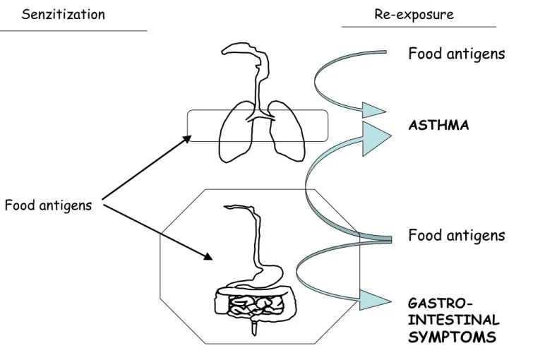
Senzitization Re-exposure
Food antigens
ASTHMA GASTRO-INTESTINALSYMPTOMS
Food antigensFood antigens
Figure 6 : Asthma and gastro-intestinal symptoms induced by food ingestion or food inhalation
2. Interplay between the GI and respiratory tracts in food allergy
Sensitization to food allergens generally occurs in the gastro-intestinal tract and primarily induces diarrhea or other gastro-intestinal symptoms. However, we will see that the respiratory tract is closely involved in both the sensitization and reactivity phases of food allergy.
2.1. Asthma induced by food ingestion or food inhalation
Asthma is an inflammatory disorder of the airways resulting in symptoms of coughing, wheezing, and shortness of breath associated with evidence of bronchial reversibility and hyperreactivity 92. Asthma associated with exposure to aeroallergens such as pollen or house dust mite is common. However, it is now recognized that food allergens have also a significant role in the etiology of asthmatic symptoms in individuals with food allergies (Figure 6). Most commonly, asthma has been described in individuals with food allergies after ingestion of the offending food 93, 94. Other studies reported asthma symptoms after exposure to aerosolized forms of the offending food: aerosolized fish, milk or egg proteins 17,
95, peanut airborne particles 96, 97 or legume particles 98.
Sensitization to these foods is commonly thought to occur in the gut. However, the respiratory tract may represent another site of sensitization to food allergens. In this regard, Gonzalez R et al. characterized soybean proteins responsible for respiratory allergies 99. The allergens recognized by IgE Abs from patients suffering from respiratory allergy were different from the allergens recognized by IgE Abs from patients suffering from allergy to soybean without respiratory symptoms. Thus, it is possible that respiratory allergy to soybean resulted from a distinct sensitization process than food allergy restricted to the gut.
23 Furthermore, the high incidence of occupational asthma in the food industry 100 indicates that inhalation of food could be an important cause of allergic sensitization 101.
2.2. The GI and respiratory tracts as sites of sensitization and reactivity
to food allergens
2.2.1. NALT and Peyer’s patches: morphologic and functional similarities
Nasopharyngeal-associated lymphoreticular tissues (NALT) and Peyer’s patches (PPs) in gut-associated lymphoreticular tissues (GALT) are organized secondary lymphoid tissues, part of the mucosa-associated lymphoid tissues (MALT). PPs and NALT share morphologic and functional similarities. Both are involved in local IgA production 102, 103 and are the portal of the mucosal immune system, through which most microbes and foreign substances enter the body. Generation of an immune response to food proteins partly depends on the uptake by microfold (M) cells, located in the epithelium of NALT and PPs. Antigens are transported to the underlying lymphoid tissue, which contain all of the immunocompetent cells that are required for the generation of an immune response: macrophages, DCs, B cells and T cells 104.
However, unique features of NALT and PPs may have a strong impact on the allergy process.
2.2.2. Distinct features of NALT and Peyer’s patches
Recent studies indicate that NALT organogenesis is different from that of PPs in terms of both kinetics and cytokine requirements 105. Furthermore, there are differences in the initiation of the immune response to food antigens in the NALT or in PPs. The GI tract is covered by a monolayer of tightly joined epithelial cells whereas respiratory tract tissues are covered by multi-layered squamous epithelial cells lacking tight junctions, making it likely that environmental antigens are more effectively sampled by the NALT than in the GALT. Accordingly, it is now well established that lower doses of antigens are needed for induction of immune responses by the nasal versus the oral route. Airway epithelial cells, more accessible to allergens than their intestinal counterparts, produce a large range of cytokines including IL-6, IL-8, IL-10, TNF-α, GM-CSF, RANTES, and CCL11, which are also secreted by classical APCs. While this could explain the higher airway responses to allergen when compared to reactions in the GI tract, contribution of immune innate factors to airway allergic responses is still poorly understood.
Additional differences in the initiation of the immune response in the NALT or in PPs are demonstrated in experimental studies using nasal or oral sensitization protocols. Nasal sensitization induces the expression of high levels of the homing and chemokine receptors CCR10 and α4β1-integrin by B cells, allowing them to efficiently traffic to the respiratory and
genito-urinary tracts 106, 107. In contrast, orally-induced B cells express CCR9 and α4β7
-integrin and migrate to sites, such as the small intestine 108. Few studies have addressed
lymphocyte trafficking in food allergy, although this phenomenon may be of particular importance to understand mechanisms by which symptoms appear in organs distant from the sensitization site. Abernathy-Carver et al. 109 demonstrated the role of T cells expressing the cutaneous lymphocyte addressin (CLA+) in milk-induced eczema. However, homing and
- Circulating IgE Blood Reactivity phase in the lung Release of mediators and cytokines
AHR
Eo - Circulating cells Homing/chemokine receptors Th2 - Systemic distribution of digested proteins Mucus production contraction of airway musclemucus secretion smooth-muscle cell proliferation
Eosinophil and mast cell degranulation Sensitization phase in the GI Food protein GI tract α4β7integrin L-selectin, CCR7 CCR3
Figure 7 : Factors contributing to food allergy-induced asthma
Eo : eosinophil
chemokine receptors expressed by T cells that were primed in PPs and migrate to the respiratory tract are currently not known. Similarly, no study has identified homing and chemokine receptors expressed by NALT T-cells sensitized to food allergens.
2.3. Mechanism of food-induced asthma
2.3.1. IgE-mediated asthma
Migration of immune cells and circulating antibodies play a major role in the pathogenesis of the food-induced asthma (Figure 7). As described earlier, sensitization to food allergens in the gut results in the differentiation of B cells secreting specific Abs, including IgE. These antigen-specific IgE can bind to mast cells and basophils in the GI tract, or they may reach other mucosal surfaces, including the lung, through the bloodstream.
The secondary exposure may occur by direct inhalation of food in aerosols or after systemic distribution of digested proteins 110. As in the GI tract, Th2 cell secretion of IL-5 and CCL11 regulate eosinophilic infiltration into the respiratory tract 111. Release of mediators such as histamine and cysteinyl leukotrienes from eosinophils and mast cells primarily causes contraction of airway muscle, increase microvascular permeability, stimulate mucus secretion, and induce smooth-muscle cell proliferation 4. All these phenomenon lead to airway obstruction and airways hyperresponsiveness (AHR).
IL-17 IL-8 IL-6 IL-4 IL-5 IL-13 BHR ASTHMA eosinophils Th2 CD4+ cells neutrophils
Epithelial cells and macrophages Figure 8 : Mechanisms of eosinophil-mediated and eosinophil-independent asthma
2.3.2. Non-IgE-mediated asthma
Until recently, asthma was exclusively regarded as an IgE-mediated atopic disease, with Th2 responses, IgE, and eosinophilic airway inflammation, resulting in enhanced bronchial reactivity 112. There is now increasing evidence that other inflammatory mechanisms may be involved in producing airway hyperreactivity, or AHR (Figure 8). Thus, asthma could also be characterized by a massive infiltration of neutrophils in the lung 113, 114. It has been shown that IL-1, IL-6 and IL-17 secreted by epithelial cells and macrophages play a major role in this neutrophil-associated asthma 115, 116. Although neutrophil-induced hyperresponsiveness has been demonstrated in humans, the mechanisms involved are still not known.
Some food may have a non specific role in the induction of asthma or in the preparation of bronchi to react to other non specific stimuli. In this regard, few studies have investigated the effect of food proteins on APCs and epithelial cells and how they could affect innate and adaptive immune responses. Whereas those cells are the first to encounter the antigen, their contribution to both the sensitization phase and the reactivity phase of the allergic response are poorly understood.
29
3. Food allergy in the legume family
Peanut is one of the eight proteins that account for 90 % of allergic reactions to foods. Lupine, which has historically been consumed in limited areas of the world, is not recognized as a main food allergen. However, the recent generalization of its use as an additive in floor or fortified pasta makes its study of particular interest in the evaluation of the allergenicity of a novel food.
3.1. Peanut and lupine as primary allergens
3.1.1. Primary allergy to peanut
Recent studies from the United Kingdom and the United States indicate that the prevalence of peanut allergy, which affects 0.8 % of young children and 0.6 % of adults, has doubled during the last 5 years 3, 117. This food allergy is of special importance because it has a tendency to be present early in life 118 and to induce more severe symptoms than other foods 119, 120, even after exposure to trace quantities of allergens 121, 122. In fact, peanut allergy is the allergy most frequently associated with fatal food-induced anaphylaxis 19, 21. Whereas clinical tolerance develops to most food allergens over time 2, peanut allergy is considered to be a lifelong threat 14 and few cases of resolution have been reported 123, 124. A minority of patients might even redevelop clinical reactivity after a negative peanut challenge result 123. Several studies have addressed the cellular responses to peanut allergens. As one could expect, the majority of T cells specific for peanut proteins isolated from the peripheral blood of allergic patients belonged to the Th2 subset 125, 126. Dietary avoidance remains the only
effective way to prevent most food allergies at this time, but it is difficult to achieve in the case of peanut because of hidden traces in consumer products 14. In addition, peanut avoidance would be of little help to prevent potential cross-reactive reactions between peanut and other members of the same botanical family or even unrelated proteins.
3.1.2. Primary allergy to lupine
3.1.2.1. Lupine in human consumption
Lupine is a legume belonging to the genius Lupinus, which includes 450 species. Sweet lupine seeds are traditionally eaten as appetizers in the Mediterranean countries. Sweet lupine is also increasingly used as additive to flour for bread, cookies and pasta 127 for both its nutritional and technological properties.
3.1.2.2. Lupine allergens
Immunoblotting using serum from lupine allergic patients has helped identify potential allergens among lupine proteins. In one study, the SDS-PAGE profile of Lupinus albus showed bands ranging from 16.5 to 175 kDa and the immunoblot analysis showed bands with molecular mass in the range of 30 to 80 kDa, the 45 kDa band appearing to be the major IgE-binding protein 98. In another study, the most important IgE-binding lupine proteins have a molecular mass of 71, 59 and 34 kDa and the less important 24 and 17 kDa
31 Immunoblotting of lupine antigen recognition by peanut-allergic patient sera were also performed. In Hefle's study 129, the SDS-PAGE of Lupinus albus showed numerous bands between 10 and 66 kDa. The IgE-binding proteins had a molecular mass of 21 kDa and 35 to 55 kDa. They are reported to be heat-stable. The band at 21 kDa was a major allergen in three patients but a minor one in three other patients. On the oher hand, the SDS-PAGE of Lupinus albus showed numerous bands between 14 and 75 kDa in Moneret-Vautrin and colleagues study 130. Serum IgE bound to protein bands at 65, 43, 38 and 13 kDa. All sera bound IgE at 43 kDa, which is reported to be the major lupine flour allergen.
Hefle et al. 127 demonstrate that different extracts of the same legume vary in allergenic potency and allergen composition. Thus, one obstacle to the characterization of the lupine allergen is the composition of food extract used for SDS-PAGE. There is no standardized source of material, method of protein extraction or condition of storage of the food extract, which is currently a complex mixture of unknown substances.
3.1.2.3. Case reports of allergic reactions to lupine
Peanuts and soybeans are the major legumes involved in human food allergy 131, 132 and scarce data exist on adverse reactions to other legumes, such as lupine. Only five studies about isolated cases of primary allergy to lupine have been published until now. The first of these reports was in 1997 by Gutierrez et al 133, who described a 25-year-old patient who exhibited symptoms of immediate urticaria related to kiss of girlfriend who had previously eaten lupine seeds. Similarly, Romano et al 134 reported a case of allergic rhinitis and asthma after ingestion of lupine seeds. The first case report of respiratory symptoms after the inhalation of lupine in dust was described in a 3-year-old child 98. Occupational allergy to
airborne lupine flour was reported in two recent studies. Moneret-Vautrin et al 128 described a patient with nasal and ocular allergic symptoms after handling of lupine flour used for prick tests and oral challenges in the diagnosis of food allergies. The symptoms disappeared after she stopped handling lupine flour. In the last study 101, three in seven subjects working in an agricultural research center reported naso-ocular symptoms immediately after being exposed to lupine.
Although these five studies describe lupine-induced allergic symptoms with tolerance to the other members of the legume family, lupine allergy is considered to be rare 134. However, the risk of cross-reaction between lupine and other legumes should be considered.
3.2. Cross-reactivity between peanut and other legumes
In general, high amino acid sequence homologies exist among legume species for a given protein 76. Theoretically, any identical epitope, hidden in different molecules, can lead to allergenic cross reactivity, or in other words elicite an allergic response as a result of an allergen binding to IgE antibody that has been produced through sensitization to another allergen 135. In fact, cross-reactive reactions between peanut and other members of the legume family have been extensively studied.
3.2.1. Cross-reactivity in the legume family is clinically irrelevant in most cases
Using RAST and RAST inhibition, Barnett et al. 135 tested sera from 40 peanut allergic patients against 10 other legumes and found IgE binding to multiple legumes for 38 % of them. Extensive immunologic cross reactivity among legumes was later confirmed by Bernhisel-Broadbent et al. 22 who studied 62 children with allergy to one legume and
33 demonstrated by SDS-PAGE/immunoblotting that 79 % had IgE binding to more than one legume, with a band at 20 kDa in all legumes except the green bean. However, on verifying the clinical reactivity to each legume by DBPCFC, they found that less than 5 % of patients reacted to more than one legume. Results of DBPCFC in other studies, including a wide variety of legumes 136-138 demonstrated that clinical cross-reaction is rare. One study reported
cross-reactions between peanut and soybean 139 but a recent review underlined the limitations of that study 140. Clinical cross-reactivity between peanut and soya seems actually to be uncommon 14, 131 despite the recent identification of shared IgE epitopes 86.
3.2.2. Some particular legumes may favor cross-reactive reactions
Particular types of legumes, like lupine, better known in the Mediterranean countries and not included in the previous studies, may be less allergenic but more cross-reactive. Although scarce data exist on adverse reaction to lupine, several reports point out the risk of cross-reactive allergies between lupine and other legumes, especially peanut.
In 1994, Hefle et al 129 described the first case of allergy in a 5-year-old girl with peanut sensitivity to lupine, after ingestion of spaghetti-like pasta fortified with sweet lupine seed flour. Skin-prick tests and RAST with extracts of lupine fortified pasta were positive in 5 out of 7 peanut sensitive patients who had also clinical symptoms with garden pea. However, no oral challenge with lupine was performed. Three other studies linked lupine allergenicity to other legume proteins. Matheu et al 141 reported the case of a chickpea-allergic woman who was tolerant to other legumes but developed urticaria and angiodema the first time she ate lupine seeds. Clinical cross-reactivity with other legumes (lentils, white beans, and green beans) progressively developed over a 5-year period. Moneret-Vautrin et al 130 evaluated clinical reactivity to lupine by DBPCFC in 24 peanut-allergic subjects. Lupine-specific IgE
was demonstrated in 11 patients, of whom 7 had clinical reactivity to lupine. This group also reported a case of acute asthma after lupine oral challenge in a patient with allergy to peanut
142 and based on these observations, they conclude that lupine flour is a high risk-allergen in
patients presenting peanut allergy.
3.3. Peanut allergy and asthma
As seen above for food allergies in general, the respiratory tract is closely involved in both the sensitization and reactivity phases of peanut allergy.
3.3.1. Aerosolized peanut as an allergic reaction-trigger
Allergic respiratory symptoms have been described in peanut-allergic patients after inhalation of peanut airborne particles in school 120 or on airline flights 96, 97. In this regard, a study about the distribution of airborne peanut allergens in the environment has just been published 143. In fact, the health care problem associated with peanut allergy is worsened by its link to life threatening asthma. Peanut is one of the food most commonly implicated in severe respiratory reactions 131, 144. The reported mortality from peanut reactions has shown that many, if not all, of the known fatalities have occurred in individuals who were known to have asthma 19, 145. Individuals with peanut allergy also present serious risks of asthma in association with inhalation of cross-reactive food allergens 142. However, the mechanisms
underlying the increased risk of life threatening asthma in the peanut-allergic population remain unclear. One possible explanation is that chronic inhalation of aerosolized food, such as that achieved in the food industry, might worsen asthma in subjects with food allergy 146.
35
3.3.2. Aerosolized peanut as a sensitizing agent
As indicated above, both the oral and inhalational routes of exposure can trigger severe adverse reactions in peanut allergic patients. It is also accepted that sensitization to food allergens can occur in the gastrointestinal tract or as a consequence of inhalational exposure. In this regard, most individuals who react to peanut do so, on their first known exposure. Since IgE-mediated allergic reactions require prior exposure resulting in sensitization, it has been hypothesized that the allergens are transferred in utero or via breast milk 147. However, no study settled the question as to whether sensitization could be achieved through inhalation of peanut airborne allergens. Peanut allergy may also be associated with inhalational sensitization to other allergens. In this regard, it is frequently associated with pollen allergy
148 and in particular with birch pollen 149, grass pollen 28 and plane tree pollen 150. It is
possible that this link only reflects a co-sensitization due to an atopic background. Alternatively, there may be a causal pathophysiological mechanism 151. For example, the role of inhalant sensitization to cross-reactive pan-allergens, like lipid transfer proteins 152 or profilins 153 in peanut allergy, has yet to be investigated 154, 155.
4. Animal models of food allergy
The double-blind placebo-controlled food challenge (DBPCFC) remains the gold standard method for identification of the offending foods in human allergies 156-158. Unfortunately, the procedure is associated with obvious limitations such as its high cost and risk of fatal allergic reactions 159. The less traumatic in vivo skin-prick test together with the
in vitro ELISA and radioallergosorbent assays (RAST) were proven useful as alternative
diagnostic tools. However, discrepancies between the in vivo and in vitro assays are common
160-162. In addition, neither the DBPCFC nor the skin-prick test address the mechanisms
implicated in primary or cross-reactive immunological reactions to food allergens. Furthermore, the use of these diagnostic tools to test on human the allergenicity of each novel food introduced in the human market raises important ethical issues. Thus, animal models are needed to better understand mechanisms of food allergies and develop more effective diagnostic and therapeutic strategies, as well as to predict the allergenicity of novel foods.
37
4.1. Models to investigate mechanisms of food allergies
Earlier studies used the Guinea pig model 163. However, most recent investigations used other animal models, including rodent (i.e., rat, mouse) and nonrodent (i.e., dog, pig). Each model presented specific advantages and drawbacks.
4.1.1. Studies in mice
4.1.1.1. Advantages and limitations of the mouse models of food allergy
The advanced understanding of the immune system in mice and its similarity with the human immune system in mechanisms such as Th1 and Th2 responses 33, 34 make murine models particularly attractive for the study of mechanisms involved in food allergy. For example, mice Th2 responses result in IgG1 production, while Th1 responses lead to IgG2a production. Therefore, quantification of mouse IgG1 and IgG2a provides an insight on Th1 and Th2 responses, respectively. Furthermore, the availability of reagents and the increasing number of knock-out and transgenic mouse strains provide valuable tools to investigate the role various immune cells and their products (i.e., cytokines/chemokines) in food allergies.
Despite similarities between the immune systems in mouse and human, major differences need to be considered. For example, in human, IgE is the only immunoglobulin isotype that directly triggers degranulation of mast cells and subsequent manifestation of anaphylaxis. The role of IgG is primarily limited to potential competitors for binding of allergens. In contrast with human, mouse IgG1 also directly trigger degranulation of mast cells and together with IgE play an important role in Passive Cutaneous Anaphylaxis. 164
4.1.1.2. Dissecting mechanisms: systemic sensitization / OVA + alum
Most studies in mice involved systemic sensitization with ovalbumin, a well-defined chicken egg allergen, in the presence of an adjuvant such as alum. This approach favors the initiation of vigorous serum IgG1 and IgE responses. Following sensitization, the mice were mucosally exposed to the antigen, in order to address the effector phase of the allergic reaction. These models have been useful in dissecting the mechanisms required for the induction of allergic gastrointestinal inflammation, including the role of CCL11 165, IL-5 166 and STAT6 167.
4.1.1.3. Mimicking human food allergy: oral sensitization / food extract + CT
Other investigators attempted to develop murine models that more closely mimic the gastrointestinal sensitization, which is the prime route of sensitization to food proteins in humans. These studies used protein extracts, instead of purified allergens that were administered by the oral route. As mice have a strong innate tendency to develop tolerance to orally administered proteins 168, the Th2-inducer adjuvant cholera toxin was used in order to generate an environment that mimics the atopic background of allergic patients. In this regard, the reported murine models of cow's milk hypersensitivity 169 and peanut anaphylaxis
170, both mimic the clinical and immunological features of peanut and cow's milk allergy in
humans.
The models of cow’s milk and peanut allergy used by Li et al. proved useful tools to investigate the influence of genetic factors on food allergy 171 and to test new immunotherapeutic strategies. For example, the oral administration of IL-12 172 or the