T
T
H
H
È
È
S
S
E
E
En vue de l'obtention du
D
D
O
O
C
C
T
T
O
O
R
R
A
A
T
T
D
D
E
E
L
L
’
’
U
U
N
N
I
I
V
V
E
E
R
R
S
S
I
I
T
T
É
É
D
D
E
E
T
T
O
O
U
U
L
L
O
O
U
U
S
S
E
E
Délivré par l’Université Toulouse III-Paul SabatierDiscipline ou spécialité : Physiopathologie, Biologie et Médecine du Développement et de la Reproduction
JURY
Professeur BUJAN Louis MD, PhD Professeur CHAP Hugues MD, PhD Docteur MIEUSSET Roger MD, PhD Professeur RIVES Nathalie MD, PhD Professeur JIMENEZ Clément MD, PhD Professeur GUICHAOUA Marie-Roberte MD, PhD
Ecole doctorale : Biologie Santé Biotechnologies
Unité de recherche : Groupe de Recherche en Fertilité Humaine, EA 3694 Directeur(s) de Thèse : Pr. BUJAN Louis et Dr. MIEUSSET Roger Rapporteurs : Pr. GUICHAOUA Marie-Roberte, Pr. JIMENEZ Clément et
Pr. RIVES Nathalie
Présentée et soutenue par Gulfam AHMAD Le 21 Décembre 2011
Titre : Température et spermatogenèse chez l’homme : conséquences potentielles d’une hyperthermie modérée des testicules et des épididymes sur l’intégrité du génome des
In the name of God, mos In the name of God, mosIn the name of God, mos
In the name of God, most Gracious, most Compassionatet Gracious, most Compassionatet Gracious, most Compassionate t Gracious, most Compassionate
« Nous sommes les enfants d’un monde Dévasté qui s’essaient à renaître dans Un monde créer. Apprendre à devenir Humain est la seule radicalité »
Raoul Vaneigem
« The reward of deeds depends upon the intentions And every person will get the reward according to what he has intended » Muhammad PBUH
« May I become at all times, both now and forever, A protector for those without protection,
A guide for those who have lost their way, A ship for those with oceans to cross, A sanctuary for those in danger, A lamp for those in the dark, And a servant to all those in need As long as living beings exist, And suffering afflicts them,
To
My father Chaudhry Atta-Ullah Baryar (may his soul rest in peace) Sughran, my mother, who dedicated her whole life for her kids
Huma, my wife, for great patience, oceans away, waiting for me to finish this task
ACKNOWLEDGEMENTS
I feel immense pleasure to be at this stage of my PhD thesis where I can look back and really appreciate my compassionate professors, colleagues, institutions, friends and family that make me able to write this page.
I would start with Prof. Louis Bujan, my supervisor, andrologist, Director of Human Fertility Research Group and President of French Federation of CECOS. There is much too to say about him, I encountered many ups and downs during the past four years and he has been helping me all the time giving me great courage to move on. His meticulousness, commitment and expertise went a long way to achieve my goal. This thesis would have not been completed without his continuous and sustained guidance at each step. I am really indebted to his efforts and all that he did for me to accomplish this research.
Dr Roger Mieusset, andrologist, head section of Male Sterility Center and co-director of
my thesis. I express my profound gratitude for his competitive and vast expertise on the subject which I learned and tried to apply during my PhD work. His critical insight and exceptional command on the project really helped me a lot to think and judge scientifically.
Dr Myriam Daudin, for her amical attachment and kindness that she really practiced
during my study period. I gratefully acknowledge the able guidance of Dr Nathalie Moinard in laboratory techniques especially in problem solving, helping me when needed
My sincere thanks to Camille Esquerré for her invaluable help in my experimental work, being a good office mate and a good friend.
Françoise Cendres, for helping me out in sample collection, processing, organizing
volunteers’ visits and staying late at work with me. All the technicians of CECOS Midi-Pyrenees for their help during sample freezing and storage and providing me a comfortable and familial ambiance in the laboratory.
Marie Walschaerts, for her statistical help and friendly time we had during our PhD
works. My sincere thanks to Marie Foulon for her help in the administrative work.
These acknowledgments would be incomplete if I do not pay thanks to all the members of EA 3694, whole staff of CECOS. This research would have not been a reality without the
help of these health professionals who were extremely supportive and cooperative in providing me all the practical logistics to accomplish the study.
Special thanks to the staff of Male Steriltity Center and Urology-Andrology, student residents for their help and, the volunteers who participated in the study to share their contributions in scientific inventions.
Finally the Higher Education Commission Islamabad, Pakistan for granting me the PhD fellowship and University Hospital of Toulouse for the research funds.
TABLE OF CONTENTS RESUME... 9 ABSTRACT ... 10 ABBREVIATIONS ... 11 LIST OF TABLES... 13 LIST OF FIGURES... 14 INTRODUCTION ... 16
1. PHYSIOLOGY OF MALE REPRODUCTION ... 19
1.1. OVERVIEW ... 19
1.1.1.HUMAN MALE REPRODUCTIVE TRACT... 19
1.1.1.1. Anatomical and functional organisation of testes... 19
1.1.1.1.1.. Interstitial compartment... 19
1.1.1.1.2. Tubular compartment... 20
1.1.1.2. Anatomical and functional organisation of epididymis ... 20
1.1.1.3. Male reproductive accessory glands... 22
1.1.1.3.1. Seminal vesicles ... 22 1.1.1.3.2. Prostate ... 22 1.1.1.3.3. Bulbourethral glands ... 23 1.1.2.SPERMATOGENESIS... 25 1.1.2.1. Spermatogoniogenesis ... 27 1.1.2.2. Meiotic phase ... 27 1.1.2.3. Spermiogenesis ... 28 1.1.2.4. Spermiation ... 31
1.1.3.ROLE OF SERTOLI CELLS IN SPERMATOGENESIS... 34
1.1.4.ROLE OF EPIDIDYMIS IN SPERM MATURATION... 36
1.1.5.TESTICULAR BLOOD SUPPLY... 36
1.1.5.1. Testicular artery... 36
1.1.6.REGULATION OF SPERMATOGENESIS... 37
1.1.6.1. Intrinsic or auto/paracrine regulation ... 37
1.1.6.2. Extrinsic or endocrine regulation ... 38
1.1.6.3. Thermoregulation of spermatogenesis ... 40
1.1.6.3.1. Pampinifrom plexus ... 40
1.1.6.3.2. Scrotum ... 40
1.2. LITERATURE REVIEW ... 43
2.1.TEMPERATURE AND MALE FERTILITY... 44
2.1.1. Testicular temperature and sperm parameters in animals... 44
2.1.2. Testicular temperature and sperm parameters in men ... 44
2.1.3. Heat stress factors... 46
2.13.1. Exogenous factors ... 46
2.1.3.2. Endogenous factors... 46
2.2.TEMPERATURE AND SPERM CHROMATIN INTEGRITY... 51
2.2.1. Evidence from animal studies ... 51
2.2.1.1. Temperature and sperm chromatin/DNA abnormalities... 51
2.2.1.2. Temperature, pregnancy rate and fate of embryo... 53
2.2.2. Human studies... 62
2.2.2.1. Temperature and sperm DNA/chromatin alterations... 62
2.2.2.2. Temperature, pregnancy rate and fate of embryo... 62
2.2.2.3. Male mediated heat stress... 62
STUDY ... 64
1. STUDY OBJECTIVES ... 65
1.1.PRINCIPAL OBJECTIVE... 66
1.2.SECONDARY OBJECTIVES... 66
2. MATERIALS AND METHODS ... 67
2.1.STUDY DESIGN... 68
2.3.1. Justification of the model ... 70
2.4.CHRONOLOGY OF EXPLORATIONS... 72
2.4.1. Clinical examinations/consultations ... 73
2.4.2. Semen collection ... 73
2.5.SAMPLE COLLECTION AND PROCESSING... 76
2.6.STATISTICS... 80
3. RESULTS... 81
3.1. SPERM CHROMATIN ... 82
3.1.1.ARTICLE:MILD INDUCED TESTICULAR AND EPIDIDYMAL HYPERTHERMIA ALTERS SPERM CHROMATIN INTEGRITY IN MEN... 83
3.1.2.SUMMARY... 84
3.2. SPERM MORPHOLOGY ... 119
3.2.1.INTRODUCTION... 120
3.2.1.1. Abnormalities of sperm morphology... 120
3.2.1.2. Decline in sperm morphology over the period of time and associated factors ... 123
3.2.1.3. Testicular hyperthermia and sperm morphology ... 124
3.2.2.METHODOLOGY... 127
3.2.3.STATISTICS... 128
3.2.4.RESULTS... 129
3.2.4.1. Multiple anomalies index (MAI) of sperm morphology ... 129
3.2.4.2. Head defects... 132
3.2.4.3. Mid piece defects... 138
3.2.4.4. Tail defects ... 142
3.2.5.DISCUSSION... 147
3.3. ACID ANILINE BLUE STUDY ... 151
3.3.1.INTRODUCTION... 152
3.3.2.MATERIALS AND METHODS... 154
3.3.2.1. Acid aniline blue stain... 154
3.3.4.RESULTS... 157
3.3.4.1. During hyperthermia... 157
3.3.4.2. After hyperthermia ... 157
3.3.5.DISCUSSION... 160
RESUME
La spermatogenèse chez l’homme et chez les autres mammifères nécessite une température inférieure à la température du corps. Ainsi, une augmentation de la température des testicules produit des effets délétères sur la fertilité masculine. Une réduction de la concentration spermatique, de la mobilité, de la vitalité et de la morphologie des spermatozoïdes a été rapportée chez les animaux et les hommes lorsque le scrotum ou le corps étaient exposés à des températures plus élevées. Dans les modèles animaux, des températures supérieures à la normale entraîneraient des altérations de l'intégrité de la chromatine des spermatozoïdes conduisant à un retard du développement embryonnaire précoce, à une réduction du taux de grossesse et à des fausses couches. Dans ce contexte, nous avons étudié les effets d'une légère augmentation (+2 °C) de la température des testicules et des épididymes sur l'intégrité de la chromatine des spermatozoïdes chez les hommes.
Nous avons mené un protocole expérimental chez 5 hommes féconds volontaires induisant une augmentation modérée de la température des testicules et des épididymes en remontant les testicules en position supra scrotale et en les maintenant dans cette position durant 15 heures par jour, pendant 120 jours consécutifs. Les paramètres spermatiques classiques ont été évalués selon les recommandations de l’OMS. L’indice de fragmentation de l’ADN du spermatozoïde (DFI) et la haute colorabilité de l’ADN (HDS) ont été analysés par le test du « Sperm Chromatine Structure Assay » (SCSA) et le test du bleu d'aniline a été utilisé pour évaluer la maturité de la chromatine. Les résultats du SCSA ont montré une augmentation significative du pourcentage de DFI et du pourcentage de HDS du sperme respectivement dès 20 jours (J20) et J34 d’hyperthermie. Les taux de DFI et de HDS sont restés plus élevés pendant les 120 jours d’hyperthermie comparés aux valeurs avant augmentation de la température (valeurs contrôles). Le test du bleu d'aniline a montré une augmentation non significative du pourcentage de spermatozoïdes avec une chromatine immature à J34 d’hyperthermie, atteignant une valeur significative à J73. Cette augmentation est demeurée constante pendant toute la durée d’hyperthermie traduisant un pourcentage de spermatozoïdes avec une chromatine immature supérieur à celui évalué avant hyperthermie. Après l’arrêt de l’hyperthermie, le DFI et le HDS sont revenus à leurs valeurs pré-exposition plus précocement (J73) que le pourcentage de spermatozoïdes avec une chromatine immature (J180). L’indice d’anomalies multiples (IAM) des spermatozoïdes est augmenté dès J9. La mobilité, la numération et la vitalité des spermatozoïdes ont été significativement diminuées par rapport aux valeurs avant hyperthermie, respectivement à partir de J20, J34 et J34 et sont restées diminuées pendant toute la durée de l'hyperthermie. Après l’arrêt de l'hyperthermie, tous les paramètres spermatiques sont revenus aux valeurs contrôles à partir de J73.
Nous avons montré, pour la première fois chez l’homme, qu'une légère augmentation de la température des testicules et des épididymes a un effet sur l'intégrité de la chromatine du spermatozoïde avant même la baisse de la numération des spermatozoïdes. Compte tenu des résultats obtenus dans les modèles animaux, les résultats de ce protocole expérimental pose la question des conséquences possibles de l’altération de la chromatine sur le développement embryonnaire alors que les paramètres du sperme ne sont pas incompatibles avec une fécondation au cours de la contraception masculine, durant les phases de récupération et d'inhibition. De plus, nos résultats pourraient être d’intérêt en infertilité masculine, en assistance médicale à la procréation et dans les fausses couches à répétition.
Mots clés: spermatozoïdes, fragmentation de l'ADN, hyperthermie, SCSA, épididyme, température
ABSTRACT
In man and other mammals, spermatogenesis is ensured at a temperature lower to body temperature. An increase in temperature has deleterious effects on male fertility. Reduced sperm out put, motility, viability and morphology have been reported in animals and men at higher than normal scrotal or body temperatures. Increased temperature caused alterations in sperm chromatin integrity resulting in retarded early embryo development, reduced pregnancy rates and miscarriages in animal models. In this context, for the first time in men, we investigated the effects of a mild increase (+2 °C) in testes and epididymal temperature on sperm chromatin integrity.
We designed an experimental protocol to induce mild testes and epididymal hyperthermia in five healthy fertile volunteers. Testes were lifted up and maintained at supra scrotal position 15 hours daily for 120 consecutive days. Classical sperm parameters were assessed according to World Health Organization (WHO) guidlines. Sperm DNA fragmentation index (DFI) and high DNA stainability (HDS) were analysed by “Sperm Chromatin Structure Assay” (SCSA) and acid aniline blue staining technique was used to assess the sperm chromatin maturity.
The results of SCSA showed a significant increase in the percentage of sperm DFI and HDS as early as day 20 (D20) and D34 respectively during hyperthermia which remained higher compared with the values before hyperthermia (controls) during the entire period of hyperthermia. Aniline blue test showed a non-significant increase in the percentage of sperm with immature chromatin as early as D34 during hyperthermia reaching significance at D73 and remained higher than control during the entire period of hyperthermia. After cessation of hyperthermia sperm DFI and HDS returned to the respective control values earlier (D73) than the percentage of aniline blue positive sperm (D180).
Multiple anomalies index (MAI) of spermaozoa was significantly increased, compared to control, as early as D9 of hyperthermia and remained increased throughout the entire period of hyperthermia. Sperm motility, count, and viability were significantly decreased compared with the respective controls as early as D20, D34 and D34 respectively during hyperthermia and remained decreased during the entire hyperthermia period. After cessation of hyperthermia all parameters returned to respective control values at D73. We report, for the first time in men, that a mild increase in testes and epididymal temperature largely impaired sperm chromatin integrity before any drop in sperm count. Based on the results from animal models the findings of present experimental protocol raise the question of possible consequences of altered sperm chromatin integrity on embryo development when sperm parameters were compatible with natural fertilization during male contraception particularly during inhibition and recovery phases of spermatogenesis. Moreover, our results may have clinical implications in male infertility, repeated miscarriages as well as assisted reproductive technologies.
Key words: sperm, DNA fragmentation, hyperthermia, SCSA, sperm count, epididymis, testicular temperature, male contraception, sperm chromatin.
ABBREVIATIONS
GPC Glycerophosphochiline DNA Deoxyribonucleic acid RNA Ribonucleic acid
FSH Follicle stimulating hormone BTB Blood testes barrier
IVF In vitro fertilization
IGF-1 Insuline like growth factor 1 INSL3 Insuline-like factor 3
TGFβ Transforming growth factor beta NGF Nerve growth factor
GnRH Gonadotropin releasing hormone LH Luteinizing hormone
DFI DNA fragmentation index SCSA Sperm chromatin structure assay
TUNEL Terminal deoxnucleotidyl transeferase dUTP nick end labelling assay Hsp Heat shock protein
COMET Single cell gel electrophoresis test hCG Human chorionic gonadotropin
NADPH Nicotinamide adenine dinucleotide phosphate ICSI Intracytoplasmic sperm injection
ART Assisted reproductive techniques ROS Reactive oxygen species
HEPES N-2-hydroxyethylpiperazine-N'-2-ethane-sulfonic acid HDS High DNA stainability
DPBS Dulbecco’s phosphate buffer saline PFA Paraformaldehyde
NaOH Sodium hydroxide MAI Multiple anomalies index WHO World health organization AAB Acid aniline blue test
FISH Fluorescence in situ hybridization
PSSC Premature separation of sister chromatids PEB Cisplatin, etoposide and bleomycin SSC Saline sodium citrate
LIST OF TABLES
Introduction
Table 1: Synopsis of factors discussed to induce genital heat stress. Table 2: Principal animal studies on heat stress and male fertility.
Materials and methods
Table 3: Chronology of consultations, semen samplings and principal tests applied.
Sperm chromatin (Article)
Table 1: Semen parameters before, during and after mild induced testicular and
epididymal hyperthermia in men
Sperm morphology
Table 4: Percentage of sperm head anomalies before, during and after a mild induced testicular and epididymal hyperthermia in men.
Table 5: Percentage of sperm mid piece anomalies before, during and after a mild induced testicular and epididymal hyperthermia in men.
Table 6: Percentage of sperm tail anomalies before, during and after a mild induced testicular and epididymal hyperthermia in men.
LIST OF FIGURES
Introduction: Physiology of male reproduction
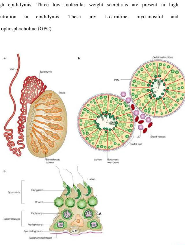
Figure 1: Distribution of causes of involuntary childlessness between men and women.
Figure 2: (a) Cross-section through a testis, showing the location of the seminiferous tubules, the vas deferens and the epididymis. (b) A diagrammatic cross- section through a testicular tubule, showing the germ cells (green) at di fferent stages of maturation. (c) A single Sertoli cell with its
associated germ cells.
Figure 3: Organs of male reproductive tract.
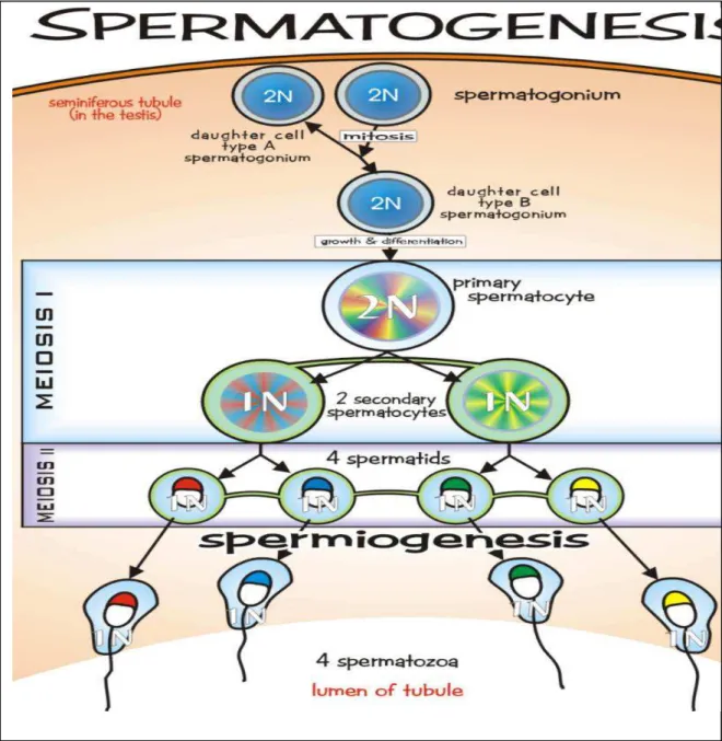
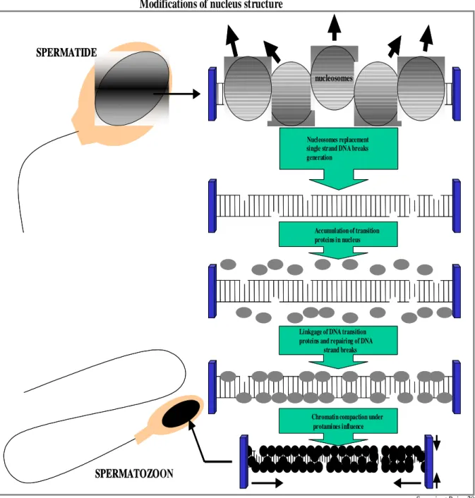
Figure 4: Germ cell division, proliferation and differentiation during spermatogenesis. Figure 5: Nuclear compaction during spermiogenesis.
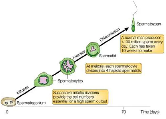
Figure 6: Stages of spermatogenesis: time course of human spermatogenesis. Figure 7: Spermatogenic cycle in man.
Figure 8: Schematic diagram illustrating the structural units of the adherens junctions in testicular germinal epithelium.
Figure 9: Intrinsic and extrinsic regulations of spermatogenesis.
Figure 10: Counter current mechanism of heat exchange a model of ram.
Study
Figure 11: Schematic representation of testicular and epididymal hyperthermia induction.
Figure 12: Chronology of semen sampling. Figure 13: Representation of the SCSA analysis.
Sperm chromatin (Article)
Figure 1: Chronology of semen sampling.
Figure 2: Total sperm and round cell counts (x106/ejaculate) measured during three
study periods, before, during and after mild induced testicular and epididymal hyperthermia in men.
Figure 3: Analysis of sperm high DNA stainability (HDS) and DNA fragmentation
index (DFI) by SCSA during three study periods; before, during and after mild induced testicular and epididymal hyperthermia in men.
Sperm morphology
Figure14: Normal human spermatozoa flat and cross-sectional view.
Figure 15: Sperm moophology: classifications of normal and abnormal human spermatozoa.
Figure 16: Multiple anomalies index (MAI) of sperm morphology before, during and after a mild induced testicular and epididymal hyperthermia in men.
Acid aniline blue test
Figure 17: Aniline blue staining: sperm with blue heads are aniline blue positive while with transparent heads are aniline blue negative.
Figure 18: Mean percentage of aniline blue positive spermatozoa before, during and after a mild induced testicular and epididymal hyperthermia in men.
Reproduction is a biological process by which new offsprings are produced from their parents. It is a fundamental feature of every known life and is opposite to death. The known methods of reproduction are grouped as: asexual and sexual reproduction. In asexual reproduction, an individual can reproduce without the involvement of another individual of that species like in plants and bacteria. Sexual reproduction requires the equal involvement of the two individuals or gametes, one of each from opposite sex. To produce a healthy offspring, the quality and genetic integrity of both gametes is equally important. When the quality of any of the gamete is compromised disturbances in fertility emerge. Fertility refers to the capability to conceive or induce a pregnancy. For example, when a couple fails to conceive during one year of unprotected sex the term infertility is used. Both partners, male and female, contribute significantly in infertility disturbances; however, the unknown causes of infertility are not negligible too.
Figure 1. Distribution of causes of involuntary childlessness between men and women (Modified from Nieschlag et al., 2010).
Disturbances in the male 20% Disturbances in both partners 26% Unknow causes 15% Disturbances in the female 39%
Male factor disturbances are important when infertility is taken into account. Half of the genetic material is contributed by male gamete, therefore, any deformity or abnormality in male gamete may lead to infertility and depending upon the conditions, complete absence of spermatozoa may lead to male sterility. Among the different causes of male infertility high testicular temperature attains an important consideration. The reason is that in man and most of the mammals, testes are anatomically placed in a position where temperature is lower than body temperature, therefore, testes functioning is temperature sensitive. Any variation in testicular temperature, high or low, may change the out put of testicular performance. Human testes encounter several conditions in daily life where temperature increases. If we look at it we find an increase in the testicular temperature during drive, sleeping, sitting for longer periods, sitting postures, taking hot baths, wearing tight underwear etc. Taking into consideration certain pathological conditions like varicocele and cryptorchidism, we also find a rise in testicular temperature resulting into spermatogenesis impairment. Though, reasonable data are available on animal models expressing the deleterious effects of higher testicular temperature on spermatogenesis but majority of these experiments have been performed above physiological temperature. Further, a human model is lacking which can give a detailed picture of the temporal effects of testicular hyperthermia on spermatogenesis. In continuation to this quest, the main objective of this research work was to investigate the effects of mild increase of testicular and epididymal temperature on kinetics of spermatogenesis, sperm parameters and chromatin/DNA integrity. The important aspect of these experiments is that the testicular temperature was maintained equal or slightly less than physiological temperature of human body.
In the next section, a brief overview of physiology of male reproductive tract, testes and epididymis is described.
1. Physiology of male reproduction
1.1. Overview
1.1.1. Human male reproductive tract
In man, reproductive tract is comprised of, two testicles, two epididymides, extensions of epididymis at both sides called vas deference, accessory sex glands known as; seminal vesicles, prostate glande, bulbouretheral glands or cowper’s gland, a penis and a scrotum. The different parts of male reproductive tract play an essential role in male reproduction. The testes are the key organ in male reproduction responsible for sperm production.
1.1.1.1. Anatomical and functional organisation of testes
Testes are glandular organs suspended by spermatic cord in a cutaneous pouch like structure called scrotum. During early fetal life they are contained in the abdominal cavity but latter on before birth they descend into the scrotum.
The parenchyma of the testes is comprised of two compartments: a) interstitial compartment
b) tubular compartment
1.1.1.1.1.. Interstitial compartment
The most important cells of the interstitial compartment are the leydig cells. Leydig cells are responsible for the production of testosterone and Insulin-like factor 3 (INSL3). This compartment also contains immune cells, blood cells, lymph vessels, nerves and fibroblasts and loose connective tissue. In human testis the interstitial compartment represents 12-15
% of the total testicular volume, 10-20% of which is occupied by leydig cells. It is estimated that human testes contain approximately 200 million leydig cells.
1.1.1.1.2. Tubular compartment
This compartment contains germ cells and two types of somatic cells; the Sertoli cells and the peritubular cells. The compartment is divided into about 250-300 lobules by septa of connective tissue. Each lobule contains 1-3 highly convoluted tubules called seminiferous tubules. It is thought that a human testis contains about 600 seminiferous tubules. The average length of each seminiferous tubule is about 60 cm (range 30-80 cm), therefore, the total length of all tubuli seminiferi per man is 720 m (360 m per testis). On average this compartment represents about 60-80% of the total testicular volume.
1.1.1.2. Anatomical and functional organisation of epididymis
Epididymis is a tubular organ which receives spermatozoa from testis. They are two in number, one on each side of each testis. The epididymis can be divided into three parts according to the functional commitments of the organ:
a) caput/head
b) corpus/body
c) cauda/tail
Principal functions of epididymis are the absorption and secretion of fluids which are essential for the maturation of spermatozoa. Here spermatozoa are matured and stored prior to ejaculation. It has a pseudostratified epithelium which contains principal cells of various heights, basal cells and lymphocytes. The fixed cells are both resorptive and secretory in nature and sperm can survive for 2 weeks in this environment. Depending
upon the testicular sperm output it takes on average 12 days for spermatozoa to pass through epididymis. Three low molecular weight secretions are present in high concentration in epididymis. These are: L-carnitine, myo-inositol and glycerophosphocholine (GPC).
Figure 2. (a) Cross-section through a testis, showing the location of the seminiferous
testicular tubule, showing the germ cells (green) at different stages of maturation. (c) A single Sertoli cell with its associated germ cells (Cooke and Saunders, 2002).
1.1.1.3. Male reproductive accessory glands
The accessory glands of the male reproductive tract are the seminal vesicles, prostate and the bulbourethral glands (Fig 3).
1.1.1.3.1. Seminal vesicles
These are the paired glands in the posterior of the urinary bladder. Each gland has a short duct that joins with the ductus deferens at the ampulla to form the ejaculatory duct which then empties into the urethra. The fluid of these glands is viscous and contains fructose that provides energy to the spermatozoa; prostaglandins, which contribute to the mobility and viability of sperm, and proteins that cause slight coagulation reactions in the semen after ejaculation.
1.1.1.3.2. Prostate
The prostate gland is a firm, dense structure that is located just inferior to the urinary bladder. It is about the size of a walnut and encircles the urethra as it leaves the urinary bladder. Numerous short ducts from the substance of the prostate glands empty into the prostatic urethra. The secretions of the prostate are thin, milky colour and alkaline. They function to enhance the motility of the sperm. Enzymes of prostate gland are responsible for the liquefaction of the semen after ejaculation. Zinc and citric acid are the major markers present in the secretions of prostate gland. Zinc helps to stabilize the sperm chromatin and its deficiency may result into lowered fertility because it may lead to high
1.1.1.3.3. Bulbourethral glands
The paired bulbourethral (Cowper's) glands are small, about the size of a pea, and located near the base of the penis. A short duct from each gland enters the proximal end of the penile urethra. In response to sexual stimulation, the bulbourethral glands secrete an alkaline mucus-like fluid. This fluid neutralizes the acidity of the urine residue in the urethra, helps to neutralize the acidity of the vagina, and provides some lubrication for the tip of the penis during intercourse.
Semen
Semen (seminal plasma + sperm cells) is a slightly alkaline mixture of sperm cells and secretions from the accessory glands. Secretions from the seminal vesicles make up about 60-65 percent of the volume of the semen, with most of the remainder coming from the prostate gland. The secretions from the bulbourethral gland contribute only a small volume.
Figure 3. Organs of male reproductive tract (Modified from “lectures on medicine of
reproduction” by Pr.Bujan Louis). Erectile tissue Glans penis Urinary bladder Vas deferens Urter Testis Epididymis Cowper’s gland Prostate gland Ejaculatory duct Seminal vesicle Internal sphincter External sphincter
The next section deals with the role of testes in spermatogenesis and maturation of spermatozoa in epididymis.
1.1.2. Spermatogenesis
Spermatogenesis takes place in seminiferous tubules in the testes. It starts with the division of germ cells arranged in particular cellular associations known as spermatogenic stages and ends with the formation of spermatozoa. The whole spermatogenic process can be divided into four phases:
1) Mitotic phase: in this phase the diploid spermatogonia (germ cells) proliferate and
differentiate mitotically. This phase can also be named spermatogoniogenesis.
2) Meiotic phase: diploid spermatocytes divide and produce haploid germ cells called
spermatids.
3) Spermiogenesis: transformation of spermatids into testicular sperm.
4) Spermiation: in this phase the elongated spermatids from germinal epithelium are
Figure 4. Germ cell division, proliferation and differentiation during spermatogenesis
(http://www.tokresource.org/tok_classes/biobiobio/biomenu/reproduction/spermatogenesis .jpg).
1.1.2.1. Spermatogoniogenesis
Spermatogonia lying at the basal part of the germinal epithelium can be distinguished by their position, their morphology and stainability of nuclei. Broadly, they are classified into two types: A type and B type spermatogonia. Type A spermatogonia are further classified into Ad (dark) and Ap (pale) spermatogonia. It is believed that Ad spermatogonia do not divide but rarely (Ehmcke and Schlatt, 2006). These spermatogonia are considered to represent the testicular stem cells (Ehmcke et al., 2006). However, these spermatogonia undergo mitosis when the reserve of testicular stem cells is in danger like in case of radiation (de Rooij, 1998). On the other hand, Ap spermatogonia divide into two daughter B spermatogonia as well as Ap spermatogonia where Ap spermatogonia serve as reservoir. B spermatogonia then divide to form preleptotene spermatocytes or primary spermatocytes. Spermatogonia multiply continuously in successive mitosis but these cell divisions are usually incomplete. The daughter cells remain interconnected by cytoplasmic bridges and a syncytium of cells is formed originating from a clone of stem cells. In cases of reduced spermatogenesis A dark-type spermatogonia are usually absent and in the absence of both types of spermatogonia no spermatogenesis takes place and the germinal epithelium consists of Sertoli cells only. There may be congenital absence of spermatogonia (Sertoli cell-only Syndrome) or Sertoli cells may be destroyed by other factors such as radiations, chemotherapy etc (acquired Sertoli cell-only Syndrome).
1.1.2.2. Meiotic phase
The stage of meiosis undergoes changes in the nuclear chromatin configuration after the last spermatogonial division. This stage is comprised of two divisions, meiosis I and meiosis II. Cells before the fist division are called primary spermatocytes (spermatocytes I)
and cells before second division are called secondary spermatocytes (spermatocytes II). Leptotene stage of first prophase (meiosis I) takes places in the basal compartment of the germinal epithelium then spermatocytes pass the Sertoli cells barrier and reach the adluminal compartment. In adluminal compartment further stages of meiosis namely, zygotene, pachytene and diplotene occur. During S phase DNA reduplicates and chromosomes condensations occure during prophase. In metaphase, homologous chromosomes form pairs and crossing over takes place in the later steps. Primary spermatocytes are the largest germ cells and after first meiotic division give rise to secondary spermatocytes which contain a haploid set of chromosome but in duplicate form. Secondary spermatocytes then undergo second meiotic division and form haploid cells called spermatids. The prophase of first meiotic division lasts 3 weeks and the other phases of first meiotic division and whole of the second meiotic divisions take 1-2 days. During meiosis genetic recombination and exchange of genetic information takes place which is essential for the diversity of life. At this stage of spermatogenesis genetic modifications can occur due to disjunction or wrong pairing of homologous chromosomes. Some times large spermatocytes (megalo- spermatocytes) appear in the ejaculates due to asynapsis of homologous chromosomes.
1.1.2.3. Spermiogenesis
Haploid cells formed after second meiotic divison are called spermatids. These are mitotically inactive but they undergo differentiation and produce elongated spermatids and sperm. These processes include condensation and shaping of the cell nucleus. Here the flagellum is formed and a large part of cytoplasm is extruded. Spermiogenesis can be divided into four phases (Nieschlag et al., 2010):
a) Golgi phase: acrosomal bubbles and craniocaudal symmetry appear. b) Cap phase: spermatids become elongated and acrosome develops.
c) Acrosomal phase: during this phase the cell nucleus is further condensed and cell elongation continues.
d) Maturation phase: in this phase the extrusion of the rest of the cytoplasm occurs.
During the process of spermiogenesis remodelling of sperm chromatin occurs. Chromatin remodelling requires well planned and regulated post-translational modifications of histones which also includes acetylation (Meistrich et al., 1992; Marcon and Boissonneault, 2004), ubiquitination (Lu et al., 2010; Chen et al., 1998; Baarends et al., 1999), methylation (Godmann et al., 2007) and phosphorylation (Leduc et al., 2008). After the hyperacetylation of histones nucleosomes are destabilized to become prepared for the replacement of transition proteins with protamines (Pivot-Pajot et al., 2003; Kurtz et al., 2007). Alterations in the sperm chromatin remodelling during spermiogenesis could result into DNA fragmentation. The presence of strand breaks (nicks) in the ejaculated spermatozoa may indicate the incomplete maturation during this process (McPherson and Longo, 1993a; McPherson and Longo, 1993b; Marcon and Boissonneault, 2004). Abnormalities in the controlling mechanism of spermiogenesis could result in anomalies of chromatin packaging and DNA strand breaks which make the sperm more susceptible to post testicular assaults.
As a result of this stage a large number of spermatids are formed. Here malformations usually affect acrosome, nucleus and flagellum and some times may be combined defects resulting into a large number of malformed spematids (round headed, multinucleated spermatids, disturbances in nuclear condensation, flagellum malformations i.e. absent or multiple flagella) (Holstein et al., 2003).
Modifications of nucleus structure
Sergerie et Bujan 2004
Liaison des protéines de transition à l’ADN et réparation des cassures simple brin Accumulation de protéines de
transition dans le noyau Déplacement des nucléosomes ,
génération de cassures simple brin de l’ADN et élimination
des supers tours SPERMATIDE
SPERMATOZOON
nucléosomes
Compaction de la chromatine sous l’influence des protamines Linkgage of DNA transition proteins and repairing of DNA
strand breaks Accumulation of transition proteins in nucleus Nucleosomes replacement single strand DNA breaks generation
SPERMATIDE
SPERMATOZO
nucleosomes
Chromatin compaction under protamines influence
Figure 5. Nuclear compaction during spermiogenesis (Modified from Sergerie et al.,
1.1.2.4. Spermiation
In this phase the spermatids are released into the lumen of the seminiferous tubules. This process can be particularly affected by hormonal modifications, temperature and toxins. Sertoli cells play a role in the movement of spermatids to the border of the lumen of seminiferous tubules. The intercellular bridges are disconnected and spermatids become free cells now called spermatozoa. Residual bodies are formed comprising of smaller parts of spermatids with cumulated RNA granules, mitochondria and lipid droplets which are digested by Sertoli cells (Breucker et al., 1985).
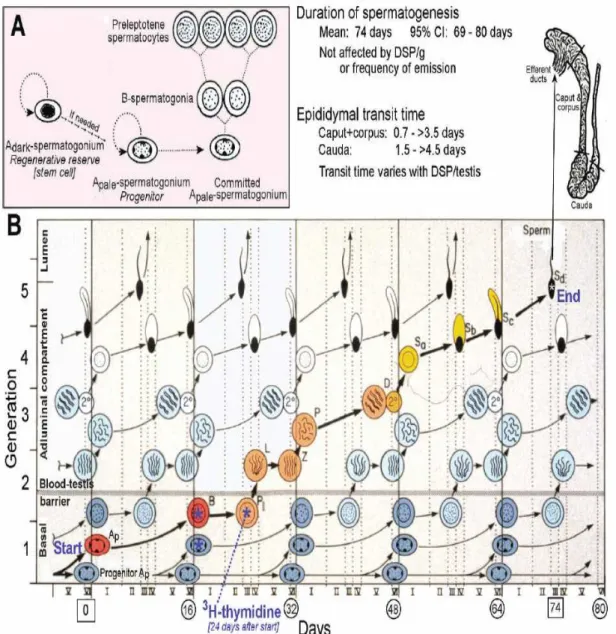
Figure 6. Stages of spermatogenesis: time course of human spermatogenesis (Cooke and
Cycle of seminiferous epithelium in human
It is believed that in adult, germ cell development follows a specific timescale, with minor biological variations. In man it takes 16 days for spermatogonia to become committed to enter spermatogenesis process. Because duration of spermatogenesis is very long (74 day in man, Fig 7) compared to the interval between commitments of spermatogonia (16 days), cohort of germ cells is layered, with the youngest near the basement membrane. These layers are sometimes termed generations. Collectively in human 6 cohorts of germ cells are found in a circumscribed area of seminiferous tubules called cellular associations. Collectively, from multiple points within the seminiferous tubules of normal human testis, more than 25 000 sperm are released each and every minute (Amann, 2008).
1.1.3. Role of Sertoli cells in spermatogenesis
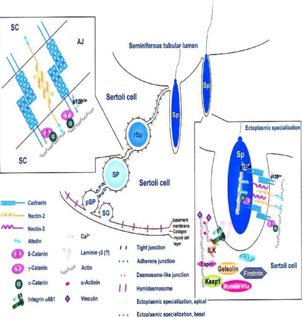
Sertoli cells synthesize and secrete several factors: proteins, cytokines, growth factors, opioids, steroids, prostaglandins, modulators of cell division etc. The morphology of Sertoli cells is strictly related to their physiological functions. They secrete inhibin and activin after puberty which work together to regulate FSH secretions. Estradiol-aromatase from Sertoli cells converts testosterone to 17 beta estradiol to direct spermatogenesis. They participate in formation of the blood testes barrier (BTB) which prevents the mixing of toxins and other harmful agents in the blood of interstitial compartment with the adluminal compartment of seminiferous tubules (Fig 8). These cells help in renewal of the germ cells and then support their stepwise development and differentiation into mature spermatozoa. During maturation of spermatozoa they also engulf the unnecessary portions of spermatozoa.
There is a specific number of Sertoli cells depending upon the species. In man 10 germ cells or 1.5 spermatozoa per Sertoli cells are observed (Zhengwei et al., 1998). About 35-40% of the volume of germinal epithelium is represented by Sertoli cells. The intatct testis with complete spermatogenesis contains 800-1200 million Sertoli cells (Zhengwei et al., 1998) or approximately 25 million Sertoli cells per gram of testis (Raleigh et al., 2004).
Figure 8. Schematic diagram illustrating the structural units of the adherens junctions in
1.1.4. Role of epididymis in sperm maturation
Sperm maturation is a process occurring in the caput and corpus of the epididymis where spermatozoa gain their fertilizing ability. The process is associated with many physiological and morphological changes in the spermatozoa (Cooper, 2007). Spermatozoa while passing through the epididymis acquire changes in morphology, sperm motility and specific motion pattern (Yeung et al., 1993; Soler et al., 2000). Epididymis plays an important role in acquiring the sperm zona binding capacity in distal parts of epididymis (Moore et al., 1992). The chromatin of sperm becomes more condensed during epididymal transit (Haidl et al., 1994; Golan et al., 1996). Also when distal epididymal spermatozoa are used in IVF cycles, higher rates of fertilization and pregnancy have been achieved (Patrizio et al., 1994).
Due to the anatomical positioning of the testes in man, the degree of increase or decrease in testicular temperature may modify the spermatogenesis. Before citing the literature on the effects of thermal variations on sperm production it will be interesting to explain the thermoregulation mechanisms of testes.
1.1.5. Testicular blood supply
1.1.5.1. Testicular artery
It originates from the aorta, runs obliquely through the abdominal wall via the inguinal canal. Then it becomes flexuous from the deep inguinal ring. In all eutherian mammals with scrotal testes, the artery is unique, except for the branches to the epididymis, two in man, superior to the head and inferior to the body and tail (Harrison and Barclay, 1948)
1.1.5.2. Testicular veins
In man venous circulation of testes includes two major drainage networks:
1) Deep venous system: drains the testes, epididymis and deferent ducts and, 2) Superficial venous network drains the scrotum (Bensussan and Huguet, 1984) Deep venous network comprises of:
a) Pampiniforme plexus b) Posterior spermatic plexus c) Veins of deferent ducts Superficial venous network comprises of:
a) Superficial veins of scrotum b) Deep veins of scrotum
1.1.6. Regulation of spermatogenesis
The process of spermatogenesis is influenced by different internal and external factors. Broadly two types of regulations are cited in literature:
a) Intrinsic regulation b) Extrinsic regulation
1.1.6.1. Intrinsic or auto/paracrine regulation
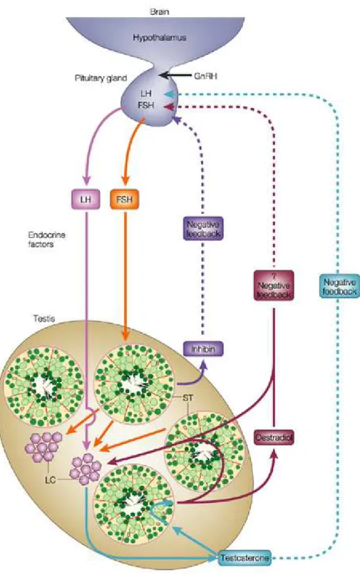
The Leydig cells in the intertubular space secrete testosterone and additional neuroendocrine substances and growth factors. These hormones, transmitters and growth factors are directed to neighbouring Leydig cells, to blood vessels, to the lamina propria of the seminiferous tubules and to Sertoli cells (Fig 9). They are involved in maintenance of the trophic of Sertoli cells and the cells of the peritubular tissue; they influence the
contractility of myofibroblasts and in that way regulate the peristaltic movements of seminiferous tubules and the transport of spermatozoa (Middendorff et al., 1997). They also contribute to the regulation of blood flow in the intertubular microvasculature. Furthermore, different growth factors (IGF1, TGFβ, and NGF) are delivered from Sertoli cells and several types of cytokines take part in a complicated circle of regulation of cell functions and developmental processes of germ cells. Taken together all these factors represent an independent intratesticular regulation of spermatogenesis. This very intricate system has been investigated mainly in animals and is not as well understood in human (Holstein et al., 2003).
1.1.6.2. Extrinsic or endocrine regulation
Local regulation of spermatogenesis in the testis requires the well known extratesticular stimuli provided by the hypothalamus and hypophysis. Pulsatile secretion of gonadotropin releasing hormone (GnRH) of the hypothalamus initiates the release of luteinizing hormone (LH) of the hypophysis. As a result of this stimulus Leydig cells produce testosterone. Testosterone influences not only spermatogenesis in the seminiferous tubules of the testis but is also distributed throughout the body and provides feedback (after aromatization in estradiol) to the hypophysis and hypothalamus related to the secretory activity of Leydig cells. Stimulation of Sertoli cells by the pituitary follicle stimulating hormone (FSH) is necessary for the maturation of germ cells. The Sertoli cells themselves secrete inhibin in the feedback mechanism directed to the hypophysis. The extratesticular influences are a necessary basis for the function of intratesticular regulations (Fig 9) (Holstein et al., 2003).
Figure 9. Intrinsic and extrinsic regulations of spermatogenesis (Cooke and Saunders,
1.1.6.3. Thermoregulation of spermatogenesis
In man testes are at a temperature 2-3 °C lower than the body temperature. This decrease in the testicular temperature is necessary for the production of fertile spermatozoa. Any change in the testicular temperature has the potential to change the fate of spermatogenesis. Therefore, temperature regulation in testicles is of prime importance for normal fertility. This temperature regulation is carried out by different structural components of which pampiniform plexus is of most importance. Secondly, scrotum in man has a special role in temperature regulation of the testes.
1.1.6.3.1. Pampinifrom plexus
This is a network formed, in the male, by veins from the testicle and epididymis, consisting of eight or ten veins lying in front of the ductus deferens and forming part of the spermatic cord. The spermatic veins emerge from the back of the testis, and receive tributaries from the epididymis: they unite and form the pampinifrom plexus which forms the chief mass of the cord. The vessels composing this plexus are very numerous, and ascend along the cord in front of the ductus deferens; below the subcutaneous inguinal ring they unite to form three or four veins, which pass along the inguinal canal, and, entering the abdomen through the abdominal inguinal ring, coalesce to form two veins (Fig 10).
1.1.6.3.2. Scrotum
Scrotum is involved in local control of temperature by three different but overlapping mechanisms, which are changes in blood flow, adaptability of the scrotal surface area and finally the evaporation of sweat.
Figure 10. Counter current mechanism of heat exchange a model of ram (Adopted from “lectures on clinical andrology” by Dr Mieusset Roger).
Scrotal blood flow
When an elevation in scrotal temperature was induced in anesthetized rats (Waites et al., 1973) or rams (air) (Fowler, 1968; Fowler and Setchell, 1971), the scrotal blood flow was increased. This increase in blood flow resulted from a mild increase in scrotal temperature which appeared as early as less than 20 minutes in rats and it became very intense (multiplied by two) when scrotal temperature passed from 33 to 37 °C in rams or rats. Furthermore, a temperature increase up to 40, 43 and 45 °C caused an increase in scrotal blood flow. However, at these higher temperatures this increase in scrotal blood flow does not appear uniformly in all regions of scrotum but mainly in posterior region (rams) (Fowler, 1968).
Variability in scrotal surface area
In rams, the degree of the dartos muscle contractions is constantly adapted to values close to normal testicular temperature in response to changes in skin temperature. Scrotum starts relaxing when scrotal skin temperature is 34 °C. When the dartos is completely relaxed the scrotal air temperature is 20% superior to its normal values, which allows exchange of fluids and evaporation (Phillips and McKenzie, 1934).
Evaporation
In men, unlike rodents, the scrotum is devoid of sweat glands, the fluid exchange occurs by simple diffusion (Buettner, 1969). Secondly, evaporation of liquids brought to the surface of the scrotal skin, sweating, or by simple diffusion causes further heat loss establishing the role of scrotum in thermoregulation (Buettner, 1969).
2.1. Temperature and male fertility
2.1.1. Testicular temperature and sperm parameters in animals
Normal functioning of the mammalian testis is ensured at a temperature 2-3 °C cooler than body temperature. Over the last century, the link between high testicular temperature and male fertility has been addressed by many scientist and clinicians. Earlier animal reports on the temperature associated testicular performance were delivered by Fukui (1923), Moore and Chase (1923) and Young (1927), showing testicular degeneration and sperm parameters alterations at a temperature higher than normal testicular temperature. Afterwards, the effects of testicular and scrotal hyperthermia and alterations in sperm parameters were reported in different animal species i.e. in rams (Dutt and Simpson, 1957; Fowler and Dun, 1966; Fowler, 1968; Rathore, 1968, 1970; Howarth, 1969; Mieusset et
al., 1992a), mice (Burfening et al., 1970; Bellvé, 1972, 1973; Meistrich et al., 1973;
Setchell et al., 1998; Zhu et al., 2004; Paul et al., 2008b; Pérez-Crespo et al., 2008), rats (Chowdhury and Steinberger, 1964) and rabbits (Howarth et al., 1965; Burfening and Ulberg, 1968). These studies have reported alterations in sperm characteristics after testicular/scrotal or whole body hyperthermia.
2.1.2. Testicular temperature and sperm parameters in men
After preliminary animal studies, researchers undertook the investigations in man. A series of studies was then carried out interrogating the adverse effects of induced hyperthermia on human spermatogenesis such as scrotal hyperthermia (Watanabe, 1959; Robinson and Rock, 1967; Rock and Robinson 1965; Robinson et al., 1968), testicular hyperthermia (Mieusset et al., 1985; Mieusset et al., 1987a; Mieusset et al., 1987c) or whole body
temperature rise (Procope, 1965). Attempts have also been made to address a male contraception method after elevating the testes through the inguinal canal thus rising the temperature near to body temperature for different periods of time in healthy men. These experiments allowed the authors to demonstrate not only drastic quantitative (Mieusset et
al., 1985) but also qualitative (Mieusset et al., 1987c) alterations in spermatogenesis.
These findings led the researchers to highlight the efficacy of this contraception method which was reversible (Mieusset and Bujan, 1994). Further, they carried out clinical trials of various suspensory designs as male contraceptives. Their results showed that men who wore one suspensory design with a rubber ring to hold the testes in the inguinal canals had ~100% effective contraception. All trial participants achieved very low motile sperm counts, between 0 and 1.6 million sperm per millilitre during experimentation (Mieusset and Bujan, 1994).
In continuation to the work on male contraception, another series of studies (Shafik, 1991a, b) described a reversible and safe male contraception method by testicular suspension. In 28 male volunteers the testes were suspended in the superficial inguinal pouch close to the scrotal neck using 2 approaches like; stitch and ball suspension for 12 months. During suspension sperm count dropped to severe oligospermia and pregnancy could not occur. However, 6 months after suspension release sperm count returned to normal, testicular biopsy showed normal spermatogenesis and pregnancy occurred (Shafik, 1991a). Further, in 1992, working on 14 men Shafik demonstrated another contraceptive technique by the use of a polyester sling applied on the scrotum. After 12 months of polyester sling men were rendered azoospermic and recovery to normal sperm parameters occured 6 months after the suspension release (Shafik, 1992). The above cited data support the idea that testicular hyperthermia effectively suppresses spermatogenesis leading to male hypofertility.
2.1.3. Heat stress factors
2.13.1. Exogenous factors
Apart from the experimental testicular hyperthermia there are other conditions associated with testicular heat stress, such as, sitting and sleeping postures, occupation, car driving, tight clothing etc. (Bigelow et al., 1998; Bujan et al., 2000; Hjollund et al., 2000; Hjollund
et al., 2002; Jung et al., 2005; Koskelo et al., 2005) previously described in a review
(Thonneau et al., 1997). In a more recent study, the authors (Mieusset et al., 2007) studied the effects of posture and clothing on scrotal temperature in fertile men. This study elucidates a linkage between scrotal temperature and the posture. The above cited studies and the studies summarised in Table 1 of this section collectively suggest that daily life style, occupation and clothings alter testicular temperature and may lead to impaired spermatogenesis (Table 1).
2.1.3.2. Endogenous factors
Fever
Fever impairs spermatogenesis due to increased testicular temperature (Table 1) (French et
al., 1973; Sheriff, 1987; Evenson et al., 2000; Sergerie et al., 2007). Evenson et al., (2000)
by using SCSA in a fertile man presenting influenza and one day fever of 39.9 °C, reported 36%, 49% and 30% denatured DNA at 18, 33 and 39 days post fever respectively. The above study also reports alterations in the normal processing of protamine 2 and a slight increase in histone to protamine ratio suggesting a fever related disruption of the synthesis of mRNA which codes protamine 2 processing enzyme. A recent case report by Sergerie et
al., (2007) reported changes in sperm parameters (total sperm count, motility and vitality)
and DNA fragmentation index (DFI) after febrile illness in a fertile man. Semen parameters were analysed before the episode of febrile illness and at days 15, 37, 58, 79 and > 180 after the fever. Total sperm count significantly decreased at 15, 37 and 58 after fever and returned back to normal by day 79 post fever. Percentage of motility significantly decreased by day 15 and 37 and returned to normal by day 58 post fever. DFI measured by sperm chromatin structure assay (SCSA) was significantly increased by 24% and 36% by days15 and 37 post fever respectively, and decreased to 15% and 8% approaching the days 58 and 79 post fever respectively.
Varicocele
Varicocele is another pathological condition which also increases the testicular temperature and may affect the sperm parameters (Table 1) but the results are contradictory (Goldstein and Eid, 1989; Lund and Nielsen, 1996; Jung et al., 2002). Lund and Nielsen reported decreased sperm quality in men with varicocele but found no significant change in the mean testicular temperature of the testicles on the same side as the varicocele, 34.6 °C (±0.7) compared to the opposite side without varicocele 34.3°C (±0.6). However, they also reported that the mean core testicular temperature of both sides was lower in donors compared with men with varicocele and those exhibiting idiopathic oligozoospermia (Lund and Nielsen, 1996). On the other hand Jung et al., (2002) found decreased semen quality as well as increased scrotal heat stress in patients with varicocele. Some more ancient studies also show discrepancy in results. For example, an elevation in testicular temperature was observed in men with varicocele (Hanley, 1956; Young, 1956) but a later study did not reveal any significant difference between the varicocele and control groups (Tessler and Krahn, 1966). Moreover, significant increase in scrotal temperature was observed in
infertile men (35±0.5 °C right, 35.1 ±0.6 °C left) having varicocele compared with controls (34.6±0.5 °C right and left) however, the difference was not significant when compared with infertile men without varicocele. Further, this study suggests the compromised sperm quality in case of higher scrotal temperature (Mieusset et al., 1987c). A more recent study supports these findings that varicocele impairs the sperm quality with maximum effects in grade III varicocele (Mori et al., 2008).
Cryptorchidism
Cryptorchidism alters sperm parameters by testicular hyperthermia and is themost frequent abnormality of male sexual differentiation. The study of Mieusset et al., (1993) showed that temperature of the undescended testis, measured in its cryptorchid location during surgical procedure for orchidopexy in boys (n = 45), was significantly higher (34.4±0.9 °C) than that of the contralateral normally descended testicle (33.2±1.2 °C). Further, they reported that among the infertile men (N=1014), 95 men were with a history of cryptorchidism and 45% of these infertile men had abnormally higher scrotal temperature (Mieusset et al., 1993). This abnormal scrotal temperature was proved to be a risk factor associated with impairement in spermatogenesis and a higher incidence of primary infertility compared with infertile men with history of cryptorchisdism but normal scrotal temperature (Mieusset et al., 1995).
In short, heat stress associated exogenous and endogenous factors cause modifications in testes and epididymal temperature which in turn impair spermatogenesis as well as sperm chromatin integrity.
Unidentified causes
Interestingly, apart from the above discussed clinical conditions, Mieusset et al., (2007) reported a thermal difference between right and left scrotum in healthy volunteers. They found that the temperature of the right scrotum was higher than the left irrespective of the position of the man. They argued that this thermal difference between right and left scrotum could contribute to the asymmetry of male external genital organs (Mieusset et al., 2007). They also reported an increased scrotal temperature in infertile men compared with fertile men without any identified factor (Mieusset et al., 1989). The exact reason of such an increase in testicular/scrotal temperature is not known, one possible factor may be the impaired thermoregulation in such men.
Table 1. Synopsis of factors discussed to induce genital heat stress (Modified from Jung and Schuppe, 2007).
Authors Source of genital
heat stress
Scrotal/testicular temperature
Semen quality/fertility parameters
(Grove et al., 2002) Disposable plastic-lined diapers
versus reusable cotton diapers
No temperature difference for the common use of cotton diapers with plastic pants No data (Hjollund et al., 2000; Jung et al., 2001; Jung et al., 2003) Genital insulation during sleep
Increased Negative influence not documented (Jung et al., 2005; Koskelo et al., 2005; Mieusset et al., 2007) Duration of sedentary posture
Increased Negative influence not documented
(Hjollund et al., 2002) Increased mean daytime scrotal temperature – Reduced (Thonneau et al., 1997; Bujan et al., 2000)
Professional drivers Increased Reduced, predominantly drivers of vans, trucks or industrial heavy machinery affected (confounders?) (Sheynkin et al.,
2005)
Sitting with portable computers in a laptop position
Increased No data
(Song and Seo, 2006) Sitting on heated floors
Increased No data
(Rock and Robinson, 1965)
Tight-fitting underwear
Increased Negative influence not sufficiently proven (Thonneau et al., 1997; Hjollund et al., 2000) Occupational exposure to high temperatures
Insufficient database Insufficient database
(Jockenhovel et al., 1990)
Sauna sessions Increased Negative influence not sufficiently proven (Watanabe, 1959) Genital heat exposure
in a water bath (>43 °C) Increased Impaired (French et al., 1973; Evenson et al., 2000; Sergerie et al., 2007) Fever episodes (3 days, >39 °C) Increased Impaired (Zorgniotti and Macleod, 1973; Jung et al., 2002)
2.2. Temperature and sperm chromatin integrity
Spermatogenesis is a complex and multi-step process which requires ~ 74 days in man for one cycle (Heller and Clermont, 1964). During this period DNA of germ cells is vulnerable to a range of insults and errors. DNA damage occurring in the male germ line is thought to contribute in birth and developmental defects and may have consequences for the later life of the offspring (Aitken and De Iuliis, 2007). The potential for insults to the integrity of sperm DNA acquired great concerns after the reports on transgenerational effects of radiation on murine spermatogenesis (Haines et al., 2002). Although other factors such as chemotherapy, toxins or pathological conditions can affect sperm chromatin but higher testicular temperature is a factor which may contribute significantly to sperm DNA/chromatin alterations as demonstrated in several studies (Karabinus et al., 1997; Sailer et al., 1997; Ahmadi and Ng, 1999; Love and Kenney, 1999). In this section studies on scrotal or whole body hyperthermia and its consequences on sperm chromatin and male fertility are addressed.
2.2.1. Evidence from animal studies
2.2.1.1. Temperature and sperm chromatin/DNA abnormalities
Several animal studies have been reported on the effects of higher scrotal/whole body heating on sperm chromatin. For example, the scrotal regions of mice were exposed to different degrees of temperature i.e. 38, 40 and 42 °C for 60 minutes to investigate the effects of hyperthermia on sperm chromatin. Mice were sacrificed after the heat exposure and the changes recorded were found to be directly proportional to the degree of heat with maximum effects at 42 °C. A decrease in the testicular weight, testicular haploid cells and
an increase in the diploid cells was observed. Chromatin integrity assessed by SCSA was severely compromised at 42 °C and these changes were observed at all stages of spermatogenesis (Sailer et al., 1997). However the temperature in this study was higher than the physiological temperature and the spermatozoa were obtained after killing the animals which could enhance the adverse effects. In experiments of Karabinus et al., (1997) the scrotal insulation of Holstein bulls was obtained continuously for 48 hours (temperature achieved not reported), samples were collected after 3 days interval post scrotal insulation, freezed and thawed for sperm chromatin structure analysis by SCSA. The results showed decreased stability of bull sperm chromatin in the ejaculated spermatozoa which were in the testes at the time of heat induction. In another study mice were given a single heat shock by dipping in water bath after sedation at 43 °C for 20 minutes. Sperm DNA damage was analysed by TUNEL assay with a recorded damage as early as 8 h after heat treatment with expression of heat shock proteins Hsp 70-1 and Hsp 70-3 (Rockett et al., 2001). Furthermore, a significant DNA damage of epididymal spermatozoa has been reported as early as 1 h post heat treatment (42 °C for 30 min) as assessed by COMET and sperm chromatin structure assay (SCSA) (Banks et al., 2005). In a more recent study of Paul et al., (2008b) male mice were subjected to a single heat stress at 38 °C, 40 °C and 42 °C for 30 minutes. Sperm chromatin structure assay performed on epididymal spermatozoa showed a temperature dependant increase in the number of sperm with impaired chromatin. The damage was slight at 38 °C while it was more profound at higher temperatures i.e. 40 °C and 42 °C. Moreover, percentage of TUNEL positive epididymal spermatozoa have been shown to be significantly higher in mice exposed to heat stress during 30 min at 42 °C compared with controls. Spermatozoa were recovered on 7, 14, 21, 28 or 60 days after heat stress. Maximum DNA damage was observed 14 days post treatment (Perez-Crespo et al., 2008). These data reveal that alterations in sperm