HAL Id: tel-01656730
https://tel.archives-ouvertes.fr/tel-01656730
Submitted on 6 Dec 2017HAL is a multi-disciplinary open access archive for the deposit and dissemination of sci-entific research documents, whether they are pub-lished or not. The documents may come from teaching and research institutions in France or abroad, or from public or private research centers.
L’archive ouverte pluridisciplinaire HAL, est destinée au dépôt et à la diffusion de documents scientifiques de niveau recherche, publiés ou non, émanant des établissements d’enseignement et de recherche français ou étrangers, des laboratoires publics ou privés.
TLR2/1 Orchestrate Human Plasmacytoid Dendritic
Cells Response to Gram+ Bacteria
Salvatore Raieli
To cite this version:
Salvatore Raieli. TLR2/1 Orchestrate Human Plasmacytoid Dendritic Cells Response to Gram+ Bacteria. Innate immunity. Université Paris-Saclay, 2016. English. �NNT : 2016SACLS495�. �tel-01656730�
1
NNT : 2016SACLS495
T
HESE DE DOCTORAT
DE
L’U
NIVERSITE
P
ARIS
-S
ACLAY
PREPAREE A
L
’U
NIVERSITE
P
ARIS
-
SUD
E
COLED
OCTORALE N° 582
CBMS Cancérologie : biologie - médecine - santé
Spécialité de doctorat : Aspects moléculaires et cellulaires de la biologie
TLR2/1 orchestrate human plasmacytoid dendritic cells
response to Gram+ bacteria
Par
Salvatore Raieli
Thèse présentée et soutenue à Paris, le 5 Décembre 2016 Composition du Jury :
CHAPUT GRAS Nathalie Institut Gustave Roussy Président du jury
JOSIEN Regis ITUN, CHU Nantes Rapporteur
POULIN Lionel Centre d’Infection et d’Immunité de Lille Rapporteur HERBEUVAL Jean-Philippe Université Paris Descartes Examinateur
3
Per aspera sic itur ad astra Marcus Tullius Cicero
4
Acknowledgement
Who built Thebes of the 7 gates ?
In the books you will read the names of kings. Did the kings haul up the lumps of rock ? And Babylon, many times demolished, Who raised it up so many times ? […]
The young Alexander conquered India. Was he alone ?
Caesar defeated the Gauls.
Did he not even have a cook with him ? Philip of Spain wept when his armada went down.
Was he the only one to weep ?
Frederick the 2nd won the 7 Years War. […] Questions From a Worker Who Reads. Bertolt Brecht (1935)
Bertolt Brecht wrote this poem explaining that behind each enterprise there is more than one man, that every challenge requires a great team.
I want to thank Vassili for giving me the opportunity to develop a scientific, critical mind and for the great freedom to explore and test different challenges.
I want to thank the jury for their kind availability at this fundamental time in my personal growth. I want to thanks the rapporteurs for their valuable comments and the critical reading of the manuscript allowing me to improve it.
I would like to thank the U932 unit for creating such a pleasant frame where to work.
I want to thank the Institut Curie Cytometry platform members: Zofia, Sophie and Annick for their time and their great help.
I would like to thank Vassili’s “team family” who have supported me during the last few years : Lucia for her great suggestions, Max for his suggestions and his jokes, Paula, Solana, Coline, Philemon, Floriane, Maude, Philemon, Ares, Antonio, without forgetting past members: Alix, Mahe, Irit, Marine, Gerome, Eve. When will we have the next “aperol” in the lab?
5
I want to thank my coffee team (PhD metabolism is mostly based on coffee consumption): Matteo (I have to be short... I cannot write ten pages about why I want to thank you), Nicolas (my great Spanish teacher who gave me great suggestions also in science), François (who corrected my awful French), Francesca (after-lunch meeting mandatory) and another Nicolas (you are not coming to sort the cells... just for the coffee). Guys, how are you standing my peculiar sense of humor? I want to thank my colleagues who made an effort to put up with me after lab time: Pamela, Leticia, Andres, Rodrigo, Emanuel, Antonella (south American communities whom I hope will accept me as a citizen), Michaelis, Vasco, Ester (I know, I am annoying),
I want to thank my English reviewer, I know it was a hard battle to found my errors (I have to be short I have much more to thank you): Lidia (my best Portuguese friend), Matthew (my great pool companion), Siba (my God, you should have killed me!), Anvita (crazy, annoying and super nice person) and Emilia Maggio (for her incredible corrections).
I want to thank my French class companion: Angelo (who has a terrible sense of humor, just like me... or even worse), Federica and Pablo (I have to thank you for many other things...I have not forgotten).
I want to thank the friends with whom I grew up together and their respective mates: Angelo (one of the best person I’ve ever met, could you imagine back then that we would do a PhD?) and Rosemary and Pino (brother, without your dinners Paris would have been an sadder place) and Azzurra and Marta.
I want to thank Francesco, who, like a swallow, migrates seasonally back and forth from Rome to Paris.
Thanks to my parents, Maria and my Vincenzo, my sister Chiara, my grandmothers Marisa and Candida, my uncles (Teresa, Beppe, Anna, Giuseppina and Massimo) and my cousins (Marialuisa, Pietro, Daniele, Francesco, Andrea, Natalia): I should write another chapter to be enough extensive to thanks you. I want to thank my aunt Anna – you should have been here but I know that in one way you are with us.
I want to thank my girlfriend Gigliola, who has supported me during my PhD and without you it wouldn’t be the same story, how we did not kill each other writing our theses at the same time? Thanks again, all of you for these 4 faboulus years.
7
Table of contents
Preamble ... 10
List of abbreviations ... 15
INTRODUCTION ... 19
1. Plasmacytoid dendritic cells and their role in the immunity ... 20
1.1 Dendritic cells: A glance in the past ... 20
1.2 Dendritic cell characteristics and function ... 20
1.3 Plasmacytoid dendritic cells discovery: an intricate immunology spy story ... 22
1.4 Plasmacytoid dendritic cells characteristics ... 23
pDCs morphology ... 23
pDCs development ... 24
Localization, migration and life span of pDCs ... 26
1.5 pDCs, a node in an intricate network ... 27
Endothelial cells ... 28
T-cells ... 28
B-cells... 30
Myeloid DCs ... 30
NK cells and NKT cells ... 31
Tumor cells ... 32
1.6 Dysregulation of the Plasmacytoid dendritic cell activation ... 34
2. Bacterial sensing ... 38
2.1 Gram positive bacterial infection ... 38
S aureus: a bacterium with two souls ... 39
Listeria monocytogenes: the comet bacteria ... 40
Mycobacterium tuberculosis: an old unfriendly companion. ... 41
2.2 innate immunity in GRAM+ bacterial infection ... 44
2.3 Dendritic cells sensors repertoire... 44
8
Cytosolic DNA sensors ... 51
C-type lectin receptors ... 52
Scavenger receptors ... 52
CD14 ... 53
NOD receptors ... 54
NLRP3 ... 54
2.4 Toll Like Receptors ... 56
MyD88-dependent pathway ... 60
TRIF-dependent pathway ... 60
2.5 Toll-Like Receptor 2 and Toll Like Receptor 1 ... 60
2.6 pDCs sensing ... 67
TLR7 and TLR9 mediated sensing ... 67
3. Plasmacytoid dendritic cells and bacterial infectious disease ... 73
3.1. Type I Interferon: a friend or a foe? ... 73
3.2 Role of Type interferon in bacterial infection ... 78
3.3 Type I IFN in parasitic and fungal infections ... 83
3.4 Plasmacytoid dendritic cells in bacterial infections ... 84
4. Choose a priori your fate ... 88
4.1 Receptor regulation complexity ... 88
The Lorenz laws in biology ... 88
Complexity and plasticity ... 90
Signal integration ... 94
4.2 Toll Like Receptors dysregulation ... 96
TLR expression alteration in bacterial infection ... 97
TLR role in autoimmunity ... 98
OBJECTIVE OF THE THESIS ... 102
RESULTS ... 105
TLR2/1 orchestrate human plasmacytoid dendritic cells response to Gram+ bacteria .. Errore. Il segnalibro non è definito. ANNEXE DATA ... 132
9
8.1 Ante fact ... 145
8.2 TLR2 and TLR1 expression in pDCs ... 145
8.3 pDCs respond functionally to bacterial lipoprotein ... 147
8.4 pDCs sense GRAM+ bacteria through TLR2/1 pathway ... 149
8.5 Differential role of TLR2 and TLR1 in pDCs response to Pam3CSK4 ... 151
8.6 Differential role of TLR2 and TLR1 in adaptive immunity priming ... 153
8.7 TLR1 activates a different signaling cascade from TLR2 in pDCs ... 159
8.8 Concluding remarks ... 162
10
List of figures
Figure 1: Dendritic cells subsets in mice and humans. ... 22
Figure 2: pDCs morphology. ... 24
Figure 3:Possible developmental pathways to lasmacytoid cells. ... 26
Figure 4: Migration of pDCs in the lymph nodes... 27
Figure 5: pDCs T-cell crosstalk. ... 30
Figure 6: pDCs’ crosstalk with the immune system. ... 34
Figure 7. Involvement of pDCs in the human diseases. ... 37
Figure 8. GRAM positive and negative bacterial membrane. ... 39
Figure 9: Lysteria model of infection 41 Figure 10:. Infected DCs crosstalk with naïve CD4+. ... 44
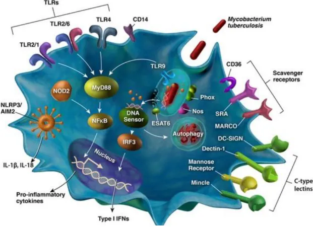
Figure 11: The different sensors present in dendritic cells. ... 55
Figure 12: Human and mouse TLRs. ... 59
Figure 13: Model of the Ligand-Induced Heterodimer of Full-Length TLR1 and TLR2. ... 61
Figure 14: Spatiotemporal differential signaling of TLR7 and TLR9 in pDCs.. ... 69
Figure 15: Type I IFN role in host protection and diseases.. ... 78
Figure 16: Effects of the Type I IFN on bacterial infections. ... 83
Figure 17: Confocal microscope picture showing that pDCs are engulfing bacteria. 2013. ... 86
Figure 18: increased presence of pDCs in TB patient LN ... 87
Figure 19: TH subsets and their role. From Craft et al., 2012. ... 92
Figure 20: Scanning electron microscopy pictures of pDCs stimulated with Aspergillus fumigatus hyphae. 94 Figure 21: TLRs expression of different blood populations. ... 135
Figure 22: Comparison of stimuli response pattern among different human blood subpopulations ... 136
Figure 23: TLRs mRNA expression change ... 138
Figure 24: Bacterial lipoproteins activate pDCs in a dose dependent manner ... 139
Figure 25: Bacterial lipoproteins treated pDCs undergo phenotypical maturation ... 140
Figure 26: Bacterial lipoprotein-treated pDCs express intracellular granzyme B ... 141
Figure 27: pDCs do not respond to NOD ligand activation ... 142
Figure 28: TLRs expression from tonsil pDCs ... 143
Figure 29: pDCs B-cells pathogenic crosstalk in SLE.. ... 157
11
List of Tables
Table 1. Toll-like receptors summary.. ... 72 Table 2. Type I, II, III family members. ... 76
13
Preamble
Two things fill the mind with ever-increasing wonder and awe, the more often and the more intensely the mind of thought is drawn to them: the starry heavens above me and the moral law within me.
Immanuel Kant, Critique of Practical Reason, 1788
When Immanuel Kant wrote this inspiring quote, fascinated by the complexity of the starry heavens, biology was moving its first steps. Nowadays, we can appreciate the intricate complexity of the living system, crafted by billions of years of evolution. In this outstanding landscape I was fascinated by the immune system and its elaborate strategies designed for confronting daily threats. This system relies on two components: innate immunity as a first line of defense, common to all the vertebrates, to quickly counterattack the invading microbes, and adaptive immunity as a sophisticated system that evolves to confront new threats. This system bases its efficiency on its complicate regulation and its capacity to be flexible to adapt to a wide range of challenges.
In the 20th century the positivistic idea of science was undermined by the formulation of Heisenberg’s Uncertainty principle and by quantum mechanics in general. In the 30’s, Gödel's Incompleteness theorems revolutionized math to the extent of affecting our faith in it. At the same time, the concept that the immune system can be detrimental to its host when its delicate balance is altered can also be considered revolutionary. As physicists have abandoned the simplicity of the Newton mechanics to delve into the uncertain space of quantum theories, more in-depth studies and a new way of thinking appear to be necessary in order to understand how all the parts of the immune system can work together in concert.
As I was captivated by this compelling challenge, my thesis is focused on the pivotal role of dendritic cells in determining the final outcome of the immune response. These cells are in a key position between innate and adaptive immunity: their repertoire of sensors allows them to sense potential threats in order to mount a specific response or shoot down excessive responses to harmless antigens. This study is mainly based on a particular subset, the plasmacytoid dendritic
14
cells (pDCs), and their interaction with GRAM+ bacteria. The unique features of pDCs and their capacities of interaction with all the immune system components influence their responses. The aim of my work was to decipher how they sense GRAM+ bacteria, which sensors are implied, and how they affect pDCs response.
This manuscript it divided in three parts: an Introduction, describing the state of the arts about pDCs, their response to bacteria and TLR2/1, to outline the framework in which results will be inserted. The results, in the form of a manuscript finalized for submission, describe how pDCs sense GRAM+ bacteria, as well as the impact of TLR2/1 on pDC function. In the discussion and perspectives results will be discussed within the wider frame of their implications and participation, with the aim of answering some open questions.
15
List of abbreviations
ALRs AIM2-like receptors
AMP adenosine monophosphate
AMPs anti-microbial peptides
APC antigen presenting cells
Aβ Amyloid β
BALF bronchoalveolar lavage fluid
BBB blood-brain barrier
BCG Bacillus Calmette-Guérin
BDCA blood dendritic cells antigen
BM bone marrow
CCL2 CC-chemokine ligand 2
CCR chemokine receptor
CD Crohn's disease
CDP common dendritic cell precursor
CLP common lymphoid precursors
CMP common myeloid precursors
CNS central nervous system
CSF cerebrospinal fluid
CTLA-4 cytotoxic T lymphocyte–associated protein 4
CXCL1 CXC-chemokine ligand 1
CXCL2 CXC-chemokine ligand 2
DAP6 domain-associated protein 6
DAMPs danger-associated molecular patterns
DCs dendritic cells
DM Dermatomyositis
dsDNA double-stranded DNA
dsRNA double-stranded RNA
DT diphtheria toxin
DTR diphtheria toxin receptor
EAE Experimental autoimmune encephalomyelitis
FLT3L fms-like tyrosine kinase-3 (Flt3) ligand
FRET fluorescence resonance energy transfer
GC germinal center
GITRL glucocorticoid-induced tumor necrosis factor receptor-ligand
HEV high endothelial venules
Hla α-hemolysin
HSC hematopoietic stem cells
16
HSV-1 herpes simplex virus type 1
IBD inflammatory bowel disease
ICD intracellular domain
ICOSL inducible T-cell co-stimulator ligand
IDO indoleamine 2,3-dioxygenase
IFN interferon
IHC immunehystochemistry
IKK IκB kinase
IL interleukine
ILT7 immunoglobulin-like transcript 7
IPCs Type 1 interferon-(α, β, ω)-producing cells (IPCs
LAIR1 leukocyte-associated immunoglobulin-like receptor 1
LAT lipoteichoic acids
LBP LPS-binding protein
LCs Langerhans cells
LDL low density lypoproteins
LLT1 lectin-like transcript 1
LN Lymph node
LPP lipoprotein
LPS lipopolysaccharide
LRR leucine-rich repeat
LTA lipoteichoic acid
MAB Mycobacterium abscessus
MAP3K8 MAPK kinase kinase 8
MDA5 melanoma differentiation antigen 5
mDCs myeloid DCs
MG Myasthenia gravis
MHC major histocompatibility complex
MIP Mycobacterium indicus pranii
MMP Matrix metalloprotease
MPO Myeloperoxidase
MS Multiple sclerosis
MTB Mycobacterium Tuberculosis
MVs called membrane vesicles
NETs neutrophils extracellular traps
NKT Natural Killer T
NMJ neuromuscular junction
NO nitric oxide
PAMPs pathogen-associated molecular patterns
17
PD-1 programmed cell death protein 1
pDCs plasmacytoid dendritic cells
PD-L1 programed death receptor-ligand 1
pETs pDCs extracellular traps
PGE2 Prostaglandin 2
PGN peptidoglycan
PI3K Phosphatidylinositide 3-kinase
PIP3 phosphatidylinositol-(3,4,5)-trisphosphate
poly(I:C) polyinosinic–polycytidylic acid PPD purified protein derivate
PPE proline-proline-glutamic
PRRs Pathogen-recognition receptors
PSA polysaccharide A
RIG-I retinoic acid-inducible gene-I
RLH RIG-like helicases
ROS reactive oxygen species
SAA Serum amyloid A
SARM Sterile α and Armadillo motif
SLE Systemic lupus erythematosus
SNP single nucleotide polymorphism
SpA Staphylococcal protein A
SS Sjögren's syndrome
SSc Systemic sclerosis
ssRNA single-stranded form of RNA
T1D Type I diabetes
TAK1 transforming growth factor-β (TGF-β)-activated kinase 1
TB tuberculosis
TB tuberculosis
TCR T cell receptor
TDM trehalose-6,6′ dimycolate
Tfh T follicular helper
TGF-β transforming growth factor β
TH T helper
TIR Toll–interleukin 1 receptor
TLR Toll like receptors
TPO Thrombopoietin
TRAIL tumor necrosis factor-related apoptosis-inducing ligand
TSTs Tuberculin skin tests
UC ulcerative colitis
19
20
1. Plasmacytoid dendritic cells and their role in immunity
1.1 Dendritic cells: A glance to the past
In 1868 Paul Langerhans observed a new cell type in epidermal tissue sections that later was named the Langerhans cell (LC). Although he considered LCs a new type of neuron cells, the link between them and the immune system was highlighted many decades later. The history of the dendritic cell (DC) began when Ralph Steinman and Zanvil Cohn discovered a new cell type in the spleens of mice in 1973. They named it dendritic cell (DC) due to its cytoplasmic extensions that reminded them of a tree. The enrichment of this population prompted them to conduct functional studies that revealed the cells’ capacitiy to induce potent proliferation and activation in T-cells. In 1985 LCs were recognized to be part of the DCs family by Gerold Schuler and Ralph Steinman. In 1991 Ralph Steinman proposed the criteria for defining a DC: 1) a dendritic morphology, (2) the constitutive expression of high levels of major histocompatibility complex (MHC) class two molecules, and (3) a capacity to induce proliferation of naïve CD4+ T cells in a mixed leukocyte reaction.
Since the discovery of DCs the scientific community’s interest has grown exponentially, and an abundance of observations and studies has shown the importance of DCs in the immune system. This importance was definitely stated in 2011 with the Nobel Prize assigned to Steinman “for his
discovery of the dendritic cell and its role in adaptive immunity”.
1.2 Dendritic cell characteristics and functions
In humans, unlike mice, where spleen and lymph nodes are available, the most accessible tissues for the study of leukocytes are peripheral blood and the skin. In 1982, after several studies, a rare population was identified in the human blood (accounting for less than 1% of the total PBMCs) that shared cytological characteristics, high levels of MHC-II and the capability of inducing potent activation of T-cells[1]. DCs are defined as lineage negative (CD3- (T-cell), CD19/20 (B-cell), and
21
CD16 (natural killer (NK) cell)) and they are MHC-II high (the high level of expression of HLA-DR is used to exclude monocytes). [2]
DCs are antigen-presenting cells with the ability to shape the adaptive immune response according to the menaces they encounter. The circulating pool of human DCs is renewed on a regular basis from the bone marrow hematopoietic stem cells (HSCs). According to the current view, CD34+ HSCs are multi-potent progenitors that give rise to common lymphoid precursors (CLPs) and common myeloid precursors (CMPs); both are capable of generating monocytes, macrophages and DCs. The progenitor cells that eventually differentiate into DCs in human (despite a better knowledge in mice) are yet to be identified [3,4]. The precursors enter the blood, acquire a mature phenotype and home to different tissues. The DCs reside in the tissues in an immature state, ready to sense the micro-environment thanks to their capacity to phagocytate and to the an abundance of sensors which they possess (that will be discussed in details below).
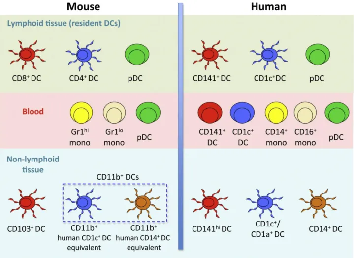
The current nomenclature divides the peripheral blood circulating DCs into three different subsets according to the expression of different surface antigens (BDCA, blood dendritic cells antigen): two myeloid DC subsets by the expression of BDCA-1(CD1c) and BDCA-3(CD141) and pDCs characterized by BDCA-2(CD303) and BDCA-4(CD304) [1
, 5].
DCs are capable of capturing antigens, expressing co-stimulatory molecules on the surface; they possess high levels of MHC-II complex, migrate to secondary lymphoid organs and secrete a high quantity of cytokines. Moreover, they can induce sustained CD4+ T-cell proliferation and shape their polarization based on the different T-helper subsets. DCs are also capable of presenting exogenous antigens to the CD8+ T-cells in a process called cross-presentation. In a different context, DCs are also capable of dampening the immune response or inducing tolerance towards certain antigens. In other words, DCs are in a unique position and they play an important role in the outcome of the whole immune response. [6,7]
22
Figure 1: Dendritic cells subsets in mice and humans. From Haniffa et al., 2012
1.3 The discovery of plasmacytoid dendritic cells: an immunology spy story
In 1958 K. Lennert and W. Remmele observed a cell, within the T-cell area of lymph nodes, which they called “T-associated plasma cells” because of its plasma cell morphology. In 1983 it was found that these cells were expressing CD4 and not B cell antigen, leading the scientists to rename them “plasmacytoid T cells”. Further observations demonstrated that these cells were not expressing T cell receptor (TCR), and the scientific community renamed them “plasmacytoid monocytes” because of their expression of MHC class II molecules. In 1996/1997, in the T-cell zone present within tonsils around the high endothelial venules (HEV), CD4+CD3−CD11c− cells with
lymphoid-23
plasmacytoid morphology were found. These cells were able to differentiate into mature DCs and were later renamed “plasmacytoid dendritic cell precursors (pDCs)” [7].
In 1970, the presence of a blood cell type capable of secreting a higher amount of type I interferon (IFN) compared with other cell types was detected. Due to this peculiar characteristic, these cells were called Type 1 interferon-(α, β, ω)-producing cells (IPCs). To start with, scientists believed NK cells were exerting this secretion. Subsequent studies excluded NK cells and B cells, T cells, monocytes and macrophages from responsibility for IFN production. These IPCs were found to express high levels of MHC-II; they were thought to belong to the DC family. In 1999 the connection between IPCs and pDCs was finally made and it was accepted that indeed they were the same cells. [8]
1.4 Plasmacytoid dendritic cells characteristics
pDCs morphology
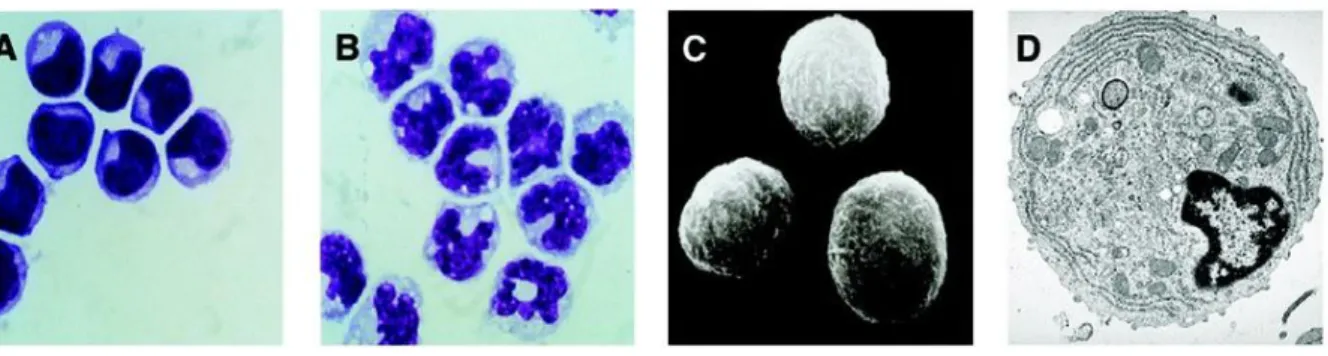
Under a light microscope, and through Giemsa staining, it is possible to observe that pDCs have a smooth, round morphology and that they resemble plasma cells. pDCs have a diameter between 8-10 µm, are smaller than monocytes and bigger than non-activated lymphocytes. Unlike monocytes, whose nucleus is horseshoe-shaped, pDCs have a kidney-shaped nucleus. They also have a basophilic cytoplasm and a dim Golgi zone. In transmission electronic microscope pictures it is possible to observe the nucleus with marginal heterochromatin, a well-developed rough endoplasmic reticulum, a reduced Golgi apparatus and the presence of many mitochondria. [9]
24
Figure 2: A. pDCs Giemsa staining. B. Monocytes Giemsa staining. C. Scanning electron microscopy
picture of pDCs. D. Transmission electron microscopy picture of pDCs. Adapted from Liu et al., 2005.
pDCs development
Despite the wide interest in understanding the development of pDCs, there are still many open questions. The administration of fms-like tyrosine kinase-3 ligand (FLT3L) into healthy volunteers appears to promote pDCs development: after the injection there was a significant increase of peripheral blood pDCs. Likewise, FLT3L transgenic mice have a higher number of pDCs compared to the wild type. FLT3L knock-out mice have fewer pDCs. In addition, FLT3L may promote the survival of pDCs in the periphery and this effect is also shown in in vitro experiments. Mainly, FLT3L is believed to promote the generation of pDCs in the bone marrow. Cultures of mouse bone marrow-derived cells give rise to a good yield of pDCs after 5-9 days when treated with FLT3L. [10] Other studies suggest that M-CSF can play a role in pDCs development: cultures of mouse bone marrow-derived cells can differentiate in pDCs when treated with M-CSF. On the contrary, other myeloid cytokines, such as GM-CSF, inhibit pDCs development. However, thrombopoietin (TPO), better known for its role in megakaryocyte/platelet development, promotes pDC development from precursors. It seems that TPO is acting in combination with FLT3L. IL-7 is also capable of promoting pDC development; their number of pDCs is reduced in IL-7 knock-out mice. It is thought that IL-7/IL-7R signaling plays a role during pDCdevelopment in the bone marrow. [11, 12, 13, 14]
25
The FLT3L signal passes through STAT3, a factor in the development of all DCs. IRF8 has proved crucial to the development of pDCs since IRF8 knock-out mice show depletion of these cells. E2-2 is specifically required for their development in both humans and mice. This transcription factor, a member of basic helix-loop-helix family, activates IRF8 and SpiB (another important gene involved in pDCs development). In the absence of E2,-2 pDCs are not found in peripheral blood. [15, 16, 17, 18,
19]
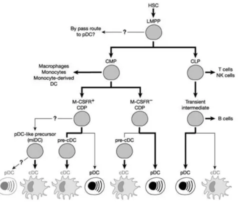
pDCs can be efficiently generated by CMP and CLP precursors in the bone marrow: most CLPs are FLT3+; on the contrary, only a small portion of CMPs is FLT3+. Other evidence showed that pDCs can have lymphoid-related developmental history, such as the rearrangement of immunoglobulin heavy chain (IgH) genes (this phenomenon can be appreciated in the other lymphoid cells outside B-cells). This is not happening in the myeloid dendritic cells. [20]
On the other hand, a common dendritic cell precursor (CDP) was found downstream of CMP, which suggested a myeloid origin for pDCs development. CDPs give origin to both pDCs and myeloid DCs. [21, 22]
In conclusion, some differences between CLP- and CDP-derived pDCs were found. CDP pDCs in the mouse present different markers of mature pDCs such as CD11c, B220 and Siglec-H but they express low levels of MHC-II. CDP pDC progenitors do not express MCSF-R and maintain high levels of E2-2. CLP-derived pDCs show differences in IL-6, TNF-α and Type I IFN production compared to their CDP-derived counterpart. The question of whether pDCs have a myeloid or lymphoid origin is still open. [23]
26
Figure 3:Possible developmental pathways to 26lasmacytoid cells. From Shortman et al., 2012
Localization, migration and life span of pDCs
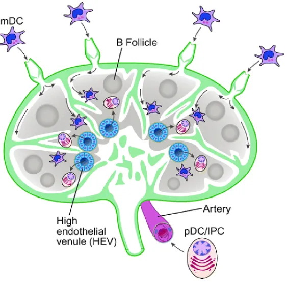
pDCs are constantly produced in the bone marrow; pDCs circulate after their exit in the peripheral blood. pDCs migrate to the T-cell area in the lymph nodes through the high endothelial venules (HEV). On the contrary, the other DCs migrate through the afferent lymphatic vessels to the lymph nodes. pDCs also migrate in a homeostatic state to mucosa-associated lymphoid tissues and the marginal zone of the spleen. pDCs can also migrate to peripheral tissues. [24, 25]
pDCs constitutively express CD62L (L-selectin for lymph node homing) and PSGL1. Moreover, their unique ability to migrate through HEV seems related to the fact that they express CXCR4 (a chemokine receptor with CXCL12 as a ligand; this chemokine is produced by HEV but it can also be expressed by the skin). pDCs also express CCR9 (the chemokine receptor for CCL25), which plays a role in the homing to the small intestine. When pDCs are activated, they express other receptors that influence their migration. They up-regulate the expression of CCR7 for lymph node homing, CCR5, CXCR3 and 1/2 integrins, receptors for CCL19 and CCl21 (a chemokine produced by the
27
HEV). pDCs also express CXCR3 and CCR9, two receptors respectively guiding the pDCs towards the skin and the small intestine. pDCs can also migrate to inflamed tissues: IL-3 stimulation leads them to express CCR6 and CCR10 that drive them towards inflamed skin or mucosa. [26, 27] Actually, pDCs migration is influenced by the environment, and different activation states lead to a different recruitment.
Figure 4: Migration of pDCs in the lymph nodes. From Liu et al., 2005
1.5 pDCs, a node in an intricate network
Although pDCs are mainly known for the high amount of Type I IFN they can secrete, it is reductive to consider their role in immunity as limited to this. In this chapter, it would be interesting to
28
summarize their capability to interact with a plethora of different cells through cytokine production and/or direct interaction. All these elements allow us to conclude that pDC activation can influence the different branches of the immune system: from innate to adaptive immunity.
Endothelial cells
After exiting the bone marrow, pDCs enter the blood stream. Endothelial cells have been shown to express different cell surface molecules and to secrete chemokines and cytokines that may aid pDCs and other leukocytes transmigration and regulate their activation state. pDCs are facilitated in their migration by endothelial cells: these cells allow them to transmigrate to lymph nodes, tumor lesions and infection sites. pDCs are also influenced by pro- or anti-inflammatory cytokines and growth factors produced by endothelial cells. For instance, endothelial cells produce IL-3 and VEGF that bind and trigger receptors on the pDCs’ surface (CD123 and BDCA4 respectively). [28] These bindings promote pDC migration and survival. Both resting and mature pDCs express ChemR23 (receptor for chemerin) on the surface. Chemerin is expressed on the surface of endothelial cells in the HEV and in the blood vessels of inflamed tissues. This interaction plays a role in pDC migration to lymph nodes and inflamed tissues. [ 29]
T-cells
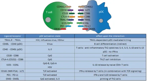
During infection, immature dendritic cells are recruited to the inflamed tissue. Upon pathogen sensing and activation by pro-inflammatory stimuli, they migrate towards the lymph node [30]. The mature dendritic cells encounter naïve T-cells in the lymph node, and induce them to differentiate into effector T-cells [31, 32]. The co-stimulatory molecules repertoire expressed and cytokines produced by DCs, differentially induced by the pathogenic stimuli, evoke a different immune response in each case. pDCs are not an exception: they influence T-cell polarization towards different subsets through different molecular interactions. pDCs are antigen-presenting cells (APCs): they are capable of presenting antigens on both MHC class I and II molecules, thus they can activate CD4+ T helper (Th) cells and CD8+ cytotoxic T cells. Due to their lack of co-stimulatory
29
molecules, resting pDCs induce T-cells anergy. Conversely, activated pDCs stimulate CD4+ T-cell polarization towards a variety of TH subsets (Th1, Th2, Th17, and Treg) [33].
pDCs express high levels of MHC molecules as well as co-stimulatory molecules such as CD80 (B7-1), CD86 (B7-2), CD40, OX40L and CD83 [34]. These molecules have a direct impact on TH polarization. According to the pathogen involved, pDCs can skew this polarization: virus-primed pDCs mostly induce a TH1 phenotype [35], while it is reported that IL-3- or TLR7-activated pDCs can induce, respectively, TH2 and TH17. pDCs can secrete Granzyme B that can affect the proliferation of T-cells. pDCs also express inducible T-cell co-stimulator ligand (ICOSL). The interaction of ICOSL with ICOS on T-cells has been shown to trigger naïve CD4+ T cells to produce IL-10. It has been suggested that this mechanism is important for generating Tregs to dampen the immune response, thus preventing excessive inflammation [36, 37]. pDCs also express programmed death receptor-ligand 1 (PD-L1); its interaction with programmed death ligand 1 (PD1) present on the T-cell surface can lead to anergy. IL-3 induces the expression of OX40L in pDCs; OX40L is a receptor associated with TH2 induction in T-cell polarization, with consequent release of 4, 5, and IL-13. Virus stimulation of pDCs can induce tumor necrosis factor-related apoptosis-inducing ligand (TRAIL) that can induce apoptosis. TRAIL expression uniquely correlates with viral load and pDCs acquire the capacity to kill HIV-infected CD4+ T cells (a process called “TRAIL-dependent pDC-mediated killing”). [ 38]
There are other molecules expressed by pDCs that can influence T-cell function, for instance, lectin-like transcript 1 (LLT1). LLT1 is a ligand of CD161 (expressed by Th1, Th17, a subpopulation of CD8+ T cells, and NK cells); its binding leads to T-cell proliferation and IFN-γ secretion, and this inhibits NK cell cytotoxicity. [39]
30
Figure 5: pDCs T-cell crosstalk. From Mathan et al., 2013
B-cells
B cells are the only cells capable of producing antibodies, and they play a critical role in the humoral response. Type I IFN induces an increase in TLR7 expression and activation markers on B-cells. IL-6 secretion by pDCs also plays a regulatory role on B-B-cells. Direct cell-to-cell interaction is also reported: CD40-CD40L and CD70/CD27 contact. These interactions result in B-cell growth, differentiation, and immunoglobulin secretion. Also, these interactions can be bi-directional, through LFA-1 or PECAM-1 (CD31), B cells stimulate Type I IFN secretion by pDCs. [40, 41]
Myeloid DCs
During inflammation, pDCs and mDCs are in contact in vivo, but this is also true at the steady state: could they act synergistically? The two DC subsets share many different functions, from the ability to present antigens to the expression of many co-stimulatory molecules. However, the two subsets are not redundant and each presents some unique features. These differences consist in the expression patterns of sensors (that will be discussed further below) but also in the production of cytokines and in the expression of co-stimulatory molecules. mDCs, for instance, are capable of
31
producing IL-1β, IL-10 and IL-12p70 that pDCs do not secrete. In a viral infection, no other cells are able to secrete such high amounts of Type I IFN as pDCs and Granzyme B production is restricted to pDCs among the DC subsets [42]. Other considerations suggest that pDCs and mDCs may cross-activate each other to reinforce the immune response. This crosstalk can be exerted by Type I IFN and TNF-α, but it seems there is a direct contact occurring. These interactions rely on CD40L/CD40, OX40L, HEVML, RANKL, CD27, CD30L, glucocorticoid-induced tumor necrosis factor receptor-ligand (GITRL), and 4-1BB. NOTCH ligands secretion by mDCs activates maturation markers on pDCs (such as CD86). After activation, pDCs express ICAM-1 (an adhesion molecule with immune stimulatory property) to match LFA-1 expression on mDCs. [43]
NK cells and NKT cells
NK cells belong to the innate immune system and are capable of recognizing both virally infected and tumor cells due to the absence of MHC on the surface. They respond rapidly by lysing. Different studies highlight the crosstalk between pDCs and NK cells through direct and cytokine-mediated interactions. In tonsil T-cell areas, pDCs and NK cells are found in close proximity. During a Herpes simplex infection it was reported, that pDCs and NK cells migrated simultaneously to the infected site. pDCs and NK cells are also found in tumor lesions. These findings support the hypothesis that these cells have many possibilities to enter into contact. Type I IFN enhances the activation and the cytolytic potential of NK cells. pDCs express GITRL on the surface to bind GITR in NK cells. This binding enhances NK cell-mediated killing and their IFN-γ production. The CD69 NK cell surface expression is IFN-α and TNF-α dependent. Furthermore, direct contact between these two cell subsets leads to up-regulation of HLA-DR on the NK cells. HLA-DR expression on NK cells seems to exert a role in handling bacterial infection (shown for instance in Mycobacterium bovis (BCG) model). [44]
This crosstalk is bi-directional, IL-2-stimulated NK cells induce pDCs to release type I IFN, also inducing up-regulation of CD83 in pDCs (but not CD80 and CD86) in a contact-dependent manner. Due to their high expression of MHC-I and lack of Nectin-2 (the ligand for NK cell activating receptors DNAM-1), pDCs are not susceptible to lysing; however, IL-3 stimulation can induce
32
Nectin-2 expression on mature pDCs. TLR stimulation induces LLT1, thus inhibiting the NK cells’ cytolytic function, which saves pDCs from being killed. [45]
Natural Killer T (NKT) cells are a T-cell subset expressing semi-invariant T-cell receptor (TCR-αβ) and surface antigens shared with NK cells. The TCR recognizes glycolipid antigens presented by the MHC class I-like molecule CD1d. iNKT cells are the best-characterized NKT subsets, as they express an invariant TCR-α chain that recognizes α-galactosylceramide (α-GalCer). Despite their lack of CD1d, pDCs crosstalk with iNKT cells directly, both via cell-to-cell interactions and by cytokine release. TNF-α and IFN-α release by pDCs enhance iNKT cell survival but not that of other NKT cell populations. pDCs interact with iNKT through the OX40L-OX40, augmenting iNKT IFN-γ release. Also pDCs cytokines augment iNKT responsiveness to CD1d mDCs presentation and induce TLR9 expression (otherwise they are unable to respond to CPG). [46]
Tumor cells
pDCs infiltrate tumors: in fact, they have been found in different types of cancer lesions (melanoma, head and neck cancers, breast, ovarian and prostate cancers). pDCs, as stated before, express CCR6 (a receptor for skin homing, CCL20 ligand is produced by keratinocytes) and the chemokine receptor’s expression level is higher in pDCs from melanoma patients compared to healthy volunteers. pDC infiltrates generally correlate with poor prognosis. pDCs infiltrating the tumor are generally in an immature state, suggesting a tolerogenic and immune-suppressive role. Prostaglandin 2 (PGE2) and TGF-β seem to play a role in maintaining pDCs at an immature stage,
inhibiting their secretion of IFN-α and TNF-α, and up-regulation of CCR7 (lymph node homing) [47]. In this environment, pDCs can indirectly support tumor cell proliferation, migration, invasion and angiogenesis through release of IL-6 and IL-8. pDCs can also support tumor growth through the induction of Tregs; the majority of Treg cells express ICOS (pDCs, as stated before, can express high levels of ICOSL supporting Tregs expansion and suppressive function). Immature pDCs express immunoglobulin-like transcript 7 (ILT7) that is activated by bone marrow stromal cell antigen 2 (BST2, which can be produced by cancer cells): this binding leads to the inhibition of IFN-α and TNF-α production in response to TLR stimulation. [48]
33
Properly activated pDCs behave differently: they induce anti-tumor T cell-mediated immune responses and kill cancer cells directly. Tumor cells are often TRAIL-sensitive: pDCs up-regulating TRAIL can induce apoptosis in these cells. [49]
On the other hand, tumors can exploit pDCs’ ability in inducing a tolerogenic response by hijacking their immune strategy. GM-CSF has been reported to activate pDCs [50] and is found at high levels in breast cancer cell line supernatant (it has also been found in colon cancer and prostate cancer cell lines). Tumors also produce GM-CSF, and this has been confirmed in tumor supernatants and
in situ through immunohistochemistry (IHC). The tumor-infiltrating pDCs express the receptor for
GM-CSF and, once stimulated, induce production of TH2 cytokines (IL4, IL5, IL10, IL13, and TNF, but lower amounts of IFN-γ), suggesting a TH2 regulatory phenotype. It has been found that concomitant high levels of pDCs and GM-CSF are characteristic of aggressive breast cancers. In fact, the role of pDCs is the result of both the micro-environment they sense and the cells with which they communicate. [51]
34
Figure 6: pDCs’ crosstalk with the immune system. Swiecki et al., 2015
1.6 Dysregulation of the Plasmacytoid dendritic cell activation
pDCs are powerful and flexible cells: a dysregulation in their function can lead to a harmful immune response. There is multiple evidence that abnormal pDC activation leads to autoimmunity.
Patients with systemic lupus erythematosus (SLE) frequently have aberrant expression of genes downstream of type I IFN stimulation, and TLR7 and TLR9 (highly expressed by pDCs) play a role in (SLE). These findings suggest a possible role of pDCs in SLE [52]. As mentioned in the first chapter, E2-2 is a critical factor for the development of pDCs from bone marrow progenitors. Knock-out mice for E2-2 do not present pDCs in vivo; in the lupus model, mice have diminished autoantibodies against RNA, dsDNA and chromatin, suggesting pDCs’ role in SLE [53]. Mice expressing the diphtheria toxin (DT) receptor (DTR) under the control of the BDCA2 promoter show a transient pDC depletion after DT administration. Depletion of pDCs in lupus-prone mice results in reduced activation and expansion of the immune cells, restricted autoantibody production, and minimized kidney inflammation [54]. Moreover, pDCs are found in SLE lesions, tending to infiltrate the damaging tissues. Some studies suggest that pDC infiltration can be used as a diagnostic criterion. It is thought that one of the mechanisms through which pDCs impact SLE is through their high type I IFN production (Type I IFN receptor knockout leads to reduced auto-antibody production) [55, 56]. The Type I IFN activates B-cells and it is one of the factors in the tolerance breaking. The germinal center (GC) is the place where antigen-reactive B cells expand and diversify. E2-2 mice have a reduced spontaneous GC reaction, fewer GCs and plasma cells, and an overall diminished GC-associated gene expression signature. Even if pDCs are able to sense nucleic acids, there are different mechanisms to prevent autoimmunity. Pathogens nucleic acids are delivered by endocytosis to TLR9 containing endosomes, and they contain unmethylated CpG motifs that activate TLR9. On the other hand, whereas self DNAs fail to access the endosomes, the high concentration of DNAses in the extracellular environment degrades the self DNAs. Pathogens' DNAs
35
In lupus, the presence of self-DNA and DNA-specific antibodies complexes is considered the trigger for pDC activation (through the binding of the low-affinity Fc receptor to IgG (FcγRIIA; also known as CD32), followed by internalization and delivery to TLR9 containing endosomes [57]. LL-37 is able to bind self-DNA to induce pDC TLR9 activation and Type I IFN release (although its role is better described in psoriasis, the LL-37 is among the most expressed genes and correlates with Type I IFN signature in LSE patients) [58]. Moreover, HGMB1 (a nuclear DNA-binding protein released by dying cells) binds multimeric aggregated DNA complexes and binds to pDC RAGE receptor, facilitating the delivery of self-DNA to TLR9 [59]. Self-RNA and auto-antibodies complexes can also activate pDCs, inducing a sustained Type I IFN production. Furthermore, terminally misfolded amyloid proteins interact with nucleic acids and these complexes activate pDCs. In mice, injection of amyloid DNA complexes or of bacterial amyloid–DNA complex induces the production of auto-antibodies [60,61].
It is hypothesized that multiple sclerosis (MS) is initiated by DC-induced activation of myelin-reactive T cells in the periphery. Activated CD4+ T cells migrate into the central nervous system (CNS), crossing the blood-brain barrier (BBB) and accumulating in the perivascular spaces where they are reactivated by resident APC (also like DCs). In the cerebrospinal fluid (CSF), and in the CNS of MS patients, accumulation of both myeloid DC and pDCs has been observed [62]. Experimental autoimmune encephalomyelitis (EAE) mice models have confirmed the role of this DC subset in the pathology. In MS patients it has been observed that pDCs accumulate in the CSF, in white matter lesions and in the leptomeninges; moreover, pDC number is increased in CSF during MS exacerbations. Stimulated pDCs from MS patients present low levels of co-stimulatory molecules (CD40, CD83 and CD86) and a lower ability to stimulate T-cells priming and proliferation. pDCs have a low capacity to produce Type I IFN and show a reduced response to TLR7 antagonists. In some contexts, Type I IFN has a high tolerogenic potential; MS patients showed improved symptoms with IFN-β treatment. Type I IFN treatment in MS patients showed that pDCs augment the CD123 expression, and this could be linked to a major tolerogenic behavior (T-regs induction). [63, 64]
36
Type I IFNs have also been implicated in the pathogenesis of Systemic sclerosis (SSc). Type I IFN is shown to induce SSc-like cutaneous sclerosis. PBMCs in SSc patients show increased Type I IFN- regulated genes signature. pDCs are also found in the lesions. Type I IFN also seems to play a role in Dermatomyositis (DM), a systemic autoimmune disease characterized by pathognomonic skin rashes and symmetric proximal muscle weakness. In DM there is a Type I IFN signature correlating with disease progression and, again, pDCs are found in the lesions. In Sjögren's syndrome (SS), another autoimmune disease of the connective tissue, up-regulation of the type I IFN pathway and an increase of pDCs in the lesions have been shown. It has been suggested that pDCs also play a role in vitiligo and in psoriasis, a chronic T-cell-mediated disease. pDCs infiltrate early psoriatic skin, and this correlates with the expression of markers typical of the early psoriasis phases. In mice models, blocking Type I IFN receptor or depleting pDCs improves the symptoms [65, 66]. In psoriasis, there is a large production of cationic anti-microbial peptides (AMPs). AMPs make up a family of peptides including cathelicidins and the defensins; these molecules are all amphiphatic or, rather, they contain a patch of positive charges clustered on one side. The AMPs bind to negatively-charged phospholipids, leading to the disruption of the microbial membrane. However, these peptides can bind other negatively-charged molecules, such as RNA and DNA fragments; this binding leads to the formation of aggregates protected from extracellular degradation. This complex can be endocyted, and is recognized by TLR7 and TLR9. Activated pDCs seem to recruit pathogenic TH17 cells to the skin, and TH17 IL-17 secretion seems to activate a pathogenic loop inducing AMPs release by keratinocytes [67]. Interestingly, pDCs were also found to be involved in Type I diabetes (T1D): an important role of Type I IFN in the initiation of the destructive autoimmunity was shown, and this correlated to the increased Type I IFN expression in pDCs in the pancreatic lymph nodes [68, 69].
In conclusion, this work presented the ability of pDCs to directly (through different surface molecules) or indirectly (through cytokine production) interact with many different cell types involved in the innate or the adaptive immune responses. These interactions shape or modulate the response of other cell types, showing that pDCs represent a knot in an intricate network. In this way, the activation of pDCs influences the innate and adaptive responses. pDCs aberrant
37
activation is involved in the local and systemic autoimmunity insurgence (it was hereby described a wide range of pathologies where there is altered pDCs activation: systemic lupus erythematosus, multiple sclerosis, systemic sclerosis, psoriasis etc…). These features show that pDCs are powerful cells, playing an important role in the immune system, and the relevance of deeply understanding how their behavior changes according to the environmental conditions.
38
2. Bacterial sensing
2.1 Gram positive bacterial infection
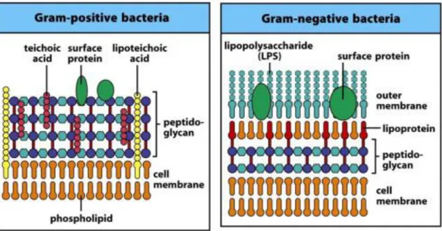
Bacteria are generally classified in two groups: GRAM+ and GRAM- Bacteria. GRAM+ bacteria become purple-coloured after GRAM staining while GRAM- bacteria appear red or pink. In this manuscript, I will focus on GRAM+ bacteria because they present a lipoprotein with a particular role in the pathogenesis on their surface. These lipoproteins are recognized by TLR2/1, and the role of TLR2/1 in pDCs is one of the questions of this thesis.
GRAM+ bacteria present different characteristics: cytoplasmic lipid membrane, thick peptidoglycan layer, lipoteichoic acids, rigid cell walls and a smaller volume of periplasm than that in GRAM- bacteria.
GRAM+ bacteria, comprehending both extracellular (for istance S. aureus) and intracellular (L. monocytogenes, M. tuberculosis) pathogens, developed different strategies to escape the immune system, many of them involving the exploitation of the immune sensors. GRAM These surface receptors are important in the recognition of extracellular bacteria. However, there are different mechanisms that allow engulfing the pathogen and permitting the activation of the intracellular receptors. In the same extent, intracellular pathogens also lead to the activation of surface receptors. Several studies highlighted their importance in the sensing process. In this chapter, I present the dendritic cells repertoire and a deeper charaterizationTLR2 and TLR1, which consist the main object of this thesis.
39
Figure 8. GRAM positive and negative bacterial membrane. From janeway et al., 2008
S aureus: a bacterium with two souls
S. aureus has been defined as “pathobiont”, a microbe that can switch from commensal state to a
pathogenic state according to certain conditions (that are still unknown). S. Aureus is the most common bacterium isolated from inpatient cultures and among the most common microbes from outpatients samples [70]. Moreover, S. aureus is considered the most potent skin pathogen; it colonizes about 30 to 50% of healthy adults intermittently and 10 to 20% persistently.
S. aureus can cause localized infections, like pneumonia or abscess, or a systemic risk like sepsis or
toxic shock syndrome (TSS). In particular, it is a major risk for patients suffering from severe burns [71]. In 2005, 20,000 deaths and 500,000 hospitalizations cases were estimated in USA to be due to
S. aureus infections. These numbers have grown during the last decade, due to the increased
frequency of antibiotic multi-resistant strains. Despite an increase in mortality and morbidity, and
S. aureus being considered a major health care problem, 25% of the population host this
40
cohort studies have been carried out, the genetic factors explaining why a quarter of the population chronically hosts S. aureus in the nasal swabs are not clear. [74]
S aureus can induce TSS, an acute toxin-mediated illness caused by the expression of bacterial
pyrogenic exotoxins called super-antigens (SAg). SAgs binding to the β chains of the T cell receptor (TCR) in the T-cells and to the MHC-II on the DCs generates the over-activation of an enormous number of T-cells. [75] This over-activation causes what is called a “cytokine storm”, a massive release of cytokines with a TH1 profile characterized by high levels of interferon-γ (IFN-γ) and TNF-α. The patient suffers hemodynamic alterations, tissue ischemia that can lead to multi-organ failure, cardiovascular collapse, and death. Paradoxically, despite the large incidence of people carrying S. aureus in the nose, there is a remarkably low frequency of those developing TSS. Some groups have proposed that an evolutionary pressure has caused S. aureus to regulate is own pathogenicity. [76]
Listeria monocytogenes: the comet bacteria
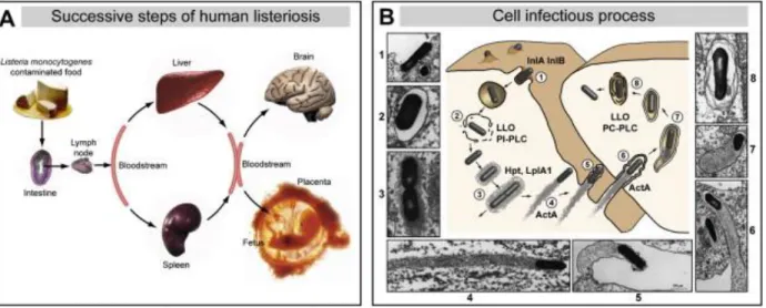
Listeria monocytogenes is responsible for a severe food-borne disease characterized by meningitis,
meningo-encephalitis, materno-fetal and perinatal infections. Generally, Listeriosis is considered an opportunistic infection in immune-compromised patients. Listeria bacteria cross the intestinal epithelial barrier, migrate through lymph and blood vessels to reach liver (they replicate inside hepatocytes) and spleen. Successively, the bacteria can disseminate in the brain and in the placenta, showing their capacity to cross the blood–brain barrier and the materno-fetal barrier. Listeria is capable of escaping neutralization by phagocytic cells and invading non-phagocytic cells.
Lysteria bacteria enter in the cells through a “zipper” mechanism, whereby bacterial surface
ligands interact with receptors in the host cells. The bacteria’s entrance into the cells is concluded with the pathogen entrapped in a sort of vacuole. In about 30 minutes, Lysteria bacteria are able to escape from the vacuole and start to replicate in the cytosol. Lysteria bacteria are also capable of recruiting actin and starting actin polymerization, which results in a network of branched
41
filaments. The polymerization process is located at one bacterial pole, allowing the bacteria to move in the cytoplasm at 10 μm per minute. Lysteria bacteria reach the membrane, inducing the formation of protrusions that allow them to invade neighboring cells and start a new infection cycle. This system protects the bacteria from the host’s defenses. [77]
Resident macrophages are responsible for the initial killing of the bacteria by phagocytation and secretion of TNF-α and IL-12. NK cells are the initial source of IFN-γ, necessary to augment the macrophages’ killing capacity. Innate immunity plays an important role in the initial control of infection, but T-cells are necessary for the final clearance of bacteria. CD4 TH1 and CD8+ memory cells are important for the protective response. [78]
Figure 9: Lysteria model of infection A. Progression of Lysteria infection. B. Cell invasion process.
Cossart et al., 2008
Mycobacterium tuberculosis: an old unfriendly companion.
Mycobacterium tuberculosis is an extremely successful pathogen that seems to have coevolved with the human species during an intimate relationship lasting thousands of years. This evolutionary success relies on the mycobacterial genome, designed to encode genes allowing
42
bacteria to escape the immune response. Moreover, mycobacterial strains resistant to antibiotics are increasingly widespread, constituting a serious threat for global human health. Tuberculosis causes approximately 1.5 million deaths per year: every year about 9 million new TB cases are diagnosed and it is estimated that 1/3 of the world’s population is latently infected with M. tuberculosis. The only existing vaccine is the bacille Calmette-Guerin (BCG), shown to be protective only against the most severe disseminated form of the disease in children but with no effect in adult lung tuberculosis. MTB affects the lungs and is transmitted through aerosols produced during coughing and sneezing of patients with active disease. After its entrance in the lung, MTB is phagocytated by alveolar macrophages where it prevents its own active killing. MTB starts to replicate inside the macrophages and the phagosome. In most individuals, a delayed adaptive immune response is able to contain the pathogen in granulomas for a long time. In these heterogeneous structures consisting mainly of infected macrophages and T cells, M. tuberculosis encounters stress induced by reactive oxygen species and nutrient deprivation, which drives the bacilli into a latent stage. Resuscitation from this containment occurs upon disturbance of the immune system, such as HIV infection or a deficiency in IFN-γ or TNF-α production.
Inhibition of macrophage effector function and phagosome maturation: M. tuberculosis resides
primarily in the macrophages, and bacilli use different sophisticated mechanisms to replicate and resist killing. In infected macrophages the fusion of phagosomes with lysosomes is abrogated (through avoidance of exposure to lysosomal hydrolases, low pH, and other components of lysosomes). The phagosomes fail to incorporate vacuolar ATPases, resulting in no acidification. New studies have found a serine/threonine kinase encoded by the M. tuberculosis pknG gene that is responsible for the inhibition of phagosome-lysosome fusion. [79]
Prevention of macrophagy autophagy: autophagy is a homeostatic process for eliminating
damaged organelles; a double membrane forms an autophagic vacuole that engulfs organelles or unwanted structures within the cell. The vacuole is successively fused with the lysosomes. This
43
process can be induced by IFN-γ. Since this process can kill Mycobacteria, the bacteria express proteins to block it. [80]
Infection and modulation of APCs: while macrophages are the most important site for intracellular
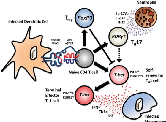
replication, also DCs can be infected by Mycobacteria. Dendritic cells can present antigens, and prime a T-cells response against the pathogen. Dendritic cells infected by the bacteria are not sufficiently activated: they migrate to the lymph nodes and, instead of priming a protective immune response, they seem to help spread the bacteria to other tissues (as a kind of “Trojan horse”). Dendritic cells fail to mature in response to Mycobacteria infection, with reduced expression of MHC-II molecules on the surface and without a sufficient load of mycobacterial antigens. [81]
Modulation of the CD4 response: TH1 cells are crucial to a protective response, as their IFN-γ
production stimulates the macrophages’ killing activity, leading to bacterial clearance. Despite some positive reports, the importance of the TH17 cells in the protective immunity development is still debated. By inducing reduced IL-12 production by monocytes and macrophages, Mycobacterium Tuberculosis affects TH1 development. [82]
44
Figure 10:. Infected DCs crosstalk with naïve CD4+. Goldberg et al., 2014
2.2 Innate immunity in GRAM+ bacterial infection
Pathological microorganisms enter the human body from different sites. These invasions are initially countered by innate immunity. Only when the innate host defenses are bypassed, evaded, or overwhelmed is an adaptive immune response required. Infections are generally rare, despite the fact that we are continuously exposed to potentially pathogenic agents, as many of them fail to overcome the epithelial barrier, and the ones that succeed to infiltrate themselves are in a great part eliminated. Epithelia are more than a simple physical barrier. Tears and saliva, for example, contain lysozyme (an antibacterial enzyme that hydrolyzes β-glyosidic linkages in the cell wall peptidoglycan), in the intestine antibacterial and antifungal peptides called cryptidins or α-defensins are secreted (by Paneth cells), in the respiratory and skin tract other antimicrobial peptides (β-defensins) are present. On the other hand, cationic peptides exist which are able to kill bacteria by damaging bacterial cell membrane. In the fluid coating the lung epithelial surfaces, two
45
proteins (surfactant proteins A and D) are present which bind to and cover the surfaces of bacteria, facilitating phagocytosis by macrophages. [83]
During bacterial infection, phagocytic cells (macrophages and neutrophils) constitute the first line of defense against many common microorganisms and are essential for the control of common infections. The sensing of different pathogen by these cells leads to phagocytosis and successive killing of the microbe. However, infectious organisms cannot always be eliminated, as some bacteria have evolved their capacity to escape and others are not recognized. The lymphocytes in the adaptive immune system have evolved to provide a more versatile means of defense, increasing innate immunity capacity to kill and, in addition, providing increased protection against subsequent re-infection by the same pathogen. The cells of the innate immune system play a fundamental role in the initiation and subsequent guidance of adaptive immune responses, as well as the effective removal of pathogens targeted by an adaptive immune response. Moreover, as the adaptive immune response acts a with a delay of 4-7 days, the innate immune response plays a critical role in controlling infection during this period [84].
Like other pathogens penetrating the epithelial surfaces of the body for the first time, bacteria encounter cells and molecules that can activate innate immune response. Phagocytic macrophages and neutrophils express receptors on their surface, able to recognize and bind different constituents of common bacterial membranes. Activation through these receptors triggers the engulfment of bacteria by macrophages and leads to the release of biologically active molecules – cytokines and chemokines – which are a starting point for the inflammation.
Local inflammation and phagocytosis of invading bacteria may also be triggered by complement activation on the bacterial cell surface. Complement is a system of plasma proteins that, when binding on microbial surfaces (but not on host cells), initiate a cascade of proteolytic reactions. The coating of microbes with the resulting fragments leads to their recognition and binding by phagocytic receptors on macrophages. Complement allows phagocytic cells to recognize
46
pathogens that would otherwise escape phagocytosis and, thus, destruction. The process also releases small peptides that contribute to inflammation. Complement can be activated through three different pathways: binding C1q to the pathogen surface; mannan-binding lectin recognition of mannose on bacteria surface and consequent triggering of complement cascade; or spontaneous and stochastic activation of the cascade on the pathogen surface. Moreover, complement’s final components are also able to damage bacteria by creating pores on their membranes. The membrane-attack complex has a hydrophobic external face and a hydrophilic internal channel. The diameter of this channel is about 100 Å, allowing the free passage of solutes and water across the lipid bilayer. The pores created by complement destroy the ionic balance and allow the entrance of lysozyme, leading to pathogen killing [85]. Complement can be subverted by pathogenic bacteria, such as S. Areus, expressing Staphylococcal protein A (SpA), an abundant cell wall-anchored surface protein. SpA binds tightly to the complement binding (Fcγ) portion of IgG and stimulates an abnormal B lymphocyte proliferation, provoking their clonal expansion and subsequent cell death [86, 87].
Inflammation is characterized by increased local blood flow and fluid leakage. Chemokine attracts leukocytes. Among the first cells to migrate are the neutrophils (recruited in large number to the infected tissue), which possess different receptors for bacteria recognition and for their subsequent engulfing and destruction. Kinetically, monocytes concomitantly migrate or arrive a short time after neutrophils. Monocytes quickly differentiate into dendritic cells and macrophages.
Dendritic cells are also able to recognize and engulf bacteria (though less efficiently than phagocytic cells). This pathogen recognition leads to dendritic cells activation, maturation and migration to the lymph nodes, where they present the antigens to the T-cells. Dendritic cells provide all three signals necessary for T-cells activation: antigen presentation, co-stimulatory molecules, and cytokines.