HAL Id: tel-01249563
https://tel.archives-ouvertes.fr/tel-01249563
Submitted on 4 Jan 2016HAL is a multi-disciplinary open access
archive for the deposit and dissemination of sci-entific research documents, whether they are pub-lished or not. The documents may come from teaching and research institutions in France or abroad, or from public or private research centers.
L’archive ouverte pluridisciplinaire HAL, est destinée au dépôt et à la diffusion de documents scientifiques de niveau recherche, publiés ou non, émanant des établissements d’enseignement et de recherche français ou étrangers, des laboratoires publics ou privés.
Emanuela Monetti
To cite this version:
Emanuela Monetti. Role of ion channels in programmed cell death induced by hyperosmotic stresses in plant cells. Vegetal Biology. Université Paris Sud - Paris XI; Università degli studi (Florence, Italie). Dipartimento di biologia vegetale, 2014. English. �NNT : 2014PA112323�. �tel-01249563�
ECOLE DOCTORALE: SCUOLA DI DOTTORATO:
Sciences du Végétal Scienze Agrarie e Ambientali
PhD Thesis
Emanuela Monetti
ROLE OF ION CHANNELS IN PROGRAMMED CELL
DEATH INDUCED BY HYPEROSMOTIC STRESSES IN
PLANT CELLS
PhD defense on November 17
th2014
Mazars Christian Research director in CNRS, Toulouse Reviewer
Baluska Frantisek Professor at the at the University of Bonn Reviewer
Dron Michel Professor at université Paris-Sud Examiner
Kawano Tomonori Professor at university of Kitakyushu Examiner
Mancuso Stefano Professor at the University of Florence Tutor Bouteau François Maître de conférences at the Université Paris Diderot Co-Tutor
The present work was done in the Laboratoire
Interdisciplinaire des Énergies de Demain (LIED-UMR
8236) of Université Paris Diderot-Paris 7 and in the
LINV-DiSPAA
(Department
of
Agri-Food
and
Environmental Science) of University of Florence
i
Abstract
The work presented in the present thesis relates to the role of ion channels in response to (ionic and non-ionic) hyperosmotic stresses and their interactions with signaling events leading to PCD in plant. Early cell responses such as cytosolic calcium increase and ROS production classically involved in PCD process, seems not to be involved in hyperosmotic-induced cell death in BY2 tobacco and A. thaliana cultured cells. When BY2 tobacco cells were subjected to hyperosmotic stress, an early influx of sodium through non-selective cation channels participates in the development of PCD through mitochondrial dysfunction and NADPH-oxidase-dependent O2·– generation. On the
contrary, non-ionic hyperosmotic stress resulted in an early decrease in anion currents. To further investigate the role of anion channels in non-ionic hyperosmotic stress further experiments were conducted by using A.thaliana cells of the anion channel mutant SLAC1. Results showed that the delayed activation of SLAC1 channels was involved in the non-ionic hyperosmotic stress induced pathway leading to cell death. Interestingly, the early anion channel activity decrease could participate to signalisation or osmotic adjustment allowing cell adaptation and survival, when a second set of events, namely superoxide anion (O2•-) generation by NADPH-oxidase and anion
channel activation could participate in PCD development of a part of the cell population. In addition, the potential role of small peptides belonging to the FMRFamide-like peptide (FLP) family described in metazoan in osmoregulation in A.
thaliana was investigated. By using synthetic peptides, based on FLPs homolog genes
existing in A. thaliana, it was possible to demonstrate that these putative FLPs are involved in hyperosmotic stress response. Overall, the present work shed light on the importance and the complexity of ion channels regulation in the signaling pathways and the processes leading to PCD.
Résumé
Le travaux présenté dans cette thèse concerne le rôle des canaux ioniques de la membrane plasmique en réponse à des stress salins et non salins ainsi qu’aux interactions possibles avec d’autres événements de signalisation conduisant à la mort cellulaire programmée (PCD). Nous avons montré que les réponses cellulaires précoces:
ii
tels que l`augmentation du calcium cytosolique et la production de ROS, classiquement impliqués lors de la PCD, ne semblaient pas être impliqué dans la mort cellulaire induite par les stress hyperosmotiques chez les cellules en culture de tabacco BY2 ou d’A.
thaliana. Nous avons montré que, dans les cas de stress salin chez les cellules de BY2
un influx précoce de sodium à travers des canaux cationiques non spécifiques participe au développement de la PCD en entraînant un disfonctionement mitochondrial et la production de O2• - par des NADPH oxydases. Dans le cas de stress hyperosmotique
non-ionique, nous avons observé une diminaution précoce de l’intensité des courants anioniques. Afin de poursuivre l’étude du rôle des canaux anioniques lors du stress hyperosmotique non salin, nous avons utilisé des cellules A.thaliana nous permettant de travailler avec le mutant de canal anionique SLAC1. Nous avons constaté que l’activation retardée des canaux SLAC1 participait au développement de la PCD induite par un stress hyperosmotique non salin. La réduction précoce de l'activité des canaux anioniques pourrait participer à la signalisation ou l'ajustement osmotique permettant l'adaptation et la survie cellulaire alors que des évènements retardés, à savoir la production d'anion superoxyde (O2• -) par les NADPH-oxydases et l'activation des
canaux anioniques pourraient participer au développement de la PCD d'une partie de la population cellulaire. Nous avons aussi étudié le rôle potentiel des petits peptides appartenant à la famille des peptides FMRFamide décrite chez les métazoaires à l'osmorégulation chez des cellules d’A. thaliana. Des génes susceptibles de coder de tels peptides sont en effet présent dans le génome d’A. thaliana. En utilisant des peptides synthétiques, nous avons montré que ces FLPS putatifs pourraient participer aux
réponses induites losr de stress hyperosmotique chez les plantes. Ce travail illustre la complexité et l'importance de la régulation des canaux ioniques dans les voies de signalisation et les processus conduisant à la PCD.
Riassunto
Il lavoro presentato in questa tesi riguarda il ruolo dei canali ionici nella membrana plasmatica in risposta a stress di tipo iperosmotico (salino e non salino) e il loro coinvolgimento nelle vie di segnalazione che portano alla PCD in pianta. Le risposte precoci (es. aumento di calcio citosolico e produzione di ROS) a stress di tipo iperosmotico, coinvolte negli eventi che portano alla PCD, non sembrano essere
iii
implicate nel modello da noi studiato (cellule di tabacco BY2 e di Arabidopsis). Abbiamo dimostrato che nel caso di stress ionico l’influsso di sodio attraverso i canali NSCC (Non Selective Cation Channels) partecipa allo sviluppo della PCD inducendo depolarizzazione mitocondriale e produzione di O2.- da parte dell’enzima NADPH
ossidasi. Nel caso di stress non ionico abbiamo riscontrato una precoce diminuzione delle correnti anioniche. Per studiare meglio il ruolo dei canali anionici abbiamo usato cellule di A.thaliana che ci permettono di lavorare su mutanti. Abbiamo dimostrato che i canali SLAC1 risultano coinvolti negli eventi che portano alla PCD in seguito a stress di tipo non ionico. La diminuzione di correnti anioniche potrebbe partecipare alla regolazione osmotica che permette l’adattamento cellulare. Un’attivazione delle correnti anioniche e generazione di O2.- da parte dell’enzima NADPH-ossidasi potrebbe
partecipare allo sviluppo della PCD di una parte della popolazione cellulare. Abbiamo inoltre indagato il potenziale ruolo di piccoli peptidi appartenenti alla famiglia FMRF amide-like peptide (FLP) nella regolazione di stress osmotico in colture cellulari di
A.thaliana. Utilizzando peptidi sintetici, abbiamo dimostrato che questi peptidi
potrebbero partecipare alla regolazione di stress osmotico nelle piante. Questo lavoro mette in luce l’importanza e la complessità della regolazione dei canali ionici nelle vie di segnalazione degli eventi che portano alla PCD.
v
I would like to thank my supervisors François Bouteau and Stefano
Mancuso for encouraging me and providing critical advice. All your help
and enthusiasm made possible for me to initiate this research project.
Thank you all the past and present members of both laboratory for assisting
me to learn the new equipment and techniques and for many fun
discussions
Index
Abstract i
Acknowledgement v
Chapter 1
1 Hyperomotic stress 1
1.2 General plant responses to osmotic stresses 2
1.2.1 Osmosensor 3
1.3 Signalisation in response to osmotic stress 7
1.3.1 Reactive Oxygen Species (ROS) signalling 7
1.3.1.1 Plant ROS generation 8
1.3.1.1.1 Singlet Oxygen (1O2) 8
1.3.1.1.2 Superoxide anion (O2.-) 9
1.3.1.1.3 Hydrogen peroxide (H2O2) 9
1.3.1.1.4 Hydroxyl radical (HO•) 10
1.3.1.1.5 Nitric oxide (NO●) 11
1.3.1.2 Sources of ROS in plant cells 11
1.3.1.2.1 Chloroplasts 11
1.3.1.2.2 Peroxisomes 12
1.3.1.2.3 Mitochondria 13
1.3.1.2.4 Apoplast 13
1.3.1.3 ROS scavenging during osmotic stress 14
1.3.1.3.1 Enzymatic control of ROS level 14
1.3.1.3.1.2 Superoxide dismutase (SOD1.15.1.1) 16
1.3.1.3.1.3 Catalase (CAT,1.11.1.6) 16
1.3.1.3. 1.4 Guaiacol Peroxidase (GPX,1.11.1.7) 16
1.3.1.3. 1.5 Ascorbate-Glutathione Cycle 16
1.3.1.3.2 Non-enzymatic control of ROS level 17
1.3.1.3. 2.1 Glycine-Betaine 17
1.3.1.3. 2.3 Polyamines and other secondary metabolities 19
1.3.2 Calcium signaling in response to osmotic stress 20
1.3.2.1 Polyphosphoinositides (PPIs) 21
1.3.2.3 Salt Overly Sensitive (SOS) 22
1.3.4 Protein kinases 23
1.3.4.1 MAPKs pathway 23
1.3.5 Plant ion channels 24
1.3.5.1 Plant cation channels 25
1.3.5.1.1 Potassium channels 25
1.3.5.1.2 Shaker-like potassium channels 27
1.3.5.1.3 Twin-Pore K+/KCO 29
1.3.5.1.4 Kir-like 30
1.3.5.1.5 Non selective cation channels (NSCC) 31
1.3.5.2 Anion channels 33
1.3.5.2.1 Slac 33
1.3.5.2.2 IRACs channels 34
1.3.5.2.3 R-type anion channels 35
1.3.5.3 Mechanosensitive ion channels 36
1.4 Osmotic stress, cell volume regulation and cell death 37
1.5 Programmed Cell Death 39
1.6 Aim of this thesis 40
Chapter 2
2.1 Introduction 43
2.2 Deciphering early events involved in hyperosmotic stress-induced
programmed cell death in tobacco BY-2 cells 44
Chapter 3
3.1 Introduction 63
3.2 Dual responses of cultured plant cells to hyperosmotic stress 64
Chapter 4
4.3 Could FaRP-like peptides participate in regulation
of hyperosmotic stress responses in plants? 103
Chapter 5
Conclusions and perspectives 107
Chapter 1
1
1 Hyperomotic stress
In nature, plants are subjected to different biotic and abiotic stresses. Among abiotic stresses, drought and salinity are the primary causes of crop loss worldwide. Global warming, due to climate change, will enhance these environmental stresses that may severely affect crop productivity (Peters et al., 2013). Drought, defined as soil and/or atmospheric water deficit, reduces plants water potential and turgor. Among drought effects are loss of water, which is involved in stomatal closure and limitation of gas exchange that results in reduced plant growth and productivity and could lead to death. It is expected an increase of drought frequency mainly due to climate change, as a result of decreasing regional precipitations and increasing evaporation driven by global warming (Seneviratne et al., 2012; Sheffield et al., 2012). Since 1970 significant increases in drought extent and severity have already been estimated for Africa, southern Europe, east and south Asia and eastern Australia (Lobell and Gourdji, 2012; Sheffield and Wood, 2008). Water deficit and aridity conditions lead also to salinization of the soil. Salinity has also been identified as one of the major threats because of its degrading effects on landscapes (Ghassemi et al., 1995). Increased salinization of arable lands is expected, resulting in 50% land loss by the year 2050 (Wang et al., 2003).
Drought and salinity have osmotic, ionic and nutritional constraint effects on plants. These effects lead to growth retardation, metabolic disturbances and oxidative stress. Plants may tolerate and adapt to these stressors with different mechanisms including changed leaf architecture, osmotic adjustment, ion exclusion and compartmentalization and a more efficient reactive oxygen species (ROS) scavenging systems. However, depending on the strength and duration of these stresses and on the plant’s genetic features, it can be a matter of life or death since there is always the so-called “stability limit”, which when exceeded leads the organism to death. In Figure 1 is shown the main abiotic stress that affects plants in nature.
2 Figure 1: Overview of abiotic stresses that affect plants (Vickers et al., 2009)
1.2 General plant responses to osmotic stresses
Plants have evolved sophisticated systems to detect and respond to osmotic stresses (Hirayama and Shinozaki, 2010). The general plant osmotic stresses response include a complex signaling pathway, that involves: (i) the reception of the stress, through the osmosensors that are probably located at the plasma membrane level; (ii) the signal transdution (i.e. osmosignaling) to the nucleus where (iii) the last step of controlled expression of specific genes occurs. The molecular basis of osmosensing is not fully understood in plants and little is known about the receptors that detect osmotic stress, much information is available for numerous transcription factors acting in the regulation of gene expression. The signals transductions, amplify and integrate diverse external signals to generate responses such as changes in enzyme activity, gene expression or ion-channel activity.
3
1.2.1 Osmosensor
Osmosensing is pivotal for mantaining a normal water status and all cellular functions in plants. All living cells developed sophisticated tools to sense and respond quickly to osmotic stresses. Osmosensors are proteins whose primary role is to monitor fluctuations in external osmolarity and start an activation of signalling pathways for osmo-adaptation. (Osakabe et al., 2011; Yamaguchi-Shinozaki and Shinozaki, 2006). In general, an osmosensor can be defined as a component that is altered by osmotic stresses. The molecular basis of osmosensing in plants is not fully understood and little is known about the receptors that detect osmotic stress. Signals emanate from several osmosensors are involved in a combinational way that allows highly specific outcomes despite the lack of exclusively specific osmosensors (Kültz, 2012). It has been hypothesized that osmosensing involve the sensing of cell volume, shape, membrane tension or macromolecular crowding by osmosensor proteins (Burg et al., 2007; Kumar
et al., 2013; Schliess et al., 2007). In animal cells slight osmolality can be sensed by
calcium sensing receptors and transient receptor potential (TRP), in particulary TRPV4 in vertebrates (Liedtke et al., 2000; Nilius et al., 2004; Strotmann et al., 2000; Wissenbach et al., 2000). Although is not very clear whether TRP is involved in volume regulation, it was found that it mediates the Ca2+ increase which subsequently stimulates the Regulatory Volume Decrease (RVD) with the activation of K+ channels (Numata et
al., 2007). Interestingly, a recent study made a parallelism between plant and animal
kingdom after finding a Calcium permeable Stress-gated cation Channel 1 (AtCSC1) in
A. thaliana (Hou et al., 2014). The analysis of whole-cell two-electrode voltage clamp
(TEVC) on Xenopus oocytes expressing this gene, led to the conclusion that this protein is a ion channel, which is permeable to various cations, including Ca2+,K+ and Na+. Interestingly, the C-terminal of AtCSC1 comprising domain of unknown function 221 (DUF221) belongs to transmembrane channel-like (TMC) proteins that are cation channel components. Since CSCs are a family of cation channels permeable to calcium triggered by physical signals such as hyperosmotic stress, and these properties are reminiscent of TRPs in animals, these channels are candidates for involvement in osmo- or mechano-sensitive calcium signaling processes in plants (Hou et al., 2014).
Receptor-like kinases (RLKs), which form a large gene family in plants, contain Ser/Thr kinase as a cytosolic domain while having structural elements similar to animal
4
receptor tyrosine kinases (RTKs) (Osakabe et al., 2013; Shiu and Bleecker, 2001). The RLKs are involved in water stress responses, and this receptor like kinase (RLK) family is composed by more than 600 members, with the leucine rich-repeat (LRR)-RLKs forming the largest subgroup (Gish and Clark, 2011; Osakabe et al., 2011). Several RLKs localized in the plasma membrane are known to be involved in the early steps of osmotic-stress signaling in a variety of plant species (Osakabe et al., 2013). Several studies have been performed to explore the involvement of RLK gene in environmental stresses (Chae et al., 2009; Lehti-Shiu et al., 2009). RLKs involvement in sensing environmental cues and abiotic stresses have been reported for various plant species, such as Arabidopsis (Bai et al., 2009; Osakabe et al., 2005; Osakabe et al., 2010; Tanaka et al., 2012), rice (Oryza sativa) (Ouyang et al., 2010), Medicago truncatula (De Lorenzo et al., 2009), and soja (Yang et al., 2010). In particular in plants there are some RLKs such as RPK1, CYSTEINE-RICH RLK (CRK36), PROLINE-RICH-EXTENSIN-LIKE RLK4 (PERK4), and GHR1 (GUARD CELL HYDROGEN PEROXIDE-RESISTANT1), that studies have been reported to control water stress signalling directly in Arabidopsis (Bai et al., 2009; Osakabe et al., 2005; Osakabe et al., 2010; Osakabe et al., 2013). Interestingly, de Lorenzo et al. (2009) identified a salt stress-inducible LRR-RLK gene (SRLK) in M. truncatula, and root growth inhibition by high salinity stress was reduced in SRLK RNA interference (RNAi) transgenic Medicago roots. Epidermal cell-specific expression of the SRLK gene was observed in roots under salt stress, and SRLK was shown to control the expression level of several salt-responsive genes. These findings suggest that SRLK activates the signalling pathway involved in the adaptive response of Medicago roots to salt stress (De Lorenzo
et al., 2009; Osakabe et al., 2013). It tempting to suppose that plants as well as
vertebrate cells, have evolved a sophisticate mechanism at the plasma membrane to sense osmotic changes. Clearly, it is not surprising that plants have evolved mechanisms to sense osmotic forces at the plasma membrane, as these are the first cell component to come in contact with the stress. In animal cells, because they lack a cell wall, as well as the membrane itself cannot provide a mechanical barrier, the actin cytoskeleton has been proposed to control shape after osmotic challenge in animal cells (Papakonstanti et
al., 2001). In different cell lines, a rapid organization of the actin filaments of cytoskeleton has been observed as a result of osmotic stress. This type of organization
5
varies between different cell types, but generally leads to an increase of the F-actin (Pedersen et al., 1999). Recent studies have demonstrated that microtubule-associated (MT) to the cytoskeleton are regulated by osmotic shrinkage. Nevertheless, the mechanisms that link the sensing of the perturbation of the volume to the rearrangement of the cytoskeleton have not been completely elucidated. In plants there is a strong and transient response of microtubules to hyperosmotic stress: microtubules first disappear, but soon they are replaced by massive bundles, called macrotubules (Komis et al., 2002). The formation of macrotubules can be suppressed by oryzalin, which at the same time blocks osmoadaptation, demonstrating that this microtubule response is not a product of adaptation but represents an essential event. A pharmacological study on this phenomenon revealed that inhibitors of phospholipase D, such as n-butanol or N-acetylethanolamine suppress both macrotubule formation and osmotic adaptation (Komis et al., 2006). In vertebrate cells, the integrins have an important role in osmotic sensing. Different studies had shown (Komis et al., 2002) that integrins were activated as a result of disruption of the cell volume, and has been proposed that they serve as the volume sensors after swelling or shrinkage. It was demonstrated an involvement of integrins in the regulation of osmosis and in the movement of ions across cell membranes, in particular they control the flow of calcium, potassium and chlorine, either directly (Hoffmann and Pedersen, 2011) or indirectly (Hoffmann and Pedersen, 2011; Shakibaei and Mobasheri, 2003). It is difficult to transfer these mechanisms to plants, since they have not yet identified canonical integrins in plant cells (Baluška et
al., 2003) and the situation in plant cells differs fundamentally. In plants, the shape is
maintained thanks the cell wall, where elongate load-bearing elements (cellulose microfibrils) are embedded in an amorphous matrix (hemicelluloses, pectins, proteins). In these cells, the interphasic plant cytoskeleton is not directly required to support cell shape and therefore it is free to adopt additional functions. In plants, cortical microtubules are connected with the extracellular matrix through transmembrane proteins that have not been identified so far, but it seems that they share some analogies with animal integrins (Baluška et al., 2003). This link seems to stabilize cortical microtubules. For instance, removal of the cell wall makes microtubules more cold-sensitive in tobacco cells (Akashi et al., 1990). Moreover, cobtorin, a compound identified from a screen that specifically perturbs the parallelity of microtubules and
6
microfibrils (Yoneda et al., 2007) has meanwhile been found to target the cell wall pectins.
7
1.3 Signalisation in response to osmotic stress
Reactive Oxygen Species (ROS), Calcium (Ca2+), Mitogen activated protein kinases (MAPK) and ion channels are recognized to be important in plant signalisation responses to osmotic stress.
1.3.1 Reactive Oxygen Species (ROS) signalling
The ROS signalling network controls a wide range of biological processes such as growth, development and responses to biotic and/or abiotic stimuli (Mittler et al., 2011). ROS are produced as a normal cellular process in all aerobic organisms and include free radicals such as superoxide anion (O2-) hydroxyl radical (•OH), as well as
non-radical molecules like hydrogen peroxide (H2O2) and singlet oxygen (1O2). ROS
are produced by reduction of molecular oxygen (O2) by means of the electron transport
activities of chloroplasts, mitochondria, and plasma membranes (Foyer et al., 1995; Heyno et al., 2011). Also NADPH oxidases respiratory burst oxidase homologues (RBOHs), is involved in ROS production (Suzuki et al., 2011; Torres and Dangl, 2005). During osmotic stress, drought and salinity, ROS production increases (Serrato et al., 2004). During water deficit there is an accumulation of abscissic acid (ABA) in leaves, which minimize water loss by stimulating stomatal guard cell closure (Li et al., 2000). The CO2 available for photosynthesis is reduced and it can misdirect electrons in the
photosystem and lead to the formation of ROS. Unlike biotic stress, where the so called oxidative burst is frequently involved in the events that lead to programmed cell death (PCD), the role of ROS production during osmotic stress needs to be demonstrated. Under abiotic stress ROS seem to be involved in a dual process depending on their cellular concentrations. At low levels ROS have functions as components of a pathway that lead to stress defense/acclimation responses (Vranova et al., 2002). When reaching a level of phytotoxicity ROS become detrimental and participate in the events triggering to cell death (Mittler, 2002).
8
1.3.1.1 Plant ROS generation
1.3.1.1.1 Singlet Oxygen (1O2)
Singlet oxygen (1O2) is a highly reactive short-lived product (half-life,
approximately 200 ns) with a strong oxidizing potential (Apel and Hirt, 2004; Vellosillo
et al., 2010). 1O2 is produced constitutively by a photochemical excitation of the
chlorophyll of photosystem II with oxygen (Krieger-Liszkay et al., 2008). In Figure 2 is shown the generation of 1O2 by excited chlorophyll (Fischer et al.,2013). 1O2 can be also
produced by phytoxins during plant-pathogen interaction. and by peroxidases (POX) (Kanofsky, 2000; Kawano et al., 1998).
Figure 2: 1O2 generation by excited chlorophyll (from Fischer et al.,2013)
Peroxidases (POXs) could also be involved in 1O2 generation (Kanofsky et al., 1988;
Kawano et al., 1998). Peroxidases are involved in a wide range of physiological processes, such as lignification, suberization, auxin catabolism, wound healing and defense mechanisms against pathogen infection (Kawano, 2003). The plant POXs in the apoplastic space are often involved in ROS generation. Peroxidases are considered essential to catalyze the generation of aromatic oxyl radicals from several aromatic compounds (Kawano, 2003; Takahama and Yoshitama, 1998) and this POX-dependent production is often associated to ROS generation (Kawano, 2003).
9 1.3.1.1.2 Superoxide anion (O2.-)
The first reaction of molecular oxygen is a reduction to form thesuperoxide anion (O2.-).
O2 + e- O2
.-O2.- is the product of the reduction of oxygen by one electron. An enzymatic sources
include NAD(P)H oxidases located on the cell plasma membrane of peroxisomes. Dismutation of O2.- produces hydrogen peroxide (H2O2), which may be reduced to water
(H2O) or partially reduced to hydroxyl radical (OH•).
The partial reduction of O2 may generate different Reactive Oxygen Species
(ROS).Moreover, ROS may be produced through transition metal-mediated pathways in the Fenton reaction or in the net Haber-Weiss reactions. A schematic representation of the sequential ROS production is shown in Figure 3
Figure 3: ROS production by multistep reduction of oxygen.(from Appel & Hirt, 2004; Gechev et
al.,2006). Grey lines show the Haber-Weiss reactions.
1.3.1.1.3 Hydrogen peroxide (H2O2)
The hydrogen peroxide production represents an evolutionary strategy performed by plants. In response to various environmental stimuli there is an increase of O2 production, which is subsequently converted to hydrogen peroxide (H2O2).
10
As the O2.- is relatively unstable, it can be converted to O2 or in reaction with a proton to
H2O2, spontaneously or catalyzed by the enzyme superoxide dismutase (SOD):
O2 + 2H+ + 2e- H2O2
H2O2 is a relatively long-life ROS, it can pass through the cellular membranes and act as
signalling molecule in cell-to-cell communication because it’s diffusable between cells (Bienert et al., 2006).It has been shown that ROS participate in various types of cell-to-cell communication in animals and plants. For example in animal cell-to-cells ROS regulate the formation of intracellular gap junctions and signal transduction (Upham and Trosko, 2009) involved in apoptosis in adjacent animal cells (Widel, 2012).
1.3.1.1.4 Hydroxyl radical (HO•)
The hydroxyl radical (HO•), one of the strongest oxidant, is highly reactive and interact with proteins, nucleic acids, lipids and phospholipids. OH• is involved in radical reactions and then is the major responsible for irreversible changes of macromolecules and damages to organelles. OH• has a very short time-life (10-9s) and interacts exclusively in the environment surrounding the site of production.
H2O2 takes part in the formation of HO•, which is produced in the reaction between
H2O2 and Fe2+ acting as a metal catalyst(Fenton reaction). The equation below
represents the main way of HO• production:
O2●- + Fe3+ Fe2+ + O2
H2O2 + Fe2+ Fe3+ + HO● + HO- (Fenton reaction)
O2●- + H2O2 O2 + HO● + HO- (Haber-Weiss reaction)
1.3.1.1.5 Nitric oxide (NO●)
Nitric oxide is a radical synthesized from arginine by NO synthase (NOS) in mammalian cells (Wendehenne et al., 2001). In plants the system responsible for its synthesis is unknown yet. Its derivate peroxynitrite ONOO- can react with O2- and
11
peroxynitrite is highly reactive and toxic in animals, it is not involved in NO-mediated cell death in plants (Delledonne et al., 1998; Gaupels et al., 2011).
1.3.1.2 Sources of ROS in plant cells
As mentioned above, ROS are produced by plant cells under normal conditions as products of metabolic processes and as responses to various environmental stresses. Different plant cell compartments are involved in ROS production. Chloroplasts, mitochondria and peroxisomes are considered the main ROS producers during abiotic stresses(Asada, 2006b; Bose et al., 2013; Mittler, 2002; Mittler et al., 2004). A schematic representation of ROS production in plant cell compartments is shown in Figure 4.
Figure 4:Sources of ROS production in plant cells (Adapted from Bose et al.,2013)
1.3.1.2.1 Chloroplasts
In the light chloroplasts produce ROS through photosystem I (PSI) and PSII electron transport chain (ECT) that are the main reactions centres (Asada, 2006a; Foyer and Noctor, 2003). The antenna pigments are the main producers of O2·-by the Mehler
12
reaction at PSI (Asada,2006). After, H2O2 is produced spontaneously or by a reaction
catalyzed by SOD and then further reduced by the action of ascorbate peroxidases (APXs) in the water–water cycle (Asada, 2006). In addition singlet oxygen 1O2 can be
produced during photosynthesis (Laloi et al., 2004). It was shown an enhanced 1O2
generation at PSI during water stress (Miller et al., 2010). The reduced CO2 availability
due to stomata closure and the excessive exposure to light are involved in this enhanced
1O
2 generation.
1.3.1.2.2 Peroxisomes
The peroxisomes are a type of microbody with a single membrane. Peroxisomes are intracellular organelles mediating a wide variety of biosynthetic and biodegradative reactions. These organelles generate large quantities of ROS (Nyathi and Baker, 2006). Peroxisomes are also involved in senescence (Rio et al., 2006) and heavy metal toxicity (Romero-Puertas et al., 1999). Peroxisomes can be divided into three groups: glyoxysomes, leaf peroxisomes and not specialized peroxisomes. Glyoxysomes (specialized peroxisomes) are storage organelles involved in lipid mobilization. Like mitochondria and chloroplasts, peroxisomes produce radicals O2.- due to their
metabolism. There are two O2- generation sites in peroxisomes, in the organelles and in
peroxisomal membranes. In the organelles the xanthine oxidase (XOD) catalyzes the oxidation of xanthine and hypoxanthine to uric acid. In the peroxisomal membranes, an electron transport chain composed by NADH and cytochrome b generates O2.-. Also the
monodehydroascorbate reductase (MDHAR) generates the O2.-. The H2O2 is produced
during different processes, including the photorespiration glycolate oxidase reaction, fatty acids β-oxidation and disproportionation of O2.–(Foyer and Noctor, 2009).
Peroxisomes can also produce nitric oxide (Corpas et al., 2011). Peroxisomes seem to be involved in ROS dependent response to drought. Riviero et al. (2007) have shown that transegenic tobacco plants expressing the isopentenyltransferase (IPT) gene, encoding an enzyme that catalyzes the rate-limiting step in cytokinin (CKs) biosynthesis, under control of a drought-induced senescence-associated-receptor-protein-kinase (SARK) promoter, had an enhanced drought tolerance (Miller et al., 2010; Rivero et al., 2007). In SARK-IPT transgenic plants there is an increase of
13
modulation of ROS metabolism genes of peroxisomal ascorbate–glutathione (AsA– GSH) cycle genes (Miller et al., 2010).
1.3.1.2.3 Mitochondria
In plants mitochondria, complexes I and III are the main sites of ROS production and they are overall the main sites of ROS production (Møller, 2001), especially in non-green tissues as roots, where they are considered as the main source of ROS generation (Bose et al., 2013)B. The enzyme alternative oxidase (AOX), contributes to H2O2
generation (Apel and Hirt, 2004). Moreover, mitochondria are involved in NO generation due to the reduction of nitrite via a nitrite reductase that has been shown to be dependent on the mitochondrial electron transport (Planchet et al., 2005). It is known that the mitochondrial ROS production enhances during abiotic stresses, especially drought and salinity (Pastore et al., 2007). During drought stress the respiration rate increases to compensate the reduced rate of chloroplast ATP synthesis, consequently ROS production in mitochondria is enhanced.
1.3.1.2.4 Apoplast
The apoplast represents an important site for H2O2 production in response to
various abiotic stresses, such as drought and salinity (Hernandez et al., 2001; Jubany-Mari et al., 2009).
Their production depends on several groups of enzymes, including cell wall peroxidases and plasma membrane NADPH oxidases (e.g.AtRbohD and AtRbohF encode two major NADPH oxidases expressed in guard and mesophyll cells in Arabidopsis), that generate O2.- by oxidizing NADPH and transferring the electron to oxygen (O2) (Sagi and Fluhr,
2006).This ROS apoplastic generation is also known as “oxidative burst” during the hypersensitive responses under a pathogen attack and also regulate cell growth, development, and programmed cell death (PCD) (Foreman et al., 2003; Gapper and Dolan, 2006),(Laloi et al., 2007; Sagi and Fluhr, 2006; Torres et al., 2002). During drought and salt stress, accumulation of H2O2 could be involved in acclimation
responses of plant to drought and salt stresses. It was shown that the reduction of stress-induced apoplastic ROS formation is associated with a decrease in leaf elongation. This response was observed after NaCl but not osmotic treatments. It was found that the
14
apoplast is a presumed site of singlet oxygen 1O2 procuction by apoplastic peroxidases
(POXs) (Kawano et al., 1998).
1.3.1.3 ROS scavenging during osmotic stress
As mentioned above, ROS are continuously produced as metabolic products of various pathways localized in different cellular compartments (Foyer and Noctor, 2013). Due to their reactivity, toxicity and involvement in different processes such as growth, development and programmed cell death, plants have evolved complex different ROS scavenging mechanisms that mainly consist in enzymatic and non-enzymatic pathways to control ROS levels. In plant cells, specific ROS producing and scavenging systems were found in different organelles, such as chloroplasts, mitochondria and peroxisomes. ROS-scavenging pathways from different cellular compartments are coordinated with each other (Pang and Wang, 2008).
3.1.3.1 Enzymatic control of ROS level
The antioxidative defense system comprises several enzymes such as: superoxide dismutase (SOD),catalase (CAT), guaiacol peroxidase (GPX), enzymes of ascorbate-glutathione (AsA-GSH) cycle: ascorbate peroxidase (APX), monodehydroascorbate reductase (MDHAR), dehydroascorbate reductase (DHAR) and glutathione reductase (GR) (Foyer and Noctor, 2013). Figure 5 shows the localization of ROS generation and scavenging pathways.
15 Figure 5: Localization of reactive oxygen species (ROS) generation and scavenging pathways in plant cells. The water–water cycle detoxifies O2·- and H2O2 and alternative oxidase (AOX; Immutans) reduces
the production rate of O2·-in thylakoids [top left; in some plants iron superoxide dismutase (FeSOD)
might replace CuZnSOD in the chloroplast]. ROS that escape this cycle and/or are produced in the stroma undergo detoxification by SOD and the stromal ascorbate–glutathione cycle. Peroxiredoxin (PrxR) and glutathione peroxidase (GPX) are also involved in H2O2 removal in the stroma (middle right). Excited
chlorophyll (Chl) in its triplet state at the light-harvesting complex (LHC) can generate1O2 when the
electron transport chain is over-reduced. ROS produced in peroxisomes during fatty acid oxidation, photorespiration or other reactions are decomposed by SOD, catalase (CAT) and ascorbate peroxidase (APX) (top right). SOD and other components of the ascorbate–glutathione cycle are also present in mitochondria. In addition, AOX prevents oxidative damage in mitochondria (bottom right). In principle, the cytosol contains the same set of enzymes found in the stroma (bottom left). NADPH oxidases [respiratory burst oxidase homologs (RBOHs)] are the major producers of ROS-associated signals required in a wide range of biological activities. The enzymatic components responsible for ROS detoxification in the apoplast and cell wall (CW) are only partially known, and the ROS-scavenging pathways at the vacuole and nucleus are unknown. Membrane-bound enzymes are depicted in white, GPX pathways are indicated by dashed lines and PrxR pathways are indicated by dotted lines in the stroma and cytosol. Although the pathways in the different compartments are mostly separated from each other, H2O2 can easily diffuse through membranes and antioxidants such as glutathione and ascorbic acid
can be transported between the different compartments. DHA, dehydroascrobate; DHAR, DHA reductase; FD, ferredoxin; FNR, ferredoxin NADPH reductase; GLR, glutaredoxin; GR, glutathione reductase; GOX, glycolate oxidase; GSH, reduced glutathione; GSSG, oxidized glutathione; IM, inner membrane; IMS, IM space; MDA, monodehydroascorbate;MDAR, MDA reductase; PGP, phosphoglycolate phosphatase; PM, plasma membrane; PSI, photosystem I; PSII, photosystem II; RuBP, ribulose-1,5-bisphosphate; Rubisco, RuBP carboxylase oxygenase; Trx, thioredoxin; tyl, thylakoid. (from Miller et al.,2009)
16 1.3.1.3.1.2 Superoxide dismutase (SOD1.15.1.1)
Superoxide dismutase (SOD), which dismutates O2.- into H2O2, is present in all
aerobic organisms (Scandalios,1993). The enzyme SOD is classified into three groups: The Fe-SOD present mainly in chloroplasts; Mn-SOD in peroxisomes and mitochondria, Cu/Zn-SOD in the cytosol and chloroplasts.
The data of SOD enzyme activities have shown an increase under various environmental stresses such as drought and salinity. The SOD activity increase is correlated to plant capacity to face environmental stress. For instance, in case of salinity the halophytes show a greater SOD enzyme activity if compared to glycophytes (Jithesh
et al., 2006; Uzilday et al., 2014)
1.3.1.3.1.3 Catalase (CAT,1.11.1.6)
The enzyme CAT is involved in decomposition of H2O2 into water and oxygen
(Willekens et al., 1997). The enzyme CAT has a very high turnover rate. As SOD also CAT has different isoforms, but the main one is sited in peroxisomes. The enzyme CAT is able to scavenge H2O2 produced by various reactions: photorespiratory oxidation,
β-oxidation of fatty acids, and other enzyme systems such as XOD coupled to SOD (Corpas et al., 2008; del Rio et al., 1977). Some studies have shown that CAT-deficients plants had an increased susceptibility to salt, ozone and paraquat (Willekens
et al., 1997).
1.3.1.3. 1.4 Guaiacol Peroxidase (GPX,1.11.1.7)
Guaiacol peroxidase (GPX, EC 1.11.1.7), belongs to a family of enzymes present in fungi, plants and vertebrates. These proteins contain an heme group and oxidize several substrates such as guaiacol and pyragallol in the presence of H2O2
(Penel et al., 1992; Vianello et al., 2007). Different isoenzymes of GPX were found in vacuoles, cell wall and cytosol. GPX is involved in different biosynthetic processes including lignification of cell wall, degradation of IAA and protection against abiotic and biotic stress.
1.3.1.3. 1.5 Ascorbate-Glutathione Cycle
The ascorbate-glutathione (AsA–GSH) cycle plays an important role in the scavenging system for ROS. This pathway includes four enzymes such as ascorbate
17
peroxidase (APX), monodehydroascorbate reductase (MDsAR), deydroascorbate reductase (DAsAR) and glutathione reductase (GR). APX reduces H2O2 to H2O,
prevents the accumulation of toxic level of H2O2, while GR reduces oxidized (GSSG)
to GSH using NAD(P)H as an electron donor. The Figure 6 schematizes the ascorbate-glutathione cycle.
Figure 6: Ascorbate-Glutathione cycle (from Foyer and Noctor 2011)
1.3.1.3.2 Non-enzymatic control of ROS level
Ascorbate and glutathione can reduce the superoxide produced by Mehler reaction (Noctor and Foyer, 1998). Plants have evolved non-enzymatic antioxidants that together with the enzymatic pathways play an important role in ROS scavenging under normal and stress conditions for example drought. The major non-enzymatic antioxidants are mentioned below.
1.3.1.3. 2.1 Glycine-Betaine
Glycine-betaine are compatible solutes accumulated in response to various environmental stresses such as drought, salt, or cold (Mahajan and Tuteja, 2005). They are associated with osmotic balance and can have a role in ROS homeostasis. Glycine-betaine stabilize and enhance both structure and function of oxygen-evolving complex of PSII against dissociation of the regulatory extrinsic proteins (the 18 kD, 23 kD and
18
33 kD proteins (Papageorgiou and Murata, 1995). Exogenous application of Glycine-betaine to Arabidopsis roots, that does not produce these compounds constitutively, has been shown to alleviate OH-generated K+ leakage (Cuin and Shabala, 2007). Moreoveor, Booker et al.(2009) have shown that overexpression of glycine-betaine synthesing enzyme Betaine aldehyde dehydrogenase (BADH) in Arabidopsis and rice(non-accumulators of glycine-betaine) results in an enhanced salinity and ozone stress tolerance in transgenic plants (Booker et al., 2009)
1.3.1.3. 2.2 Proline
Proline is known to accumulate in plants in response to environmental stress and represents a solute involved in plants protection from water deficency. Chen and Dickman (2005) demonstrated that the amino acid proline is a potent scavenger of ROS (Chen and Dickman, 2005). Moreover, this amino acid is capable to mitigate the impacts of drought, salt and temperature stress in plants. For example, proline accumulation can influence stress tolerance in multiple ways (Fig 7).
Figure 7: Multiple functions of proline in plants. Proline is used for protein synthesis, has protective functions as an osmolyte, contributes to the maintenance of the redox balance, can regulate development and is a component of metabolic signaling networks controlling mitochondrial functions, stress relief and development. Abbreviations: APX, ascorbate peroxidase; CAT, catalase; PCD, programmed cell death (from Szabados and Savouré, 2010)
19 1.3.1.3. 2.3 Polyamines and other secondary metabolities
Polyamines (PA) are polycationic compounds present in most living organisms (Alcázar et al., 2010). These compounds seem to be involved in plant growth and development, PCD, signaling, gene expression and adaptation to environmental stresses (Kuznetsov and Shevyakova, 2007; Takahashi and Kakehi, 2010). Recently it was shown an important role for 1,3-diaminopropane (DAP) in A. thaliana drought stress response (Jammes et al., 2014). The 1,3-diaminopropane in plants is co-produced with H2O2 from metabolism of the classical polyamines. Acetylated 1,3-diaminopropane
seems to have a role in adaptation to drought avoiding the complete stomatal closure to maintain CO2 diffusion to photosynthetic tissues. Indeed, during stomata closing there is
an increase in water use efficiency for the plant and the CO2 is cut off. This diminution
in CO2 may conduce to stomatal (re)opening despite high ABA and the increased
liability of dehydration, and even death (Jammes et al.,2014). The model proposed by Jammes et al. (2014) shown that during slow-soil-water availability, acetylation of DAP by acetyltransferases, identified earlier as NATA1 is involved in regulation of membrane transport mechanisms involved in readjusting CO2-H2O homeostasis. While
an intense drought leads to suppression of NATA1 expression (Jammes et al., 2014). Carotenoids are natural pigments, which are synthesized by plants, with antioxidant property, indeed they are involved in quenching of 1O2 and peroxy-radicals (Neubauer
and Yamamoto, 1994). Other secondary metabolites such as sorbitol, mannitol, myo-inositol, ononitol or pinotol are accumulated during drought and salinity stresses (Williamson et al.,2002) acting as antioxidants.
20
1.3.2
Calcium signaling in response to osmotic stress
Calcium (Ca2+) is an important second messenger in regulation of different cellular processes and Ca2+dependent signaling is widely conserved among eukaryotes. (Lecourieux et al., 2006; Xiong et al., 2014).
In plant cells, [Ca2+]cyt is maintained low around 100nM respect to the vacuole (200 nM)
and apoplast (300 nM) and it was proposed that this mechanism may provide the possibility to quickly import calcium inside the cell when required for signalling (Lecourieux et al., 2006). Changes in plant [Ca2+]cyt are observed in response to salinity
and drought (Parre et al., 2007; Ranf et al., 2008). The signalling pathway in which is involved Ca2+ is complex because it processes multiple signals simultaneously. Increase in [Ca2+]cyt are sensed by Ca2+-sensor proteins and it transforms them in downstream
pathways by binding or activating different targets.
Among the effector proteins there are calmoduline (CaM), CaM-binding proteins, Ca2+ -dependent protein kinases(CDPK) (Wheeler and Brownlee, 2008). The signals from CaM can induce gene expression of specific transcription factors (Tuteja and Mahajan, 2007). CDPKs were found in osmotically stressed plants, in fact a CDPK was reported to be expressed earlier and for longer in a drought tolerant species respect to the sensitive ones. In plant several isoforms of CDPKs exist and this can be explained with the need of fine tuning responses to diverse abiotic stresses.
Calcium permeable channels can be considered as the upstream elements in the Ca2+- dependent signalling pathways, thus their activation is quite important. These channels are placed in plasma membrane, tonoplast, as well as in the ER (Kudla et al., 2010), and their activity is coupled with Ca2+-ATPase whose main function is to maintain calcium homeostasis by pumping out calcium (White, 2000). On plasma membrane also Ca2+ voltage dependent channels are present and their activation depend on depolarization of membrane such as depolarization-activated Ca2+channel (DACC)and the hyperpolarization-activated Ca2+channel (HACC) (Tuteja and Mahajan, 2007). Cytosolic calcium increase can be due also to Ca2+ mobilization from organelles, event mediated by second messengers, IP3or cADPR ((Hetherington and Brownlee, 2004), that acts as ligand for calcium gated channels in vacuolar or ER membranes or by Ca2+ -antiporters (Lecourieux et al., 2006).
21
It has also been reported that the rise in [Ca2+]cyt in plant could be provided by
non-selective cation channels (NSCCs) that allow the cation passage across membranes with limited discrimination between cations (Demidchik and Maathuis, 2007). These NSCCs play also a crucial role in salinity stress (Demidchik and Maathuis, 2007). Indeed, Ca2+ blocks some NSCC and consequently reduces Na+ influx into intact tissue to maintain a correct K+/Na+ ratio (Demidchik and Maathuis, 2007).
A recent study has demonstrated that Ca2+-dependent wave signaling may play an important role in the process of systemic signaling in response to salt stress (Choi et al., 2014). It was found that local salt stress at the root tip level leads to the spreading of a Ca2+ wave that propagates preferentially through cortical and endodermal cells to distal shoot tissues, triggering systemic molecular responses in the shoot. In particular, local treatment with LaCl3, a putative plasma membrane channel blocker, inhibits the
transmission of the Ca2+ waves and in parallel also inhibits the systemic molecular responses. Up to now, the Cyclic Nucleotide-Gated Channels(CNGC) and the Glu Receptors (Dodd et al., 2010) involved in Ca2+flux (Vincill et al., 2012) are identified, but none have been shown to function in NaCl-induced [Ca2+]cyt elevation. Recently a
role was proposed for plant anexin in Ca2+flux under salinity (Laohavisit et al., 2013). The anexin1 (AtANN1) seems to have an role in the generation of root and root epidermal NaCl-induced [Ca2+]cyt elevation.
1.3.2.1 Polyphosphoinositides (PPIs)
Polyphosphoinositides (PPIs) are present in all eukaryotes (Michell, 1975). The Phosphatidylinositol 4,5-bisphosphate [PtdIns(4,5)P2] is the best known PPI and it is the substrate for signal-activated phospholipase C (PLC), with the latter being an enzyme that hydrolyzes inositol-1,4,5-trisphosphate (InsP3) and diacylglycerol (DAG). InsP3 releases Ca2+ from an intracellular store by activating a ligand-gated Ca2+- channel (Munnik and Vermeer, 2010) which triggers several physiological changes in response to osmotic stress, including stomata closure (Bartels and Sunkar, 2005). Furthermore, in plant the first phospholipid based signalling pathway in response to osmotic stress is the PLC pathway, which is activated within minutes after both salt and water stresses (Munnik and Vermeer, 2010). Osmotically-triggered PtdIns(3,5)P2 responses have been reported for Chlamydomonas, tobacco pollen tubes, epidermal
22
strips of pea leaves and suspension-cultured cells of tomato and alfalfa (Munnik and Vermeer, 2010; Zonia and Munnik, 2004). In mammalian cells, during osmotic stress, the PtdIns (4,5) P2 regulates many ion channels and transporters (for example NHE1, VRAC, ENaC) (Hilgemann et al., 2001; Suh and Hille, 2005; Voets and Nilius, 2007). All phosphatidylinositols phosphorylated at position 3 are synthesized by the phosphatidylinositol 3 kinase (PI3K), capable of phosphorylating inositol in position D3 (Toker, 1998). Several studies suggest that the activity of this enzyme is related with the Regulatory Volume Decrease Channel (RVDC, topic discussed more in detail in the next section). Current knowledges indicate that the PI3K plays a role as a sensor of cellular swelling, as the phosphorylation of PI in position 3 of the ring, by means of PI3K, strongly depends on the curvature of the membrane; therefore it would appear that the activity of this enzyme can be regulated by the mechanical deformation of the membrane itself due to changes in cell volume (Hübner et al., 1998).
1.3.2.3 Salt Overly Sensitive (SOS)
Exposure to salinity activates the Salt Overly Sensitive (SOS) pathway. The SOS pathway is involved in removal of Na+ from the cytosol by activation of Na+/K+ exchangers (antiporters), which are membrane proteins localized in the vacuolar or plasmamembrane. The Ca2+ spike after salt stress induces the SOS pathways. SOS3 encodes amyristoylated calcium-binding protein that appears to have the function of calcium sensor in the perception of an increase of cytosolic Ca2+. The complex SOS3-Ca2+ is able to interact with the protein kinase SOS2 which phosphorylates SOS3 proteins. SOS3–SOS2 interactions recruit SOS2 to the plasma membrane, leading to activation of the downstream target SOS1, an Na+/H+ antiporter allowing extrusion of excessive Na+ from the cytosol (Ji et al., 2013).
23
1.3.4 Protein kinases
1.3.4.1 MAPKs pathway
Mitogen-activated protein kinase (MAPKs) appears to be a key mechanism for intracellular signal transduction in both eukaryotic and prokaryotic. In general, the cascades of MAP kinase enzymes are characterized by the presence of three functionally related protein kinases: (i) the activation of MAP kinase requires phosphorylation of tyrosine and threonine residues, preserved in the so-called “TEY (Thr,Glu,Tyr) activation loop” by the MAPK kinases (MAPKK);; (ii) in turn, the MAPKK are activated through phosphorylation of threonine residues of conserved threonine and or serine by a MAPKK kinase (MAPKKK) (Bartels and Sunkar, 2005); (iii) and finally the activated form of MAPK then migrates from the cytoplasm to the nucleus in order to directly activate transcription factors (TF). In vertebrates 14 MAP kinase genes have been identified that define 7 distinct MAP kinase pathways, involved in different abiotic and biotic stresses (Pearson et al., 2001). For example, in vertebrates, TF involved in osmotic stress response is the NFAT5 that activates the transcription of genes, which are indirectly involved in cell volume regulation, such as
the sodium chloride-betainecotransporter (SLC6A12)the sodium/myo-inositol
cotransporter (SLC5A3) and the taurine transporter (SLC6A6) (Lee et al., 2011). As in vertebrates, in plants there are several MAP kinase genes, that define distinct MAP kinase pathways vital in the signaling of different abiotic and biotic stresses. For example, in Arabidopsis thaliana there are MPK3, MPK4 and MPK6 kinases involved in response to shock osmotic stress. Indeed, Kim and coworkers (Kim et al., 2011) have shown a role for MPK4, and observed that mpk4 loss-of-function mutants and MpK4-overexpressing transgenic plants (MpK4 OXs) produce aberrant responses following osmotic stress. In-gel kinase assays for MPK3 phosphorylation in mpk4 mutants and MpK4 OXs demonstrated that MpK4 plays an important role in osmotic-stress responses via regulation of MPK3 activity; in fact, after osmotic stress in Mpk4 mutant, there is a decline in phosphorylation rate meanwhile in MpK4 OXs the level of phosphorylation remains higher, even when compared with wild type. In addition, it was also found that these components also cross-talked with ABA-dependent stress-responses genes, in particular NCED3, a gene that encodes a key regulator of the ABA signaling pathway. NCED3 gene expression is reduced in mpk4 mutants and increased
24
in MpK4 OXs compared with WT plants. All together, these results strongly suggest that MpK4 might mediate osmotic-stress response and promote NCED3 expression(Kim et al., 2011).
1.3.5 Plant ion channels
The ion channels activity is generally associated to the earliest observed events following the perception of a stimulus. The ions (eg. K+,Na+, etc) are transferred in the direction of their electrochemical gradient, from one compartment to another. The opening/closure of these channels depends on various stimuli, there are two main types of ion channels:gated and non-gated. Under physiological conditions, ion channels perform three basic functions: (i) are involved in osmotic homeostasis (ii) the transduction of signals, amplifying and propagating electric signals for example, or carrying secondary messengers, and (iii) control of the membrane potential. The following sections describe an overview of the different classes of ion channels present in plants, focusing mainly on plasma membrane channels, on how their structures and activities are linked to other signaling events, and finally, on their roles and implications in signaling pathways leading to PCD process.
25
1.3.5.1 Plant cation channels
1.3.5.1.1 Potassium channels
Potassium (K+) represents up to 10% of the total dry weight of the plant. It is the most abundant cation in the cytosol as it is compatible with the structure of proteins, even at high concentrations (Kronzucker and Britto, 2011; Maathuis and Amtmann, 1999). The cell uses this cation in several important functions such as neutralization of the anion electric control of membrane polarization and osmoregulation. Plants have a variety of transport systems for K+(Fig.8).
Figure 8:Phylogenetic relationship and topology of Arabidopsis thaliana potassium channels. A: the genome of the model plant Arabidopsis encodes for 15 K+-selective channels, which are subdivided into three structural classes: The class of voltage-dependent Shaker-like potassium channels (Kv), the twin
pore channels (TPKs;K2P), and Kir-like potassium channel (KCO3). In addition, it encodes for a
cation-nonselective two-pore channel (TPC1). TM, transmembrane domain; P, pore region. B: despite their structural resemblance, plant Shaker-like Kv channels split up into outward (left), inward (middle), and
26
weak (right) rectifiers according to their gating properties. Typical voltage-clamp recording from heterologously (Xenopus oocytes) expressed Kv channels are shown (from Hedrich,2012)
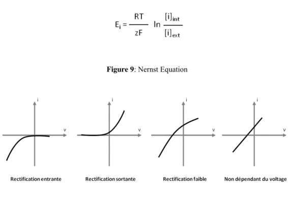
In plants, as in animal cells, the activity of potassium channels is determined by the cell membrane electrical properties. Two K+ permeable conductances have been characterized and they differ in the voltage range by which they are activated. One is activated by membrane hyperpolarization (with potential more negative than EK= Ei,
thermodynamic equilibrium potential of K+) and it is mainly involved in K+ entry (Fig 9). This incoming rectifying conductance is commonly called IRC Inward Rectifying Conductance (Fig 10). The other K+ conductance is activated at potential levels more positive than EK membrane and it’s involved in the efflux of K+ out of the cell. This type
of conductance is called ORC for Outward Rectifying Conductance.
Figure 9: Nernst Equation
Figure 10: Relation between current and voltage (I/V). It is shown the different type of K+channels in plants (Adapted from Véry & Sentenac, 2002)
The ORC is characterized by slow sigmoidal activation current (Fig 8). The ion channels belonging to this family are mainly selective for K+ if compared to Na+ and are blocked by Ba2+ and tetraethylammonium (TEA). In Arabidopsis, the presence of ORC
27
was reported in guard cells, root hairs and cultured cells of different plants (Bouteau and Tran, 2012; Forestier et al., 1998; Roelfsema and Hedrich, 2010). The K+ channels are multimeric proteins. Their transmembrane subunits, named α subunits are characterized by the presence of one or two pore domains P. Three families of subunits α forming selective channels K+ have been identified in plants areas: Shaker, TPK and Kir-like (Véry and Sentenac, 2003). They all have counterparts in animal cells.
3.5.1.2 Shaker-like potassium channels
Shaker-like potassium channels, inhibited by TEA, are divided in three groups depending on their voltage sensitivity (Véry and Sentenac, 2002). The first class is called Potassium Inward-Rectifying channels (KIR) including AKT1, KAT1, KAT2, and
SPIK. They are responsible of K+ uptake after membrane hyperpolarization. In the second class there are the potassium channels that depending in K+ electrochemical gradients mediate both uptake and release.AKT2 belongs to this family. The third is the Potassium Outward-Rectifying channels (KOR) including GORK and SKOR that mediate K+ release upon membrane depolarization. The Shaker family contains nine genes and are composed of four subunits arranged around a central pore (Zimmermann and Sentenac, 1999).Seven of the nine Shaker Arabidopsis proteins were characterized so far and it was found to be highly selective for K+. The available information suggests that these channels are the main transport systems involved in the nutrition K+ and / or the regulation of the K+ equilibrium. Among the Shaker ion channels GORK(outward-rectifying potassium-selective channel) is expressed in guard cells and root hairs, while SKOR is localized in the vasculature, namely, the xylem parenchyma cells, where it is involved in solute loading in the xylem network (reviewed by, (Hedrich, 2012). Fig. 11 shows a schematic representation of the expression patterns of Arabidopsis K+ -selective channels.
28 Figure 11: Expression patterns of Arabidopsis K+-selective channels (from Lebaudy et al.,2007).
29 1.3.5.1.3 Twin-Pore K+/KCO
In mammalian cells, the KCNK are characterized by a hydrophobic core composed by four transmembrane segments and two P domains (Fig 12). The characteristic of this class of ion channel is that they have a weak voltage activation (Patel and Honore, 2001). In Arabidopsis, five channels (TPK1, TPK2 ,TPK4, and TPK5 TPK6) have structural homologies to mammalian ion channels (Voelker et al., 2006; Zimmermann and Sentenac, 1999). These channels are localized in the tonoplast (Czempinski et al., 2002; Schönknecht et al., 2002; Voelker et al., 2006), except AtTPK4, which is localized at the plasma membrane and it’s specific for the pollen tube (Becker et al., 2004).
30 1.3.5.1.4 Kir-like
In mammalian cells, this family is characterized by IRK conductance and has an hydrophobic core formed of two segments and a trasmembrane domain (fig 13) .These channels don’t show voltage-dependence, the inward rectification is due to a blockage of outward current by Mg2+ and polyamines (Ficker et al., 1994; Matsuda et al., 1987). In Arabidopsis thaliana, a channel located at the tonoplast was found (Voelker et al., 2006) to share similarity with the two P domain channels (KCO family) and was therefore named KCO3 (Czempinski et al., 1999).
Figure 13: Structural conformation of Kir-like ion channels in Arabidopsis (Adapted from Lebaudy et al., 2007)
31 1.3.5.1.5 Non selective cation channels (NSCC)
Non-‐selective cation channels are macromolecular pores at plasma membrane that allow the ions Na+, K+, Ca2+ fluxes.
Under saline conditions Na+ enters in the cells through non-selective cation channels (NSCCs,) (Demidchik and Tester, 2002), depolarizing the plasma membrane (Chen et
al., 2007; Pandolfi et al., 2010; Shabala and Cuin, 2008; Wegner et al., 2011).
Even though no definitive molecular candidates have clearly emerged for NSCCs, it seems that various classes of NSCCs could be responsible for influx of Na+ under salt stress, especially a depolarization activated class of NSCCs (Demidchik and Maathuis, 2007). There are some candidate genes belonging to family of glutamate receptors (GluR) (Lacombe et al., 2001) and the family of CNGC (= Cyclic Nucleotide Gated Channel) (Mäser et al., 2001). The NSCC may be activated by many factors such as the voltage (hyperpolarization or depolarization), calcium, mechanical variations, cyclic nucleotides or glutamate (Demidchik et al., 2002). Most studies have focused on the NSCC of roots. They are responsible for the influx of Na+ at the root level (Demidchik and Maathuis, 2007; Demidchik and Tester, 2002). NSCCs can therefore be subdivided according to their voltage dependence into voltage-insensitive, depolarization-activated o rhyperpolarization-activated NSCCs (reviewed by(Demidchik and Maathuis, 2007)): Depolarization-activated NSCCs (DA-NSCCs)
Voltage-independent NSCCs (VI-NSCCs)
Hyperpolarization-activated NSCCs (HA-NSCCs)
The DA-NSCCs were found in Arabidopsis cultured cells(Cerana and Colombo, 1992),
Arabidopsis leaf mesophyll (Shabala et al., 2006), Arabidopsis root epidermis (Shabala et al., 2006), Arabidopsis guard cells (Pei et al., 1998), Hordeum vulgare root xylem
(Wegner and De Boer, 1997), Thlaspi spp. Mesophyll cells (Piñeros and Kochian, 2003), Arabidopsis pollen tubes (Becker et al., 2004), Phaseolus vulgaris seed coats (Zhang et al., 2002) and Phaseolus vulgaris cotyledon epidermal cells (ZHANG et al., 2004). The voltage dependence of DA-NSCCs is similar to outward rectifying K+ -selective channels (ORC). The DA-NSCCs can be blocked by extracellular Ca2+, TEA, nifedipine, diltiazem and verapamil (reviewed by (Demidchik et al., 2002; Shabala et
32
The voltage-independent NSCCs (VI-NSCCs) are insensitive to plasma membrane voltage, this suggests that they equally conduct outward and inward currents. They can be divided into two groups on the basis of blockage by cations such as Ca2+, Ba2+,Mg2+ and Zn2+. This class of ion channels was found in Aster tripolium guard cells (Véry et
al., 1998), Arabidopsis mesophyll cells (Shabala et al., 2006), Arabidopsis cells
(Amtmann et al., 1997), Arabidopsis pollen tubes (Becker et al., 2004), expanding
Pisum sativum leaf epidermis (Elzenga and Van Volkenburgh, 1994) and motor cells of Samanea saman (Yu et al., 2001). DA-NSCCs and VI-NSCCs are the primary
candidates for Na+ entry.
The Hyperpolarization-activated NSCCs (HA-NSCCs) conduct mainly Ca2+ and were described as hyperpolarization-activated Ca2+ channels (HACaCs) (Demidchik and Maathuis, 2007). This class of NSCC is involved in Ca2+ influx important for plant elongation, growth of root hairs and cells in the elongation zone (Demidchik and Maathuis, 2007). Gelli et al. (Gelli et al., 1997) have shown that some type of HA-NSCCs are sensitive to elicitors, this suggest that these channels may be involved in Ca2+signalling, which is part of the early response to pathogen attack (Gelli et al., 1997).