HAL Id: tel-00924475
https://tel.archives-ouvertes.fr/tel-00924475
Submitted on 6 Jan 2014HAL is a multi-disciplinary open access
archive for the deposit and dissemination of sci-entific research documents, whether they are pub-lished or not. The documents may come from teaching and research institutions in France or
L’archive ouverte pluridisciplinaire HAL, est destinée au dépôt et à la diffusion de documents scientifiques de niveau recherche, publiés ou non, émanant des établissements d’enseignement et de recherche français ou étrangers, des laboratoires
New roles of STAT5 factors in chronic myeloid leukemia
cell maintenance
Luana Casetti
To cite this version:
Luana Casetti. New roles of STAT5 factors in chronic myeloid leukemia cell maintenance. Human health and pathology. Université René Descartes - Paris V, 2013. English. �NNT : 2013PA05S018�. �tel-00924475�
UNIVERSITÉ PARIS 5 – PARIS DESCARTES
É
COLE DOCTORALE BIOLOGIE ET BIOTECHNOLOGIEThèse
pour l’obtention du Diplôme de
DOCTEUR DE L’UNIVERSITÉ PARIS 5
S
PÉCIALITÉ: HÉMATOLOGIE
ETONCOGENÈSE
Soutenue
le 28 Novembre 2013
par
Luana CASETTI
New roles of STAT5 factors in chronic myeloid leukemia
cell maintenance
Thèse dirigée par
Mme le Docteur Isabelle DUSANTER-FOURT
JURY:
Mme le Professeur
Catherine LACOMBE Président
M. le Professeur
Stefan CONTANTINESCU Rapporteur
M. le Professeur
François-Xavier MAHON Rapporteur
M. le Professeur
Philippe ROUSSELOT Examinateur
“Strive not to be a success, but rather to be of value”
Je tiens tout d’abord remercier l’ensemble des membres de mon jury de thèse, Madame le Professeur Catherine Lacombe pour avoir accepté d’en être la présidente, Monsieur le
Professeur François-Xavier Mahon et Monsieur le Professeur Stefan Constantinescu pour
avoir accepté d’être rapporteurs de ma thèse, et Monsieur le Professeur Philippe Rousselot et Monsieur le Docteur Jérôme Kluza pour avoir accepté de faire partie de ce jury.
Je remercie Isabelle Dusanter-Fourt pour m’avoir accueillie au sein de son équipe et pour avoir dirigé cette thèse. Elle m’a encadrée, guidée et conseillée au cours de ces trois ans. Elle a toujours été compréhensive, disponible, patiente et attentive en toutes circonstances et finalement on a partagé plein de beaux moments au niveau professionnel mais aussi personnel. Je la remercie de m’avoir fait partager sa vision des choses sur de nombreux sujets, scientifiques et autres. Merci aussi pour toutes les corrections du manuscrit, pour ses précieux conseils et son esprit synthétique que moi, en tant qu’italienne, je n’ai pas du tout. Elle m’a aussi donné l’opportunité d’interagir avec beaucoup de jeunes scientifiques en me faisant participer à beaucoup de congrès et réunions en France et à l’étranger. C’est aussi grâce à ça que j’ai acquis une maturité que je n’avais pas avant.
Je remercie tous les membres passés et présents de l’équipe. Avec une pensée particulière je remercie Aurore pour avoir partagé avec moi tous les jours de ma thèse. Ensemble on n’a pas seulement travaillé, mais aussi couru, rigolé, dansé, chanté et pleuré. Je la remercie aussi pour avoir été très patiente avec moi, pour m’avoir aidée avec tous les trucs technologiques et, bien sûr, pour m’avoir appris à parler correctement français. Merci pour cette belle amitié. Enfin, «I’m glad you came!».
Un très grand merci à Sévérine pour m’avoir transmis toutes ses connaissances, pour m’avoir guidée vers le bon chemin et, bien sûr, pour ses éclats de rire qui faisaient du bien au quotidien.
Je remercie Evelyne pour les heures passées à trier mes cellules, pour ses conseils scientifiques, pour sa bonne humeur et pour nous la transmettre chaque jour. Merci aussi pour sa sincérité, sa manière directe de dire les choses et de nous faire les petites remarques quotidiennes. Ceci nous a finalement permis d’instaurer une confiance très rare les uns avec les autres et de faire un bon travail d’équipe.
Merci à Brigitte pour sa disponibilité, son rire contagieux et pour avoir animé mes journées avec ses histoires marrantes et particulières. Même si elle nous a rejoints depuis peu
Je remercie également Geoffrey pour son aide technique, pour m’avoir toujours réconfortée en me faisant rigoler et pour avoir dirigé la « Cake Session » (que j’ai gagné en plus!). On a passé une très bonne année ensemble et j’aurai toujours un bon souvenir de nos moments dans le labo et en dehors du labo.
Je remercie Claude pour ses encouragements et sa force de caractère, elle m’a appris beaucoup.
Merci également à Alexandre pour son enthousiasme, pour sa disponibilité et pour être toujours prêt à me dépanner avec un sourire.
Je remercie Audrey pour être toujours gentille, souriante et réjouissante, c’est très agréable d‘avoir partagé le bureau avec elle!
Merci à Fred pour ses longs discours qui changent les idées et nous en donnent des nouvelles.
Merci à Cécile pour m’avoir supportée avec un sourire tous les matins quand j’arrivais au labo déjà stressée et pour nos petites pauses café qui me remotivaient pour la journée.
Merci aussi à Katy, car, même si elle ne fait pas partie de notre équipe, elle m’a soutenue, écoutée et motivée jour après jour, elle a été pour moi un grand exemple à suivre.
Merci également à Azzedyne pour son écoute et son thé à la menthe, Serge pour ses apéros qui nous changeaient les idées, je remercie aussi Fabio, Yasmine, Ayda, Isabelle
Vigon, Véronique Chauvin, Marie-Christine, Jean-Marc et Alain, car leur présence a
également contribué à me faire évoluer et a rendu cette thèse plus enrichissante.
Je remercie le Pr. Frédéric Morinet, pour son envie de partager ses connaissances et ses compétences et pour avoir contribué à étoffer ma culture générale. Je le remercie aussi pour m’avoir incluse dans ses dernières publications qui pourraient, pourquoi pas, me donner des bonnes opportunités futures.
J’ai été très heureuse de faire partie de cette belle équipe.
Je ne peux pas citer tout le monde, mais je tiens à remercier également l’ensemble des personnes du 1er étage de Mechain, car chacune m’a aidé, à un moment ou à un autre, à embellir les journées les plus difficiles.
Merci aussi à tous les amis de toutes nationalités que j’ai rencontré dans cette merveilleuse ville avec lesquels j’ai partagé des très bonnes soirées, des moments sportifs, des sorties culturelles et de longs voyages à la découverte de la France. Ces 4 années sur Paris n’auraient pas été pareilles sans eux.
Merci à ma famille pour son soutien sans faille. Merci à mes cousins Antonietta,
Margherita, Gianluigi, Cristiano, Nazaret, Vittorio, mon petit adoré Samuel et mes tantes, Zia Angela, Zia Adelina, et tous les autres, pour leurs conseils et leur confiance, pour
m’avoir toujours soutenue même de loin et pour être présents dans ma vie, jour après jour. Vous êtes vraiment essentiels pour moi.
Merci à Chiara pour m’avoir appris l’importance des séries dans la vie des doctorants, pour me réjouir avec ses chansons et ses discours infinis chaque jour. Merci aussi pour son support face à chaque problème de la vie ou du labo.
Merci à Rosa, ma Colocataire depuis des années. Notre complicité et notre soutien est quelque chose d’assez rare et fort et le résultat d’une amitié enviable. J’espère qu’il y aura encore plein de pages à écrire dans notre bouquin « Le pagine della nostra vita ».
Merci à Sara, la copine de toute une vie. Sa présence, son écoute et sa compréhension m’ont été de grand soutien durant tout ce travail de thèse et pour surmonter les périodes difficiles loin de mon pays. En elle je retrouve la force qui me permet d’éclairer tout d’un coup même les moments les plus sombres.
A Fabrizio, merci pour tout. Pendant ces 3 ans sa présence a été essentielle pour moi. Que dire de plus, je le remercie déjà chaque jour. Merci aussi pour avoir voulu que ce manuscrit soit parfait.
Merci à mes parents, qui ont toujours cru en moi, qui m’ont supportée, qui m’ont appris à être sérieuse, persévérante, intègre, attentive aux opportunités de la vie et qui m’ont toujours encouragée à partir et faire mes choix professionnels.
TABLE OF CONTENTS ... 7
ABBREVIATIONS ... 13
INTRODUCTION PART I ... 19
1. The Hematopoietic system ... 21
1.1 HEMATOPOIESIS AND HSCs ... 21 1.2 HEMATOPOIETIC DEVELOPMENT ... 23 1.2.1 Embryonic hematopoiesis ... 23 1.2.2 Adult hematopoiesis ... 24 1.2.3 Regulators of HSC generation ... 25 1.3 BIOLOGY OF HSCs ... 27
1.3.1 HSCs as quiescent cells: a protection mechanism that favors their resistance ... 27
1.3.2 Regulation of self-renewal and maintaining of HSCs ... 28
1.3.2.1 Cell‐intrinsic regulators of adult HSC maintaining ... 29
1.3.2.2 Cell‐extrinsic regulators of HSCs ... 30
1.4 IDENTIFICATION AND ISOLATION OF HUMAN HSCs ... 31
1.4.1 The sources of human HSCs ... 31
1.4.2 Experimental systems to identify HSCs ... 32
1.4.3 Isolation of candidate human HSCs by their surface antigen markers ... 33
1.5 HEMATOPOIETIC STEM CELL NICHE ... 35
1.5.1 Historical perspective on the hematopoietic stem cell niche ... 35
1.5.2 Cell type forming HSC niche ... 36
1.5.3 Microenvironment-dependent signals which regulate hematopoiesis ... 40
1.5.4 Hypoxia and reactive oxygen species (ROS) ... 43
2. The Chronic Myeloid Leukemia (CML) ... 45
2.1 WHAT IS CANCER? ... 45
2.1.1 Leukemia ... 45
2.2 THE CML DISEASE ... 46
2.3 THE BCR-ABL ONCOGENE ... 47
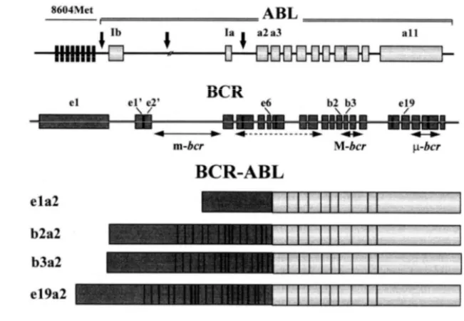
2.3.1 Structure of the various BCR-ABL fusion genes ... 48
2.3.2 BCR-ABL fusion proteins ... 49
2.3.3 Signaling pathways regulated in BCR-ABL cells ... 51
2.3.4 BCR-ABL and promotion of DNA mutations ... 54
2.4 MANIFESTATION, DIAGNOSES AND STAGING OF CML ... 55
2.4.1 The phases of CML ... 56
2.4.2 Incidence of CML pathology ... 57
2.5.1 Imatinib, the first generation TKI ... 57
2.5.2 Mechanisms of resistance to IM ... 58
2.5.3 Nilotinib and Dasatinib, the second generation TKIs ... 61
2.5.4 New therapeutic agents ... 62
2.5.5 Targeting CML stem cells... 63
2.5.6 Allogenic hematopoietic stem cell transplantation (Allo-HSCT) ... 63
2.6 MODELS TO STUDY CML ... 64
2.6.1 In vitro models: CML cell lines and primary cells ... 64
2.6.2 In vivo models: a new CML mouse models ... 64
2.7 MICROENVIRONMENTAL REGULATION OF CML CELLS ... 65
3. The JAK/STAT signaling pathway and STAT5 factors ... 67
3.1 JAK AND STAT PROTEIN STRUCTURES ... 67
3.1.1 The JAK kinases ... 67
3.1.2 The STAT factors... 69
3.2 CANONICAL AND NON-CANONICAL JAK/STAT SIGNALLING ... 71
3.2.1 Canonical JAK/STAT signaling ... 71
3.2.2 Non-canonical JAK/STAT signaling ... 73
3.3 NEGATIVE REGULATORS OF JAK/STAT PATHWAY ... 75
3.4 STAT TARGET GENES ... 76
3.5 STAT5 FACTORS ... 77
3.5.1 STAT5 molecular structures ... 77
3.5.2 Known roles for STAT5A and STAT5B ... 79
3.5.3 STAT5 in pathophisiology ... 81
3.5.3.1 Hematopoietic neoplasms ... 82
3.5.3.2 Solid cancers ... 82
4. STAT5 factors in CML development and maintaining ... 85
4.1 STAT5 AS A CRUCIAL FACTOR FOR CML: A HISTORICAL PERSPECTIVE 85 4.1.1 First results obtained in in vitro models ... 85
4.1.2 First results obtained in in vivo models ... 86
4.1.3 STAT5 as a new therapeutic target: the use of Pimozide ... 86
4.2 STAT5 PHOSPHORYLATION IN CML: WHAT’S NEW ... 87
4.3 STAT5 AND TKIs RESISTANCE ... 87
4.4 STAT5 AND CELLULAR LOCALIZATION IN CML CELLS ... 88
5. Objectives ... 89
SUMMARY ARTICLE I ... 97
ARTICLE I ... 99
Differential Contributions of STAT5A and STAT5B to Stress Protection and Tyrosine Kinase Inhibitor Resistance of Chronic Myeloid Leukemia Stem/Progenitor Cells ... 99
GENERAL DISCUSSIONS AND PERSPECTIVES ... 11919
ANNEXES ... 129
AB : Actin Binding
ABC : Adenosine triphosphate-Binding Cassette
ABL : Abelson murine Leukemia
AGM : Aorta-Gonad Mesonephros
AGP1 : Alpha1-Acid Glycoprotein-1
Allo-HSCT : Allogenic Hematopoietic
Stem Cell Transplantation
AML : Acute Myeloid Leukemia
AP : Accelerated Phase
BAD : Bcl-2-Associated Death promoter
BCR : Breakpoint Cluster Region
BCR-ABL : Breakpoint Cluster Region-Abelson murine Leukemia
BM : Bone Marrow
BP : Blastic Phase
CB : Cord Blood
CCR : Chemokine Receptor
CCyR : Complete Cytogenic Response CLP : Common Lymphoid Progenitor
CML : Chronic Myeloid Leukemia
CMP : Common Myeloid Progenitor
CNL : Chronic Neutrophilic Leukemia
CP : Chronic Phase
CXCL12 : C-X-C motif Chemokine
DBD : DNA Binding Domain
DDsB : DNA Double strand Break EGF : Epidermal Growth Factor
EPO : Erythropoietin
ER : Endoplasmic Reticulum
ERK : Extracellular Regulated Kinase
FACS : Fluorescence-Activated Cell Sorting
FDA : Food & Drug Administration
FISH : Fluorescent In Situ Hybridization
Flt3-l : Fms-related tyrosine kinase 3 ligand
FNIII : Fibronectin type III
FOXO : Forkhead box O
G-CSF : Granulocyte-Colony Stimulating
Factor
GH : Growth Hormone
GLA : Glutamic Acid
GM-CSF : Granulocyte Macrophage-Colony Stimulating Factor
Grb2 : Growth factor receptor bound protein 2
HIF : Hypoxia-Inducible Factor
HIM : Hematopoietic Inductive Microenvironment
HLA : Human Leukocyte Antigen
HPV : Human Papilloma Virus
HRR : Homologous Recombination
Repair
HSC : Hematopoietic Stem Cell
HSP25 : Heat-Shock Protein 25 HSPC : Hematopoietic Stem and
Progenitor Cell
ICAM : Intercellular Adhesion Molecule Ig : Immunoglobulin
IL : Interleukin IM : Imatinib Mesylate
JAK : Janus Kinase
JAK/STAT : Janus Kinase/Signal
Transducers and Activator of Transduction
JNK : Jun Kinase
KO : Knock-Out LG : Laminin G
LTC-IC : Long Term Culture-Initiating
Cell
MAPK : Mitogen Activated Protein
Kinase
M-bcr : Major breakpoint cluster region m-bcr : Minor breakpoint cluster region MDR : Multi-Drug Resistance
MGF : Mammary Gland Factor
MMP : Matrix Metalloproteinase
MMR : Major Molecular Response MMR : Mismatch Repair
MSC : Mesenchymal Stem Cell
mTOR : Mammalian Target of
Rapamycine
NCK : Non-Catalytic region of tyrosine Kinase adaptor protein
NER : Nucleotide Excision Repair
NK : Natural Killer
NOD : Non-Obese Diabetic
NOD-SCID : Non-Obese Diabetic-Several
Combined
Immunodeficiency
NOG : NOD/SCID/IL-2Rg null
NOX : NADPH Oxidase
NTS : Nuclear Translocalization Signal
OCT1 : Organic Transporter1
PB : Peripheral Blood
PCR : Polymerase Chain Reaction
PDGFR : Platelet-Derived Growth Factor
Receptor
PHD : Prolyl-Hydroxylase PI3K : Phosphoinositide 3-Kinase
PIAS : Protein Inhibitors of Activated STATs PLC : Phospho-Lipase C PPR : Parathyroid hormone-related Peptide Receptor PRL : Prolactin PtdSer : Phosphatdylserine
PTP : Protein Tyrosine Phosphatase
PV : PolycythemiaVera RacGAP : Rac GTPase Activating
Protein
RanBMP : Ran-Binding Protein in the Microtubule organization center
ROS : Reactive Oxygen Species
RTK : Receptor Tyrosine Kinase
RT-PCR : Reverse Transcriptase-Polymerase Chain Reaction
S/TK : Serine/Threonine Kinase
S6K : S6 Protein Kinase
SAPK : Stress Activated Protein Kinase
SCF : Stem Cell Factor
SCID : Several Combined Immunodeficiency
SCN : Severe Congenital Neutropenia
Sdf1 : Stromal derived factor
SHP-1 : Src Homology region 2
domain-containing Phosphatase-1
SOCS : Suppressors Of Cytokine Signaling
STAT : Signal Transducers and
Activator of Transduction
TAM : Tyro3, Axe, Mez
TBLA : Trabecular Bone-Like Area TK : Tyrosine Kinase
TKI : Tyrosine Kinase Inhibitor
TLR : Toll Like Receptor
TPO : Thrombopoietin
VEGF : Vascular Endothelial Growth
INTRODUCTION
PART I
1.
The Hematopoietic system
1.1 HEMATOPOIESIS AND HSCs
The hematopoiesis is the process that leads to the formation of all blood cells. These cells are required for a multitude of tasks, such as protection from infections, removal of damaged tissues but also to transport vital molecules such as oxygen throughout the body and to prevent bleeding. In a healthy adult person, approximately 1011–1012 new blood cells are produced every day in order to maintain steady state levels in the peripheral circulation.
All cellular blood components derive from Hematopoietic Stem Cells (HSCs). HSCs are multipotent stem cells operationally defined by their capacity to reconstitute the entire blood system of a recipient and, at the same time, are capable of renewal. The self-renewal is the cellular process by which a stem cell divides into one mother cell that is identical to the original undifferentiated stem cell and may be either symmetric or asymmetric in its nature. A symmetric self-renewing division refers to the process whereby both daughter cells retain mother cell characteristics. This type of cell division expands the stem cell pool and is therefore thought to be important after transplantation or after hematopoietic injury. In the asymmetric self-renewal division the 2 daughter cells adopt different cell fates, resulting in only one cell maintaining stem cell property and the other one can commit to any of the alternative differentiation pathways that lead to the production of one or more specific types of blood cell.
Most of the mature blood cells have a limited life span, therefore HSCs are required throughout life to continuously replenish multilineage progenitors and precursors committed to individual hematopoietic lineages.
The process of hematopoietic differentiation is a hierarchical process that starts from the most immature HSCs and schematically leads to the generation of 2 kinds of progenitors with restricted potential: the Common Lymphoid Progenitor (CLP) and the Common Myeloid Progenitor (CMP). Progenitor cells can divide only a limited number of times; they are pushed to differentiate in their precursor cells whose fate is even more restricted and they will give rise to mature blood cells, such as mature erythroid, megakaryocytic, myeloid (monocyte/macrophage, eosinophil and neutrophil), mast, dendritic or lymphoid cells. (Fig.1)
Figure 1: Hematopoiesis scheme. Hematopoietic Stem Cell (HSC) is capable of self-renew and generates all
the blood cells. Population cell numbers rise with increasing maturity. This diagram omits for simplicity natural killer and dendritic cells. Abbreviations: CFU-s (colony-forming unit spleen), CLP (common lymphoid progenitor), CMP (common myeloid progenitor), MEP (megacaryocyte-erythroid progenitor cell), GMP (granulocyte-macrophage progenitor cell), BFU-E (burst-forming unit-erythroid), Meg-CFC (megacaryocyte forming cell), Eo-CFC (eosinophil forming cell), GM-CFC (granulocyte-macrophage colony-forming cell), CFU-E (colony-colony-forming unit-erythroid), G-CFC (granulocyte colony-colony-forming cell), M-CFC (Monocyte-Macrophage colony-forming cell), Mast-CFC (Mast colony-forming cell).
1.2 HEMATOPOIETIC DEVELOPMENT
During mammal development there are different and sequential sites of hematopoiesis. Primitive hematopoiesis starts in the yolk sac and move later to the aorta-gonad mesonephros (AGM) region and to the fetal liver. Just before birth, cycling HSCs migrate from the liver to the developing bone marrow (BM), where they remain after birth.
The properties of HSCs in each site differ, probably reflecting different niches that support HSC expansion and/or differentiation and intrinsic characteristics of HSCs at each stage.
1.2.1 Embryonic hematopoiesis
De novo generation of hematopoietic cells starts shortly after mesoderm formation and continue through gestation. Hematopoiesis in embryo occurs in several tissues that include the yolk sac, the AGM region, the placenta and the liver (Fig.2A-2B). Embryonic hematopoiesis consists of three different waves which lead to the generation of HSCs.
- The first wave is called “primitive hematopoiesis” and it takes place in the yolk sac. This wave generates short-lived primitive nucleus bearing erythrocytes necessary to carry oxygen to the rapidly growing embryo. These cells co-localize with blood vessels, forming the so called “blood islands”. HSCs and vascular cells are believed to originate by a precursor cell called hemangioblast (Palis and Yoder 2001). These cells may be capable of producing both hematopoietic and endothelial cells. The first wave of primitive hematopoietic cell generation begins at embryonic day (E)7.5 in the mouse system and is highly conserved across vertebrate species, including men, where it begins at 16-20 days of gestation (Tavian and Peault 2005)
- The second wave of hematopoiesis is when the circulation starts and hematopoietic cells are detectable in the AGM region. These hematopoietic progenitors are functionally more complex than primitive progenitors; indeed they can produce erythroid, myeloid and lymphoid cells. At this development stage in AGM region, we can find the firsts definitive HSCs that are able to regenerate hematopoiesis in adult recipients (Medvinsky and Dzierzak 1996) and at this stage starts the so called “definitive hematopoiesis”.
- The third wave provides for the generation of fetal HSCs which are detectable in placenta, circulation and later in the fetal liver, which remains the main hematopoietic organ until birth.
From fetal liver HSCs migrate and colonize bone marrow just prior to birth. At this moment there is a switch from fetal to adult hematopoiesis: the high proliferative fetal program switches to the adult program, where HSCs are quiescent and mainly in G0/G1 cell cycle.
1.2.2 Adult hematopoiesis
Figure 2: Hematopoietic stem cell development in mouse embryo. Depiction of a mouse embryo at E10.5, at
the time when the firsts HSCs are generated in the aorta. A) Sites generating hematopoietic cells are shown: the extraembryonic yolk sac and placenta, the intraembryonic aorta and liver, and the umbilical and vitelline arteries that connect the placenta and the yolk sac to the aorta. The dotted line indicates the transverse section showed in the panel B. B) Depiction of a transverse section with the AGM region in the rectangle. The AGM region is flanked on the dorsal side by the neural tube and the somites and on the ventral side by the gut and the peritoneum. A hematopoietic cluster is indicated on the ventral wall of the aorta.
Prior to birth, HSCs move to the BM where they engraft in small cavities of trabecular bone, close to the endosteal interface between bone and marrow. In adult humans, the major sites of hematopoiesis are the bones of axial skeleton (cranium, sternum, ribs and vertebrae) in addition to the ilium. In children, hematopoiesis occurs in the marrow of the long bones such as the femur and tibia, then it ceases between 5 and 7 years of age and the red (hematopoietic active) BM is replaced by yellow (hematopoietic inactive) adipose tissue.
after birth under steady-state conditions. However, extramedullary splenic hematopoiesis can occur in times of hematopoietic stress.
In humans, by four weeks after birth, HSCs stop cycling and acquire largely a
dormant phenotype, in order to prevent cell exhaustion. In response to blood cell loss HSCs
are able to enter in cycle and to replenish the hematopoietic system.
Adult hematopoietic hierarchy is the consequence of progressive HSC maturation culminating with the mature myeloid and white blood cells. Lineage specification largely takes place within the bone marrow as well; mature cells are then mainly found in blood circulation but also in thymus, spleen and lymph nodes.
1.2.3 Regulators of HSC generation
The hematopoietic fate is dictated by the microenvironment and by the intrinsic program of the cells. As it will be discussed later, there is a cross-talk between HSCs and the other cells that surround them, and they form together the microenvironment called “niche”. In the embryonic microenvironment, the underlying mesenchymal cells produce signaling molecules that impact directly or indirectly on emerging hematopoietic cells. These molecules regulate different signaling pathways in HSCs, such as BMP-TGF, JAK/STAT, Wnt and Notch pathways. The molecular mechanisms of these signaling pathways will be discussed below in the “Hematopoietic stem cell niche” paragraph.
In addition to the microenvironment, HSCs possess an intrinsic molecular program that specifies their stemness properties. It consists in the activation/repression of specific genes and signaling cascades that dictates the choice of the hematopoietic cell fate. A lot of intrinsic regulators of HSC generation have been described and the very first discovered come from genetic deletion experiments in mice. These 3 transcription factors are Scl, Gata2 and
Runx1 and to date, they are the best known factors involved in HSC generation. Germ line
deletion of each single gene leads to mid-gestation, embryonic lethality and profound anemia. Interestingly, these factors seem to work in complexes to regulate gene expression during hematopoietic development. Below, you can find a more detailed description of these crucial and more studied factors.
Scl/Tal
Scl/Tal1 is a basic helix–loop–helix (bHLH) transcription factor. It can form heterodimer complexes with E-proteins and binds specific DNA motifs by its bHLH domain,
in order to regulate gene expression. Scl is normally expressed in hematopoietic tissues of the developing embryo. Scl germ line deletion in mice show an early embryonic lethality at day E 9.5, with a completely absence of blood formation (Robb et al. 1996, Shivdasani, Mayer and Orkin 1995). Thus, Scl is an essential factor during primitive hematopoiesis (first wave) of the embryonic development.
Gata2
The Gata2 transcription factor is a member of the Gata family of factors. There are 6 evolutionarily conserved proteins, Gata1 to Gata6, and they have two highly conserved zinc finger (ZnF) domains. Only Gata1, 2 and 3 are relevant to the hematopoietic system: Gata1 for the erythroid/megakaryocytic lineage and for mastocytes differentiation, Gata2 for hematopoietic stem and progenitor cells, and Gata3 for T-lymphocytes.
Gata2 is highly expressed in hematopoietic cells of the AGM region, fetal liver and placenta. Gata2 knock-out (KO) mice exhibit embryonic lethality at day E10 and E10.5 because of a profound anemia. Moreover, in Gata2 deficient mice, primitive hematopoietic progenitor numbers are decreased and no HSCs are produced (Ling et al. 2004, Tsai et al. 1994).
Runx1
Runx1/AML1 belongs to the transcription factor family that includes also Runx2 and Runx3. It is often found in chromosomal rearrangement in acute myeloid leukemia (AML) cells. Runx1 binds to regulatory elements of several genes and provides for tissue specific gene expression of molecules known to be important in hematopoiesis. Among them, there are the genes that encode for GM-CSF, IL3 and CSF1 receptor. Deletion of Runx1 transcription factor in mice leads to embryonic lethality at E12.5. Runx1 KO mice show normal morphogenesis and yolk-sac erythropoiesis, but they exhibit a block of HSCs generation in AGM region, a block of fetal liver hematopoiesis and a lack of all hematopoietic tissues (Wang et al. 1996, Okuda et al. 1996). These results suggest that Runx1 regulate specific genes which are essential for definitive hematopoiesis of all lineages. Moreover, Runx1 seems to act in concert with Gata2. Indeed, Runx1+/- and Gata2+/- single heterozygous mice are viable and present a normal hematopoietic blood profile, but Runx1+/-:Gata2 +/-double heterozygous mice embryos die and fetal livers contain less hematopoietic progenitors than single heterozygous mice (Wilson et al. 2010). These findings show that Runx1 and
Gata2 have a crucial role in hematopoietic development and that they can form a protein complex that regulate expression of genes essential for HSCs generation
Finally, the specific temporal and spatial sequence of extrinsic signals and the combination and/or the levels of intrinsic signals, play a role in the differential transcription factor expression and production of the distinct waves of hematopoietic cells in the developing embryo and steady as well as stress hematopoiesis in adults.
1.3 BIOLOGY OF HSCs
1.3.1 HSCs as quiescent cells: a protection mechanism that favors their
resistance
Hematopoiesis is the tightly regulated process of blood formation. Short-lived mature blood cells are replenished every day by HSCs through a large number of hematopoietic progenies, as multipotent hematopoietic progenitors and more restricted lineage-committed progenitors. HSCs are the first identified and best characterized adult stem cells, and they have served as model for other kind of adult stem cells.
A common property of adult HSCs is quiescence in term of cell cycle. Quiescence and slow cell cycle progression are critical for sustaining self-renewing HSC compartment throughout life. Moreover, quiescence is responsible for the protection of HSC pool from various stress insults. Indeed, quiescent cells are protected from replication-associated mutation generation and they are also more resistant to radiation than other cells. This feature is due to expression of repair, anti-oxidant and anti-apoptotic machinery. Ito and colleagues demonstrated that the Ataxia Telangiectasia Mutated (ATM) gene, which maintains genomic stability in response to DNA damage, has an essential function in the reconstitutive capacity of HSCs, but is not as important for the proliferation or differentiation of progenitors. Atm -/-mice show progressive bone marrow failure resulting from a defect in HSC function that was associated with elevated reactive oxygen species (ROS). Treatment with anti-oxidative agents restored the reconstitutive capacity of Atm-/- HSCs, resulting in the prevention of bone marrow failure (Ito et al. 2004). These results show that the self-renewal capacity of HSCs
depends on inhibition of oxidative stress and its associated DNA damages which can alter genomic stability.
In addition, HSCs present a different metabolic status compared to their progenitors: they show low oxidative phosphorylation and high glycolytic activity to synthesize ATP, meaning that HSCs favor anaerobic enzymatic pathways to synthesize ATP. These data demonstrate that HSCs are in hypoxic conditions and are metabolically different from their progenitor cells (Fig.3). This specific metabolic status permits to the more immature stem cells to be protected throughout life and is responsible for their resistance to anti-cancer drug treatments and to stress insults such as oxidative and genotoxic insults.
Figure3: cycling HSPCs have increased ROS levels. HSCs are quiescent and may proliferate to generate
short-cycling HSCs that give rise to multipotent progenitors and differentiated cells. Most immature HSCs exhibit very low ROS levels and they are protected from oxidative insults. On the other side, more proliferative cells, such as cycling HSCs and progenitors, present increased ROS levels compared to immature HSCs. Low ROS in quiescent cells prevent oxidative stress and favor their resistance and long-live capacity.
1.3.2 Regulation of self-renewal and maintaining of HSCs
Hematopoiesis must be rapidly and meticulously regulated, in order to face to a variety of situations, ranging from normal homeostasis to acute blood loss, infection and to avoid over-production of mature cells. A wide variety of factors critical for HSC regulation have been identified, including cytokines, growth factors, transcription factors, chromatine modifiers and cell cycle regulators. All these factors have been long-time studied as they
significant impact on the collection of HSCs for transplantation and on gene therapy strategies. Below, some examples of both intrinsic and extrinsic regulators that have been shown to be important in adult HSC maintaining and self-renewal are reported.
1.3.2.1 Cell-intrinsic regulators of adult HSC maintaining
Valuable knowledge about factors influencing self-renewal, has been gained from gene manipulation studies, which have identified multiple proteins that play important roles in the regulation of HSC self-renewal, including transcription factors, epigenetic modifiers, and cell cycle regulators. The most studied genes are reported below.
Homeobox genes
One of the first genes described involved in HSC self-renewal is HoxB4. The homeobox (Hox) genes encode transcription factors that regulate embryonic body patterning and organogenesis. Several members of the homeobox gene family are expressed during hematopoietic differentiation. Over-expression of HoxB4 by retroviral infection of murine bone marrow cells leads to expansion of HSCs in vivo and in vitro, therefore appearing to be a positive regulator of HSC self-renewal (Sauvageau et al. 1995, Krosl et al. 2003). Surprisingly, HoxB deficient mice do not exhibit any major defects in hematopoiesis, possibly because of the compensatory effects by HoxA and/or HoxC genes (Bijl et al. 2006).
It is also interesting to note that the over-expression of the HoxB4 paralogue HoxC4 in primitive hematopoietic cells also enhances the proliferation of both HSCs and committed progenitors, although the HoxC4's effect may be weaker than HoxB4's (Daga et al. 2000, Auvray et al. 2012). HoxB4 is a potent enhancer of primitive hematopoietic cell growth and this action may be explained by its interaction with c-Myc gene and the cell cycle machinery (Antonchuk, Sauvageau and Humphries 2001).
Ikaros
The Ikaros gene product is a zinc-finger transcription factor. Ikaros proteins modulate transcription by recruiting co-repressor complex to the promoter of target genes. Ikaros displays crucial functions in the hematopoietic system and mice homozygous for Ikaros dominant negative mutation (DNA-binding domain mutated) possess no measurable repopulating activity at all, conferring to Ikaros factors an essential role in the maintaining of the HSC pool (Nichogiannopoulou et al. 1999).
Tel/Etv6
The transcription factor Tel (Translocation Ets leukemia; also known as Etv6 [Ets variant gene 6]), the product of a locus frequently involved in translocations in leukemia, is a selective regulator of HSC survival. Following inactivation of Tel/Etv6, HSCs are lost in the adult bone marrow. Intriguingly, its inactivation in hematopoietic lineages does not affect neither their proliferation nor their survival (Hock et al. 2004), meaning that it acts selectively on immature hematopoietic cells.
Gfi1
Gfi1 (Growth factor independence 1) is a Zinc-finger repressor. When Gfi1 is deleted in mice, HSC frequencies are significantly reduced and Gfi1-/- bone marrow cells are severely impaired in competitive long-term reconstituting abilities after transplantation. Gfi1 deficient bone marrow cells show a surprisingly high proportion of actively cycling HSCs, suggesting that Gfi1 restrains HSCs proliferation increasing their self-renewal ability (Zeng et al. 2004).
The Polycomb group proteins (PcG)
The Polycomb group proteins are a family of proteins that can remodel chromatin in order to epigenetically silencing gene expression. Members of the Polycomb repressor complex 1 (PRC1), especially Bmi1, have been implicated in HSCs self-renewal. Part of the mechanism by which PRC1 sustains HSC self-renewal is by repression of genes that promote lineage specification, cell death and cell cycle arrest (Park et al. 2003, Molofsky et al. 2003).
1.3.2.2 Cell-extrinsic regulators of HSCs
Adjacent cells and blood supply HSCs self-renewal and differentiation decision, bringing a number of cytokines, growth factors or hormones. Among these, Stem Cell Factor (SCF), Thrombopoietin (TPO), Fms-related tyrosine kinase 3 ligand (Flt3-l), Interleukin-11 (IL-11), Interleukin-3 (IL-3), Interleukin-6 (IL-6) ,Granulocyte Macrophage-colony stimulating factor (GM-CSF) and combination of these, have been used for in vitro HSC expansion. These cytokines allow short-term HSC maintenance for a few days but by themselves are not sufficient to sustain HSC self-renewal, leading to a progressive depletion of long-term repopulation capacity and differentiation of HSCs (Glimm and Eaves 1999, Le
the receptors for TPO and SCF, c-mpl and c-kit respectively, are expressed on repopulating HSCs. Moreover, mice with genetic mutation in TPO or c-mpl genes have a dramatically reduction in HSCs, demonstrating a physiological role for TPO and its receptor in regulating HSC production and function (Solar et al. 1998, Kimura et al. 1998) .
The cocktail of SCF, Flt3, and TPO (mouse and humans) or IL-11 (mouse) is defined as the best cytokine cocktail medium supporting short-term Hematopoietic Stem and Progenitor Cells (HSPCs) proliferation and survival in in vitro cultures.
These cytokines are present in blood and are also synthesized by HSC surrounding cells within the niche. These cells also secrete other HSC regulators which lead to the activation of proper signaling pathways in HSCs. Among these, there are the Tie2-Ang1,
BMP-TGF, Sdf1/CXCR4, Notch, Wnt and JAK/STAT signaling pathways. The role of the
niche and of these crucial factors in HSC pool maintaining will be discussed below in the “Hematopoietic stem cell niche” paragraph.
1.4 IDENTIFICATION AND ISOLATION OF HUMAN
HSCs
The stem cell population constitutes only a small percentage of the total number of hematopoietic cells in bone marrow and blood. Identifying HSCs will allow for comprehension of their biology. Moreover, the availability of stem cells is extremely useful for bone marrow transplantation and for gene therapy. Since few decades ago, world-wide efforts have been done in order to isolate, characterize and biologically analyze HSCs.
1.4.1 The sources of human HSCs
The known sources of human HSCs are the bone marrow (BM), the umbilical cord
blood (CB) and the peripheral blood (PB). In the BM, HSCs are rare cells representing the
0.01% (1 every 10.000 cells) of the all population. Even if the BM represents the source with the majority of HSCs, it has been known for decades that stem and progenitor cells circulate, in low percentage, also in the blood stream. Researchers have observed that HSCs can be mobilized to the blood stream upon granulocyte-colony stimulating factor (G-CSF) treatment, increasing their percentage in peripheral blood. Another source of HSCs recognized by
researchers since the late 1980s is the umbilical cord blood. This tissue is responsible for the support of the developing fetus during pregnancy, is delivered along with the baby and is usually discarded, so it is easier to be obtained compared to BM or PB. These features make the CB the most used sample as source of HSCs.
1.4.2 Experimental systems to identify HSCs
The characterization of human HSCs has been possible by making use of experimental systems that have permitted their identification on the basis of their biological properties. In vitro and in vivo approaches that permit the identification and isolation of human HSCs are summarized below.
In vivo models
The most stringent assay to identify cells with high stemness potential is by testing their blood system reconstitution capacity in irradiated mice. It consists in injecting stem and progenitor cells into recipient mice and evaluates their capacity to give rise to all blood lineages. However, it is important to underline that to study human hematopoiesis in in vivo models the host should not eliminate the xenograft via an immune reaction and, should provide a permissive microenvironment for engraftment and multilineage differentiation of donor cells. Immunodeficient mice present these criteria and have been modified to improve their model function. Early studies were done in SCID (Several Combined
Immunodeficiency) mice that lack of T- and B-cell defect (McCune et al. 1988). However,
these mice still possess macrophages and NK cells that can mediate rejection of xenografts. More recently, SCID mice were crossed to non-obese diabetic (NOD) mice that display partially deficient NK cell, antigen-presenting cell, and macrophage functions. NOD-SCID
mice are currently used in in vivo studies of hematopoiesis and they display a 10-20-times
better engraftment capacity than the SCID mice. However, NOD-SCID mice engraftment can be monitored for about 6 months because of the limited life-span of these mice. More recently, the NOD/SCID/IL-2Rg null (NOG) strain was shown to exhibit significantly higher engraftment potential than other immunodeficient mouse strains (Shultz et al. 2005).
In vitro assay
self-renewal, and differentiation. The best and stringent in vitro assay of HSC activity is the
long term culture-initiating cell (LTC-IC) assay and it was reported in 1989 by Sutherland
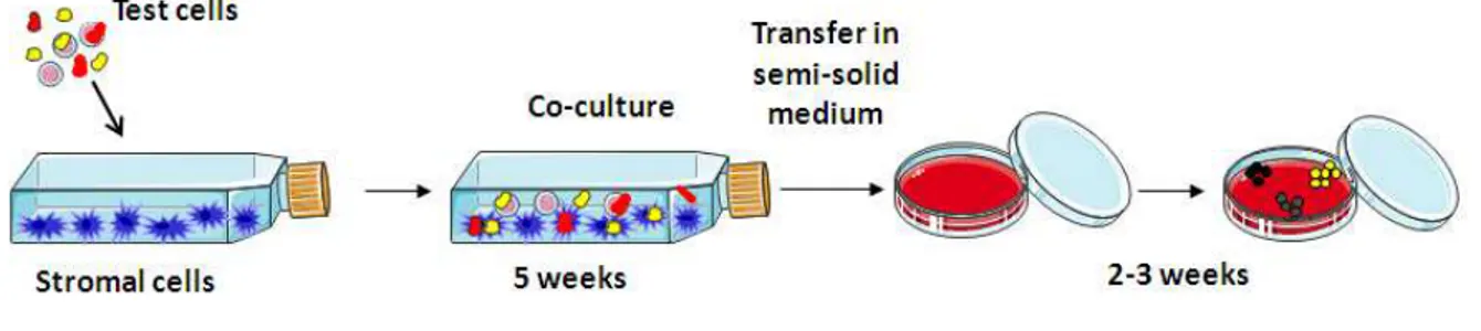
and colleagues (Sutherland et al. 1989). This test has provided an approach for the investigation of the regulation and maintenance of HSCs under conditions that mimic the marrow microenvironment. LTC-IC approach provides proper evidence of HSC activity without recurring to in vivo models. In a first step, candidates HSCs are cultured on bone marrow-derived stromal cells, in a 30% serum medium for 5 weeks or longer. In a second step, cells are transferred in a semi-solid medium of methyl cellulose containing cytokines, for about 2 weeks. Each HSC will be able to give rise to multiple differentiated colonies. Finally, the total number of myeloid, erythroid and multilineage clonogenic progenitors is evaluated and used to provide a quantitative assessment of the number of LTC initiating cells originally added (Fig.4). This long-lasting test is now largely used and it is the most stringent assay to identify human most immature hematopoietic stem cells.
Figure 4: The LTC-IC in vitro assay to identify primitive hematopoietic cells. In the LTC-IC assay, stem and
progenitor cells are co-cultured with bone marrow stromal cells for at least 5 weeks. Then, they are transferred in a semi-solid medium of methyl cellulose containing cytokines for about 2 weeks. The number of clonogenic progenitors raised is evaluated and used to provide an assessment of the number of LTC initiating cells.
1.4.3 Isolation of candidate human HSCs by their surface antigen markers
In vivo and in vitro studies together permitted the identification of a population of HSCs that was consequently characterized by the presence of different antigen markers. Detecting the expression of these marker panels allows separation of specific HSC-enriched cell populations via techniques like fluorescence-activated cell sorting (FACS).
Most of the studies on HSC characterization have been done in mice, but it is now clear that there are considerable differences in distribution of surface markers between human and mouse hematopoietic cells, which makes identification of human counterparts of mouse stem and progenitor cells more complicated. Importantly, mouse LongTerm (LT)-HSCs are
CD34-/low, CD38+ and CD90 (Thy1)low, whereas human HSCs are hCD34+, hCD38- and hCD90+.(Iwasaki and Akashi 2007)
The major representative and most used marker for human hematopoietic stem and progenitor cells was the CD34 antigen. The role of CD34 in human HSPCs is poorly known; studies in mice in which human CD34 antigen was ectopically expressed on the cellular surface reveal a role for CD34 in adhesion to BM stromal cells, elucidating a possible role for CD34 in regulation and compartmentalization of stem cells (Healy et al. 1995). However, mutant mice do not exhibit any defect in peripheral blood counts and answer to hematopoietic stress as well as wild-type mice (Cheng et al. 1996). In conclusion, CD34 seems to not be essential for hematopoiesis mice. Moreover, the fact that mouse LT-HSCs in steady-state bone marrow do not express significant levels of mCD34 raised an important question of whether hCD34+ cells could mark all long-term self-renewing human HSCs. Human CD34+ cell population is a heterogeneous population made of stem cells but also of progenitor cells. Among the CD34+ cells, just the 1-10% does not present any marker of lineage differentiation (Lin-) as CD3, CD4, CD8, CD11b and CD14. Oppositely, the majority of CD34+ cells (90– 99%) co-express the CD38 antigen, and this subset contains most of the lineage-restricted progenitors. Indeed, CD34+CD38- cells and not CD34+CD38+ cells are highly enriched for LTC-IC (Petzer et al. 1996) and contain NOD-SCID- repopulating cells (Bhatia et al. 1997). However, although most human HSCs are described as CD34+, the existence of CD34- HSCs in human and rhesus monkey have been described (Goodell et al. 1997). Recently, Anjos-Afonso and colleagues showed that in addition to CD34+ HSPC population, there is a rare CD34- population with severe combined immune-deficiency repopulating capacity. This CD34
-CD38-CD93hi population contains cells that not only function as HSCs, but that can also be
placed above CD34+38- population in hematopoietic hierarchy (Anjos-Afonso et al. 2013). Thus, CD34-CD38-CD93hi population contain more bona fide HSCs than CD34+CD38- population and the CD34 marker might be used as a positive marker anymore, but rather as a negative marker of most immature HSCs in addition to CD38 negative and CD93 positive markers.
Another well-established marker for HSCs enrichment is the CD90 (Thy-1) antigen, which is present in humans and not in mice (Baum et al. 1992). Other markers as the
CD45RA negative marker was assumed to be specific for the most immature human HSC
population (Majeti, Park and Weissman 2007).
CD34+CD38- cell. However, recent evidences seem to favor the hypothesis that most immature HSCs possess the CD34 negative marker and that CD34-CD38-CD93hi population can be placed at the top of hematopoietic hierarchy.
1.5 HEMATOPOIETIC STEM CELL NICHE
HSCs reside as rare cells in the bone marrow and depend on their microenvironment for regulation of self-renewal and differentiation. This microenvironment, which consists of hematopoietic and non-hematopoietic (stromal) cells, is a specific anatomic location called
niche. A lot of efforts have been done to decipher the interplay between a hematopoietic cell
and its niche. A historical perspective starting from the first concept of niche formulated in 1971 until the notions we have nowadays on this complicated but intriguingly system is reported below.
1.5.1 Historical perspective on the hematopoietic stem cell niche
The first experiments evidencing the concept of niche were performed in 1968 in Trentin’s laboratory. They demonstrated that both bone marrow and spleen stromal cells have an active role in the regulation of differentiation of HSCs into all blood lineages type. Moreover, they demonstrated that marrow and spleen organ stroma are geographically segmented into microenvironments and that in spleen stroma the predominant microenvironment dictates erythroid differentiation, whereas in marrow stroma the predominant microenvironment dictates neutrophilic granuloid differentiation. The hematopoietic organ stroma were thereby termed hematopoietic inductive microenvironments (HIMs) (Trentin 1971).
Trentin formulated the concept of HIMs as a differentiation inducing microenvironment. This observation was expanded by Schofield in 1978. Schofield observed that HSCs need to reside in the bone marrow to retain their infinite potential, whereas those homed to the spleen were more restricted to their capacity to sustain hematopoiesis. He proposed that, in addition to a differentiation microenvironment, there was also a specific hematopoietic microenvironment which fixed the stem cells in place and prevented their maturation, allowing the stem cells to proliferate and retain their stemness. Schoefield
formulated that “The stem cell is seen in association with other cells that determine its
behavior” (Schofield 1978). The concept that stem cells fate is determined by surrounding
cell structure is nowadays referred to as the stem cell microenvironment or the stem cell
niche.
In 2003, two reports published on Nature journal demonstrated by in vivo experiments in two different mouse models, that bone-forming osteoblasts were critical component of the niche, being capable of influencing HSC size and number (Calvi et al. 2003, Zhang et al. 2003). However, few years later, Kiel and colleagues suggested the role of another hematopoietic stem cell niche, which was rather formed by the endothelial cells within the bone marrow (Kiel et al. 2005).
Nowadays, it is clear that numerous elements of the BM intersect each other to appropriately regulate HSC fate choices and that it is inappropriate to say that HSCs are regulated just by osteoblast or by endothelial cells. Many other cells interplay with HSC and regulate their fate, as monocyte-macrophages, megakaryocytes and nervous system cells. Understanding how niche regulate HSCs is becoming more and more attractive for stem cell regenerative therapies, where cell proliferation and differentiation must be controlled in vitro, so that sufficient quantity of the proper stem cells can be produced prior to being introduced back into the patients.
1.5.2 Cell type forming HSC niche
The stem cell niche is the functional and anatomical ‘node’ that allows integration of signals from the periphery into the appropriate stem cell behavior. Osteoblasts and endothelial cells of the bone marrow niche have been long time studied, as the two major components of the niche that support maintenance, proliferation and differentiation of HSCs. Two different niches were proposed: the endosteal niche, where HSCs are closed to osteoblasts, and the perivascular niche, where HSCs are closed to vascular endothelium in marrow sinuses. Much has been debated over the distinction of a single niche, yet, to this day, consensus has not been reached in determining either over the other. This is because the periosteal region of the BM is a sponge-like trabecular complex structure where dormant HSCs are influenced not only by osteoblasts and vasculature but also by other stromal structures in quite close proximity (Fig.5). To date, it is in fact known that in addition to osteoblasts and endothelial cells, there are also reticular stromal cells, macrophages, megakaryocytes, adipocytes,
HSCs. Therefore, the concept of the BM niche should be viewed as a conjoined influence of
different cellular components.
The most studied cell types present in the BM niche and how they are able to regulate HSC maintenance are discussed below.
Osteoblasts and mesechymal stem cells (MSCs)
Osteoblasts are mononucleated cells that are responsible for the bone formation. Bone tissue undergoes a constant process of remodeling and is constantly being reshaped by osteoblasts, which produce matrix and minerals, and osteoclasts, that are responsible for the resorption of the tissue.
Different cells of the osteoblastic lineage are present in the bone marrow: mesenchymal stem cells (MSCs) (the multipotent stromal cells that give rise to osteogenic lineages), osteoprogenitor cells, osteoblast and osteocytes.
Osteoblasts are usually found in layer along the endosteum (the connective tissue around the medullary cavity) and the periosteum (the membrane that lines the outer surface of the bone). In vitro results obtained in 1996 seem to favor the hypothesis that human osteoblast can support HSCs in ex vivo culture system (Taichman, Reilly and Emerson 1996). Based on these observations, Zhang and colleagues and Calvi and colleagues examined the osteoblast regulation of the niche by two different in vivo approaches. Zhang and colleagues made use of a mouse model in which the Bone Morphogenetic Protein Receptor1A (BMPR1A) was conditionally deleted. They found that in these mice there was an ectopic formation of a trabecular bone-like area (TBLA) and an increase of N-Cadherin-positive osteoblasts. The formation of the TBLA correlated with an increase of the number of HSCs and these HSCs were associated with osteoblast by N-Cadherin (Zhang et al. 2003). Calvi and colleagues examined the effects of the constitutively activated parathyroid hormone-related peptide receptor (PPR) under the control of a specific osteoblast-collagen promoter. They found that in PPR transgenic mice there was a size increase of the trabecular bone which correlated with an increased number of osteoblasts and an increased number of HSCs in conjunction with the trabecular bone. Moreover, they found that these HSCs had a more activated Notch1 pathway than HSCs of wild-type mice. (Calvi et al. 2003).
However, studies in which osteoblast number is decreased do not show a reduction in HSCs. Vinjic and colleagues made use of a mouse model in which deletion of osteoblast was possible by the expression of thymidine kinase under the control of an osteoblast-specific promoter and the consecutive treatment with gancyclovir. In gancyclovir-treated mice there
was a decreased of marrow size and a decreased number of HSCs in the bone marrow. However, they found an increased extramedullary hematopoiesis in spleen and in the liver, resulting in an increase in peripheral HSCs and active primary in vitro hematopoiesis (Visnjic et al. 2004).
Recently, also mesenchymal stem cells (MSCs) have been shown to play a role in HSC regulation by secretion of specific factors. For instance they express, in association with osteoblasts, the tissue inhibitor of metalloproteinase-3 (TIMP3), to influence not only HSC quiescence and fate determination, but also normal bone formation and maintenance (Shen et al. 2010, Nakajima et al. 2010). Moreover, agrin, a proteoglycan involved in neuromuscular junctions, is expressd by MSCs and differentiated osteoblasts, while its receptor, alpha-dystroglycan, is expressed by HSCs. Agrin was shown to support the proliferation of hematopoietic progenitor cells and a microenvironment devoid of agrin results in progenitor cell apoptosis as well as reduced hematopoiesis (Mazzon et al. 2011).
In conclusion, both MSCs and osteoblasts contribute to HSC regulation in the endosteal niche of the bone marrow.
The endothelial cells
In bone marrow, endothelial cells form a barrier between the marrow and the blood circulation. They are the site of entrance of all blood cells that leave the blood circulation to enter into bone marrow and the site of exit for cells that are ready to enter the blood stream.
A decade ago it was shown that endothelial cells regulate proliferation of hematopoietic progenitor cells and long-term culture initiating cells by elaboration of lineage-specific cytokines in in vitro system (Rafii et al. 1995). These results were then validated by the use of human brain endothelial cells (Chute et al. 2002) and heart, lung and liver endothelial cells (Li et al. 2004). Moreover, in vivo confocal imaging of bone marrow mice revealed that HSCs can localize to specific subset of the marrow microvasculature where cells persist or increase in number (Sipkins et al. 2005). Finally, another group found that the majority of HSCs are located in the sinusoidal endothelium region and that only the 16% of these cells localize at the periendosteal region (Kiel and Morrison 2006).
Endosteal monocytes and macrophages
Also monocytes and macrophages can importantly contribute to HSC regulation in the BM niche. In a study of G-CSF mobilization of HSCs from the BM, Winkler and colleagues
macrophages, recapitulated the HSC mobilization into blood (Winkler et al. 2010). Reduced endosteal monocyte-macrophage populations coincide with reduced levels of HSPC-active factors, like CXCL12, in the niche, providing a potential mechanism of how these myeloid cells function in HSC regulation (Chow et al. 2011). Together, these results establish that bone marrow monocytes and macrophages are pivotal to maintain the endosteal HSC niche and that the loss of such monocytes/macrophages leads to the egress of HSCs into the blood.
Cells of the nervous system
In 2011 Yamazaki and colleagues proposed that also particular nervous system cells can play a role in niche regulation of HSCs. They found that non-myelinating Schwann glial cells in BM are TGF-beta-producing cells and express HSC niche factor genes. Moreover, they are in contact with a substantial proportion of HSCs. A nerve denervation reduces the number of these active TGF-beta-producing cells and leads to rapid loss of HSCs from BM. Thus, they propose that glial cells are components of BM niche and maintain HSC quiescence by regulating activation of latent TGF-beta (Yamazaki et al. 2011).
Reticular stromal cells
CXCL12 abundant reticular (CAR) cells are a population of reticular cells, which express CXCL12 at high amounts, with several long processes and scattered throughout adult BM niche. Most cells expressing high amounts of CXCL12 are not in close proximity to the bone surface but rather they are scattered throughout the trabecular space of the BM cavity (Sugiyama et al. 2006). When BM CXCL12-ecpressing cells are ablated in vivo, HSC number and size decrease, they are more quiescent and they express myeloid genes (Omatsu et al. 2010). Thus, CAR cells are required for proliferation of HSCs as well as maintenance of HSCs in an undifferentiated state thanks to their ability to express CXCL12 and consequently to retain HSCs in BM, preventing their mobilization to vasculature.
Megakaryocytes
Another mature cells type with key HSC regulatory roles are the megakaryocytes (MKs). TPO, through its receptor c-mpl, favors platelet productions from MKs as well as HSC self-renewal. MKs control the availability of TPO and in an environment where the number of MKs is increased, like in Myb or p300 mutated mice, TPO is less available and HSCs show an increase in cell cycle and a decrease in number (de Graaf et al. 2010).
In conclusion, the bone has a peculiar role in hematopoiesis regulation and it is composed of different kind of cells that create a cross-talk with HSCs and impact their residency and function. Research is evolving to better understand how these cells are anatomically and functionally organized, in a 3D structure, and how the BM architecture changes with disease. If the BM niche can be mimicked in vitro, this will not only allow for HSC expansion and differentiation in vitro, but will also increase understanding and ability to manipulate HSCs for more robust engraftment and mobilization. Although more research is required to ascertain how they interact with each other, MSCs, osteoblasts, nerves, reticular stromal cells and endothelial cells, as well as macrophages and megakaryocytes should all be considered key components of the HSC niche, playing a critical role in regulating HSC fate.
1.5.3 Microenvironment-dependent signals which regulate hematopoiesis
HSCs are normally quiescent cells that reside in the bone marrow. In response to hematopoietic stress, they are able to divide, to differentiate and to replenish the hematopoietic system. The different choices of a HSC are regulated by its microenvironment and this tight regulation involves different microenvironment-dependent signaling pathways. The most important are described below.
Tie2-Ang1 signaling pathway and N-Cadherin
Tie2-Angiopoietin1 (Ang-1) signaling pathway is required to maintain HSC quiescent at the endosteal surface. Indeed, HSCs that express the tyrosin kinase receptor Tie2 are quiescent and anti-apoptotic; the interaction of these cells with the Tie2-ligand, Ang-1, permit the maintaining of in vivo long-term repopulating activity of HSCs. Moreover, Ang1 enhances the ability of HSCs to remain quiescent and to adhere to the bone (Arai et al. 2004).
One target of Tie2 signaling is N-Cadherin; this adhesion protein was shown to be implicated in niche-HSC interaction too. Over-expression of N-Cadherin in HSCs promotes quiescence and preserves HSC activity during serial bone marrow transplantation. Moreover, N-Cadherin knock-down inhibits the HSC localization to the endosteal surface of bone marrow and decreases long term engraftment. Oppositely, N-Cadherin knocked down cells in the spleen do not exhibit any activity defects, meaning that this adhesion molecule is essential for proper HSC regulation in bone marrow niche (Arai et al. 2012).
VEGF
The vascular endothelial growth factor (VEGF) is a stimulator of angiogenesis and a key mediator of the interaction between osteoblasts and vascular endothelial cells. VEGFR2 is the main receptor expressed by the osteoblasts, osteoclasts as well as endothelial cells within the bone marrow. VEGF can couple osteogenesis and angiogenesis by stimulating survival and proliferation of endothelial cells and osteoblastic lineage commitment of osteoprogenitors (Maes et al. 2010). VEGF was also shown to be essential for HSC maintaining by an autocrine loop control (Gerber et al. 2002) and for the correct hematopoietic niche formation (Chan et al. 2009).
BMP-TGF pathway
Bone Morphogenetic Proteins (BMP) is a group of growth factors that interact with their receptors (BMPR) at the cellular surface and activate mobilization of the SMAD proteins. BMP4 is expressed in cells of the hematopoietic stem cell microenvironment, including osteoblasts, endothelial cells and megakaryocytes. BMP4 is implicated in stromal cell regulation of embryonic HSC in the AGM region (Durand et al. 2007) as well as adult HSC in vivo (Goldman et al. 2009). One receptor of BMP is BMPR1 and it is found to be highly expressed by osteoblasts. As described before, BMPR1 deficiency strongly decreases the number of HSCs capable of repopulating activity (Zhang et al. 2003).
Sdf1/CXCL12 and CXCR4
Stromal derived factor (Sdf1), also called C-X-C motif chemokine (CXCL12), is a chemotactic molecule produced by stromal cells and its receptor, CXCR4, is highly expressed on earliest HSCs.
Sdf1 is capable to regulate the attachment of the HSCs to the niche and consequently to modify the number of HSCs in the blood circulation, by regulating soluble-kit release via the matrix metalloproteinase1 (MMP1) (Heissig et al. 2002). Moreover, Sdf1/CXCR4 signaling is not only essential to confine HSCs to their proper niche, but also to maintain these cells in a quiescent status (Nie, Han and Zou 2008).
Notch pathway
The Notch receptor is a single-pass transmembrane receptor protein, composed of a large extracellular domain and a smaller intracellular domain. In mammalian, members of the
Delta-like (DLL1, DLL3, DLL4) and Jagged (JAG1, JAG2) families serve as ligands for Notch signaling receptors.
In murine hematopoietic stem cells, LSK (Lin-, Sca1+, c-Kit+) cells, an over-expression of the active intracellular Notch1 domain is responsible for self-renewal (Varnum-Finney et al. 2000). Moreover, in vitro results obtained from cell line cultures, show that the interaction between Jagged1(JAG1) ligand and Notch1 sustains the long-term maintenance of hematopoietic progenitor cells and impairs differentiation, suggesting a role in cell fate decision for JAG1 and Notch signaling pathway (Li et al. 1998). These in vitro results were strengthened by in vivo data in which Notch activation was shown to have a role in lymphoid over myeloid lineage commitment (Stier et al. 2002). More recently, it has been shown that Notch1 is expressed on HSCs and Notch ligands as DLL1 and JAG1 are expressed by osteoblastic cells, meaning that Notch1 could be a crucial mediator of HSC-niche crosstalk (Nobta et al. 2005, Calvi et al. 2003). Notch signaling seems to be very important for niche dependent HSC regulation.
Wnt pathway
Wingless (Wnt) signaling pathway is activated by the binding of a Wnt-protein to a Frizzled family ligand. Two different Wnt downstream signaling pathways may exist: the β-catenin-dependent canonical and non-canonical pathway. Both canonical and non-canonical pathway promotes self-renewal of HSCs in an intrinsically or extrinsically way, respectively (Luis et al. 2009, Murdoch et al. 2003). Moreover, blocking Wnt signaling in the niche, by the expression of a Wnt inhibitor under the control of a specific osteoblastic promoter, increases the number of proliferating HSCs and reduces their ability to reconstitute the hematopoietic system of irradiated recipient mice. This effect is microenvironment-dependent because if these HSCs issue from transgenic mice are transplanted in wild type mice, they are completely capable to reconstitute hematopoiesis (Fleming et al. 2008). These findings show that Wnt/β-catenin activity is crucial for the cross-talk between HSC and their niche and the consequently maintain of HSCs.
The JAK/STAT pathway
Last but not least, another molecular pathway involved in HSC maintenance by the niche is the JAK/STAT (Janus family kinase–signal transducer and activator of transcription) pathway. It is a common downstream pathway of extracellular signaling (cytokine, growth
factors, hormones...) and plays critical role in transmitting a variety of biological functions by regulating transcription of target genes.
This pathway will be discussed in greater detail in the last paragraph.
Figure 5: Schematic picture of the HSC microenvironment. HSCs are located in a sponge-like trabecular
structure and there is a conjoined influence of different cellular compartments. Among these there are osteoblasts, endothelial cells, mesenchymal stem cells (MSC), CXCL-12 abundant reticular cells (CAR), macrophages, megakaryocytes and adipocytes. Under steady state, they all interact with HSCs by secreting specific factors such as TGF-beta, CXCL-12 and Ang-1, and they promote the maintaining of their stemness properties. Once HSCs start cycling to produce new hematopoietic differentiated cells, they move to blood vessels, where they cross the endothelial barrier and reach the blood stream.
1.5.4 Hypoxia and reactive oxygen species (ROS)
As discussed before, the proper interaction between HSCs and bone marrow niche is crucial to prevent cell exhaustion from excessive proliferation. In addition, the niche protects HSCs from stress, like accumulation of reactive oxygen species (ROS) and DNA damages. Indeed, in the bone marrow niche there is an O2 gradient from below 1% in hypoxic niche to