HAL Id: tel-03122468
https://tel.archives-ouvertes.fr/tel-03122468
Submitted on 27 Jan 2021HAL is a multi-disciplinary open access
archive for the deposit and dissemination of sci-entific research documents, whether they are pub-lished or not. The documents may come from teaching and research institutions in France or abroad, or from public or private research centers.
L’archive ouverte pluridisciplinaire HAL, est destinée au dépôt et à la diffusion de documents scientifiques de niveau recherche, publiés ou non, émanant des établissements d’enseignement et de recherche français ou étrangers, des laboratoires publics ou privés.
The LINC complexe is a mechanotransducer that
regulates catenin signaling during
epithelial-mesenchymal transitions
Pietro Salvatore Carollo
To cite this version:
Pietro Salvatore Carollo. The LINC complexe is a mechanotransducer that regulates catenin signaling during epithelial-mesenchymal transitions. Cellular Biology. Université de Paris, 2019. English. �NNT : 2019UNIP7131�. �tel-03122468�
Université de Paris
Ecole doctorale FIRE 474
Institut Jacques Monod / Mécanotransduction: de la surface de la cellule à son
noyau - chef d’équipe: Dr. Nicolas Borghi
The LINC complex is a mechanotransducer that
regulates catenin signalling during epithelial-mesenchymal transitions
Par Pietro Salvatore CAROLLO
Thèse de doctorat de Biophysique-Biologie Cellulaire
Dirigée par Dr. Nicolas BORGHI
Présentée et soutenue publiquement à Paris le 19/11/2019
Président du jury: Antoine GUICHET - Directeur de Recherche - IJM, Paris Rapporteurs: Catherine COIRAULT - Directrice de Recherche - Institut de Myologie, Paris Philippe CHAVRIER - Directeur de Recherche - Institut Curie, Paris Examinateurs: Christophe GUILLUY - Chargé de Recherche - IAB, Grenoble Sandrine ETIENNE-MANNEVILLE - Directrice de Recherche - Institut Pasteur, Paris Directeur de thèse: Nicolas BORGHI - Chargé de Recherche - IJM, ParisTitle: The LINC complex is a mechanotransducer that regulates catenin
signalling during epithelial-mesenchymal transitions
Abstract
In multicellular organisms, cells generate and experience mechanical forces. As a consequence, these forces can regulate cellular behaviour as well as tissue organization through a process known as “mechanotransduction”, by which cells convert mechanical stimuli into biochemical signals. In animal cells, the nucleus is mechanically coupled by the cytoskeleton to cell adhesion complexes, such that extracellular mechanical cues can affect the position and shape of the nucleus. Such mechanical coupling is provided by outer nuclear transmembrane proteins, nesprins, whose KASH domain interacts with inner nuclear transmembrane SUN proteins in the perinuclear space. The cytoplasmic domain of nesprins can bind to the cytoskeleton and the nucleoplasmic domain of SUNs to the nucleoskeleton to form the so-called LINC complex: Linker of Nucleoskeleton and Cytoskeleton. Mutations in, or loss of LINC complex proteins impair nuclear envelope integrity, nucleus anchoring, chromosome positioning, DNA repair, genome transcription and replication and, in addition, LINC complex disruption negatively impacts nuclear translocation of transcription co-factors. However, it is still unclear whether the consequences of a dysfunctional LINC complex result from an impairment of mechanotransduction.
In this thesis, we focused on nesprin-2 giant (nesprin2G), which forms a complex with and regulates the nuclear localization of β-catenin, a major transcription co-factor in several morphogenetic processes. Upon induction of epithelial-mesenchymal transition (EMT) -a process through which epithelial cells can gradually acquire increased motility and decreased intercellular adhesion-, epithelial cell packing regulates β-catenin signalling. We thus hypothesized that the LINC complex could participate in this mechanical regulation. To this aim, we combined molecular tension microscopy, involving a genetically encoded FRET biosensor, with mechanical, genetic and pharmacological perturbations of fibroblastic and epithelial cells in culture.
We found that the LINC complex is mechanosensitive to cell packing. Moreover, nesprin2G tension increases upon induction of partial, but not complete EMT, thereby defining two mechanisms of β-catenin nuclear translocation. Upon induction of complete EMT, relaxed nesprin2G recruits α-catenin at the nuclear envelope, which results in nuclear translocation of both catenins. Upon partial EMT however, tensed nesprin2G does not recruit α-catenin and only β-catenin translocates to the nucleus. Finally, we found that α-catenin sequesters β -catenin in the nucleus in a transcriptionally less active form. We thus propose that, in a manner dependent on the EMT program, mechanosensitive nesprins may capture, at the nuclear envelope, the catenins and fine-tune their nuclear translocation and activities.
Titre: Le complexe LINC est un mécanotransducteur qui régule la
signalisation de la caténine au cours des transitions
épithélo-mésenchymateuses
Résumé
Dans les organismes multicellulaires, les cellules génèrent et subissent des forces mécaniques. En conséquence, ces forces peuvent réguler le comportement cellulaire ainsi que l'organisation des tissus grâce à un processus appelé «mécanotransduction», par lequel les cellules convertissent les stimuli mécaniques en signaux biochimiques. Dans les cellules animales, le noyau est couplé mécaniquement par le cytosquelette aux complexes d’adhésion cellulaire, de sorte que des signaux mécaniques extracellulaires puissent affecter la position et la forme du noyau. Un tel couplage mécanique est assuré par les protéines transmembranaires nucléaires externes, les nesprines, dont le domaine KASH interagit avec les protéines SUN transmembranaires nucléaires internes dans l'espace périnucléaire. Le domaine cytoplasmique des nesprines peut se lier au cytosquelette et le domaine nucléoplasmique des SUN au nucléosquelette pour former le complexe dit LINC: Linker of Nucleoskeleton and Cytoskeleton. Les mutations ou la perte de protéines du complexe LINC altèrent l'intégrité de l'enveloppe nucléaire, l'ancrage du noyau, le positionnement des chromosomes, la réparation de l'ADN, la transcription et la réplication du génome et, de plus, la rupture du complexe LINC a un impact négatif sur la translocation nucléaire des co-facteurs de transcription. Cependant, il n’est pas clair si les conséquences d'un complexe LINC dysfonctionnel résultent d'une déficience de la mécanotransduction.
Dans cette thèse, nous nous sommes concentrés sur la nesprine-2 géante (nesprine2G), qui forme un complexe avec et régule la localisation nucléaire de la β-caténine, un co-facteur de transcription majeur dans plusieurs processus morphogénétiques. Lors de l'induction de la transition épithélium-mésenchyme (TEM) -processus par lequel les cellules épithéliales peuvent acquérir progressivement une motilité accrue et une adhésion intercellulaire réduite- la compaction des cellules épithéliales régule la signalisation de la β-caténine. Nous avons donc émis l’hypothèse que le complexe LINC pourrait participer à cette régulation mécanique. À cette fin, nous avons combiné la microscopie de tension moléculaire, impliquant un biosenseur FRET codé génétiquement, à des perturbations mécaniques, génétiques et pharmacologiques de cellules fibroblastiques et épithéliales en culture.
Nous avons constaté que le complexe LINC est mécanosensible à la compaction des cellules. De plus, la tension de nesprin2G augmente lors de l'induction d'une TEM partielle, mais pas d’une TEM complète, définissant ainsi deux mécanismes de translocation nucléaire de la β-caténine. Lors de l'induction de la TEM complète, la nesprine2G détendue recrute l'α-caténine au niveau de l'enveloppe nucléaire, ce qui entraîne une translocation nucléaire des deux caténines. Cependant, en cas de TEM partielle, la nesprine2G sous tension ne recrute pas d'α-caténine et seule la β-caténine effectue une translocation dans le noyau. Enfin, nous avons constaté que l'α-caténine séquestrait la β-caténine dans le noyau sous une forme transcriptionnellement moins active. Nous proposons donc que, d'une manière dépendant du programme de TEM, les nesprines mécanosensibles peuvent capturer, au niveau de l'enveloppe nucléaire, les caténines et réguler finement leur translocations et activités nucléaires.
Mots-clés: Mécanotransduction/Enveloppe nucléaire/Complexe LINC/α et β-caténines
/TEM/Biosenseur FRET/Microscopie
Acknowledgments
I would like to thank my PI, Dr. Nicolas Borghi, for his precious mentoring and scientific support provided to me throughout my PhD years.
I am thankful to all my co-workers, present and past, in the lab with whom I shared the researcher’s daily routine.
I am very grateful to Imagoseine Facility at Institut Jacques Monod and, especially, I would like to thank Xavier Baudin, whose constant help let me carry out microscopy experiments.
My thanks go also to Dr. Matthieu Piel and Dr. Valérie Doye, my TAC members, whose insightful advices have been important for my PhD project.
Last, but not least, I deeply thank my mother and father, Maria Antonia and Filippo, and my brother, Riccardo, for their constant emotional support and I am very, very grateful to my fiancé, Viviana, whose presence in my everyday life is simply fundamental.
General table of contents
Chapter 1: Mechanotransduction: a growing field in biology
Premise ... 9
1 Mechanotransduction through history: pioneering works ... 10
2 Molecular scale mechanistic models of mechanotransduction ... 14
3 Mechanotransduction in the cell: mechanosensors and mechanical signalling ... 16
3.1 Focal Adhesions ... 173.2 Adherens junctions ... 20
3.3 Cytoskeleton ... 21
3.4 Signalling upon mechanotransduction ... 24
In summary. ... 26
Chapter 2: The nuclear envelope: what wraps the cell’s nucleus
1 Nuclear envelope’s structure ... 27
1.1 Outer nuclear membrane (ONM), inner nuclear membrane (INM) and nuclear lamina. ... 271.2. The LINC complex: nesprin and SUN proteins ... 31
1.3. The LINC complex: when the LINC is perturbed ... 38
1.4 The nuclear pore complex: structure and nucleo-cytoplasmic shuttling ... 42
2 Nuclear mechanotransduction ... 47
3 Nuclear envelope associated pathologies: the envelopathies ... 52
In summary ... 55
Chapter 3: Alpha- and beta-catenins: structure and functions
1 What are the catenins? ... 56
2 β-catenin’s structure ... 57
3 β-catenin’s signalling activation ... 58
3.1 Activation through Wnt ... 583.2 Mechanical activation ... 61
4 α-catenin’s structure ... 67
5 α-catenin regulates β-cat signalling ... 67
6 Cancerous diseases associated to impaired functions/signalling in β-and α-catenins
... 72
6.1 β-catenin. ... 726.2 α-catenin ... 73
In summary ... 73
Chapter 4: The Epithelial-Mesenchymal Transition (EMT)
1 Epithelial versus mesenchymal cells ... 74
2 The EMT ... 76
3 The EMT is induced following wound healing and HGF exposure ... 79
4 Partial EMT: meaning and experimental evidence ... 81
In summary ... 84
Thesis objectives
... 85
Chapter 5: Materials and Methods
1 Cell cultures ... 86
2 Plasmid constructs ... 87
3 Transient genetic perturbations ... 90
4 Pharmacological perturbations ... 91
5 Wound Healing and HGF scatter assays ... 91
6 Nuclear confinement, cell stretching and confined collective migration ... 92
7 β-catenin dependent gene transcription measurements ... 94
8 FRET (Förster Resonance Energy Transfer): background concepts, tension evaluation
and image acquisition ... 95
8.1 Background concepts ... 958.2 Tension evaluation ... 97
8.3 Image Acquisition ... 99
9 Immunostaining ... 101
10 Fluorescence imaging and analysis ... 101
11 Statistics ... 102
Chapter 6: Results
1 The LINC complex is simultaneously under specific tension and unspecific
compression ... 103
2 Tension on the LINC complex is exerted by contractile cytoskeletal networks and it is
balanced by cell adhesion ... 104
3 Tension on the LINC complex is sensitive to extracellular mechanical cues ... 105
4 Cells sense cell packing within the nucleus through the cytoskeleton and the LINC
complex ... 107
5 Tension in the LINC complex responds to cell packing upon induction of partial EMT
... 109
6 Tension in the LINC complex does not respond to cell packing upon induction of
complete EMT ... 110
7 Nesprin cytoplasmic domain defines two mechanisms of β-catenin nuclear
translocation differentially activated upon induction of partial or complete EMT ... 111
8 Relaxed, but not tensed nesprin2G recruits α-catenin to the nuclear envelope ... 113
9 Nuclear localization of α-catenin causes β-catenin nuclear retention, but in a
transcriptionally less active form ... 114
In summary ... 115
Chapter 7: Discussion and conclusion
Discussion ... 117
Conclusion ... 120
Chapter 8: Annex 1 and 2
Annex 1
1 Evaluation of E-cadherin tensional state upon nucleo-cytoskeleton coupling
impairement/enhancement. ... 121
2 Evaluation of DNA damage upon LINC complex disruption ... 124
3 Evaluation of c-myc expression upon LINC complex disruption ... 125
4 Generation of a nanobody construct for miniNesprin 2G mechanical manipulation.
... 127
Annex 2
Molecular Tension Microscopy of E-cadherin during Epithelial-Mesenchymal Transition
... 129
Bibliography
... 143
CHAPTER 1
Mechanotransduction: a growing field in biology
Table of contents
Premise ... 9
1 Mechanotransduction through history: pioneering works. ... 10
2 Molecular scale mechanistic models of mechanotransduction. ... 14
3 Mechanotransduction in the cell: mechanosensors and mechanical signalling. ... 16
3.1 Focal Adhesions. ... 173.2 Adherens junctions. ... 20
3.3 Cytoskeleton. ... 21
3.4 Signalling upon mechanotransduction. ... 24
In summary. ... 26
Premise
The study of biological phenomena in living organisms has been greatly focused, over the past 50 years, on unravelling the subtle molecular mechanisms underlying cellular functions. Indeed, scientists have made a great effort in deciphering the “hidden secrets” of the cellular genome and proteome, bearing in mind that understanding how nucleic acids and proteins work and interact together is the key to explain every shade of cell’s behaviour. Despite the incomparable breakthroughs achieved with this approach in life sciences, a wider view to see and interpret cellular biology has been lacking for decades. Indeed, explaining cell’s behaviour and functions by only using an “omic” approach, which involves a large-scale study of genomes and proteins, is somehow not complete, since a cell is not a single entity out of context but, on the contrary, it is surrounded by an environment, to which cell interacts by receiving inputs and giving outputs. If we just thought about the simplicity of the unicellular organisms or the complexity of the animals, we would be astonished by the fact that cells can literally shape and regulate themselves according to the plethora of stimuli they receive. From bacterial clones on a Petri dish to animal and plant cells organized in structured tissue and organs, it is extremely clear that cells must have developed some “tools” to be used, accordingly with a specific genetic and protein repertoire, to explore and interact among themselves as well as with their surroundings.
It is thus clear that the only “omic” approach is not sufficient to explain cell’s behaviour in relation to its external environment and, therefore, a more specific and accurate way to study intracellular/extracellular interactions is needed.
Since cells respond to physical stimuli, we can therefore talk about “mechanobiology” and specifically “mechanotransduction”, which is basically the way cells sense and respond to mechanical signals - coming from the cell’s interior or its surroundings -, by converting them into biochemical signals (Donald E. Ingber, 2006; Ingber, 2003; Maurer & Lammerding, 2019). Actually, the term mechanotransduction refers to both physical stimuli and the consequent biochemical signalling, which can generate confusion. To be more accurate, we can talk about “mechanotransmission” and “mechanosensing” (Maurer & Lammerding, 2019), both of them part of the larger phenomenon that is the mechanotransduction. “Mechanotransmission” is the transmission, throughout the cell, of the
mechanical cues via the cytoskeletal elements (such as actin filaments, microtubules and intermediate filaments). “Mechanosensing” is, instead, the actual process of transduction through the activation of mechansensors or mechanotransducers, which fundamentally are the protein complexes located at one specific side of the cell, such as the Focal Adhesions (Fas) (N. Wang, Butler, & Ingber, 1993) on adherent animal cells, or the stretch-sensitive ion channels onto the plasma membrane of prokaryotes and eukaryotes (Martinac, 2004). Not only proteins are able to discriminate mechanical signals, indeed some organelles, such as the nucleus, are mechanosensitive (Isermann & Lammerding, 2013; N. Wang, Tytell, & Ingber, 2009).
Once mechanosensors are active, downstream signalling pathways can be activated, thus regulating multiple cellular functions.
To understand the great revolution that mechanotranduction has represented, over the years, in biology, it is worth to trace its historical path. Therefore, in the following paragraph, I will briefly discuss some works which have been extremely important in bringing pieces of evidences on mechanotransduction and its consequences.
1 Mechanotransduction through history: pioneering works
The first hints of mechanotransduction date back to the end of the 19th century, when the German
surgeon Julius Wolff noticed that the trabecular bone forms load-bearing struts and continuously reshapes interstitially (Wolff, 2010a, 2010b, 2011; WOLFF & J, 1892).In his work, Wolff looked at the process of bone ossification due to mechanical loading/unloading as a consequence of geometric changes induced by mechanics (Wolff, 2010a, 2010b, 2011; WOLFF & J, 1892).
Some years later, at the beginning of the 20th century, D’Arcy W. Thompson proposed that the
distribution of the surface tension is at the base of the organization of cells in tissues (Thompson, 1995). But Wilhelm Roux (1850-1924) was the first to associate mechanics with biology. Indeed, he proposed that direct physical forces caused biological processes: compression induces bone, tension induces connective tissue and, ultimately, shear plus compression or tension induces cartilage (Hamburger, 1997). Which can be therefore summarized as “form follows function” (Wall et al., 2018).
Despite these very early studies and theorizations, including the 1939 work by Moore and Burt on the forces causing gastrulation in embryos of “Dendraster excentricus” (Moore & Burt, 1939), mechanotransduction remained a relatively unexplored field till the 70s, when Beloussov et al. dissected embryos of “Rana Temporaria” to study tissue shape alterations (Beloussov, Dorfman, & Cherdantzev, 1975). From such experiments, the scientists were able to discriminate between two categories of deformations. The first one accounted for deformations happening just after dissection, which were identified as passive relaxations of previously established elastic tensile stresses; the second category listed deformations which were slow to happen. During these latter, cells elongated and migrated and, occasionally, isolated fragments of the dissected tissue underwent complex morphodifferentiations. Researchers considered these phenomena as the result of the contractile systems within the cell, either pre-existing or induced de novo. In the meantime, Rodan et al. demonstrated that compressive forces in the order of 60g/cm2 (that is of physiologic magnitude) determined a reduced concentration of both cyclic AMP (cAMP) in the epiphyses of 16-day-old chick embryos’ tibiae (Fig 1A). In addition, they looked at cAMP and cyclic GMP (cGMP) levels in cells extracted from the distal, middle and proximal parts of the same epiphyses and subjected these cells to an equivalent hydrostatic pressure as above. Also in this case, cAMP plus
cGMP levels varied in respect to a control condition where pressure was not applied (Fig 1B). The authors thus concluded that the bone immediately responds to the applied pressure by altering the levels of cAMP and cGMP (Rodan, Bourret, Harvey, & Mensi, 1975) . Fig 1: Pressure applied, over time, on epiphyses of 16-day-old chick embryos’ tibiae alters cAMP levels in bone (A) and cAMP plus cGMP levels in cells extracted from bone (B). In A, the significant differences are boxed in red. Adapted from (Rodan et al., 1975) The next year, Bourret and Rodan demonstrated that the drop in cAMP concentration under pressure is due to calcium intake with a consequent inhibition of the membrane-associated adenyl cyclase (Bourret & Rodan, 1976).
The above studies gave an evidence which implied that, somehow, mechanical stimuli can affect cell’s function, which results in variations in cellular metabolism.
A direct demonstration of the influence of mechanotransduction on cell’s metabolism dates back to Frangos’ work. This study revealed that human endothelial cells, subjected to a pulsatile flow shear stress, increased the production of the potent platelet aggregation inhibitor prostacyclin (PGI2)
(Frangos, Eskin, McIntire, & Ives, 1985) (Fig 2A). Some years later, the group of McIntire showed that in HUVEC cells subjected to arterial shear stresses of 15 and 25 dynes/cm2, the tissue plasminogen
activator (tPA) secretion rate was higher (2.1 and 3.0 times higher, respectively) than in in control condition with no shear stress(Diamond, Eskin, & McIntire, 1989) (Fig 2B). A B
A
B
PGI2 tPAFig 2. Shear stress influences cell’s metabolism. A: cumulative production of PGI2 in human endothelial cells subjected to pulsatile (straight line), steady (dashed line) and near-zero (dotted-dashed line) flow shear stress. From (Frangos et al., 1985). B: cumulative tPA production in HUVEC cells maintained in static culture (black circles) or exposed to different steady laminar shear stresses of 4 (open circles), 15 (open triangles) and 25 (black squares) dynes/cm2. Note that with 4 dynes/cm2 the authors reproduced venous shear stress. From (Diamond et al., 1989).
Mechanical cues thus do regulate cell’s metabolism and as postulated by Diamond et al. in the case of tPA secretion, they must control mRNA transcription (Diamond et al., 1989) Therefore, can physical stimuli influence gene expression as well? A first answer to this question was given by Farge group in 2002. Indeed, Rauch et al. demonstrated that mechanically-inhibited endocytosis of BMP2
(bone morphogenetic protein 2) signalling protein determines nuclear translocation of Smad1
(mother against Dpp), with increased expression of Jun b gene in C2C12 mouse myoblasts (Rauch, Brunet, Deleule, & Farge, 2002). One year later, Emmanuel Farge showed the direct implication of mechanical forces on developmental gene expression. By applying a constraint to the fruitfly
Drosophila Melanogaster embryos, the researcher was able to show that Twist gene and protein
expression, involved in fruitfly development, is sensitive to mechanical forces via the nuclear accumulation of Armadillo, the homolog of mammalian β-catenin in Drosophila (Farge, 2003) (Fig 3A and B).
Fig 3: Mechanical stress induces Twist protein ectopic expression in Oregon R Drosophila embryos (A) via nuclear accumulation of Armadillo (B). Adapted from (Farge, 2003).
Force sensing has been also demonstrated to determine the differentiation fate of naive mesenchymal stem cells (MSCs). Indeed Engler et al. showed that naive MSCs differentiated towards neurons, myoblasts or osteoblasts if plated on a substrate with stiffness mimicking the one found in brain (0.1-1kPa), muscle (8-17kPa) or bone (25-40kPa), respectively (Engler, Sen, Sweeney, & Discher, 2006) (Fig 4A). In addition, this lineage specification was shown to be dependent on nonmuscle myosin II (NMM II) activity. Indeed, MSCs treated with the myosin II inhibitor blebbistatin failed to differentiate when seeded on the above substrate (Engler et al., 2006)(Fig 4B). Thus the activity of NMM II is required for matrix-lineage commitment.
Fig 4: Substrate stiffness determines MSCs differentiation fate. A, above: schematic representing the different and increasing tissue stiffness (or elasticity “E”) from brain to bone; middle: schematic depicting the substrate used to plate MSCs (h: substrate thickness); below: fate specification, over time, of MSCs seeded on the above substrate (see “middle”) with increasing stiffness, mimicking brain (0.1-1kPa), muscle (8-17 kPa) or bone (25-40 kPa) elasticity, respectively. Scale bar: 20µm B: Microarray expression map for genes involved in neurogenic (left), myogenic (middle) or osteogenic (right) differentiation in MSCs at the indicated stiffness. Note that the presence of blebbistatin blocks lineage commitment. Adapted from (Engler et al., 2006).
The first demonstration of the existence of a protein able to act as a mechanosensor was given by Sheetz team, which demonstrated that the Focal Adhesion (FA) protein p130Cas undergoes tyrosine phosphorylation by Src family kinases (SFKs) after a 10% biaxial stretch in HEK cells (Sawada et al., 2006) (Fig 5A). Moreover, phosphorylated p130Cas involved the activation of the small GTPase RAP1 (Sawada et al., 2006) (Fig 5B), which is important in different signalling pathways as well as in integrin signalling (Hattori & Minato, 2003). Thus, a direct connection between mechanical cue sensing and consequent activation of intracellular signalling was demonstrated.
Fig 5: p130Cas is tyrosine phosphorylated upon stretch with consequent RAP1 activation. A: p130Cas phosphorylation (box in red) is mediated by SFKs (inhibited by CGP77675) upon stretch. B: Following p130Cas phosphorylation (box in red), the small GTPase RAP 1 is activated (box in red). Note that depletion (siRNA) of
A B
A
p130Cas causes a significant reduction of RAP1 activation, even in presence of stretch. Adapted from (Sawada et al., 2006)
From the above studies, it is clear that the phenomenon of mechanotransduction influences many aspect of cell’s behaviour -ranging from shape and metabolism to gene expression and cell fate decision- and, therefore, a deep comprehension of how it mechanistically works is of great importance in cell biology.
In the next paragraph, I will briefly discuss the different mechanistic models proposed to explain mechanotransduction.
2 Molecular scale mechanistic models of mechanotransduction
Daily, cells in our body continuously sense and respond to a plethora of mechanical stimuli. For instance, physical exercise generates and models muscle cells (Fitts & Widrick, 1996), which implies that muscles precursors must respond to mechanical forces, with their consequent expansion/contraction and differentiation (Lim, Jang, & Kim, 2018; Torgan, Burge, Collinsworth, Truskey, & Kraus, 2000) Endothelial cells in blood vessels are constantly subjected to a shear stress due to blood flow (Baratchi et al., 2017), whose change can determine atherosclerosis (Hajra et al., 2002). Also hearing and touch rely on mechanotransduction: vibration or pressure are forces sensed and interpreted by sensory neurons (Lim et al., 2018). It is intuitive to say that all the above forces cause a deformation in cell’s structure and this deformation must be converted into biochemical inputs to execute cell’s responses. We can thus conceive two ways of transferring physical stimuli to generate biochemical signals. The first one is referred to as the “tethered model”; instead we can call the second one as the “lipid bilayer model” (Lim et al., 2018).
In the “tethered model”, when proteins tethered to the cell-cell contacts or to the interface between cell-extracellular matrix (ECM) are pulled by mechanical forces opposed to the tethering site, they can stretch with consequent conformational changes. These changes can involve the exposure of a protein-binding site or the disruption of a protein-protein interaction (Lim et al., 2018; Orr, Helmke, Blackman, & Schwartz, 2006) (Fig 6A and B). The latter can result in the release of a biochemical messenger, such as a growth factor, in the cell’s interior with the consequent activation of signalling pathways.
In the “lipid bilayer model”, cells can be entirely as well as locally deformed by applied mechanical forces and this can result in stretching and/or bending of the lipid bilayer in the plasma membrane. As such, this deformation in the cellular membrane can result in conformational changes in the integral plasma membrane proteins, with following changes in protein-protein interactions or enzymatic activity (Lim et al., 2018; Orr et al., 2006). Notably, the lipid bilayer model has been used to explain the opening or closing of “mechano-gated” ion channel (Haswell, Phillips, & Rees, 2011) (Fig 6D), even though their activity can be modulated by the tether model as well (Fig 6C).
Fig 6: Tethered (A, B and C) and lipid bilayer (D) models used to describe conformational changes in mechanosensor proteins, leading to the activation of signalling pathways. A: A protein is stretched upon force application with the unmasking of a protein-binding site (in red). B: Mechanical force causes the transforming growth factor β (TGF β) release from the latency-associated peptide (LAP). C: NOMPC (no mechanoreceptor potential C) opening is associated with structural changes in the S6 helices. D: TRAAK and TREK-2 opening after plasma membrane stretching. Red arrows indicate force direction. Adapted from (Lim et al., 2018)
With the tethered model, one can explain, for instance, the molecular stretching of p130Cas, which results in its following phosphorylation (Sawada et al., 2006), or the binding of the Focal Adhesion (FA) protein vinculin to talin (another FA protein) after talin stretching, in vitro (Del Rio et al., 2009) (Fig 6A). Also, the tethered model can be used to understand the transforming growth factor β (TGF β) release from the “latency-associated peptide” (LAP) (Lim et al., 2018) Mechanical forces applied to LAP induce its conformational change, with consequent TGF β release (Buscemi et al., 2011) (Fig 6B). The tether model can also explain the opening of some mechano-gated ion channels, such as the NOMPC (no mechanoreceptor potential C, also referred to as TRPN1), a mechanosensing-involved channel present in Drosophila (Walker, Willingham, & Zuker, 2000) (Fig 6C). Basically, the channel is made of four subunits, each of which comprises six alpha helices (S1-S6), with helices S5 and S6 forming the pore domain, where each S6 helix from each channel subunit blocks the passage of ions (Jin et al., 2017).
When a stretching force is applied to the channel, structural changes in the S6 helices cause the opening of the pore domain, with the subsequent entry of ions (Jin et al., 2017) (Fig 6C).
The lipid bilayer model can be used to describe the opening of some other mechano-gated channels, such as the mammalian K+ channels TREK-1 (F Maingret, Patel, Lesage, Lazdunski, & Honoré,
1999; Patel et al., 1998), TRAAK (François Maingret, Fosset, Lesage, Lazdunski, & Honoré, 1999) and TREK-2 (Lesage, Terrenoire, Romey, & Lazdunski, 2000), each of them made of four transmembrane domains (TM1-TM4). It has been demonstrated that TRAAK and TREK-2 have an “up” and a “down” conformation (Brohawn, 2015; Brohawn, Campbell, & MacKinnon, 2014; Dong et al., 2015), corresponding to the “open” or the “close” state, respectively. In the “up” conformation, TM4 in the channels is moved upwards, whereas, in the “down” conformation, TM4 is moved downwards. These movements cause the opening (upon mechanical stretching) or closing of the channel, respectively, thus regulating ion entry (Fig 6D). A B C D S6 he lix TRP
It is evident that cell’s mechanosensors can work according to the tethered model or the lipid bilayer model, thus impacting cellular functions through mechanotransduction. In the next paragraph, I will outline how mechanotransduction works in the cell, taking into account mechanosensing and mechanotransduction signalling.
3 Mechanotransduction in the cell: mechanosensors and mechanical signalling
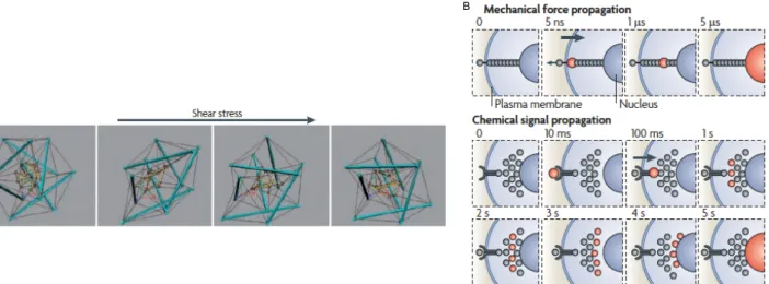
As previously shown, mechanosensors can undergo conformational modifications if a mechanical stimulus is applied, with subsequent changes in cell’s functions. As a consequence, cell’s cytoskeleton can be imagined as a “hard-wired” network (N. Wang et al., 2009), which can induce variations in cellular, cytoskeletal and nuclear structures upon mechanical forces (Ingber, 2003) (Fig 7A). The meaning of “hard-wired” stands in the fact that cytoskeleton’s filaments are strong enough to deal with mechanical stress cells undergo, thus keeping their shape stable (N. Wang et al., 2009). As such, cell’s shape stability and mechanical force transmission throughout cell’s interior are dictated by the level of isometric tension (also referred to as “prestress”) in the cytoskeleton. We can define the prestress as the result of a force balance between opposing structural elements in the cell (namely cytoskeletal filaments and extracellular adhesions). Such a phenomenon happens because cell tenses and thus stiffens cytoskeleton’s filaments relative to the surrounding cytoplasmic regions (Donald E. Ingber, 2006; Ingber, 2003; N. Wang & Suo, 2005). By virtue of the prestress in the cell’s cytoskeleton, mechanical cues are faster in propagation than diffusion-based chemical signals (N. Wang et al., 2009) (Fig 7 B).
Fig 7: In cells, the “hard-wired” model accounts for mechanical force propagation (A), which is much largely faster than chemical signal propagation (B). Note that mechanical force propagation is in the order of micro-seconds, whereas chemical signal propagation is in the order of seconds. Adapted from (N. Wang et al., 2009).
In adherent animal cells, the first layer of extracellular mechanostranduction is represented by the interaction between ECM and cellular membrane via Focal Adhesions (FAs) (Fig 8). Indeed, FAs are multiprotein complexes through which extracellular mechanical stimuli can be perceived and, thus, transmitted to the cell’s interior (Martino, Perestrelo, Vinarský, Pagliari, & Forte, 2018). Due to their complexity in structure, FAs can be split in a transmembrane and in an intracellular layer. Basically, the transmembrane layer is composed of integrins, which establish cell-ECM contacts, connected to the cytoskeletal actin filament via the intracellular layer of scaffolding, docking and signalling proteins (Martino et al., 2018) (Fig 8). ECM mechanics and structure influence FAs core
A
protein composition. Moreover, integrin clustering impacts the recruitment of proteins in FAs (Cavalcanti-Adam et al., 2007; Schiller & Fässler, 2013).
Fig 8: Focal Adhesion structure. See text for details. ACTN, actinin; FAK, focal adhesion kinase; IT,integrin; PAX, paxillin; TLN, talin; VASP, vasodilator-stimulated phosphoprotein; VCL, vinculin; ZYX, zyxin. Adapted from (Nardone et al., 2017). 3.1 Focal Adhesions Assembly of FAs follows the interaction of the transmembrane proteins integrins with ECM proteins, such as fibro- and vitro-nectins, collagens and laminins. Basically, integrins are heterodimeric proteins made of α and β subunits (Fig 9A) and their assembly is dictated by ECM’s molecular composition. Moreover, the combination of 24 α and 9 β subunits and alternative splicing events regulate integrins specificity in mammals (Martino et al., 2018). The affinity between integrins and ECM can be modulated by both “inside-out” signalling and mechanical cues, which cause a high-affinity conformational change (Chen, Lou, Evans, & Zhu, 2012). This, in turn, elicits integrins activation (Fig 9B), due to increased integrin’s affinity for extracellular ligands, with consequent clustering and reinforcement of molecular partners at the level of cell-ECM contacts (Oria et al., 2017; Strohmeyer, Bharadwaj, Costell, Fässler, & Müller, 2017), thus forming FAs. In these cellular structures, integrins connect the ECM to the actin cytoskeleton by interacting, through their cytoplasmic domain, with several docking proteins, which build up the inner core of FAs. As stated before, ECM’s molecular composition regulates the specific assembly of the integrins’ subunits, which then causes specific cellular signalling (Seetharaman & Etienne-Manneville, 2018). Because of the great number of subunit combinations that can be generated and the relative signalling pathways that can arise, integrins are “focal- points” of cellular mechanosensing (Martino et al., 2018).
Fig 9: Integrins are heterodimeric proteins made of α and β subunits. A: integrins in their inactive form. B: following “inside-out“ signalling, as well as mechanical cues, integrins are activated with consequent recruitment of FAs docking proteins, such as talin. Outside: extracellular side; inside: intracellular side. Adapted from (Shattil, Kim, & Ginsberg, 2010).
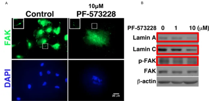
Another molecule involved in FAs composition is Focal Adhesion Kinase (FAK) (Fig 10), which is one of the first proteins to be recruited following externally applied forces. FAK is activated via autophosphorylation, which is thought to elicit intracellular mechanotranduction with following activation of cytoplasmic mechanotransducers (Lachowski et al., 2018). Contraction of the cytoskeleton as well as cell spreading can increase FAK activation, whose phosphorylation can be also enhanced by external applied mechanical forces (such as substrate rigidity or cell stretching) (Friedland, Lee, & Boettiger, 2009; Seong et al., 2013). Within the cell, FAK/cytoskeletal network interplay is strictly regulated in order to maintain tension at some critical cellular locations and regulate force transmission towards the nucleus (J. Zhou et al., 2015). Indeed, when cell polarizes or cell’s nucleus deforms, FAK activation locally happens at cellular sites, thus eliciting cytoskeleton local rearrangement and nucleus squeezing (Jung et al., 2012). FAK activity has also been shown to influence nuclear morphology, as recently demonstrated by Yang team. Indeed, the authors found that in A549 lung carcinoma cells, inhibition of FAK with the competitive ATP-inhibitor PF-573228 (Slack-Davis et al., 2007) resulted in nuclei with aberrant shapes (Fig 11 A), which the authors claimed to be the a consequence of the decreased expression of the nuclear lamins A and C (Chuang et al., 2019) (Fig 11 B).
Fig 10: Schematic representing focal adhesion kinase (FAK). Starting from the N-terminus, FAK ‘s structure displays an autoinhibitory FERM domain, comprising three lobes (F1-F3), followed by a first proline-rich domain (Pro-1), a linker domain, a central kinase domain, two other proline-rich domains (Pro-2 and Pro-3) and the focal adhesion targeting domain (FAT). Tyrosine phosphorylation sites (Y) are displayed. Adapted from (Lawson & Schlaepfer, 2013). α β β α A B N ter C ter
Fig 11: FAK inhibition induces nuclear aberrant morphology due to the decreased expression of nuclear lamins A and C. A: In A549 lung carcinoma cells treated with FAK inhibitor PF-573228, FAK distribution at the FAs is greatly reduced compared to control (see insets in the FAK immunostaing) and this correlates with aberrant nuclear morphology (nuclei counterstained with DAPI). B: PF-573228 decreases protein levels of nuclear lamins A and C as well as of active (phosphorylated) FAK (p-FAK) (boxes in red) in A549 lung carcinoma cells. Scale bar 20μm. Adapted from (Chuang et al., 2019). FAs also comprise the 270 kDa protein talin, which binds both filamentous actin (F-actin) and the FA protein vinculin. Talin is also responsive to force. Indeed upon mechanical force application, it unfolds and unmasks cryptic hydrophobic biding sites for vinculin (Del Rio et al., 2009; Hirata, Tatsumi, Lim, & Sokabe, 2014). Once vinculin is recruted to talin, talin/F-actin interaction is stable with consequent force transmission into cell’s interior (Humphries et al., 2007).
Vinculin recruitment in FAs is directly dependent on the forces applied to the FAs (Dumbauld et al., 2013). Vinculin is basically made of a head domain, with which is connected to talin (Price et al., 1989), a rod-like tail domain, with which vinculin is also connected to paxillin (Turner, Glenney, & Burridge, 1990) -a docking protein residing in the intracellular layer of FAs- and a flexible linker between head and tail domains (Fig 12). The tail domain in vinculin is important for force transmission to the actin cytoskeleton (Dumbauld et al., 2013).
Fig 12: Schematic representing vinculin’s structure. Vinculin head is made of 3 domains (D1-D3), whereas one domain (D5) composes vinculin tail. A flexibile linker is in between vinculin head and vinculin tail. aa=amino acids. N: N-terminus. C: C-terminus. Adapted from (Bays & DeMali, 2017).
Other proteins present at the FAs sites are Zyxin, which promotes filamentous-actin (F-actin) polymerization, α-actinin, which cross-links F-actin and p130Cas which, following cell stretching, is phosphorylated to regulate signalling pathway (Sawada et al., 2006).
A
10µM
BTalin interaction Paxillin interaction
3.2 Adherens junctions
Besides the ECM, in cohesive animal tissues, cells can interact among them via intercellular adhesion complexes, whose the best understood example is represented by adherens junctions (AJs) (Nicolas Borghi & James Nelson, 2009). Ajs mediate cell-cell interaction via “classical cadherins” (Fig 13), single-span transmembrane glyocoproteins which share a highly conserved cytoplasmic domain (M Takeichi, 1995). Classical cadherins are named according to the tissue they derive: this is why one can talk about E-cadherin (from epithelial tissue), N-cadherin (from neural tissue) and R-cadherin (from retinal tissue), for instance (Baranwal & Alahari, 2009). Hereafter, with “cadherins” I will refer to classical cadherins. With the five extracellular repeats domains, cadherins present on one cell engage the cadherins residing on the opposing cell via Ca2+ dependent trans-binding (Shapiro & Weis, 2009; M
Takeichi, 1988; Masatoshi Takeichi, Atsumi, Yoshida, Uno, & Okada, 1981)(Fig 13). Cadherins’ cytoplasmic domain is involved in the interaction with p120 and β-catenins; the further interaction of β-catenin with α-catenin permits the link of cadherins to the actin cytoskeleton (Baranwal & Alahari, 2009; Nelson, 2008) (Fig 13).
Fig 13: The adherens junction. See text for details: TM: transmembrane domain. p120 ctn: p120 catenin. F-actin: filamentous actin. Adapted from (Baranwal & Alahari, 2009)
AJs are involved in mechanical coupling of plasma membrane with the cytoskeleton via cadherins’ cytoplasmic domain (Tabdanov, Borghi, Brochard-Wyart, Dufour, & Thiery, 2009) and, moreover, if mechanically stimulated via cadherins’ extracellular domain, cells stiffen in a vinculin-dependent manner (le Duc et al., 2010). In addition, E-cadherin was demonstrated to be under cytoskeleton generated tension in epithelial MDCK type IIG cells. Indeed, by using a FRET TSMod biosensor (Grashoff, Hoffman, Brenner, Zhou, Parsons, Yang, McLean, Sligar, Chen, Ha, & Schwartz, 2010) inserted into full length as well as β-catenin binding domain deleted E-cadherins (EcadTSMod and EcadTSModΔcyto, respectively), Borghi et al demonstrated that the FRET index measured at the level of the cell-cell contacts (namely adherens junctions) was lower (meaning higher tension) in MDCK cells stably expressing EcadTSMod compared to those cells stably expressing EcadTSModΔcyto Cytoplasm TM TM TM TM TM TM TM TM C F-actin
(N. Borghi et al., 2012) (Fig 14A). This reported variation in tension through FRET was associated to the lost connection between the cytoskeleton and the truncated E-cadherin construct, suggesting that E-cadherins are under constitutive cytoskeleton-generated tension at the cell-cell contacts. This was then confirmed by the authors treating MDCK cells stably expressing EcadTSMod with the actin polymerization inhibitor cytochalasin B (Cooper, 1987) (Fig 14B). Compared to untreated cells, drug-treated cells displayed a higher FRET index difference, meaning that tension in E-cadherin decreased (Fig 14B). Thus, the authors concluded that E-cadherin is under constitutive actin cytoskeleton-generated tension at the cell-cell contacts (N. Borghi et al., 2012).
Fig 14: E-cadherin is under constitutive cytoskeleton-generated tension at the cell-cell contacts. A: FRET index measurements in MDCK type IIG cells stably expressing EcadTSMod (full length) and EcadTSModΔcyto (deletion of the β-catenin binding domain) constructs at the cell-cell contacts (AJs). Due to the lack of cytoskeleton connection, cells expressing EcadTSModΔcyto show a higher FRET index, which means lower tension, compared to EcadTSMod expressing cells. B: cytochalasin b treatment induces actin polymerization inhibition, which determines a reduction in tension (measured as FRET index difference) applied by the cytoskeleton to E-cadherin at the cell-cell contacts. Adapted from (N. Borghi et al., 2012).
3.3 Cytoskeleton
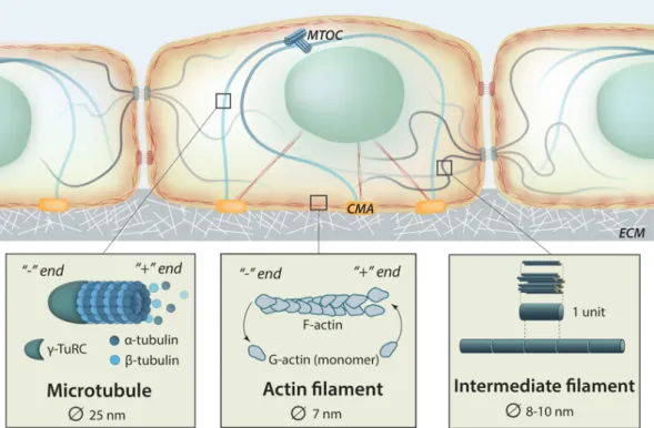
Force transmission, as well as force generation, within the cell is assured by the presence of the cytoskeleton, whose tensional state is highly regulated (Discher, Janmey, & Wang, 2005). The cytoskeleton gives the cell mechanical support and, among others, regulates its shape and motility (Fletcher & Mullins, 2010). Impairment of cytoskeletal structure has been shown to impact gene expression and cell signalling (Dupont et al., 2011; Jaalouk & Lammerding, 2009). Basically, the dynamic cytoskeletal network comprises actin filaments (F-actin, filamentous actin, also referred to as microfilaments), microtubules (MTs, made of α and β tubulin monomers) and intermediate filaments (IFs) (Fig 15). Through this organized filaments network, the cytoskeleton is able to keep the integrity of cell’s organelles (Martino et al., 2018). A B
Fig 15: Schematic representing cell’s cytoskeleton with insets showing microtubules and actin filaments with their dynamical polymerization and intermediate filaments. MTOC: microtubule organizing center. CMA: cell-matrix adhesion. ECM: extracellular matrix. Adapted from Mechanobio.info.
Microfilaments and the motor protein myosin II, important for cytoskeletal contractility, are cross-linked by α-actinin in complex structures know, in mammals, as stress fibers (SFs), which have been shown to transmit force from ECM to cell and from cell to ECM when FAs are pulled (Cramer, Siebert, & Mitchison, 1997; Naumanen, Lappalainen, & Hotulainen, 2008; Pellegrin & Mellor, 2007).
SFs are defined as dorsal SFs, transverse arcs and ventral SFs (Naumanen et al., 2008). Dorsal SFs, which lack myosin II, have just structural function at the level of FAs -to which are connected- and do not contract (Tojkander, Gateva, & Lappalainen, 2012).Transverse arcs interact with dorsal SFs and are, instead, contractile and indirectly connected to FAs through dorsal SFs (Martino et al., 2018). Lastly, ventral SFs are both contractile and directly connected to the FAs (Martino et al., 2018) (Fig 16). Contraction of SFs has been shown to elicit vinculin recruitment to the FAs (H. Yamashita et al., 2014). It has been demonstrated that the Nuclear Envelope (NE) is connected to FAs via a particular subtype of actin fiber, namely the perinuclear actin cap, which wraps around the nucleus (Khatau et al., 2009) (Fig 16). Mechanical stimuli have been shown to be directly transferred from the FAs to the nucleus via the perinuclear actin cap (D. H. Kim et al., 2012; Q. Li, Kumar, Makhija, & Shivashankar, 2014).
Fig 16: Stress fibers and formation of the perinuclear actin cap. Transverse arcs and dorsal fibers are cross-linked via α-actinin (orange dots). Transverse arcs contraction promotes the movement of the cross-linking points and the stress fibers from cell’s periphery in front of cell’s nucleus (bleu and white arrows). Transverse arcs and dorsal fibers move stress fibers above the nucleus (purple arrows), thus leading to the perinuclear actin cap formation. From (Tanja Mierke, 2018).
Actin dynamics is regulated by the Rho/ROCK pathway. Indeed, the ROC Kinase (ROCK) activates myosin II via myosin light chain (MLC) phosphorylation or MLC phosphatase inhibition (Feng et al., 1999; Mutsuki et al., 1996), thus resulting in F-actin contraction.
In addition, Rho/ROCK pathway activates the formin Diaphanous (mDia) which, in turn, elicits microfilament polymerization (Watanabe et al., 1997) (Fig 17). Rho/ROCK pathway also regulates F-actin stabilization by activating LIM kinase (LIMK) (Ohashi et al., 2000), which phosphorylates and inhibits the actin-severing protein cofilin (Bamburg, Harris, & Weeds, 1980)(Fig 17).
Fig 17: Rho/ROCK pathway regulates actin cytoskeleton dynamics by 1) inducing actin polymerization through mDia activation; 2) stimulating F-actin contraction via either direct MLC phosphorylation or 3) inhibition of MLC phosphatase; 4) determining F-actin stabilization via LIMK activation which, in turn, inhibits cofilin through
phosphorylation. mDIA: Diaphanous; MLC: myosin light chain; LIMK: LIM Kinase. SF: stress fiber. From (Martino et al., 2018).
It has been shown that microfilaments interact with MTs (Dugina et al., 2016) ,which are the stiffest cytoskeletal filaments. MTs regulate intracellular trafficking, mitotic spindle formation and cellular polarity (Fletcher & Mullins, 2010; J. Zhang, Guo, & Wang, 2014). Moreover, MTs have been shown to be mechanoresponsive when mitotic cells are stretched. Indeed, Fink et al. demonstrated that mitotic spindle aligns parallel to the stretch direction (Fink et al., 2011).
Lastly, IFs are more flexible and stable than microfilaments and MTs. Also, post-translational modifications govern IFs’ dynamics and implication in different cellular pathways (Snider & Omary, 2014).
3.4 Signalling upon mechanotransduction
Mechanical cues, perceived by the cell via mechanosensors, can let other mechanorensposive proteins undergo conformational changes and/or post-translational modifications with consequent nuclear shuttling (Martino et al., 2018). An example of this shuttling was given by Gottardi et al., who showed that the tight junction protein zonula occludens-1 (ZO-1) accumulates into the nuclei of subconfluent MDCK epithelial cells but not in confluent monolayers (Gottardi, Arpin, Fanning, & Louvard, 1996). Furthermore, Lewis et al found that cell-cell adhesion induced by integrins determines the translocation of the FA tyrosine kinase c-Abl from the nucleus to the early focal contacts and then back to the nucleus, in C3H 10T1⁄2 mouse fibroblasts plated onto fibronectin (Lewis, Baskaran, Taagepera, Schwartz, & Wang, 1996). Recently, β-catenin, which interacts with the E-cadherin at the AJs, has been demonstrated to undergo nuclear accumulation upon E-cadherin tension relaxation in MDCK type IIG epithelial cells (Gayrard, Bernaudin, Déjardin, Seiler, & Borghi, 2018). The FA protein Zyxin, was also demonstrated to shuttle between nucleus and the cytoplasmic focal contacts in primary CEFs (chicken embryo fibroblasts) (Nix & Beckerle, 1997). Moreover, it has been suggested that, once in the nucleus of rat aortic smooth muscle cells (SMCs) upon cyclic stress, zyxin may regulate few mechanoresponsive genes, such as endothelin B receptor (ETB-R) and matrix protein tenascin-C (Cattaruzza, Lattrich, & Hecker, 2004). Another FA protein demonstrated to undergo nuclear translocation is paxillin, whose nuclear shuttling has been shown to be regulated, in fibroblasts, by cell geometry and be independent of ECM composition (Sathe, Shivashankar, & Sheetz, 2016).
Other mechanoresponsive proteins not associated to FAs demonstrated to undergo nuclear shuttling are the transcriptional co-activators Yes-associated protein (YAP) and WW Domain-Containing Transcription Regulator Protein 1 (WWTR1/TAZ), playing the role of downstream effectors in the Hippo pathway (Huang, Wu, Barrera, Matthews, & Pan, 2005; Oka & Sudol, 2009) (Fig 17), known to limit organ size in animals (Yu, Zhao, & Guan, 2015).
YAP and TAZ are intended as molecular relays for ECM mechanics. Indeed, Dupont et al showed that human mesenchymal stem cells (MSCs) grown on hard substrates displayed YAP/TAZ nuclear accumulation, compared to YAP/TAZ cytoplasmic localization in MSCs grown on soft subtrates (Dupont et al., 2011). Moreover, YAP localization has been also shown to be regulated by E-cadherin. Indeed, Kim et al found that inducible expression of E-cadherin in human breast cancer cells (MDA-MB-231), grown at high density, determined cytoplasmic localization of YAP, which was instead nuclear in the parental, E-cadherin negative cells (N.-G. Kim, Koh, Chen, & Gumbiner, 2011). YAP
nuclear translocation has been also associated with cell area. To this regard, Nardone et al showed that adipose tissue-derived mesenchymal stem cells (AD-MSCs), grown onto fibronectin-coated micropatterns, presented an increasing YAP nuclear localization when cell’s area increased (Nardone et al., 2017) In addition to mechanical regulation, Hippo pathway can be also controlled by G-protein-coupled receptor signalling and cell’s energy status (Y. Zhang, Zhang, & Zhao, 2018) (Fig 18), as well as by insulin/IGF signalling (IIS) (Straßburger, Tiebe, Pinna, Breuhahn, & Teleman, 2012). When Hippo pathway is active, MST1/2 phosphorylates SAV1, MOB1A/B and LATS 1/2, whose kinase activity is thus activated. Consequentially, LATS1/2 phosphorylates YAP/TAZ, which can be either retained in the cytoplasm by interacting with 14-3-3 protein or degraded via E3 ligase SCF β-TRCP. When Hippo pathway is not active, YAP/TAZ are then no more phosphorylated, which results in their nuclear accumulation. Once in the nucleus, YAP/TAZ can bind to transcription factors, such as TEADs or others having PPXY motif, in order to allow gene transcription (Y. Zhang et al., 2018) (Fig 18). Fig 18: Hippo pathway regulation. See text for details. Adapted from (Y. Zhang et al., 2018).
It has also been shown that a complex of YAP/TAZ/SMAD can undergo nuclear shuttling via cell density regulation. Indeed, YAP/TAZ/SMAD complex translocates into the nucleus of sparse HaCaT cells, which do not perceive cell-cell connection (Grannas et al., 2015).
Co-transcriptional activity of nuclear YAP/TAZ has been connected to cell growth and tumor spreading (Zanconato et al., 2015). Nevertheless, recent findings proved that YAP activity in mechanotransduction is related to its capability to directly elicit transcription of genes involved in cell-matrix interaction and ECM composition (Nardone et al., 2017), as well as cytoskeleton integrity during cardiomyocyte renewal and regeneration (Morikawa et al., 2015).
In summary
• Mechanotransduction refers to the way cells sense and respond to external and internal
mechanical cues by converting these physical stimuli into biochemical signals.
• Mechanosensors or mechanotransducers are the protein complexes, as well as cell’s organelles, being able to transduce mechanical stimuli.
• Pioneering works showed that mechanotransduction is involved in tissue shaping, cellular metabolism, embryo development, gene expression and lineage commitment.
• Mechanistically, two ways of transferring physical stimuli can be conceived: the tethered model and the lipid bilayer model.
• Mechanical force propagation in cells is possible since cell’s cytoskeleton is prestressed. • Extracellular matrix, focal adhesions and adherens junctions participate in
mechanotransduction.
• Signalling upon mechanical cues is highly regulated and can imply protein shuttling between nucleus and cytoplasm.
CHAPTER 2
The nuclear envelope: what wraps the cell’s nucleus
In this chapter, I will discuss the structure of NE focusing on its molecular components in normal as well as in diseased conditions. Nucleo-cytoplasmic shuttling and mechanotransduction in the nucleus will be also taken into account.
Table of contents
1 Nuclear envelope’s structure ... 27
1.1 Outer nuclear membrane (ONM), inner nuclear membrane (INM) and nuclear lamina. ... 271.2. The LINC complex: nesprin and SUN proteins. ... 31
1.3. The LINC complex: when the LINC is perturbed. ... 38
1.4 The nuclear pore complex: structure and nucleo-cytoplasmic shuttling. ... 42
2 Nuclear mechanotransduction. ... 47
3 Nuclear envelope associated pathologies: the envelopathies. ... 52
In summary. ... 55
1 Nuclear envelope’s structure
1.1 Outer nuclear membrane (ONM), inner nuclear membrane (INM) and nuclear lamina.Cell’s nucleus is surrounded by the nuclear envelope (NE) consisting of two different lipid membranes, the outer nuclear membrane (ONM) and the inner nuclear membrane (INM). INM and ONM are in continuity with the endoplasmic reticulum (ER) (Schirmer & Gerace, 2002) and separated by the perinuclear space (PNS) (Fig 19), which is 30-50 nm in width (Maurer & Lammerding, 2019). This structure allows the chromatin to be well separated, in the nucleus, from the cytoplasm (Osorio & Gomes, 2013) and the presence on the NE of the selective barrier of the nuclear pore complex (NPC) permits to control the inwards/outwards nuclear shuttling of molecules larger than 30 kDa (Maurer & Lammerding, 2019).
Fig 19: The nucleus, surrounded by the inner nuclear membrane (INM) and the outer nuclear membrane (ONM), which, with the nuclear pore complex (NPC), form the nuclear envelope (NE). Underneath the INM there lays the nuclear lamina. Note that INM and ONM are in continuity with the endoplasmic reticulum (ER). On ER surface, ribosomes are displayed in ochre. PNS: perinuclear space. Adapted from (Schirmer & Gerace, 2002). Underneath the INM, there lays the nuclear lamina (Fig 20), a protein meshwork with a thickness of 10-30 nm (Turgay et al., 2017). Basically, the proteins composing the nuclear lamina, called lamins, are type V nuclear intermediate filaments and are present in the greatest part of the differentiated cells (Maurer & Lammerding, 2019).
Fig 20: Nuclear lamina is located underneath the inner nuclear membrane (INM) and interacts with chromatin as well as with different proteins. ER: endoplasmic reticulum. ONM: outer nuclear membrane. Adapted from: cellbiology.med.unsw.edu.au
In mammals, lamins are fundamentally divided into A-type lamins and B-type lamins (Janin & Gache, 2018) (Fig 21). A-type lamins, which are encoded by the gene LMNA, produce the major isoforms lamin A and C and other less common ones (Maurer & Lammerding, 2019). Lamins A and C are generated by an alternative splicing event occurring on exon 10. As such, lamins A and C share a similar structure from the N-terminus till the amino-acid position 556 at the C-terminus. However, while lamin C presents just five additional amino-acids, lamin A shows an extra domain encoded by exons 11 and 12 of the LMNA gene (Janin & Gache, 2018). Lamins A and C have a development-dependent regulation and are present during cellular differentiation (Maurer & Lammerding, 2019). Regarding B-type lamins, they are divided into lamins B1 and B2 encoded by LMNB1 and LMNB2
PN S PN S ONM INM
genes, respectively (Maurer & Lammerding, 2019). Being themselves intermediate filaments, nuclear lamins dimerize via their central rod domain (Herrmann & Aebi, 2016) (Fig 21). Fig 21: Lamins A and B structures. With the rod domain, nuclear lamins dimerize. Ig:immunoglobulin. Adapted from(Dittmer & Misteli, 2011)
Nuclear lamins interact with different partners, such as chromatin, transcription factors and LEM (LAP2, Emerin and MAN1) proteins, the latter being critical in gene regulation (Dorner, Gotzmann, & Foisner, 2007). It is thus clear that nuclear lamins are important in terms of genome regulation and chromatin organization (de Leeuw, Gruenbaum, & Medalia, 2018; Gruenbaum & Foisner, 2015).
Lamins and heterochromatin interact either in specific lamin-associated-domains (LADs)-genomic regions involved in molecular contact with nuclear lamina (Pickersgill et al., 2006; van Steensel & Belmont, 2017)-or through the LEM protein lamin-associated protein 2 (LAP2) and its partner, BAF (barrier to autointegration factor) (Goldman, Gruenbaum, Moir, Shumaker, & Spann, 2002; Shumaker, Lee, Tanhehco, Craigie, & Wilson, 2001). Both A-type and B-type lamins, by binding nucleoporin 153 (Nup153) – one of the protein composing the NPC – place the NPC at the level of the NE (Maurer & Lammerding, 2019). Nuclear lamina was postulated to have elastic extensibility and limited compressibility, as proposed by Dahl et al. following osmotic swelling experiments involving
Xenopus oocyte nuclear envelopes (Dahl, 2004). In addition, Disher team also showed that 3D
migration of lung carcinoma-derived A549 cells is influenced by Lamin A levels (Harada et al., 2014). Indeed, Harada et al. found that cell migration through 3-μm pore Transwell filters increased upon Lamin A depletion and reached a peak when depletion was partial (Fig 22). On the contrary, Lamin A overexpression impeded migration (Harada et al., 2014)(Fig 22).
Fig 22: Lamin A expression influences 3D migration. Note that the highest migration level (≈4.6 fold) is reached when Lamin A depletion is partial (0.5 Lamin A expression on x axis). Overexpression of Lamin A impedes migration. Pre-coating filters with fibronetcin (+FN) does not exert any variation on migration compared to uncoated filters (-FN). Adapted from (Harada et al., 2014).
Among those proteins interacting with lamins, we find emerin, which is encoded by the EMD gene (Bione et al., 1994; Janin & Gache, 2018). Beside being involved in gene regulation (Holaska, Lee, Kowalski, & Wilson, 2003; Wilson et al., 2005), emerin regulates cell signalling as well as nuclear architecture (Berk, Tifft, & Wilson, 2013). With its LEM domain located at the N-terminus (Fig 23), emerin interacts with BAF, which, in turn, is important for post-mitotic nuclear assembly, cell viability and cell cycle progression (Janin & Gache, 2018). Additionally to its presence at the nuclear lamina, emerin has been also found at the level of ONM and ER, where it directly interacts with the centrosome (Salpingidou, Smertenko, Hausmanowa-Petrucewicz, Hussey, & Hutchison, 2007).
Fig 23: Emerin’s structure. The interaction with BAF (barrier to autointegration factor) occurs at the LEM (LAP2, Emerin and MAN1) domain. With the TM (transmembrane) domain, emerin is inserted into the inner nuclear membrane (INM). APC-L: Adenomatous poliposis coli-like domain. N: N-terminus. C: C-terminus. Adapted from (Berk et al., 2013).
Emerin has also a role in β-catenin’s activity regulation. Indeed, by using human fibroblasts, both wild-type or null for emerin, Markiewicz et al. found that a β-catenin antibody co-immunoprecipitated emerin in cell extracts from wild-type human fibroblasts but did not in cell extracts from emerin null human fibroblasts (Fig 24A),thus demonstrating that emerin interacts with β-catenin. Moreover, the researchers showed that this interaction is mediated by an APC-like domain in emerin (Fig 24B) whose deletion increased β-catenin’s activity (Fig 24C) and stimulated β-catenin nuclear accumulation in HEK 293 cells (Fig 24D). The authors thus concluded that emerin negatively regulates β-catenin’s activity (Markiewicz et al., 2006).
A549 cells migrating through 3-µm pore Transwell filters
Fig 24: Emerin interacts with β-catenin and regulates its activity and nuclear accumulation. A: emerin co-immunoprecipitates with β-catenin in cell extracts from wild type human fibroblasts but it did not in emerin null human fibroblasts B: cell extracts from HEK 293 cells co-transfected with β-catenin plus GFP-emerin wt (wild type emerin), GFP-emerinΔ (emerin with deletion in the APC-like domain) or GFP alone were used for immunoprecipitation with an anti-GFP antibody. In GFP-emerinΔ extracts, β-catenin was not co-immunoprecitated with the anti-GFP antibody (see asterisk in “pellet” lane). In A and B red boxes to indicate (co)immunopreciptated emerin and β-catenin; H and L to indicate “heavy” and “light” IgG chains, respectively. C: luciferase assay to estimate β-catenin’s acitivity in HEK 293, co-transfected with wt (wild type) β-catenin and TOPGLOW (to measure β-catenin’s acitivity) plus the GFP-emerin (or GFP alone) constructs (GFP-em: wild type emerin; GFP-emΔ: APC-like domain deleted emerin). Note that GFP-emΔ increases β-catenin’s acitivity compared to GFP-em. D: in HEK 293 cells co-transfected with β-catenin plus GFP-wt em (wild type emerin), β-catenin localizes at the cell-cell contacts (β-catenin immunostaining panel,above), whereas in the presence of GFP-emΔ (APC-like domain deleted emerin) co-transfected β-catenin is mostly nuclear (β-catenin immunostaining panel, middle). ** p<0.001. Adapted from(Markiewicz et al., 2006).
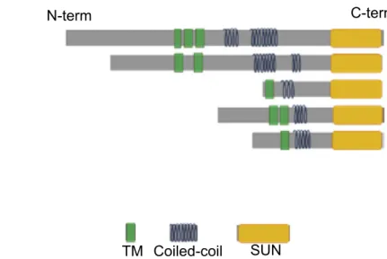
1.2. The LINC complex: nesprin and SUN proteins Embedded into the ONM and the INM, the LINC (Linker of Nucleoskeleton and Cytoskeleton) complex spans the NE and assures coupling between the cytoskeleton and the nucleus (Crisp et al., 2006; Starr & Han, 2002).This function is made possible because of the presence of the nesprin (nuclear envelope spectrin repeat proteins,) proteins (Q Zhang et al., 2001) and the SUN (Sad1p and UNC-84 homology) domain proteins(Lygerou, Christophides, & Séraphin, 1999; Malone, Fixsen, Horvitz, & Han, 1999).
A B
C D