DOI: 10.5897/AJB2015.14611 Article Number: 618135153292 ISSN 1684-5315
Copyright © 2015
Author(s) retain the copyright of this article http://www.academicjournals.org/AJB
African Journal of Biotechnology
Full Length Research Paper
Assessment of four different methods for selecting
biosurfactant producing extremely halophilic bacteria
ARIECH Mounira
1,2* and GUECHI Abdelhadi
21
Departement of Microbiology and Biochemistry, Faculty of Sciences, Mohamed Boudiaf University,M’sila, 28000,
Algeria.
2
Departement of Microbiology, Faculty of Natural Sciences and Life, Ferhat Abbas Setif-1-University, Setif, 19000,
Algeria.
Received 1 April, 2015; Accepted 25 May, 2015
Biosurfactants are surface active agents with broad range commercial applications in various industries
and have considerable advantages over their chemical counterparts. In this study, drop collapse assay,
oil spreading assay, emulsification assay and surface tension measurement were compared for their
reliability and ease of use to screen biosurfactant producing six extremely halophilic bacteria isolated
from saline soil of Chott El Hodna-M’sila (Algeria), which is considered as a thalassohaline environment.
Results from screening methods revealed that, CH2 and CH5 strains are potential candidates for
biosurfactant production. Among these, there was a very strong negative linear correlation between the
emulsification index and surface tension (r
s= - 0.987) and a strong negative linear correlation between
the oil spreading technique and surface tension (r
s= - 0.971) and a weaker negative correlation between
the drop collapse method and surface tension (r
s= - 0.807), suggesting that strains highly active in any
one of these methods were active in other three methods. As every method has its advantages and
disadvantages, a combination of different methods is appropriate for a successful screening.
Key words: Biosurfactant, drop collapse, emulsification stability, extremely halophilic bacteria, oil spreading,
surface tension.
INTRODUCTION
Biosurfactants are natural surface active agents
produced by bacteria, yeast, and fungi, having very
different chemical structures and properties (Rone and
Rosenberg, 2001; Chen et al., 2007). These
biosurfac-tants are amphiphilic molecules consisting of hydrophobic
and hydrophilic domains that find application in an
extremely wide variety of industrial processes involving
emulsification, foaming, detergency, wetting, dispersing
or solubilization (Rodrigues et al., 2006).
Nowadays, biosurfactants are used in industries as a
cosmetic and special chemical substance, food,
phar-maceutics, agriculture, cleansers, enhanced oil recovery
*Corresponding author. E-mail: [email protected]. Tel: +213 779 01 65 57. Fax: +213 35 55 05 24.Author(s) agree that this article remains permanently open access under the terms of the Creative Commons Attribution License 4.0 International License
and bioremediation of oil-contaminated environments and
can be effectively used in numerous processes, including
enhanced oil recovery, transportation in pipelines,
cleaning of oil storage tanks, refining and product
formulation (Urum and Pekdemir, 2004; Mukherjee et al.,
2006).
They are potential alternatives of chemically
synthe-sized surfactant in a variety of applications because of
their advantages such as lower toxicity, higher
biode-gradability, better environmental compatibility, lower
critical micelle concentration, each of production, ability to
be synthesized from renewable resources, higher
foaming, higher selectivity, specific activity at extreme
temperature, pH and salinity (Chen et al., 2007;
Nitscheke and Costa, 2007).
For this reasons a variety of methods for the screening
of new biosurfactant producing microbes has been
developed and successfully applied. Since the 1970th
there have been various trials in this field (Walter et al.,
2010). Hence, for efficient detection of potential
biosur-factant producers, combination of various screening
methods are required which was evaluated during this
study.
Thus, the main goal of this study was to introduce novel
extremely halophilic bacteria with potential for
biosurfac-tant/bioemulsifier production isolated from saline soils of
Chott El Hodna-M’sila (Algeria). The potential of these
isolates in producing biosurfactant was examined by drop
collapse assay, oil spreading assay, emulsification assay
and surface tension measurement.
Finally, this study is considered as a valorization of this
environment which is a representative type of wetland in
the Mediterranean by the extent of its size and its
watershed, its location in the arid zone is another
advantage to justify the rarity of this type of natural
environment in one piece having undergone little or no
significant changes by man.
Chott El Hodna is a model representative of the
presence of several types of soil, bioclimates and
biological species. For this reason, it was classified as
humid zone of international importance by the RAMSAR
convention in 2001. It presents a considerable ecological
interest and accommodates a large number of animals
and plants species diversifies of interest for biodiversity of
the region El Hodna (Boumezbeur, 2002).
MATERIALS AND METHODS Area study
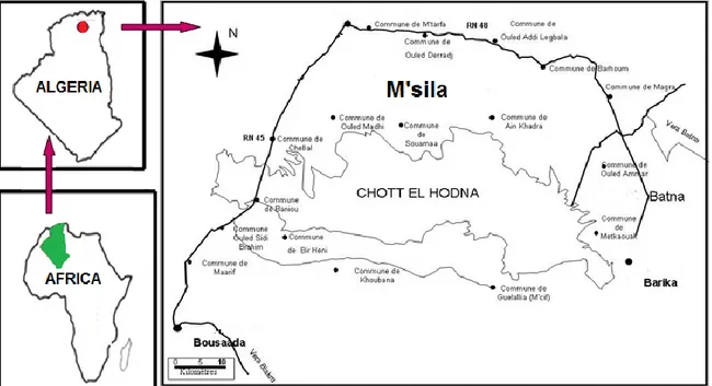
Chott El Hodna is a shallow saline lake in Algeria; it is located within an endorheic basin in North-Central of Algeria (lat. 35°18’/35°32’, long. 4°15’/5°05’) (Figure 1). The peripheral zone of this Chott is characterized by wadis and oases which surrounds the central zone of the sebkha with a complete absence of vegetation. The basin of Hodna has an area of 8500 Km2, but what is called the
Chott has an area of 1100 Km2 and is located about 400 m above
sea level. It gets water only in winter and dry in summer, salt crusts
cover its full extent. Due to the high evaporation, it becomes regularly a desert of salt (Boumezbeur, 2002).
Sample collection and physicochemical analysis
Water samples (200 ml) were collected at 10 cm depth from the water surface with 250 ml sterile flasks. Samples were gathered during the winter season (January). A total of five water samples were collected from this lake, and dispatched on the day of collection in ice box to the laboratory. Physicochemical properties of the samples taken from Chott El Hodna were analysed according to the methods described by Rodier et al. (1996). Chemical properties included compositional estimation of Ca2+ and Mg2+: by a
complexometric method using ethylene-diamine-tetra-acetic acid (EDTA), HCO3 and Cl- by a titremitric method, SO-24 by UV/Vis
spectrophotometry (Beckman/DU, 520), Na+ and K+ by spectrophotometry with flame ionisation (Jenwey PEP7) and nitrate by the sodium salicylate method. Physical parameters included pH and temperature measured in situ using a portable instrument. In addition, the colour and odour of the lake water were noted. The conductivity was monitored using a conductivity meter (Rodier et al. 1996).
Isolation of extremely halophilic bacteria
Five saline soil samples were collected in sterile plastic bags from Chott El Hodna-M’sila (Algeria). Each sample was inoculated for enrichment and was streaked on SG medium. This medium contained (g/L): NaCl, 250; MgSO4.7H2O, 20; KCl, 2; sodium citrate
(trisodium salt), 3; casamino acids, 7.5; yeast extract, 1 and FeSO4.7H2O, 0.0023. The pH was adjusted to 7.3 with 1 M KOH.
For solid medium, 20 g agar/Lwas added. Plates were incubated at 37°C. After two weeks incubation, representative colonies were transferred to fresh SG medium and isolated in pure culture (Ozkan et al., 2006).
Screening for haloarchaea
Haloarchaeal strains were selected among all isolates by their susceptibility to antibiotic and a bile acid. They were streaked on the solidified medium containing chloromphenicol at 20 mg/L. The cultures were incubated for ten days at 37°C. Strains that grew on the plates with chloromphenicol were regarded as Haloarchaea (Madalin et al., 2008).
Characterisation of isolates
Phenotypic characterisation was carried out in accordance with the recommended minimal standards for the description of new taxa in the order Halobacteriales (Oren et al., 1997). Cell motility and morphology of exponentially growing liquid cultures were examined using a microscope equipped with phase contrast optics. Colony morphology was observed on agar medium after incubation at 37°C for 10 days. Gram strains were carried out as described by Dussault (1995). Cytochrome oxidase, catalase, nitrate reduction, indol and H2S production, hydrolysis of gelatin and starch were
determined as explained before (Oren et al., 1997; Ozekan et al., 2007). The optimum salt concentrations for growth were determined in media containing 0-32% NaCl by turbidity measurement, pH from 2-11 and temperature from 0-60°C. Acid production was carried out in a medium with sugars as substrates. Antibiotic susceptibility was tested according to the methods described by Stan-Lotter et al. (2002).
Figure 1. The area study is located in North-Central of Algeria.
Screening methods
Biosurfactant producing Haloarchaea were screened by using four methods. Experiments are done in three replicates.
Drop collapsing test
A modified oil collapse method was carried out using 96 well microtitre-plates containing 100 µl mineral oil, which was equilibrated for an hour at room temperature. 10 µl of supernatant of culture broth was added to the surface of a well and the picture captured after 1 min using × 10 objective lens of a microscope. Biosurfactant production was considered positive when the drop diameter was at least 0.5 mm larger than those produced by distilled water and also by culture medium as negative controls (Plaza et al., 2006; Youssef et al., 2004). The results were interpreted as follows « + » to « ++++ » corresponding to partial to complete spreading on the oil surface. Those cultures that gave rounded drops were scored as negative «-» indicative of the lack of biosurfactant production (Loganathan et al., 2010).
Oil spreading test
In oil spreading method, 50 ml of sea water synthetic was added to the large Petri plate (90 × 15 mm) followed by 20 µl of crude oil making a thin layer on the surface of the water. A 10 µl aliquot of supernatant was delivered onto the surface of oil (Morikawa et al., 2000). The triplicate assays from the same sample were determined (Rodrigues et al., 2006).
Emulsification index
After growing in standard for 7 days in an orbital shaker at 160 rpm and 40°C, cells were removed by centrifugation at 12,000 × g for 5 min at room temperature. 2 ml of the cell-free supernatant was mixed with 2 ml gazoil in a test tube (125 × 15 mm). This mixture
was shaken for 2 min and then left to stand relative emulsion volume (EV,%) and emulsion stability (ES,%) were measured in intervals up to 24 h using the following Equations:
Emulsion formed by the isolates were compared to those formed by a 1% (w/v) solution of synthetic surfactant sodium dodecyl sulfate in deionised water as positive control and with sterile medium as negative control (Kebbouche-Gana et al., 2009). A criterion cited for emulsion stabilizing capacity is the ability to maintain at least 50% of original emulsion volume 24 h after formation (Nasr et al., 2009).
Surface tension measurement
The surface tension measurements of cell free supernatant were determined in a tensiometer (TD1C LAUDA). The values reported are the mean of three measurements. All measurements were made on cell-free broth (50 ml) collected at different time intervals after centrifugation (10 000×g for 25 min) at room temperature. The criterion used for selecting biosurfactant-producing isolates was the emulsification and the reduction of the surface tension of the medium to below 40 mN m1- (Ainon et al., 2013).
Statistical analysis of the correlation between different tests A general rank correlation test according to Spearman, was conducted to determine the correlation between each of the four methods. The Spearman rank correlation coefficient, rs ranged Emulsion height (mm)× cross-section area(mm2)
EV,% = ×100 Total liquid volume (mm3)
EV,% at time t,h
ES,% = ×100
EV,% at 0h
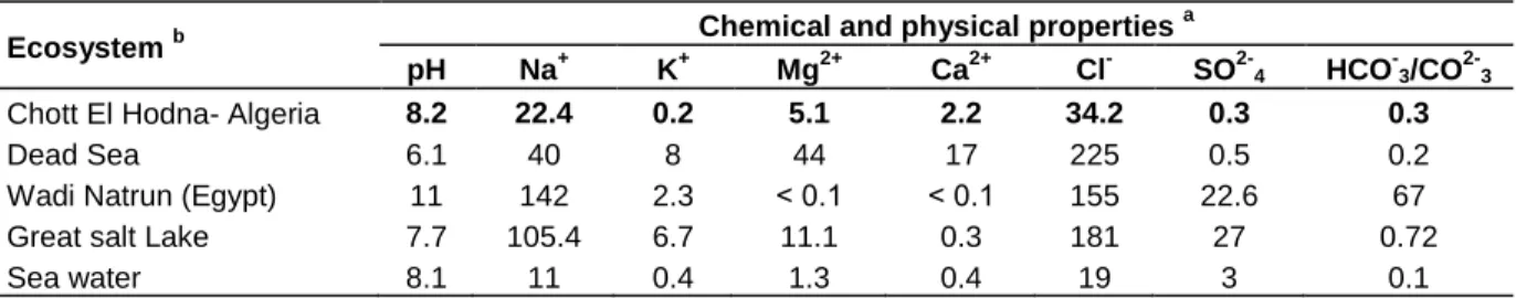
Table 1. Chemical and physical properties of Chott El Hodna Salt Lake compared to other hypersaline and marine ecosystems.
Ecosystem b Chemical and physical properties a
pH Na+ K+ Mg2+ Ca2+ Cl- SO2-4 HCO-3/CO2-3
Chott El Hodna- Algeria 8.2 22.4 0.2 5.1 2.2 34.2 0.3 0.3
Dead Sea 6.1 40 8 44 17 225 0.5 0.2
Wadi Natrun (Egypt) 11 142 2.3 ˂ 0.1 ˂ 0.1 155 22.6 67
Great salt Lake 7.7 105.4 6.7 11.1 0.3 181 27 0.72
Sea water 8.1 11 0.4 1.3 0.4 19 3 0.1
a
Ions are represented as g per litre. For Chott El Hodna : Conductivity of the brine sample was 108300 µs/cm, ash content was 73.644 g/L and hardness was 725.60 °F. bChemical and physical properties of some hypersaline and marine ecosystems
(Gavrieli, 1997 ; Lmhoff et al., 1979; Post, 1981; Copin-Montégut, 1996).
between -1 (strong negative correlation) to +1 (strong positive correlation).
RESULTS AND DISCUSSION
Physicochemical properties of brine sample
Chemical and physical properties of brine sample taken
from Chott El Hodna were compared to other hypersaline
and marine ecosystems (Table 1). The results obtained
indicate that the mineral content, pH and hardness of
brine are suitable for the growth of haloarchaea.
As there is no previous description about a physical
and chemical property of Chott El Hodna, these few tests
was conducted to understand an outline of ecological
condition where halophilic microbes were thriving. Chott
El Hodna saline environment was an ideal habitat for
haloarchaea and other halophiles, which were well
adapted to gradual changes in Chott El Hodna
ecosystem and was formed by ecological changes during
last hundreds of years of evolution. The presence of all
ions and a relatively high Mg
2+ion content (Table 1)
enhances the natural enrichment of Haloarchaea (Bolhuis
et al., 2006). But it contains about 73 and 74 g/L salts,
approximately three times lower than the salinity of the
Dead Sea, which is a source of extremely halophilic
archaea. Typically, salt concentrations of 100 to 150 g/L
are required for structural stability and viability of
members of the Halobacteriaceae. Exposure to lower
concentrations, even for short periods, leads to
denaturation of the cells’ proteins, including the
glycoprotein cell wall present in most species, and the
cells lyse. Still, there are reports of the isolation of
Haobacteriaceae from low-salt environments, and some
members have a surprising ability to survive exposure to
low salt for prolonged periods. Isolates affiliated with the
genera Halococcus, Haladaptatus and Halogeometricum
obtained from a traditional Japanese salt field survived
prolonged suspension at 50 g/L salts, and cells of an
isolate related to Haladaptatus paucihalophilus even
retained viability after nine days at 30 g/L (Fukushima et
al., 2007).
To gain deeper understanding of Chott El Hodna salt
lake, physicochemical properties of brine sample were
determined,
including
pH,
conductivity
and
ion
composition (Table 1). The results indicate that the
mineral content is dominated by chlorides (34.2 g/L)
among the anions and sodium, magnesium and calcium
among the cations (22.4 g/L, 5.1 g/L, and 2.3 g/L)
respectively, which explains the slightly alkaline pH (8.2)
of these lakes. As these environments result from
evaporation of sea water (Oren, 2006), compared to the
above, we can deduce that Chott El Hodna part of
thalassohaline environment.
Extremely halophilc strains
In the current study six halophilic strains isolated from
saline soil samples named CH1, CH2, CH3, CH4, CH5,
CH6, were phenotypically characterized (Table 2), and
compared with strains described previously by Oren,
(2014). The strains studied belong to the family of
Halobacteriaceae, order Halobacteriales. These strains
were
screened
for
biosurfactant
production
and
emulsification activity.
Screening of biosurfactant production
The primary screening of biosurfactant producing
halophilic archaea was carried out using, drop collapse
and oil spreading techniques. Selecting of these methods
was due to their strong advantages including simplicity,
low cost, quick implantation and use of relatively common
equipment
that
is
accessible
in
almost
every
microbiological laboratory. Results obtained from various
screening protocols are mentioned in Table 3 and Figure
2 (A, B and C.)
Drop collapse method
Jain et al. (1991) suggested the use of the drop collapse
method as a sensible and easy to perform method which
requires a small volume (5-10 µl) of culture broth or
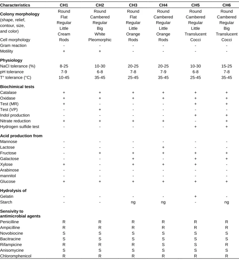
Table 2. Features of the extremely halophilic strains. Characteristics CH1 CH2 CH3 CH4 CH5 CH6 Colony morphology (shape, relief, contour, size, and color) Round Flat Regular Little Cream Round Cambered Regular Big White Round Flat Regular Little Orange Round Cambered Regular Little Orange Round Cambered Regular Little Translucent Round Cambered Regular Big Translucent
Cell morphology Rods Pleomorphic Rods Rods Cocci Cocci
Gram reaction - - - - Motility + + - - - - Physiology NaCl tolerance (%) 8-25 10-30 20-25 20-25 10-30 15-25 pH tolerance 7-9 6-8 7-8 7-9 6-8 7-8 T° tolerance (°C) 10-45 35-45 25-45 35-45 25-45 35-45 Biochimical tests Catalase + + + + + + Oxidase + + + + + + Test (MR) + - - - + + Test (VP) - + - - - - Indol production - - - - + + Nitrate reduction + + + + - +
Hydrogen sulfide test - - - - + +
Acid production from
Mannose - - - - Lactose - - - + - - Fructose - + + + + + Galactose - - + - + + Xylose + - + + + - Arabinose - - - - mannitol - - - - Glucose + + + + + + Hydrolysis of Gelatin - - - - + - Starch - - ng ng - ng Sensivity to antimicrobial agents Penicilline R R R R R R Ampicilline R R R R R R Novobiocine S S S S S S Bacitracine S S S S S S Rifampicine R R R S S R Anisomycine S S S S S S Chloromphenicol R R R R R R
CH1, Halomonas elongata; CH2, Natrialba asiatica; CH3, Ntrinema pellirubrum; CH4, Natrinema pallidum; CH5, Haladaptatus paucihalophilus; CH6 Haloplanus natans; R, resistance; S , sensible; ng, no growth.
biosurfactant solution to test the surfactant property. In
addition, it can be performed in Microplates (Tugrul and
Cansunar, 2005). This assay has been applied several
times for screening purposes (Batista et al., 2006; Bodour
Table 3. Drop-collapse test.
Strain CH1 CH2 CH3 CH4 CH5 CH6
Motor oil ++ ++++ + +++ ++++ ++
Corn oil ++ ++++ + ++ ++++ +
Olive oil + ++++ + ++ ++++ +
Flat drops with a scoring system ranging from (+) to (++++) corresponding to partial to complete spreading on the oil surface, rounded drops were scored as negative (-) indicative of the lack of biosurfactant production. Sterilized standard medium used as negative control (-) and a solution of 1% SDS used as positive control (++++).
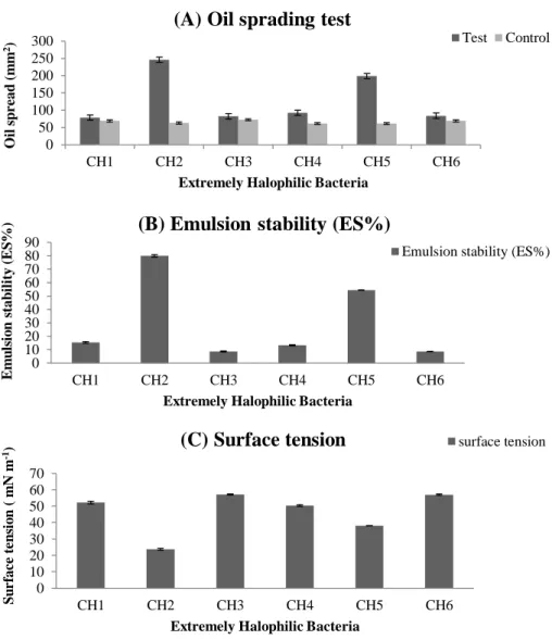
Figure 2. Oil spreading test, emulsion stability and surface tension of culture media without cells and relative volume of emulsions formed between cell culture media and diesel oil after growth of bacteria strains in standard medium for 7 days at 40°C and 200 rpm. Oil dispersion was expressed as mm2 using the sterile standard medium as control (A); emulsion stability was as a percentage (B) and surface tension was expressed as mN m-1(C). Values reported are average of three replicates.
et al., 2003; Plaza et al., 2006). The results of our
experiments indicate that all strains tested positive for
biosurfactant in the drop-collapse test reduced as shown
as Table 3. Motor oil proved better to work with than olive
oil and corn oil. Since, it has caused spreading of the
sterilized standard medium used as negative control and
0 50 100 150 200 250 300 CH1 CH2 CH3 CH4 CH5 CH6 O il s p re ad (m m 2)Extremely Halophilic Bacteria
(A) Oil sprading test
Test Control 0 10 20 30 40 50 60 70 80 90 CH1 CH2 CH3 CH4 CH5 CH6 Em u ls io n s tab il ity (ES % )
Extremely Halophilic Bacteria
(B) Emulsion stability (ES%)
Emulsion stability (ES%)
0 10 20 30 40 50 60 70 CH1 CH2 CH3 CH4 CH5 CH6 S u rfac e te n si o n ( m N m -1)
Extremely Halophilic Bacteria
(C) Surface tension
surface tension0 50 100 150 200 250 300 CH1 CH2 CH3 CH4 CH5 CH6 O il s p re ad (m m 2)
Extremely Halophilic Bacteria
(A) Oil sprading test
Test Control 0 10 20 30 40 50 60 70 80 90 CH1 CH2 CH3 CH4 CH5 CH6 Em u ls io n s tab il ity (ES % )
Extremely Halophilic Bacteria
(B) Emulsion stability (ES%)
Emulsion stability (ES%)
0 10 20 30 40 50 60 70 CH1 CH2 CH3 CH4 CH5 CH6 S u rfac e te n si o n ( m N m -1)
Extremely Halophilic Bacteria
produced plates in which drop diameter was most readily
estimated, an important factor given this test is based on
visual observation.
In this experiment, cell free culture broth was used as
the biosurfactant source. For strains which produce
extracellular biosurfactant there was a drop collapse
activity and four strains which do not produce
biosurfactant the results were negative, which also
inferred that to check the biosurfactant production of any
microbial strain, cell free culture broth should be used
instead of using culture broth with cells. This criterion will
exclude microbial strains having high cell hydrophobicity
but no biosurfactant production. Accuracy and reliability
of results obtained in the drop collapse assay in this
study were similar to the results reported by
Kebbouche-Gana et al. (2009). The isolates with more than one
positive response were exposed to comple-mentary
screening. The latter screening stage included surface
tension and emulsion activity measurements.
To further confirm the biosurfactant production of the
above strains with positive and negative results, cell free
culture broth from all six archaeal strains was subjected
to oil spreading, emulsion stability and surface tension
measurement experiments.
Oil spreading assay
Oil spreading assay results were in corroboration with
drop collapse assay results. Strains found with positive
drop collapse results were positive for oil spreading
assay also (Table 3 and Figure 2A). These results
confirmed the presence (for strains with positive results)
and absence (for strains with negative results) of surface
active compounds (biosurfactant) in the cell free culture
broth. Morikawa et al. (2000), reported that the area of oil
displacement in an oil spreading assay is directly
proportional to the concentration of the biosurfactant in
the solution. However, in this study there was no
quanti-tative study conducted on biosurfactant concentration
versus oil spreading activity, but a qualitative study to
check the presence of biosurfactant in the cell free
culture broth was in concurrence with the above
mentioned earlier report. Similar results with drop
collapse and oil spreading assay was reported by
Youssef et al. (2004), while screening bacteria from
biosurfactant production and also recommended that
both drop collapse and oil spreading assay methods as
reliable techniques for testing biosurfactant production.
These results suggested that the oil-spreading technique
is more sensitive than the other methods for biosurfactant
detection in the supernatant from a culture medium.
Emulsification capacity assay
According to Willumsen and Karlson (1997), a criterion
used for selecting biosurfactant producers is the ability to
maintain at least 50% of the original emulsion volume 24
h after formation. The results observed in this study
(Figure 2B) reveal that from six strains screened, two
(33.33%) strains showed positive emulsification activity.
CH2 and CH5 strains showed that the relative emulsion
stability formed: 80.00 ± 1.0% and 54.39 ± 0.2,
respectively. Evaluating the emulsification capacity is a
simple screening method suitable for a first screening of
biosurfactant producing microbes. It is applied in many
screenings (Chen et al., 2007), whereas, the Kerosene
can be replaced with other hydrophobic compounds. In
this study, gas oil was used as the hydrophobic
substrate. Consequently, this method gives just an
indication of the presence of biosurfactants.
Surface tension measurement
According to Bodour and Maier (1998), the criterion used
for selecting biosurfactant producers is the ability to
reduce the surface tension below 40 mN.m
-1. Surface
tension measurement of cell free culture broth revealed
that out of the six strains screened, two (33.33%) strains
(CH2 and CH5) showed reduction in surface tension and
the highest reduction was observed up to 23.7 ± 0.5
1and
38.1 ± 0.1 mN.m
-1, respectively (Figure 2C). There was a
direct correlation found between drop collapse, oil
spreading, emulsification stability and surface tension
assays. Strains highly active in any one of these methods
were active in other three methods. The direct
measurement of the surface activity of the culture
supernatant is the most straight forward screening
methods. This gives a strong indication on biosurfactant
production (Lin, 1996; Thavasi et al. 2011).
A comparison of the four methods using statistical
analysis
Table 4 shows the coefficient of correlation between the
four methods that were used to detect biosurfactant
production. The Spearman rank correlation, (r
s= - 0.987)
showed a strong negative correlation between the
emulsification index with measurement of emulsion
stability in intervals up to 24 h and surface tension. A
weaker negative correlation, (r
s= - 0.971) was detected
between the diameter of clear zone obtained with the oil
spreading technique and surface tension. However, there
was a weak negative correlation (r
s= - 0.807) between
drop collapse method and surface tension. Oil spreading
technique and emulsification stability method were
strongly correlated with Spearman rank correlation
coefficient of r
s=0.991. However, a weak correlation was
detected between drop collapse method and oil
spreading method (r
s=0.864) and between drop collapse
Table 4. Statistical correlation between different methods.
Spearman rank correlation
coefficient ( rs ) Drop collapse Oil spreading Emulsification stability Surface tension
Drop collapse 1
Oil spreading 0.864 1
Emulsification stability 0.822 0.991 1
Surface tension - 0.807 - 0.971 -0.987 1
Conclusion
Interest in biosurfactants has led to the development of a
multitude of methods for the screening of biosurfactant
producer strains. A combination of different methods is
appropriate for a successful screening. In the present
study two superior haloarchaea isolates CH2 and CH5
with biosurfactant-producing ability and the former with
emulsion capacity were isolated from saline soil of Chott
El Hodna-M’sila (Algeria). Their ability to reduce surface
tension and emulsion capacity makes them new potential
candidates for biosurfactant and bioemulsion production.
Further studies have been initiated to identify their
properties and consequently determine the potential of
their different industrial applications in particular
enhanced oil recovery application.
Conflict of interests
The authors did not declare any conflict of interest.
ACKNOWLEDGMENTS
This work was supported by the Algerian Ministry of
Higher Education and Scientific Research. The authors
would like to acknowledge all those who contributed
directly or indirectly in the development of this work.
REFERENCES
Ainon H, Noramiza S, Shahidan R (2013). Screening and optimization of biosurfactant production by the hydrocarbon-degrading bacteria. Sains. Malays. 42(5):615-623.
Batista SB, Mounteer AH, Amorim FR, Totola MR (2006). Isolation and characterization of biosurfactant/bioemulsifier producing bacteria from petroleum contaminated sites. Bioresour. Technol. 97(6):868-875.
Bodour A, Drees K, Maier R (2003). Distribution of biosurfactant-producing bacteria in undisturbed and contaminated arid southwestern soils. Appl. Environ. Microbiol. 69(6):3280-3287. Bodour A, Miller-Maier R (1998). Application of a modified drop-collapse
technique for surfactant quantitation and screening of biosurfactant-producing microorganisms. J. Microbiol. Methods 32(3):273-280. Bolhuis H, Palm P, Wende A, Falb M, Rampp M, Rodriguez-Valera F
(2006). The genome of the square archaeon Haloquadratum walsbyi: life at the limits of water activity. BMC Genome 7:169.
Boumezbeur A (2002). Atlas 2: Algerian wetlands of international
importance. Direction Generales des Forets, Ben Aknoun, Algeria pp. 14-17.
Chen C, Baker S, Darton R (2007). The application of a high throughput analysis method for the screening of potential biosurfactants from natural sources. J. Microbiol. Methods 70(6): 503-510.
Chen SY, Wei YH, Chang JS (2007). Repeted pH-stat fed-batch fermentation for rhamnolipid production with indigenous Psedomonas
aeroginosa S2. Appl. Microbiol. Biotechnol. 76(1):67-74.
Copin-Montegut G (1996). Sea Water Chimistry. Ocean Instit, Paris. pp. 319.
Dussault HP (1995). An Improved Technique for Staining Red Halophilic Bacteria. J. Bacteriol. 70(4):484-485.
Fukushima T, Usami R, Kamekura M (2007). A traditional Japanese-style salt field is a niche for haloarchaeal strains that can survive in 0.5% salt solution. Saline Systems 3:2.
Gavrieli I (1997). Halite deposition in the Dead Sea: 1960-1993. In: The Dead Sea- the lake and its setting. Oxford Univ. Press. pp.162 -171. Jain D, Collins-Thompson D, Lee H, Trevors JT(1991).A drop-collapsing
test for screening surfactant-producing microorganisms. J. Microbiol. Methods 13(4):271-279.
Kebbouche-Gana S, Gana ML, Khemili S, Fazouane-naimi F, Bouanane NA, Penninckx M, Hacene H (2009). Isolation and characterization of halophilic archaea able to produce biosurfactants. J. Ind. Microbial. Biotechnol. 36(5): 727-738.
Lin SC (1996). Biosurfactants: Recent advances. J. Chem. Technol. Biotechnol. 66(2):109-120.
Lmhoff J, Sahl H, Soliman G, Truper H (1979). The Wadi Natrun: chemical composition and microbial mass developments in alkaline brines of eutrophic desert lakes. Geomicrobiol. J. 1(3): 219 - 234. Loganathan K, Gaurav K, Kokati V, Bhaskara R (2010). Comparaison of
methods and screening of biosurfactant producing marine actinobacteria isolated from Nicobar marine sediment. Environ. Biotechnol. Div. 2(1):34-38.
Madalin E, Takashi I, Masahiro K, Gabriela P, Lucia D (2008). Halophilic archaea of Halopherax genus isolated from Anthropocentric Telega (PALADA) salt lake. Proc. Rom. Acad. 1-2:11-16.
Morikawa M, Hirata Y, Imanaka T (2000). A study on the structure function relationship of the lipopeptide biosurfactants. Biochim. Biophys. Acta 1488(3):211-218.
Mukherjee S, Das P, Sen R (2006). Towards commercial production of microbial surfactants.Trends Biotechnol. 11:509-515.
Nasr S, Soudi M R, Mehrnia M R , Sarrafzadeh M H (2009). Characterization of novel biosurfactant producing strains of Bacillus
spp. isolated from petroleum contaminated soil. Iran. J. Microbiol.
2(1):54-61.
Nitscheke M, Costa SG (2007). Biosurfactants in food industry. Trends Food. Sci .Technol. 18(5): 252-259.
Oren A (2006). Life at Haigh Salt Concentrations in: Prokaryotes. Springer 2: 263-282.
Oren A (2014). The Family Halobacteriaceae in: Prokaryotes. Springer. pp. 41-121.http:// dx. doi.org/10.1007/978-3-642-38954-2_313 Oren A, Ventosa A, Grant WD (1997). Proposed minimal standards for
description of new taxa in the order Halobacteriales. Int. J. Sustain. Bacteriol. 47(1): 233-238.
Ozcan B, Cokmus C, Coleri A, Caliskan M (2006). Characterization of extremly halophilic archaea isolated from saline environment in different parts of Turkey. Mikrobiologiia 75(6): 849-856.
Ozekan B, Ozcengiz G, Coleri A, Cokmus C (2007). Diversity of Halophilic Archaea from Six Hypersaline Environments in Turkey. J.Microbiol. Biotechnol. 17(6):985-992.
Plaza GA, Zjawiony I, Banat IM (2006). Use of different methods for detection of thermophilic biosurfactant-producing bacteria from hydrocarbone-contaminated and bioremediated soils. J. Pet. Sci. Eng. 50(1):71-77.
Post FJ (1981). Microbiology of the Great Salt Lake north arm. Hydrobiology 81-82(1):59-69.
Rodier J, Bazin C, Broutin JC, Chambon P, Champsaur H, Rodi L (1996). Analysis of the water. Dun. Par. pp. 50-85.
Rodrigues L, Banat IM, Teixeira J, Oliveira R (2006). Biosurfactants: Potential applications in medicine. J. Antimicrob. Chem. 57(2):609-618.
Rodrigues LR, Teixeira JA, Mei HC, Oliveira R (2006). Physicochemical and functional characterization of a biosurfactant produced by
Lactococcus lactis 53. Colloids Surf. B. 49(1):79-86.
Rone EZ, Rosenberg E (2001). A Review of Natural Roles of Biosurfactants. Environ. Microbiol. 3(4):229-236.
Stan-Lotter H, Pfaffenhuemer M, Legat A, Busse HG, Radax C, and Gruber C (2002). Haloococcus dombrowskii sp.nov., an Archaeal Isolat from a Permian Alpine Salt Deposit. Int. J. Syst. Evol. Microbiol. 52(5):1807-1814.
Thavasi R, Sharma S, Jayalakshmi S (2011). Evaluation of Screening Methods for the Isolation of Biosurfactant Producing Marine Bacteria.J Pet Environ Biotechnol. S1:001. http://dx.doi:10.4172/2157-7463.S1-001.
Tugrul T, Cansunar E (2005). Detecting surfactant-producing microorganisms by the drop-collapse test. World. J. Microbiol. Biotechnol. 21(7):851-853.
Urum K, Pekdemir T (2004). Evaluation of biosurfactants for crude oil contaminated soil washing. Chemosphere 57(9):1139-1150.
Walter V, Syldatk C, Hausmann R (2010). Screening concepts for the isolation of biosurfactant producing microorganisms. Adv. Exp. Med. Biol. 672:1-13
Willumsen PA, Karlson U (1997). Screening of bacteria, isolated from PAH-contaminated soils, for production of biosurfactant and bioemulsifiers. Biodegradation 7(5):415-423.
Youssef NH, Duncan KE, Nagle DP, Savage KN, Knapp RM, McInerney MJ (2004). Comparison of methods to detect biosurfactant production by diverse microorganisms. J. Microbiol. Methods 56(3):339-347.