O
pen
A
rchive
T
OULOUSE
A
rchive
O
uverte (
OATAO
)
OATAO is an open access repository that collects the work of Toulouse researchers and
makes it freely available over the web where possible.
This is an author-deposited version published in :
http://oatao.univ-toulouse.fr/
Eprints ID : 18700
To link to this article :
DOI: 10.1104/pp.15.01537
URL :
http://dx.doi.org/10.1104/pp.15.01537
To cite this version :
Trapet, Pauline and Avoscan, Laure and Klinguer,
Agnès and Pateyron, Stéphanie and Citerne, Sylvie and Chervin, Christian
and Mazurier, Sylvie and Lemanceau, Philippe and Wendehenne, David and
Besson-Bard, Angélique The Pseudomonas fluorescens Siderophore
Pyoverdine Weakens Arabidopsis thaliana Defense in Favor of Growth in
Iron-Deficient Conditions. (2016) plant physiology, vol. 171 (n° 1). pp.
675-693. ISSN 1532-2548
Any correspondence concerning this service should be sent to the repository
administrator:
[email protected]
Weakens
Arabidopsis thaliana Defense in Favor of
Growth in Iron-Deficient Conditions
1
Pauline Trapet2, Laure Avoscan, Agnès Klinguer, Stéphanie Pateyron, Sylvie Citerne, Christian Chervin, Sylvie Mazurier, Philippe Lemanceau, David Wendehenne, and Angélique Besson-Bard*
Agroécologie, AgroSup Dijon, Centre National de la Recherche Scientifique, Institut National de la Recherche Agronomique, Université de Bourgogne Franche-Comté, F-21000 Dijon, France (P.T., A.K., D.W., A. B.-B.); Agroécologie, AgroSup Dijon, Institut National de la Recherche Agronomique, Université de Bourgogne Franche-Comté, F-21000 Dijon, France (L.A., S.M., P.L.); Transcriptomic Platform of IPS2, Institute of Plant Sciences Paris-Saclay, Unité Mixte de Recherche 9213/Unité Mixte de Recherche 1403, Centre National de la Recherche Scientifique, Institut National de la Recherche Agronomique, Université Paris-Sud, Université d’Evry, Université Paris-Diderot, Sorbonne Paris-Cité, F-91405 Orsay, France (S.P.); Institut National de la Recherche Agronomique-AgroParisTech, Unité Mixte de Recherche 1318, Institut J.-P. Bourgin, Centre de Versailles-Grignon, F-78026 Versailles, France (S.C.); and Université de Toulouse, Institut National Polytechnique de Toulouse-Ecole Nationale Supérieure Agronomique-Institut National de la Recherche Agronomique, Unité Mixte de Recherche 990 Génomique et Biotechnologie des Fruits, Castanet-Tolosan, CS 32607, F–31326, France (C.C.)
ORCID ID: 0000-0001-5026-095X (S.C.).
Pyoverdines are siderophores synthesized by fluorescent Pseudomonas spp. Under iron-limiting conditions, these high-affinity ferric iron chelators are excreted by bacteria in the soil to acquire iron. Pyoverdines produced by beneficial Pseudomonas spp. ameliorate plant growth. Here, we investigate the physiological incidence and mode of action of pyoverdine from Pseudomonas fluorescens C7R12 on Arabidopsis (Arabidopsis thaliana) plants grown under iron-sufficient or iron-deficient conditions. Pyoverdine was provided to the medium in its iron-free structure (apo-pyoverdine), thus mimicking a situation in which it is produced by bacteria. Remarkably, apo-pyoverdine abolished the iron-deficiency phenotype and restored the growth of plants maintained in the iron-deprived medium. In contrast to a P. fluorescens C7R12 strain impaired in apo-pyoverdine production, the wild-type C7R12 reduced the accumulation of anthocyanins in plants grown in iron-deficient conditions. Under this condition, apo-pyoverdine modulated the expression of around 2,000 genes. Notably, apo-pyoverdine positively regulated the expression of genes related to development and iron acquisition/redistribution while it repressed the expression of defense-related genes. Accordingly, the growth-promoting effect of apo-pyoverdine in plants grown under iron-deficient conditions was impaired in iron-regulated transporter1 and ferric chelate reductase2 knockout mutants and was prioritized over immunity, as highlighted by an increased susceptibility to Botrytis cinerea. This process was accompanied by an overexpression of the transcription factor HBI1, a key node for the cross talk between growth and immunity. This study reveals an unprecedented mode of action of pyoverdine in Arabidopsis and demonstrates that its incidence on physiological traits depends on the plant iron status.
Iron, the fourth most abundant element in the Earth’s crust, is an essential micronutrient for plant growth and development, notably through its involvement in major metabolic processes such as respiration and photosyn-thesis (Guerinot and Yi, 1994; Robin et al., 2008). De-spite its abundance, iron is weakly bioavailable for organisms due to its poor solubility in soils under aer-obic conditions. Therefore, plants and other organisms had to evolve mechanisms to efficiently assimilate iron from the soil. Bacteria, but also other microbes includ-ing fungi, produce iron chelators known as sidero-phores (Ahmed and Holmström, 2014). Siderosidero-phores are low-Mr molecules that exhibit a high affinity for ferric iron (Fe3+) and are synthesized in iron-limiting conditions. For instance, fluorescent Pseudomonas spp. produce yellow-green fluorescent siderophores termed
pyoverdines (or pseudobactins) having a high affinity for Fe3+, with a stability constant of the ferric-pyoverdine complex around 1032 M21 (Meyer and
Abdallah, 1978; Albrecht-Gary et al., 1994). Once excreted, siderophores chelate ferric iron and are transported back as ferric siderophore complexes inside the bacte-rial cell, where ferric iron is released from the sidero-phore and further reduced to ferrous iron (Fe2+; Chu et al., 2010).
Higher plants evolved two main strategies to incor-porate iron (Curie and Briat, 2003). Grass plants use a strategy similar to the bacterial one, based on the re-lease of phytosiderophores (strategy II). Nongrass plants, like Arabidopsis (Arabidopsis thaliana), use the strategy I based on soil acidification through proton excretion by H+-ATPases, solubilization of Fe3+, and
reduction of the Fe3+chelate, mainly by the ferric che-late reductase FRO2. Fe2+ions, which are more soluble than Fe3+, are subsequently taken up by root transporters, including IRON-REGULATED TRANSPORTER1 (IRT1). Once in the plants, iron has to be transported, utilized, or sequestrated because of its high reactivity and toxicity through the Fenton reaction. IRT1 and FRO2 expression is induced under iron-limiting conditions, and the tran-scription factor FIT1 is required for the proper regulation of the iron-uptake system in Arabidopsis (Colangelo and Guerinot, 2004). FIT1 does not act alone to orchestrate the iron deficiency response. Indeed, several studies high-lighted the role of other basic helix-loop-helix (bHLH) transcription factors on the expression of genes involved in the adaptation to iron deficiency stress. These tran-scription factors were shown to act together with FIT1, such as bHLH38 and bHLH39 (Yuan et al., 2008), or alone, as reported for POPEYE, bHLH100, and bHLH101 (Long et al., 2010; Sivitz et al., 2012). Besides the FRO2/ IRT1 system, Fe-efficient plants also respond to iron deficiency by enhancing the root secretion of coumarin compounds, improving plant iron nutrition (Fourcroy et al., 2014).
Because iron is central for many metabolic processes and not easily available, it is the focus of serious com-petition between organisms. In mammals, there are several examples in which the virulence of infectious bacteria has been correlated to their ability to assimilate host iron through their siderophores (Saha et al., 2013). In turn, the host fights back and develops strategies to avoid iron capture by bacteria, highlighting cross-regulatory interactions between iron homeostasis and immune responses (Ong et al., 2006; Nairz et al., 2014). Similar processes seem to operate in plants. For exam-ple, the production of siderophores by the pathogenic enterobacteria Dickeya dadantii, Erwinia amylovora, and Pseudomonas syringae pv tabaci was shown to facilitate infection in their hosts, pointing out their role as viru-lence factors (for review, see Franza and Expert, 2013). Consecutively, plants establish a strategy for keeping iron that consists in lowering iron availability by
overexpressing iron-chelating proteins, notably ferri-tins, and in improving iron acquisition through IRT1 (Dellagi et al., 2005, 2009). As highlighted in animals, this iron-retention strategy used by plants to combat infections could be a key component of their immune responses. Supporting this concept, treatment of plant tissues or cell suspensions by bacterial siderophores was reported to trigger defense responses in tobacco (Nicotiana tabacum) and Arabidopsis (van Loon et al., 2008; Aznar et al., 2014).
On the other hand, siderophore secretion by bacteria is not always associated with pathogenicity and, in contrast, is even beneficial for plant health. In particu-lar, different soil pseudomonad strains, commonly called plant growth-promoting rhizobacteria, were shown to suppress soil-borne diseases (Haas and Défago, 2005; Weller, 2007). Their biocontrol activities have been correlated to their capacity to release sider-ophores, which act as competitors reducing the iron availability for plant pathogens, including Fusarium oxysporum and Pythium ultimum (Lemanceau et al., 1992, 1993; Duijff et al., 1999; Bakker et al., 2007; Van Wees et al., 2008; Ahmed and Holmström, 2014). Im-portantly, siderophores produced by other bacterial species such as Bacillus subtilis also display biocontrol activities (Yu et al., 2011). Besides their ability to com-pete with soil pathogens for iron, siderophores pro-duced by plant beneficial rhizobacteria also were shown to protect different plant species by eliciting induced systemic resistance (ISR). For example, puri-fied pyoverdines from Pseudomonas putida WCS358 were shown to trigger ISR in Arabidopsis when using P. syringae pv tomato as the challenging pathogen. They were also efficient in inducing ISR against Botrytis cinerea in bean (Phaseolus vulgaris) and tomato (Solanum lycopersicum; Meziane et al., 2005). Accordingly, De Vleesschauwer et al. (2008) demonstrated that pyo-verdine was the determinant responsible for Pseudomonas fluorescens WCS374-induced ISR in rice (Oryza sativa) against the leaf blast pathogen Magnaporthe oryzae. In-vestigations in tobacco cell suspensions indicated that the pyoverdine siderophores from P. putida WCS358 and P. fluorescens WCS417 strains are perceived by cells and mediate defense-related early signaling events (van Loon et al., 2008). However, these defense-related reactions were poorly correlated to the ability of the siderophores of interest to promote ISR. Therefore, how siderophores promote ISR remains poorly understood.
In addition to their protective function, siderophores produced by plant growth-promoting rhizobacteria were shown to enhance plant growth, probably by providing the plants with nutrients. Of particular in-terest here, Vansuyt et al. (2007) provided evidence that in its iron-chelated structure, pyoverdine from P. fluorescens strain C7R12 is assimilated by roots of Arabidopsis plantlets grown in vitro. This process was accompanied by an improvement of plant growth and chlorophyll content. This study also revealed that iron chelated to pyoverdine was incorporated in a more ef-ficient way than iron chelated to EDTA. The mechanism
1
This work was supported by the University of Burgundy (BQR project), the Burgundy State (PARI AGRALE 8 project), and the Min-istère de l’Education Nationale de la Recherche et de la Technologie.
2
Present address: Plant-Microbe Interactions, Department of Biol-ogy, Faculty of Science, Utrecht University, P.O. Box 800.56, 3508 TB, Utrecht, The Netherlands.
* Address correspondence to [email protected]. The author responsible for distribution of materials integral to the findings presented in this article in accordance with the policy de-scribed in the Instructions for Authors (www.plantphysiol.org) is: Angélique Besson-Bard ([email protected]).
P.T., L.A., S.M., P.L., D.W., and A.B.-B. conceived the original pro-ject; P.T. performed most of the experiments with the help of L.A. and A.B.-B.; A.K. provided technical assistance; P.T., L.A., D.W., and A.B.-B. designed the experiments and analyzed the data; S.P. conducted the CATMA microarray analysis; S.C. analyzed the samples for hormone contents except ethylene; C.C. analyzed the samples for ethylene con-tents; A.B.-B. wrote the article with the contribution of D.W.
by which siderophores enhance iron nutrition is little known. It has been proposed that microbial ferric sid-erophore could be reduced to release ferrous iron once transported in the apoplast of root cells, thus providing substrate for the iron transport system (Ahmed and Holmström, 2014). Also, ferric siderophore could pro-vide iron to plant phytosiderophores (Crowley, 2006). In the case of pyoverdine, Vansuyt et al. (2007) provided another explanation by showing that, in Arabidopsis, pyoverdine is incorporated by plant roots and accu-mulates in shoots. Interestingly, this incorporation does not rely on the strategy I iron acquisition system. Taken together, these observations underline the importance of pyoverdine in plant-microbe interactions but call for further studies to better understand the mechanisms involved.
In this study, we further deciphered the mode of action of pyoverdine in plants. For this purpose, we investigated the incidence of pyoverdine from P. fluorescens C7R12 on the growth and defense capac-ities of Arabidopsis plantlets grown in iron-sufficient or iron-deficient medium. Pyoverdine was provided to the culture medium in its iron-free structure (apo-pyo), thus mimicking a situation in which it is produced by the bacterial strain. We observed that while apo-pyo displayed a remarkable growth-promoting effect on plants facing iron deficiency, it clearly impaired their resistance against B. cinerea. Contrary to a P. fluorescens C7R12 bacterial strain impaired in apo-pyo production, the wild-type C7R12 reduced the accumulation of anthocyanins in plants grown under iron-deficient conditions. Analysis of the underlying mechanisms showed that the apo-siderophore triggered a strong up-regulation of genes related to development and iron uptake/redistribution in planta. Accordingly, the growth-promoting effects of apo-pyo were shown to rely on IRT1 and FRO2 expression. Furthermore, we provide evidence that, in plants exposed to iron defi-ciency, the apo-siderophore modulates the expression of genes controlling the tradeoff between growth and immunity in favor of growth. Collectively, data from our work describe an unexpected mode of action of pyoverdine in Arabidopsis plants grown in iron defi-ciency.
RESULTS
Impact of Apo-Pyo on Arabidopsis Phenotype
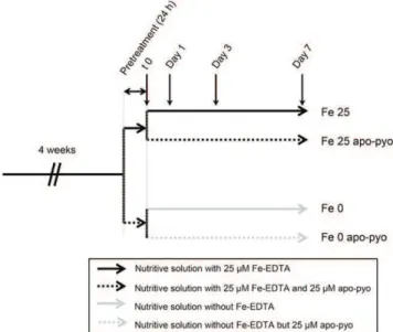
Four-week-old plants were subjected to apo-pyo treatment in iron-deficient or iron-sufficient medium as described in Figure 1. For this purpose, after 4 weeks of hydroponic culture in a classical nutrient solution containing 25 mM Fe-EDTA, plants were subjected to
24 h of pretreatment in iron-sufficient (nutritive solution with 25 mMFe-EDTA) or iron-deficient (nutritive
solu-tion without Fe-EDTA) medium. Then, each resulting batch was separated into two new batches containing fresh nutritive solution with or without 25 mMapo-pyo
to obtain four different treatments: 25 mM apo-pyo in
iron-sufficient (Fe 25 apo-pyo) or iron-deficient (Fe 0 apo-pyo) medium and the corresponding controls without apo-pyo (Fe 25 or Fe 0, according to the pres-ence or abspres-ence or Fe-EDTA, respectively). Impor-tantly, we measured the iron content of the culture media 6 h after apo-pyo addition (see Fig. 6A below). The corresponding data showed that apo-pyo addition did not result in iron enrichment in the medium, thus proving that the siderophore was purified in its iron-free structure and was not contaminated by ferri-pyo (pyoverdine chelated to iron).
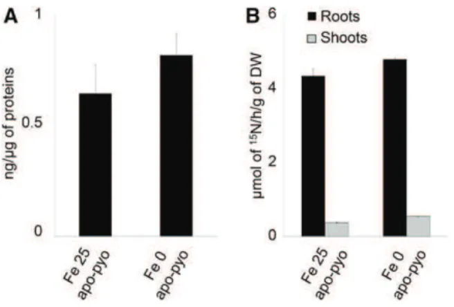
First, we checked that, in our experimental condi-tions, apo-pyo was efficiently assimilated by plants and potentially transported from roots to shoots. The presence of pyoverdine in root tissue was investigated by ELISA using rabbit polyclonal anti-pyoverdine antibodies. In root extracts of Fe 25 apo-pyo- and Fe 0 apo-pyo-treated plants, we detected 0.64 and 0.82 ng pyoverdine mg21protein, respectively (Fig. 2A). In shoots, we were not able to discriminate a positive signal from the background (data not shown). To strengthen the data, the presence of apo-pyo in plant tissues was further analyzed by isotope ratio mass spectrometry. Following the treatment of plants by [15N]apo-pyo, approximately 4.5 mmol15N h21 g21dry weight was measured in the
roots of Fe 25 apo-pyo- and Fe 0 apo-pyo-treated plants, thus confirming the results from the ELISA. In addition, a lower signal (0.37 and 0.54 mmol15N h21g21dry weight
for Fe 25 apo-pyo and Fe 0 apo-pyo treatments, respec-tively) also was measured in the shoots (Fig. 2B). These data also showed that the concentration of apo-pyo
Figure 1. Experimental design. After 4 weeks of hydroponic culture in the nutritive solution containing 25 mMFe-EDTA, plants were subjected to 24 h of pretreatment in iron-sufficient (nutritive solution with 25 mM Fe-EDTA) or iron-deficient (nutritive solution without Fe-EDTA) medium. Then, plants were treated for 7 d with 25 mMapo-pyo in iron-sufficient (Fe 25 apo-pyo) or iron-deficient (Fe 0 apo-pyo) medium. As controls, plants were cultivated in sufficient (Fe 25) or iron-deficient (Fe 0) medium without apo-pyo.
assimilated by plants did not differ according to the concentration of iron in the culture medium.
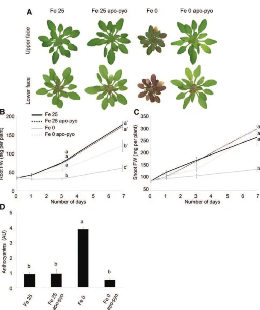
Next, the impact of apo-pyo treatment on the plant macroscopic phenotype was evaluated (Fig. 3). Fe 25-and Fe 25 apo-pyo-treated plants appeared bigger 25-and greener compared with plants facing iron deficiency. As expected, plants facing Fe 0 treatment were severely affected after 7 d of culture: they showed reduced growth, and their leaf abaxial sides displayed a violet pigmentation probably due to anthocyanin production (Fig. 3A). In comparison with Fe 0-treated plants, Fe 0 apo-pyo-treated plants exhibited a distinct phenotype that resembled those of plants grown in iron-containing medium. Indeed, after 7 d of treatment with apo-pyo, their growth was considerably higher. Quantitatively, their root masses were between those of Fe 25/Fe 25 apo-pyo- and Fe 0-treated plants (Fig. 3B), and growth of their aerial parts was similar to that of Fe 25 plants (Fig. 3C). Furthermore, although we noticed a yellowing of several leaves, Fe 0 apo-pyo-treated plants did not show violet pigmentation compared with Fe 0-treated plants (Fig. 3A). Supporting this observation, the high pro-duction of anthocyanins measured in Fe 0 plants was not observed in Fe 0 apo-pyo-treated plants, which, in con-trast, displayed an anthocyanin level similar to those of Fe 25- and Fe 25 apo-pyo-treated plants (Fig. 3D).
Collectively, these data indicate that apo-pyo is as-similated by Arabidopsis plants grown in hydroponic conditions and highlight a growth-promoting effect of the apo-siderophore in plants facing iron deficiency.
Transcriptomic Analysis of Apo-Pyo-Treated Plants We initiated a transcriptomic analysis in order to provide, to our knowledge, a first and overall view of
how apo-pyo impacts the growth of plants. For this purpose, a microarray analysis using CATMA arrays covering the entire transcriptome of Arabidopsis was performed from roots and shoots of plants 3 d after the addition of apo-pyo in the nutritive medium containing (Fe 25 apo-pyo condition) or not (Fe 0 apo-pyo condi-tion) iron. This time point was chosen on the basis of Figure 3, B and C, demonstrating that the increase of growth of the Fe 0 apo-pyo plants was noticeable from 3 d. Changes in root and shoot gene expression were determined by comparing the transcriptomes of apo-pyo-treated plants with their respective controls not treated with the apo-siderophore (i.e. Fe 25 apo-pyo versus Fe 25 and Fe 0 apo-pyo versus Fe 0).
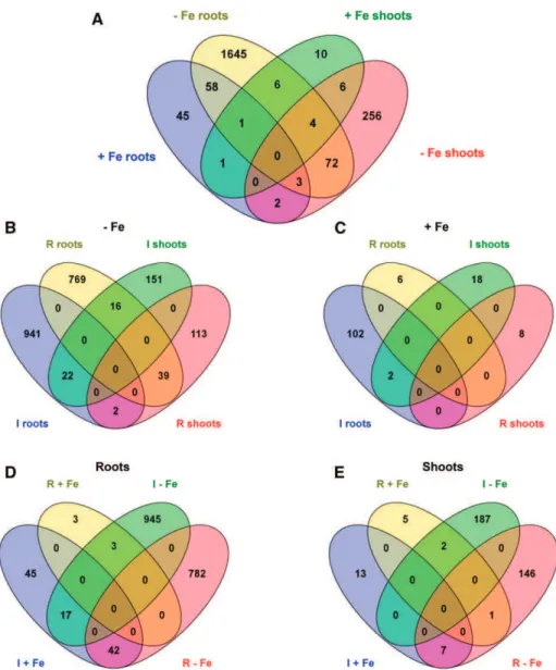
We first selected genes whose expression was mod-ulated by a log2ratio of +1.5 or greater or 21.5 or less (with adjusted P # 0.05). This analysis revealed that 2,109 genes displayed significant differential expression in response to apo-pyo (Fig. 4A; listed in Supplemental Tables S1–S4). The incidence of apo-pyo on gene ex-pression clearly differed according to the tissue and the presence or not of iron in the culture medium. Indeed, apo-pyo modulated the expression of 2,053 genes in iron-depleted medium (Fig. 4B) versus 136 genes in iron-containing medium (Fig. 4C). Furthermore, 1,837 genes showed a modified expression in the roots (Fig. 4D) versus 361 in the shoots (Fig. 4E). Regarding the percentages of induced/repressed genes, pyo-induced genes represent 55% to 56% of the total apo-pyo-modulated genes when considering the roots (Fig. 4D), the shoots (Fig. 4E), or the Fe 0 condition (Fig. 4B). In contrast, in plants grown in the Fe 25 medium, 90% of the genes were induced in response to the apo-pyo treatment (Fig. 4C).
A closer analysis indicates that 79 of the apo-pyo-dependent genes (genes 1–79 in Supplemental Table S5) could be gathered in a first cluster corresponding to genes modulated under iron deficiency in both roots and shoots. Among these genes, 22 (28%) or 39 (49%) were induced or repressed in both tissues, respectively (Fig. 4B). A second cluster includes 63 genes (genes 2, 62, 75, and 80–138 in Supplemental Table S5) modu-lated in roots in iron-deficient and iron-sufficient me-dia. Among this category, 45 (71%) were inversely regulated according to the presence or absence of iron in the culture medium. The third cluster groups 10 shoot genes (genes 49, 60, 61, 71, and 139–144 in Supplemental Table S5) that showed differential ex-pression in iron-sufficient and iron-deficient medium, nine of them being inversely modulated in the two conditions. Therefore, a large part of the genes modu-lated by apo-pyo in roots and shoots of plants culti-vated in the iron-deficient condition were similarly regulated. On the contrary, a majority of the apo-pyo-dependent genes modulated in both iron-sufficient and iron-deficient conditions were regulated in the opposite way. Using AmiGO (http://amigo1.geneontology. org/cgi-bin/amigo/term_enrichment), we identified these genes as mainly responsive to stress or stimulus.
Figure 2. Detection of apo-pyo in Arabidopsis tissues. Plants were cultivated and treated by apo-pyo as indicated in Figure 1. A, Apo-pyo was detected in roots by ELISA using rabbit polyclonal anti-pyoverdine antibodies. Signal was expressed in ng of apo-pyo per mg of total root proteins. Each value represents the mean 6SEof three measurements. Experiments were repeated three times with similar results. B, [15 N]Apo-pyo was detected using isotope ratio mass spectrometry in roots and shoots. Each value represents the mean 6SEof three measurements. Ex-periments were repeated three times with similar results. DW, Dry weight.
Next, we focused our analysis on the genes whose expression was highly induced (91 and 15 genes in roots and shoots, respectively) or highly repressed (94 and eight genes in roots and shoots, respectively) by apo-pyo in the iron-deficient condition (log2ratio of +3 or
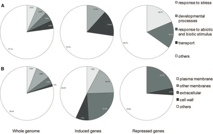
greater or 23 or less; P # 0.05). This choice was made based on the phenotypic analysis showing that apo-pyo deeply improved the growth of plants facing iron de-ficiency (Fig. 3). Genes were putatively assigned to functional categories based on the Gene Ontology an-notations from The Arabidopsis Information Resource database (http://www.arabidopsis.org/tools/bulk/ go/index.jsp; Fig. 5). Concerning the genes whose expression was induced by apo-pyo, an overrepre-sentation of genes related to developmental processes (11.7% compared with 6.2% in the whole genome) and, in particular, to cell differentiation was ob-served (Fig. 5A). These genes include a-EXPANSIN7 (AT1G12560) and a-EXPANSIN18 (AT1G62980), GLY-COSYL HYDROLASE9C1 (AT1G48930), CELLULOSE SYNTHASE-LIKE5 (AT4G15290), b-GALACTOSIDASE6
(AT5G63800), and CER4 (AT4G33790), which encodes an alcohol-forming fatty acyl-CoA reductase involved in cuticular wax biosynthesis. Moreover, this analysis revealed a predominance of genes involved in transport when comparing with the whole-genome categoriza-tion (15.6% compared with 4.8% in the whole genome). Accordingly, genes up-regulated by apo-pyo encode proteins preponderantly located in the plasma mem-brane, in endomembranes, in the cell wall, and also extracellular proteins (Fig. 5B). These four cellular component categories represent 56.8% of the genes in-duced by apo-pyo, while they represent only 20.6% of the whole genome. Importantly, this category in-cludes genes related to iron transport, including IRT1 (AT4G19690) and IRT2 (AT4G19680; Supplemental Table S6). Strengthening this evidence, other genes encoding proteins involved in iron homeostasis also were identified as up-regulated by apo-pyo in roots facing iron deficiency (Supplemental Table S6). Among these, FRO2 (AT1G01580) and the transcription factor bHLH39 (also named ORG3; AT3G56980) belonged to
Figure 3. Phenotypic analysis of Arabi-dopsis plantlets exposed to apo-pyo in iron-sufficient or iron-deficient condi-tions. Plants were cultivated and treated by apo-pyo as indicated in Figure 1. A, Rosette macroscopic phenotypes. Phe-notypes were observed 7 d after the addition of apo-pyo. Results are repre-sentative of nine experiments. B, Root fresh weights. C, Shoot fresh weights. D, Anthocyanin contents. Each value rep-resents the mean 6 SE of three mea-surements. Experiments were repeated at least three times with similar results. Significant differences between the treat-ments were determined by one-way ANOVA/LSDmethod, with different let-ters signifying distinct statistical groups (P , 0.01 for B and P , 0.0001 for C and D). AU, Arbitrary units; FW, fresh weight.
the most induced genes. Another transcription fac-tor, bHLH100 (AT2G41240), was highly induced in roots and shoots. Although to a lower extent, NAS4, encoding NICOTIANAMINE SYNTHASE4, also was found to be up-regulated. We recently showed that the corresponding protein, which catalyzes the syn-thesis of the iron chelator/in planta transporter nicotianamine, plays a key role in the plant response to iron deficiency (Koen et al., 2014). To check the microarray data, we tested the level of accumula-tion of four transcripts among the most modulated by apo-pyo in iron-deficient medium by reverse transcription-quantitative PCR (RT-qPCR). The re-sults, presented in Supplemental Figure S1, were in agreement with the CATMA transcriptomic data. In particular, we noticed a major induction of bHLH39 and AT2G38240 (encoding a 2-oxoglutarate- and Fe2
+-dependent oxygenase superfamily protein) in the
shoots and of FRO2, bHLH39, and IRT1 in the roots in response to apo-pyo.
Regarding the genes whose expression was deeply repressed by apo-pyo in roots and shoots of plants grown in the iron-deficient condition, an overrepre-sentation of genes involved in the stress response (18.7% compared with 6.3% in the whole genome) and in abiotic or biotic stimulus (16.1% compared with 5.6% in the whole genome; Fig. 5A) was ob-served. For instance, we identified the lipid transfer protein LTP3 (AT5G59320), which is predicted to encode a pathogenesis-related protein (Supplemental Table S7). As described for up-regulated genes, down-regulated genes encoding extracellular proteins (21.7% versus 4.8% in the whole genome) and proteins lo-cated in the cell wall (4.3% versus 1.3% in the whole genome) were well represented compared with the data for the whole genome (Fig. 5B). It also should be speci-fied that AtFER1, encoding FER1 involved in iron se-questration, was found to be strongly down-regulated in roots of iron-deprived plants (Supplemental Tables S6 and S7). These data further support the assumption that
Figure 4. Venn diagrams illustrating the transcriptome changes in response to apo-pyo in deficient (2Fe) or iron-sufficient (+Fe) medium in roots or shoots (log2ratio of +1.5 or greater or 21.5 or less; P # 0.05). A, General view of the number of genes commonly regulated between the four comparisons. B, Focus on the number of genes induced (I) or repressed (R) in the iron-deficient me-dium. C, Focus on the number of genes induced or repressed in the iron-sufficient medium. D, Focus on the number of genes induced or repressed in the roots. E, Focus on the number of genes induced or repressed in the shoots. Diagrams were generated using http://bioinfogp.cnb. csic.es/tools/venny/index.html.
apo-pyo impacts the iron homeostasis of plants grown in the iron-deficient condition.
The functional categorization of the root genes whose expression was strongly modulated by apo-pyo treatment (log2ratio of +3 or greater or 23 or less; P #
0.05) in plants grown in the iron-deficient condition was further assessed using AmiGO and the MapMan program. This analysis clearly confirmed an inci-dence of apo-pyo in the expression of genes involved in development and biotic stresses (Supplemental Fig. S2A). The genes gathered in the biotic stress category encode proteins that play roles in hormone signaling, in cell wall dynamics, and in signaling processes (Supplemental Fig. S2B). To complete this investigation, we performed a more systemic analy-sis by including root genes regulated by apo-pyo to a lower extent (log2 ratio of +1.5 or greater or 21.5 or
less; P # 0.05) in plants facing iron deficiency. We noticed a repression of genes encoding resistance genes (R genes), transcription factors ERF, WRKY, and MYB, salicylic acid (SA)-related genes (such as AT5G24210, belonging to the lipase class 3 protein family), and abscisic acid (ABA)-related genes (such as the lipid transfer protein LTP3; Supplemental Fig. S2C; Supplemental Table S7). The observation that genes related to the SA and ABA pathways were repressed
prompted us to compare our transcriptomic data with those recorded for hormone perturbations in Genevestigator. The online analysis revealed that many of the root genes modulated by apo-pyo in the iron-deficient condition were inversely expressed in Arabidopsis exposed to SA or ABA (Supplemental Fig. S3). These data reinforce the possibility that apo-pyo might repress the SA/ABA pathways. We also compared our transcriptomic signature with micro-array analyses searching for genes modulated in response to elicitors and biotic stresses. This comparison high-lighted that most of the apo-pyo-modulated root genes of plants facing iron deficiency were regulated in an opposite way than in response to the patho-gens Phytophthora parasitica, Sclerotinia sclerotiorum, and P. syringae pv maculicola (Supplemental Fig. S4). These results support the hypothesis that apo-pyo could dis-play biological properties related to plant defense re-sponses.
Collectively, the transcriptomic analysis revealed that apo-pyo treatment of plants facing iron deficiency impacted the expression of numerous genes. Notably, apo-pyo positively regulated the expression of genes related to development and iron acquisition (uptake/ transport). On the opposite side, several genes en-coding proteins involved in iron sequestration (FER)
Figure 5. Overrepresented functional categories of induced and repressed genes relative to the whole genome by annotation for Gene Ontology (GO). A, GO Biological process. B, GO cellular component. Induced or repressed genes refer to genes whose expression was highly modulated (log2ratio of +3 or greater or 23 or less; P # 0.05) by apo-pyo in iron-deficient medium in roots and shoots.
but also in the plant response to biotic stresses were repressed.
Perturbations of Iron Homeostasis Induced by Apo-Pyo The impact of apo-pyo on the expression levels of genes related to iron homeostasis led us to investigate its impact on the plant iron content in our different conditions (for the experimental design, see Fig. 1) 6 h and 3 d after the addition of apo-pyo.
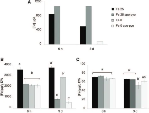
First, we measured the iron content in the nutritive medium. In the Fe 25 medium, a decrease of the iron content occurred between 6 h and 3 d after the treat-ment, indicating that plants imported part of the iron present in the medium. When apo-pyo was added in the same medium, the concentration of iron did not decrease with time (Fig. 6A). The measurement of iron contents in roots further supported these data (Fig. 6B). Indeed, compared with the Fe 25 treatment, the addition of apo-pyo in the medium containing 25 mM
iron (Fe 25 apo-pyo condition) strongly reduced the root iron concentration at 6 h and 3 d (Fig. 6B). In contrast, the iron content in roots of plants grown in the Fe 25 mMcondition without apo-pyo remained
al-most constant over time. As expected, this first set of data indicates that apo-pyo negatively impacts the dynamic of iron import from the medium by plant roots, probably by direct chelation of iron in the me-dium. Accordingly, the expression of IRT1 and FRO2 was repressed 3 d after the addition of apo-pyo in the iron-containing medium (Supplemental Table S6). The comparison of the Fe 0 and Fe 0 apo-pyo conditions also was relevant. In the absence of apo-pyo, the level
of iron at 6 h in the roots of Fe 0-treated plants was reduced compared with Fe 25-treated plants (Fig. 6B). Therefore, it is likely that plants already perceived the lack of iron in the medium. In comparison, the addi-tion of apo-pyo did not further alter the root iron content of plants grown in the iron-deficient condition. In contrast, at day 3, whereas the iron content of the Fe 0 plants was almost similar to that measured at 6 h, Fe 0 apo-pyo-treated plants showed a strong reduction in the iron content. Taken as a whole, these results indi-cate that the addition of apo-pyo in the culture me-dium containing or missing iron triggered a reduction of the root iron content.
Completing this analysis, the iron content in shoots also was measured (Fig. 6C). Six hours after the treatments, the iron content was similar in all condi-tions. After 3 d, while the apo-pyo treatments induced a strong reduction of the root iron content in culture medium containing or not containing iron (Fig. 6B), the same treatments did not impact the iron concen-tration in shoots (Fig. 6C). In contrast, the iron content in shoots of plants grown in medium lacking iron without apo-pyo (Fe 0 treatment) decreased slightly but significantly.
To further investigate the incidence of apo-pyo treatment on iron homeostasis, we performed a func-tional analysis of six key iron-related genes found to be modulated by apo-pyo: IRT1, FRO2, FER1, NAS4, bHLH100, and bHLH39/ORG3. As discussed above, with the exception of FER1, which was repressed, the expression of these genes was up-regulated in the roots in the Fe 0 apo-pyo condition. Furthermore, FRO2 and IRT1 expression was down-regulated in the Fe 25 apo-pyo condition (see above; Supplemental Table S6).
Figure 6. Iron concentrations measured in the nutritive medium and tissues 6 h and 3 d after apo-pyo treatment in iron-sufficient or iron-deficient medium. A, Iron concentrations measured in the nutritive medium. B, Root iron contents. C, Shoot iron contents. Values are means of three measurements 6SEfrom three independent experiments. Significant dif-ferences between the concentrations of iron for each time point were determined by one-way ANOVA/LSD method, with different letters signifying distinct statisti-cal groups (P , 0.001 for B and P , 0.05 for C). DW, Dry weight.
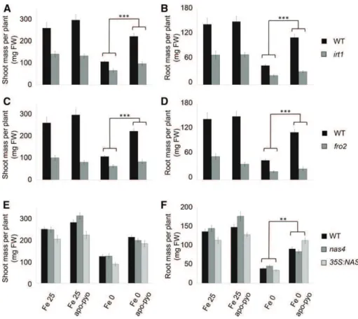
Therefore, we studied whether corresponding mu-tants could be affected in their response to apo-pyo in iron-deficient or iron-sufficient medium by measur-ing their shoot and root masses after 7 d of treatment, as described in Figure 3, B and C. The response to apo-pyo was not modified in the bhlh100 and bhlh39/ org3 mutants or in the FER1 overexpressor (ov. fer.) regardless of the iron content in the culture medium (Supplemental Fig. S5, A–D). In contrast, compared with the wild type, the irt1 and fro2 knockout mu-tants showed an impaired response to apo-pyo in iron-deficient medium in shoots (Fig. 7, A and C) and in roots (Fig. 7, B and D). Indeed, the growth-promoting effect triggered by apo-pyo in the wild type was strongly reduced in both mutants in this culture condition, highlighting the involvement of IRT1 and FRO2 in this process. Regarding NAS4, whereas the knockout nas4 mutant did not show a modified response to apo-pyo compared with the wild type (Fig. 7, E and F), the growth-promoting effect of the siderophore was significantly more pro-nounced in roots of plants overexpressing NAS4 (35S:NAS4) grown in iron deficiency (Fig. 7F). Similar to wild-type plants, apo-pyo treatment did not affect the shoot and root masses of all the mutants and overexpressors tested when grown in iron-sufficient conditions. Collectively, these data highlight an in-volvement of IRT1, FRO2, and, to a lower extent, NAS4 in the growth effect of apo-pyo in plants ex-posed to iron deficiency.
Incidence of Apo-Pyo in SA, ABA, Indole-3-Acetic Acid, and Ethylene Synthesis/Accumulation
The finding that apo-pyo modulated the expression of genes related to SA and ABA signaling and increased the growth of plants in medium deprived of iron prompted us to analyze the concentration of several hormones in our distinct conditions, namely SA, ABA, indole-3-acetic acid (IAA; the major endogenous auxin), and ethylene. Hormone measurements were performed 6 h and 3 d after apo-pyo treatment (for the experi-mental design, see Fig. 1).
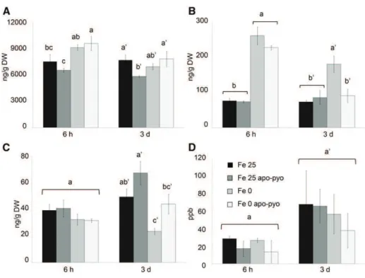
In plants grown in iron-containing medium, at 3 d, we only measured a slight decrease of SA content by apo-pyo (Fe 25 apo-pyo versus Fe 25; Fig. 8A). Con-cerning ABA, iron deficiency induced a clear increase of its concentration at 6 h and 3 d (Fe 0 condition versus Fe 25 condition; Fig. 8B). At 6 h, the addition of apo-pyo did not influence the ABA content in iron-containing or iron-missing culture medium. However, after 3 d, the higher level of ABA measured in the shoots of plants grown in iron-deficient conditions (Fe 0) was not ob-served in plants grown in the same medium but sup-plied with apo-pyo (Fe 0 apo-pyo). In this latter condition, the shoot concentration of ABA was similar to those found in the shoots of Fe 25- and Fe 25 apo-pyo-treated plants. Therefore, apo-pyo appeared to nega-tively impact ABA synthesis/accumulation in the iron-deficient condition at 3 d, the time point where apo-pyo begins to rescue the growth of plants facing this deficiency (Fig. 3). In the case of IAA, there was no
Figure 7. Growth phenotypes of mu-tants impaired in genes involved in iron homeostasis in response to apo-pyo in iron-sufficient or iron-deficient medium after 7 d of treatment. Plants were treated as described in Figure 1. A, C, and E, Fresh shoot masses measured in wild-type (WT) and irt1 (A), fro2 (C), and nas4 and 35S:NAS4 (E) plants. B, D, and F, Fresh root masses measured in wild-type and irt1 (B), fro2 (D), and nas4 and 35S: NAS4 (F) plants. Each value represents the mean 6 SEof almost 12 measure-ments from almost three independent biological experiments. Significant dif-ferences between the shoot and root masses of the wild type and the mutants in response to apo-pyo and/or iron defi-ciency were determined by two-way ANOVA/Tukey’s honestly significant dif-ference method (**, P , 0.01 and ***, P , 0.0001). FW, Fresh weight.
difference after 6 h regardless of the treatment. How-ever, after 3 d, iron deficiency induced a reduction of the content of IAA (Fe 25 versus Fe 0 treatment; Fig. 8C). The presence of apo-pyo in Fe 25 or Fe 0 medium had no significant additive effect on the IAA level. In addition, none of the treatments significantly affected the ethyl-ene content of the plantlets (Fig. 8D).
Involvement of Apo-Pyo in the Growth/Defense Balance We and others recently reported that iron deficiency induces an increased resistance of Arabidopsis plants against the fungal pathogen B. cinerea (Kieu et al., 2012; Koen et al., 2014). The findings that apo-pyo negatively regulated the levels of ABA and the expression of defense-related genes of plants in the iron-deficiency condition led us to check whether the siderophore could impact the plant resistance to this pathogen. For this purpose, leaves of plants exposed to our different conditions (for the experimental design, see Fig. 1) were inoculated with B. cinerea at day 3, and the disease symptoms were measured 4 d after the inoculation. As expected and as reported previously, the average of the necrotic lesion areas produced by B. cinerea infection was reduced in plants that suffered from iron deficiency compared with plants grown in the Fe 25 medium (22 versus 50 mm2; Fig. 9). Interestingly, the induced re-sistance conferred by iron deficiency was partly im-paired in response to apo-pyo. Indeed, compared with the condition without apo-pyo, plants grown in iron-deficient medium in the presence of the siderophore were more sensitive to the fungal infection (necrotic lesion areas about 36 mm2). In contrast, no effect of
apo-pyo was observed in plants grown in the iron-sufficient condition.
To further understand how apo-pyo impacts the growth and defense of plants grown under iron defi-ciency, we analyzed the involvement of four genes highly modulated by the siderophore and shown pre-viously to display important functions in develop-ment and/or defense. These genes are LTP3, CER4, AT5G59320 (named PLTP in this article), encoding a protein involved in lipid transport and binding and whose transcripts were shown to be induced in leaves of Arabidopsis plants subjected to a combination of drought and heat stresses (Rizhsky et al., 2004), and AT3G21460 (named GRX in this article), which encodes a glutaredoxin family protein involved in cell redox homeostasis (Finkemeier et al., 2005). The growth-promoting effects of apo-pyo observed in plants fac-ing iron deficiency were not altered in the mutants impaired in the expression of the genes of interest (Supplemental Fig. S5, A, B, E, and F).
We completed this analysis by investigating whether apo-pyo could impact the balance between growth and defense, as the siderophore had a positive effect on growth but a negative effect on the resistance to B. cinerea in plants cultivated in iron deficiency. Re-cently, it was demonstrated that this tradeoff can be based on cross talk operating between growth- and immune-related signaling pathways (Bai et al., 2012; Fan et al., 2014; Malinovsky et al., 2014). Notably, it has been shown that such cross talk could involve two transcription factors, BZR1 and HBI1, the second being a partner of the tripartite module PRE1-IBH1-HBI1 that regulates cellular elongation upstream from such ex-ternal or endogenous signals as brassinosteroids and
Figure 8. Analysis of hormone contents in the shoots (A–C) or in the air (D) 6 h and 3 d after apo-pyo treatment in iron-sufficient or iron-deficient medium. A, SA contents. B, ABA contents. C, IAA contents. D, Ethylene contents. Values are means of at least three measure-ments 6SEfrom at least three indepen-dent experiments. Significant differences between the concentrations of hormones for each time point were determined by one-way ANOVA/LSDmethod, with dif-ferent letters signifying distinct statis-tical groups (P , 0.05 for A and C and P , 0.01 for B). DW, Dry weight; ppb, parts per billion (nL L21).
temperature (Fan et al., 2014). IBH1 interacts with and inhibits HBI1, which is a positive regulator of cellular elongation. In particular, HBI1 activates ATEXPA8 and ATEXPA1, two genes encoding expansins (cell wall-loosening enzymes), by direct binding to their pro-moters. The effects of IBH1 on HBI1 are inhibited by PRE1, which sequestrates IBH1. We first checked the modulation of these different genes in a microarray experiment (Supplemental Table S8). It appeared that HBI1 and PRE1 were highly up-regulated by apo-pyo in the shoots of Fe 0 apo-pyo- versus Fe 0-treated plants, while the inhibitor IBH1 was slightly down-regulated. We also noticed an induction of the expression of ATEXPA1 and ATEXPA8. Moreover, other genes en-coding expansins also were activated in response to
apo-pyo in roots and/or shoots and could be involved in the siderophore-induced promotion of growth in iron deficiency.
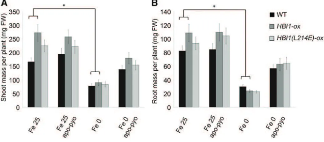
As no HBI1-deficient mutant is available (Malinov-sky et al., 2014), we tested the phenotype of HBI1-ox plants, which overexpress HBI1, and HBI1(L214E)-ox plants, which display a dominant-negative effect on HBI1 in our different conditions (for the experimental design, see Fig. 1). In the absence of apo-pyo, HBI1-ox plants had higher growth that wild-type plants in Fe 25 medium (Fig. 10). However, in response to iron depri-vation, the shoot (Fig. 10A) and root (Fig. 10B) masses of HBI1-ox plants were similar to those of wild-type plants. Finally, the growth-promoting effects of apo-pyo observed in wild-type plants facing iron defi-ciency were not statistically different in HBI1-ox and HBI1(L214E)-ox plants.
Comparison of Apo-Pyo Effects with Those Induced byP. fluorescens C7R12 Bacterial Inoculation
Four-week-old plants were subjected to apo-pyo treatment as described in Figure 1 or inoculated with the wild-type P. fluorescens strain C7R12 or with the mutant strain PL1 in iron-deficient or iron-sufficient medium. PL1 is a pyoverdine minus C7R12 mutant obtained by random insertion of the transposon Tn5 in the pyoverdine synthetase gene pvsB (Mirleau et al., 2000). PL1 is unable to produce pyoverdine but pro-duces ornicorrugatine, another siderophore.
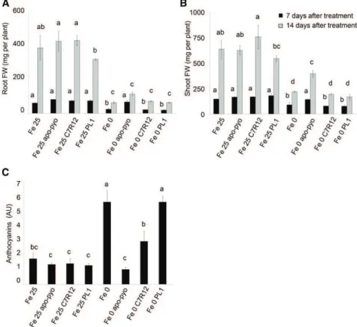
First, we compared the macroscopic phenotypic ef-fects of the purified siderophore with those of bacterial inoculation with the C7R12 or PL1 strain. As described for apo-pyo (Fig. 3), no effect of C7R12 or PL1 was visible in iron-sufficient medium after 7 d (Supplemental Fig. S7A) or 14 d (Supplemental Fig. S7B) of treatment. In-deed, root and shoot weights of the inoculated plants
Figure 9. Evaluation of the sensitivity to B. cinerea of plants treated by apo-pyo in iron-sufficient or iron-deficient medium. Each value repre-sents the mean of the area of the necrotic lesion 6SEof 30 measure-ments. Experiments were repeated four times with similar results. Significant differences between the mean lesion areas were determined by one-way ANOVA followed by one-way ANOVA/LSDmethod, with different letters signifying distinct statistical groups (P , 0.05).
Figure 10. Growth phenotypes of HBI1-ox and HBI1(L214E)-ox mutants in response to apo-pyo in iron-sufficient or iron-deficient medium after 7 d of treatment. Plants were treated as described in Figure 1. A, Fresh shoot masses. B, Fresh root masses. Each value represents the mean 6SEof almost 12 measurements from almost three independent biological experiments. Significant dif-ferences between the shoot and root masses of wild-type (WT) and mutants plants in response to apo-pyo and/or iron deficiency were determined by two-way ANOVA/Tukey’s honestly significant difference method (*, P , 0.05). FW, Fresh weight.
were equivalent to those of noninoculated plants (Fig. 11, A and B). Importantly, in contrast to the treatment with purified apo-pyo, in iron-deficient conditions, the C7R12 strain producing apo-pyo did not partially restore the growth of plants (Fig. 11, A and B; Supplemental Fig. S7). However, compared with plants grown in iron-deficient medium without apo-pyo treatment (Fe 0-treated plants), the C7R12 strain triggered a reduction of the violet pigmentation after 14 d of treatment in the iron-deprived medium (Supplemental Fig. S7B). Accordingly, the production of anthocyanins measured in C7R12-treated plants facing iron deficiency was intermediate between those measured in Fe 0- and Fe 0 apo-pyo-treated plants (Fig. 11C). Highlighting the putative involvement of apo-pyo in this process, the reduction of the anthocyanin level observed in C7R12-treated plants was not found when plants suffering from iron deficiency were inoc-ulated with the PL1 strain. Indeed, PL1-treated plants displayed an anthocyanin concentration similar to that of Fe 0-treated plants (Fig. 11C).
We also compared the sensitivity to B. cinerea of the plants treated with apo-pyo and those inoculated with the C7R12 or PL1 strain. No effect of both strains was observed in the Fe 25 medium, as described previously for apo-pyo (Fig. 12). In the medium lacking iron, C7R12-inoculated plants (Fe 0 C7R12) were more sen-sitive to the pathogen than Fe 0-treated plants, their
higher sensitivity being intermediate between those of Fe 0 apo-pyo-treated plants and Fe 0 PL1-treated plants. These data strongly suggest that the production of apo-pyo by the wild-type strain was responsible for the higher sensitivity of the plants to B. cinerea in iron-deficient medium.
DISCUSSION
Most studies dealing with the effects of bacterial siderophores on plants were performed in nonlimiting iron nutrition medium and/or using ferri-siderophores (Vansuyt et al., 2007; Aznar et al., 2014). Here, we an-alyzed the biological effects of a nonchelated sidero-phore, apo-pyo, on plants that were maintained in a medium depleted or not of iron. This enabled us to mimic a situation in which the two partners, bacterial strain and host plant, are subjected to iron deficiency and attempt to acquire iron from the rhizosphere. As a matter of fact, siderophores are known to be produced by bacteria only in iron stress conditions (Meyer and Abdallah, 1978), as in the rhizosphere environment in which pyoverdine synthesis was evidenced using a reporter gene (Loper and Henkels, 1997; Duijff et al., 1999).
We first showed that apo-pyo was effectively taken up by roots when plants were cultivated in hydroponic
Figure 11. Phenotypic analysis of Ara-bidopsis plantlets exposed to apo-pyo or inoculated with C7R12 or PL1 and grown in iron-sufficient or iron-deficient conditions. Plants were cultivated and treated by apo-pyo as indicated in Figure 1. Inoculation with C7R12 or PL1 was performed as described in “Materials and Methods.” A, Root fresh weights. B, Shoot fresh weights. C, Anthocyanin contents. Each value represents the mean 6SEof three measurements. Ex-periments were repeated three times with similar results. Significant differ-ences between the treatments were deter-mined by one-way ANOVA/LSDmethod, with different letters signifying distinct statistical groups (P , 0.05). AU, Arbi-trary units; FW, fresh weight.
solution. This phenomenon was shown previously to occur in plants cultivated in vitro and treated by ferri-pyo in a medium deprived of iron (Vansuyt et al., 2007). Moreover, using [15N]apo-pyo, we were also able to detect15N in the shoots. The fact that apo-pyo was not detected by ELISA in the shoots could be explained by the lesser sensitivity of this technique compared with
15N detection by mass spectrometry or, eventually, by
a degradation or a chemical modification that would impair its recognition by antibodies, as suggested pviously by Vansuyt et al. (2007). In both cases, this re-sult indicates that apo-pyo or a derivative is a systemic compound. Moreover, the quantity of apo-pyo assimi-lated by plants did not differ whether or not the plants were precultivated 24 h in the presence of iron. This observation indicates that plants may not adapt the rate of apo-pyo assimilation according to the availability of iron in the rhizosphere.
Apo-pyo had a striking macroscopic effect on plants facing iron deficiency. Compared with nontreated plants grown without iron, it induced a remarkable promotion of growth together with a reduction of an-thocyanin accumulation. We checked that the iron-free medium was still deprived of iron once apo-pyo was added, thus ruling out the possibility that the growth phenotype observed in the presence of the siderophore was due to iron contamination. A promotion of growth was observed previously by Vansuyt et al. (2007) in iron-deficient medium following the addition of ferri-pyo to the roots of Arabidopsis. This promotion was significantly higher with ferri-pyo than with Fe-EDTA and was associated with a higher increase in the iron content of the plants.
To strengthen these data, we performed similar ex-periments using the wild-type C7R12 strain and the
corresponding PL1 strain impaired in apo-pyo pro-duction. Similar to purified apo-pyo treatment, in the iron deficiency condition, the C7R12 strain triggered a reduction of anthocyanin accumulation when com-pared with Fe 0-treated plants. This phenomenon was due to apo-pyo production, since it was not visible in response to PL1 inoculation. However, the promotion of growth induced by the purified apo-pyo in iron-deficient conditions was not observed in Fe 0 C7R12-inoculated plants. This discrepancy may be explained by different factors. First, it is conceivable that the concentration of the apo-pyo released by the bacteria may be different from that of the purified apo-pyo (25 mM). Second, besides apo-pyo, other compounds
produced by the bacteria and recognized by the plant, such as lipopolysaccharides or exopolysaccharides, as well as molecules secreted into plant host cells, also may account for the effects of C7R12, thus making the situation much more complex. Indeed, it has been shown that numerous plant-beneficial pseudomonads such as P. fluorescens C7R12 harbor type 3 secretion system (T3SS)-like genes related to the HYPERSENSI-TIVE RESPONSE AND PATHOGENICITY1 T3SS family (Mazurier et al., 2004). This T3SS contributes to the plant-bacteria interactions and to the bacterial ef-fects in plants. The involvement and role of this system in the Arabidopsis-C7R12 bacteria interaction remain to be determined.
To investigate the mechanisms underlying apo-pyo effects, we choose to perform a large-scale transcriptomic analysis. It appeared that apo-pyo modulated the ex-pression of around 2,100 genes (log2 ratio of +1.5 or greater or 21.5 or less; P # 0.05). The number of genes modulated by apo-pyo and their induction or repression status clearly depended on the tissue and/or the presence or not of iron in the hydroponic solution. First, root genes rather than shoot genes were preferentially modulated. This result was expected, as apo-pyo was applied in the nutrient solution, directly in contact with the roots, and once assimilated by plants accumulated preferentially in roots rather than in shoots. Second, a great part of the apo-pyo-dependent genes was modulated in iron-deficient rather than in iron-sufficient conditions. Therefore, the effect of apo-pyo on gene expression appeared to be de-pendent on the plant iron status. Furthermore, the higher number of apo-pyo-modulated genes seen in plants grown in the iron-deficient medium corroborates the strong phenotypic effect triggered by the siderophore in this condition.
A focus on the apo-pyo-regulated genes in plants grown in iron-free medium showed that the most in-duced genes encoded proteins involved in develop-ment and iron acquisition processes, while the most repressed genes were related to iron sequestration and defense responses. Interestingly, Aznar et al. (2014) recently reported data from a transcriptomic analysis performed on Arabidopsis plantlets cultivated in iron-sufficient conditions in which the apo-form of the sid-erophore deferrioxamine (DFO; 1 mM) was infiltrated
in leaves. The major categories of genes modulated by
Figure 12. Sensitivity to B. cinerea of plants treated by apo-pyo or in-oculated with C7R12 or PL1 in iron-sufficient or iron-deficient medium. Each value represents the mean of the area of the necrotic lesion 6SE of 30 measurements. Experiments were repeated three times with similar results. Significant differences between the mean lesion areas were determined by one-way ANOVA followed by one-way ANOVA/ LSDmethod, with different letters signifying distinct statistical groups (P , 0.05).
DFO in leaves were related to biotic and abiotic stresses, including the immune response, whereas in roots, the DFO target genes were associated with heavy metal homeostasis. Importantly, no effect on gene modula-tion was observed when Fe-DFO complexes were used instead of iron-free DFO, and the physiological re-sponses induced by DFO were similarly observed in response to a synthetic iron chelator. Therefore, the ef-fects of DFO were due, or partly due, to its iron chela-tion ability. Inevitably, these data raise the queschela-tion of the incidence of apo-pyo on plant iron homeostasis and whether its effects also could be related to its chelating properties. The transcriptomic analysis as well as the quantification of the iron content in apo-pyo-treated plants provided first answers. In plants grown in iron-containing medium, the addition of apo-pyo did not affect the shoot iron content but triggered a strong reduction of the root iron concentration accompanied by a repression of IRT1 and FRO2 expression. Fur-thermore, many of the genes regulated by apo-pyo in roots also were found to be modulated by iron defi-ciency in roots of Arabidopsis in the study of Schuler et al. (2011; listed in Supplemental Table S9), and, using Genevestigator, we identified a profile close to those of two iron deficiency treatments (Supplemental Fig. S6). Therefore, apo-pyo triggered an iron deficiency-like response in plants grown in the iron-sufficient condi-tion. As expected, we provided evidence that this pro-cess was due mainly to the chelation by apo-pyo of the iron present in the culture medium. Intriguingly, the resulting reduction of iron availability did not impact the growth of the plants. This observation suggests that, although the apo-pyo treatment impacted the root iron content, the iron concentration of plants grown in iron-containing medium was probably sufficient to ensure normal growth. This assumption does not exclude the possibility that part of the siderophore imported by the plants could be internalized in its iron-chelated struc-ture and contribute to iron assimilation. If this is the case, this mechanism poorly accounts for the plant iron nutrition.
In plants facing iron deficiency, apo-pyo also trig-gered a strong reduction of the root iron content com-pared with nontreated plants grown in iron-free medium. However, the reduction of shoot iron con-centration observed in the latter was less pronounced in the apo-pyo-treated plants. It is striking that, in plants grown in the iron-deficient condition, apo-pyo pro-moted an important induction of genes related to iron assimilation, such as IRT1, FRO2, and ORG3, as well as a down-regulation of AtFER1. These data deeply con-trast with those obtained in apo-pyo-treated plants grown in iron-sufficient conditions and highlight that, in plants facing iron deficiency, apo-pyo strongly pro-motes the iron-uptake machinery. Furthermore, in ad-dition to the iron-uptake process, apo-pyo also induced a strong expression of genes encoding proteins related to in planta iron redistribution, such as NAS4 and OPT3 (Supplemental Table S6; Koen et al., 2014; Zhai et al., 2014). As plants were grown in iron-free conditions, it is
reasonable to assume that apo-pyo provided in the culture could be imported by plants in its iron-free structure and, once internalized in roots, could chelate cellular iron. Accordingly, due to their high affinity for iron, bacterial siderophores including pyoverdine are able to efficiently compete with host iron-binding pro-teins for iron acquisition (Saha et al., 2013). This process could account for the ability of apo-pyo to induce the overexpression of the plant iron-import system. What-ever the scenario, we provide evidence that the induc-tion of the iron-import machinery contributes to the growth-promoting effects of apo-pyo in plants facing iron deficiency. Indeed, the positive effect of apo-pyo on growth was severely impaired in mutants deficient in IRT1 and FRO2 expression. Furthermore, the positive effect of apo-pyo on growth was further induced in 35S: NAS4 plants overproducing NA, a nonproteinogenic amino acid that contributes to iron long-distance cir-culation in phloem and transfer within the cells and that is required for the proper establishment of the plant response to iron deficiency (Koen et al., 2014).
The analysis of the content of ABA in plants exposed to apo-pyo also supports the findings that apo-pyo had a major impact on iron homeostasis. First, the hormone accumulated in the shoots of plants suffering from iron deficiency in the presence or not of apo-pyo within 6 h. Second, after 3 d of apo-pyo treatment, plants grown in iron-free medium showed a drastic reduction of shoot ABA level compared with nontreated plants cultivated in the same medium. Therefore, the production of ABA appeared to be more transient in plants exposed to apo-pyo. Accordingly, the transcriptomic analysis revealed that, in the iron-free condition, apo-pyo repressed the expression of root genes related to ABA signaling. Our data should be discussed in light of the investigation of Lei et al. (2014). Those authors reported that iron defi-ciency induced a rapid ABA accumulation (within 6 h) in the roots of Arabidopsis. In that study, ABA was shown to help alleviate iron deficiency by promoting root iron reutilization and transport from the roots to the shoots. This process involved the ABA-dependent regulation of genes encoding proteins related to the reutilization of the iron stored in the vacuoles, such as AtNRAMP3, and to long-distance transport of iron, including AtFRD3, AtYSL2, and AtNAS1. In our study, the observations that, in iron deficiency, the apo-pyo-treated plants displayed an induced expression of root genes related to iron in planta transport/redistribution suggest that the mechanism described by Lei et al. (2014) could operate efficiently and, therefore, more transiently in response to the siderophore. However, none of the ABA-dependent genes described by those authors were found to be modulated by apo-pyo after 3 d of treatment, thus minimizing this possibility.
Recent investigations have reported that the plant iron status conditions its resistance against microor-ganisms. Notably, it has been found that iron deficiency activates defense-related processes and confers resis-tance against microbial pathogens, including D. dadantii and B. cinerea (Kieu et al., 2012; Koen et al., 2014).
Several studies also demonstrated that siderophores produced by microorganisms trigger an immune re-sponse that could be linked to their capacity to chelate iron from the host (Aznar and Dellagi, 2015; Aznar et al., 2015). In the same vein, we recently demonstrated that b-aminobutyric acid, a priming agent that con-fers enhanced resistance to numerous pathogens, is a powerful iron chelator triggering a transient iron defi-ciency in plants. This process could bring the plant to a defense-ready state, contributing to an efficient resis-tance (Koen et al., 2014). Further strengthening the concept that plant immunity is tightly linked to iron homeostasis, several genes, including those encoding the transcription factor MYB72 (AT1G56160) and the b-glucosidase BGLU42 (AT5G36890), were shown to function in the cross talk of root signaling pathways regulating rhizobacteria-mediated ISR and the plant response to iron deficiency (Zamioudis et al., 2014, 2015). In our investigation, in accordance with the studies listed above, we confirmed that plants culti-vated in iron deficiency displayed a higher resistance to B. cinerea. This higher resistance was reduced by the apo-pyo treatment but also by a C7R12 bacterial inoc-ulation. In response to apo-pyo, this negative effect was correlated to a general down-regulation of genes encoding proteins involved in immunity with few exceptions. In particular, the expression of BGLU42 required for rhizobia-induced ISR was slightly induced in apo-pyo-treated plants (Supplemental Table S6). Several works demonstrated that pyoverdines act as determinants in ISR against numerous pathogens. The induction of ISR is not systematic and depends on the specific pathosystem and the type of pyoverdine. For instance, some pyoverdines, such as those of C7R12, induce resistance against the rice blast, but some do not. De Vleesschauwer and Höfte (2009) suggested that these differences can be explained by the induction of iron deficiency by certain pyoverdines. In our case, despite the induction of an iron deficiency-like response in the Fe 25 medium, apo-pyo did not induce ISR in Arabidopsis against B. cinerea. Other factors are prob-ably involved in the induction of ISR.
We observed that pyoverdine did not favor the re-sistance against B. cinerea but rather partially impaired it in plants grown in the iron-deficient condition. The interpretation of this process should take into account the drastic growth-promoting activity of apo-pyo. In-deed, the antagonistic effect of apo-pyo on growth and resistance observed in plants facing iron deficiency strongly suggests that the siderophore impacts the tradeoff between growth and immunity. As recently highlighted in several studies, cross talk between growth and immune signaling operates in plants. According to the physiological contexts, notably path-ogen challenge or light conditions, such cross talk pri-oritizes one process over the other (for review, see Lozano-Durán and Zipfel, 2015). Clearly, our data in-dicate that, in plants grown in iron-deficient conditions and in response to apo-pyo, growth is prioritized over immunity. Further supporting this assumption, the
microarray analysis revealed that apo-pyo modulates the expression of the PRE1-IBH1-HBI1 module in shoots of plants facing iron deficiency. As discussed in “Results,” HBI1 is a key node for the cross talk between growth and immunity and, once activated, negatively regulates defense responses while prioritizing growth (Fan et al., 2014; Malinovsky et al., 2014). HBI1 effects are antagonized by IBH1. In turn, through its binding to HBI1, PRE1 prevents its inhibitory effect (Bai et al., 2012). Supporting a role of HBI1 in the mediation of the tradeoff between growth and immunity, its over-expression was shown to trigger the repression of the expression of genes involved in immunity and to en-hance the susceptibility of the plants to the bacterium P. syringae (Fan et al., 2014; Malinovsky et al., 2014). We found that both HBI1 and PRE1 expression in shoots were strongly up-regulated in response to apo-pyo, whereas the expression of IBH1 was slightly re-pressed. Therefore, these expression profiles reflect a physiological situation in which the balance between growth and immunity is weighted in favor of growth. To strengthen this possibility, we checked whether the growth-promoting effect of apo-pyo in plants grown in the iron-deficient condition was enhanced in the HBI1-overexpressing line HBI1-ox. We did not observe such an effect. This finding could be explained by the fact that the level of HBI1 expression induced by apo-pyo in wild-type plants was particularly high.
CONCLUSION
To conclude, this investigation provides evidence that the effects of pyoverdine depend on iron avail-ability and, consequently, on the plant iron status. Furthermore, it highlights that, in plants facing iron deficiency, the apo-siderophore modulates the tradeoff between growth and immunity in favor of growth, this mechanism being dependent on the expression of the iron uptake-related genes IRT1 and FRO2. More gen-erally, this work indicates that microorganism-derived siderophores impact the cross-regulatory interactions operating on host plants between iron homeostasis, immune function, and growth.
MATERIALS AND METHODS Plant Material and Growth Conditions
Arabidopsis (Arabidopsis thaliana) Columbia-0 ecotype was used as the wild-type control for phenowild-type comparisons for all mutants and transgenic lines described in this study. The transfer DNA mutants bhlh100 (SALK_074568C), bhlh39 (SALK_025676C), cer4 (SALK_000575C), grx (SALK_001139C), ltp3 (SALK_095248C), and pltp (SALK_063856) were obtained from the Nottingham Arabidopsis Stock Centre. Homozygosity for the mutant alleles was checked using specific primers (Supplemental Table S10). The following lines were de-scribed previously: irt1 (Vert et al., 2002), fro2 (also named frd1-1; Robinson et al., 1999), ov. fer. (Vansuyt et al., 2007), HBI1-ox and HBI1(L214E)-ox (Malinovsky et al., 2014), and 35S:NAS4 and nas4 (Koen et al., 2013).
Seeds were surface sterilized by immersion in a solution containing 1.2% (v/v) bleach and 50% (v/v) ethanol for 3 min, rinsed three times with 100% ethanol, air dried, and placed for 3 d at 4°C in the dark in 0.15% (w/v) agar.