HAL Id: hal-02086456
https://hal-mines-albi.archives-ouvertes.fr/hal-02086456
Submitted on 3 Apr 2019
HAL is a multi-disciplinary open access
archive for the deposit and dissemination of
sci-entific research documents, whether they are
pub-lished or not. The documents may come from
teaching and research institutions in France or
abroad, or from public or private research centers.
L’archive ouverte pluridisciplinaire HAL, est
destinée au dépôt et à la diffusion de documents
scientifiques de niveau recherche, publiés ou non,
émanant des établissements d’enseignement et de
recherche français ou étrangers, des laboratoires
publics ou privés.
Near Infrared Light Heating of Soft Tissue Phantoms
Containing Nanoparticles
Simon Eibner, R. Jaime, B. Lamien, R. Basto, R. Orlande, Olivier Fudym
To cite this version:
Simon Eibner, R. Jaime, B. Lamien, R. Basto, R. Orlande, et al.. Near Infrared Light Heating of Soft
Tissue Phantoms Containing Nanoparticles. Revista de Engenharia Térmica, 2014, 13 (1), pp.13-18.
�10.5380/reterm.v13i1.62059�. �hal-02086456�
NEAR INFRARED LIGHT HEATING OF SOFT TISSUE
PHANTOMS CONTAINING NANOPARTICLES
S. Eibnera, R. A. O. Jaimeb, B. Lamienb, R. L. Q. Bastob, H. R. B. Orlandeb, and O. Fudyma
aÉcole des Mines d'Albi-Carmaux
Campus Jarlard - 81013 Albi CT Cédex 09 seibner@mines-albi.fr
bUniversidade Federal do Rio de Janeiro
Departamento de Engenharia Mecânica COPPE Cidade Universitária CEP. 68503, Rio de Janeiro, Brasil
ABSTRACT
The objective of this paper is to investigate the effect of the addition of nanoparticles to soft tissue phantoms, aiming at the enhancement of photothermal therapy for cancer. The phantoms were made of Polyvinyl chloride-plastisol (PVC-P), with two different nanoparticles, namely, titanium dioxide nanoparticles (TiO2) and silica nanoparticles (SiO2). A
phantom without nanoparticles and a phantom containing a thermal paste were also manufactured for comparison purposes. The PVC-P phantom is transparent to the near infrared laser light, whereas the addition of titanium dioxide nanoparticles modified the optical properties enhancing the local heating, as demonstrated through experiments with a laser-diode and an infrared camera.
Keywords: hyperthermia, NIR laser, photothermal therapy, phantom,
photoabsorbing nanoparticles.
NOMENCLATURE
a larger ellipses semi-axis, m b smaller ellipses semi-axis, m Ba bias limit of quantity a
cp fluid specific heat at constant pressure,
J/(kg.K) D tube diameter, m E ellipses eccentricity, b/a H array height, m
K fluid thermal conductivity, W/(m.K) L array length, m
2b
L
array length to smaller ellipses axis aspect ratio
m total mass of the arrangement, kg
m~ dimensionless total mass of the arrangement nf number of fins
nt total number of tubes
N number of tubes in one unit cell Nec number of elemental channels
p pressure, N/m2
P dimensionless pressure
Pe2b Peclet number based on smaller ellipses axis
Pr fluid Prandtl number, ν/α Pa precision limit of quantity a
*
q~ dimensionless overall thermal conductance, Eq.(15)
Q overall heat transfer rate, W
Re2b Reynolds number based on smaller ellipses
axis, u∞2b/ν
S spacing between rows of tubes, m, Fig. 1 S/D dimensionless spacing between rows of tubes
(circular arrangement)
S/2b dimensionless spacing between rows of tubes (elliptic arrangement)
t time, s
tf fin thickness, m
tt tube thickness, m
T average fluid temperature, K u1,u2,u3 velocity components, m/s
U1,U2,U3 dimensionless velocity components
Ua uncertainty of quantity a
W array width, m
x,y,z cartesian coordinates, m
X,Y,Z dimensionless cartesian coordinates
Greek symbols
α thermal diffusivity, m2/s
ε mesh convergence criterion, Eq. (21) δ fin-to-fin spacing, m
θ dimensionless temperature
θ dimensionless average fluid temperature ν fluid kinematic viscosity, m2/s
ρ density, kg/m3
φf dimensionless fin density in direction z
τ dimensionless time
Subscripts
in unit cell inlet m maximum opt optimal out unit cell outlet
s solid tube wall and fin material w tube surface
∞ free stream
INTRODUCTION
In medicine, hyperthermia refers to a moderate use of heat to increase the temperature of human tissues for therapeutic purposes. For example, it has been used for the treatment of cancer in chemotherapy, for rapid drug delivery by increasing blood flow in the cancerous region, or in radiotherapy, by making cancerous cells more sensitive to radiation (Chekuri et al., 2010). Besides that, tumorous cells are more sensitive to heat than normal cells. Consequently, protocols based solely on hyperthermia have been used for irreversible damage to tumorous cells, which is caused by the rupture of the cell membrane and denaturing of proteins. Such effects take place in temperatures ranging from 41 oC
to 47 oC. Heating strategies such as laser, microwave,
radiofrequency and ultrasound waves have been used to heat a specific target region for cancer treatment. Common drawbacks related to these methods are the undesirable burning of healthy cells and the difficulty to achieve uniform temperatures in the tumor volume (Huang et al., 2008). On the other hand, extracorporeal laser heating of a specific target region containing photothermal agents is not invasive and very selective, in order to achieve hyperthermia (Feng et al., 2009; Dickerson et al., 2008). Such a method, in which photoabsorbing agents are used as light/heat converters, has received the name photothermal therapy (PTT). Primary investigations in this field dealt with the use of natural chromophores, or externally added dye molecules, as photothermal agents. Natural chromophores, however suffer from very low absorption, while small dye molecules suffer from photobleaching under laser irradiation (Huang et al., 2008).
Recent advances in nanotechnology allowed the production of nanoparticles with enhanced optical properties for a localized surface plasmon resonance. This is a resonant phenomenon in which light induces oscillations of conductive electrons at the surface of metal nanoparticles (Hirsch et al., 2003). Interestingly, such a local phenomenon depends on
the wavelength of the incident light through the geometrical properties of the nanoparticles. Therefore, proper design may induce it to occur in the near-infrared (NIR) region of the spectrum. Indeed, this is the optical window within which light penetration depth in tissue is optimal, due to minimal light absorption of water and hemoglobin. Noble metal nanoparticles, as compared to photothermal agents which have been used in the past, do not suffer from photobleaching and enable considerable laser power reduction due to their strong cross section absorption (Huang et al., 2008). Lastly, a number of
in vitro and in vivo investigations reported by
different research groups demonstrate the practical use of nanoparticles made from noble metals, such as, gold and silver, or single-walled carbon nanotubes as hyperthermic agents in photothermal therapy (Hirsch
et al., 2003; O’Neal et al., 2004; Huff et al., 2007;
Xu et al., 2011; DasGupta et al., 2009).
Commonly in biomedical investigations, new technologies are evaluated on phantoms, prior to experiments with animals or human tissues. Phantoms are liquid or solid materials designed in a way that their properties simulate those of human tissues. Materials, such as intralipid (Xu, 2003), polyacrylamide gel (Tanaka, 1981), agar gel (Korte, 1981) and polyvinyl chloride-plastisol (Spirou et al., 2005) have been used for the preparation of phantoms of soft-tissues.
In this work, an experimental analysis of PVC-P (polyvinyl chloride plastisol) phantoms containing nanoparticles and subjected to NIR laser irradiation is performed. Phantoms were prepared containing nanoparticles of titanium dioxide (TiO2) or silica
(SiO2). A phantom without nanoparticles and a
phantom containing a thermal paste were also manufactured for comparison purposes. The phantoms were characterized in terms of their optical and thermal properties, and experiments were conducted where the temperature variations of phantoms heated with a NIR laser were measured with an infrared camera.
MATERIALS AND METHODS
For the preparation of the phantoms in this work, we made use of PVC-P (polyvinyl chloride plastisol, M-F Manufacturing Co., Fort Worth, TX, USA). PVC-P is a non-toxic plastic, commonly used for making fishing lures. PVC-P does not absorb light in the near infrared region (600 to 1200nm), which makes it attractive to simulate human tissues for the application under analysis. It is a white opaque solution of monomers that polymerizes and becomes translucent (in the visible spectrum), when heated to high temperatures (Spirou et al., 2005). PVC-P phantoms can be easily prepared by heating the PVC-P liquid solution to approximately 200°C and continuously stirring the solution with a magnetic stirrer, for about 2 hours. Afterwards, the solution is
let to cool down in a mold (with a cylindrical shape in this work). The diameter of the mold is 25.4 mm and its thickness approximately 3 mm.
The preparation of the phantoms embedded with nanoparticles basically followed the same procedure used to produce regular PVC-P phantoms, but the PVC-P solution was mixed with a nanofluid before being heated. Two different commercial nanofluids were used in this work, both provided by US Research Nanomaterials, Inc. One of the nanofluids consisted of an aqueous solution of TiO2
nanoparticles, with mean diameters of 15 nm, and concentration of 15 wt %. The other nanofluid used in this work also consisted of an aqueous solution, but of silica nanoparticles (SiO2), with mean
diameters of 30 nm, and concentration of 25 wt %. The mixture of the nanofluid and PVC-P was heated to 200°C, under continuous stirring with a magnetic stirrer, for about 2 hours. Afterwards, it was cool down in the cylindrical shaped mold.
For comparison purposes, a phantom consisting of mixture of thermal paste and PVC-P was also manufactured, by following basically the same procedure described above. The thermal paste was manufactured by Implastec (Thermal Silver). The thermal paste was mixed with PVC-P, before heating. Table 1 summarizes the characteristics of the phantoms manufactured, which are presented in Fig. 1.
Figure 1. From left to right, phantoms made of: PVC-P, PVC-P and SiO2 nanoparticles, PVC-P and
thermal paste, PVC-P and TiO2 nanoparticles.
Table 1. Phantoms’ characteristics. Phantom Thickness (mm) Concentration (%w) PVC-P 2.70 - PVC-P with SiO2 nanoparticles 3.2 3.05 PVC-P with TiO2 nanoparticles 2.55 0.69 PVC-P with thermal paste 2.80 10.56
Measurement of Optical and Thermal Properties
The absorption coefficients, at wavelengths ranging from 500 to 900nm and 1300 to 2300 nm,
were measured with the spectrophotometers Varian CARY100 and Varian 3100 FT-IR, available at IMA (Macromolecules Institute, UFRJ). The thermal diffusivities, densities and specific heats of the produced phantoms were respectively measured with the NETZSCH LFA 447 NanoFlash, with a Sartorius YDK 01 densimeter mounted on a Marte AM 220 scale and with a Differential Scanning Calorimeter NETZSCH - DSC 204 F1 Phoenix.
Experimental Setup
We used in the experimental setup a near-infrared diode laser from oclaro, Inc. World Headquarters, supplying light at a mean wavelength of 829.1 nm. The diode laser is connected to a collimator F-H10-NIR-FC from Newport Corporation, by an optical fiber. The laser output power is controlled by a laser diode driver, model 525B from Newport Corporation, allowing a maximum output power of 600mW. The laser diode was cooled to avoid excessive heating.
In order to avoid transmitted light reflection from the phantom’s support, the opaque sample support was selected with very low reflectivity.
In the experiments, the laser diode was set to deliver an output power of 350 mW on continuous wave mode through the collimator. The laser diode, located 20 mm above the sample, was perpendicularly pointed to the phantom for irradiation. The phantom was exposed to the laser during 180 s in all experiments. During irradiation, an IR thermographic camera (FLIR, Thermacam SC660), placed at a distance of 40 mm above the phantom, was used to measure phantom surface temperature. Also, a thermocouple type K placed at the interface of the rear face of the phantom and the sample support was used to measure local temperature variation. The thermocouple was connected to an AGILENT 34970 A data acquisition system controlled by a computer. The experimental setup is illustrated in Fig. 2.
RESULTS AND DISCUSSION
Optical characterization of the PVC-P phantom was concerned with the measurement of the absorption coefficient at wavelengths ranging from 500 to 900nm, using Varian CARY100 Conc. UV-visible and Varian 3100 FT-IR spectrophotometers available at IMA (Macromolecules Institute, UFRJ), respectively.
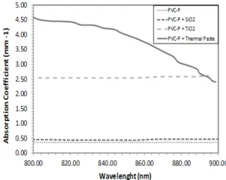
Figure 3 shows the absorption coefficients of the manufactured phantoms in the range from 800 nm to 900 nm. The PVC-P absorption coefficient is relatively close to the absorption coefficient of fat at these wavelengths (Kim et al., 2008; Siddiqi, 2009). Figure 3 shows that, in the range of 800 nm and 900 nm, the use of SiO2 nanoparticles has not
significantly affected the absorption coefficient of PVC-P. On the other hand, the absorption coefficient of the phantom containing TiO2 nanoparticles is
around six times larger than that of the pure PVC-P phantom. The absorption coefficient of the phantom containing thermal paste is also shown in this figure, only for wavelengths larger than 800 nm, due to large uncertainties at smaller wavelengths. The absorption coefficient of the thermal paste phantom is larger than that of the phantom with TiO2 nanoparticles, but
rapidly drops in the range considered for the measurements.
The thermal diffusivities, densities and specific heats of the phantoms were respectively measured with the LFA 447 NanoFlash, with a Sartorius YDK 01 densimeter mounted on a Marte AM 220 scale, and with a Differential Scanning Calorimeter NETZSCH - DSC 204 F1 Phoenix. The obtained results are presented in Tab. 2, while published values of human soft tissues are shown in Tab. 3 (Da Silva et al., 2011). From Tab. 2, it can be seen that the addition of nanoparticles or thermal paste has modified the thermal properties of the pure PVC-P. The density, thermal conductivity and thermal diffusivity of the phantom with thermal paste are larger than those of the phantom made of pure PVC-P and of the phantoms loaded with nanoparticles. The density, thermal conductivity and thermal diffusivity of the phantom with TiO2 nanoparticles are larger
than those of the phantom with SiO2 nanoparticles
and of the phantom without nanoparticles. Indeed, the addition of SiO2 nanoparticles did not modify the
thermal conductivity of the phantom made of pure PVC-P. The largest measured specific heat was for the phantom made of pure PVC-P. Densities of the phantoms with TiO2 nanoparticles and with thermal
paste are around those of fat and skin, while the thermal conductivities of all the produced phantoms are around those of fat and of skin.
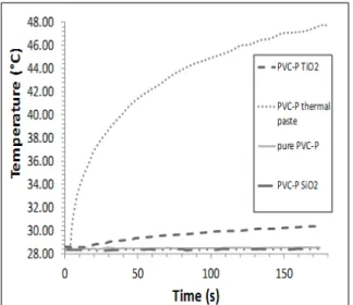
Transient temperature variations at the center of the non-heated surface of each manufactured phantoms are presented in Fig. 4. Such temperatures were measured with a thermocouple placed at the
interface of the sample and its support. As it can be seen in this figure, a temperature variation around 20
oC was achieved for the phantom containing the
thermal paste.
Figure 3. Absorption coefficient of the manufactured phantoms.
Table 2. Thermophysical properties of the prepared phantoms . Phantoms Density (Kg/m3) Thermal diffusivity (10-8) (m2/s) Thermal conductivity (W/m oC) Specific Heat (J/g ºC) PVC-P 995.1 ±0.1 8.70 ±0.32 ±0.004 0.150 ±0.0094 1.7900 PVC-P + SiO2 989.3 ±0.1 8.76 ±0.24 0.150 ±0.002 1.7200 ±0.0097 PVC-P + TiO2 1,080.8± 0.1 8.80 ±0.33 0.173 ±0.001 1.7700 ±0.0097 PVC-P + Thermal Paste 1,094.2±0.1 12.00 ±0.51 0.24 ±0.01 ±0.0049 1.7500
Table 3. Thermophysical properties of some human soft tissues.
For the phantom containing TiO2 nanoparticles,
a temperature variation of approximately 2.5 oC was
observed. On the other hand, no temperature increase was observed with the phantoms made of pure PVC-P and PVC-PVC-PVC-P loaded with SiO2 nanoparticles. This is
due to the fact that these phantoms are practically transparent to the NIR light used, as demonstrated by Fig. 3, and the slight temperature increase measured by the thermocouple is due to heat diffusion from the sample support to the phantom. As opposed to the SiO2 nanoparticles, the TiO2 nanoparticles have
Tissues Density (ρ) (Kg/m3) Thermal condutivity (k) (W/m ºC) Specific Heat (cp) (J/Kg ºC) Fat 1,200 0.203 2,678 Muscle 1,097 0.529 3,684 Skin 1,200 0.228 3,593 Bone 1,585 0.735 1,785
enhanced the absorption coefficient of PVC-P (see Fig. 3), so the NIR light was absorbed and heat was generated inside the phantom.
Figure 5 presents a snapshot of the temperature distribution taken by the infrared camera at the final time of the experiment with the phantom containing TiO2 nanoparticles. Figure 6 shows the temperature
profile along a cross section line at the top surface of the phantom containing TiO2 nanoparticles, for
different times. It can be noticed in this figure that the maximum temperature reached at each time is confined in a region slightly larger than the laser spot. However, in the outer region of the laser spot, heat is diffused and the temperature decreases to reach the initial values at the edge of the phantom.
Figure 4. Temperature variation at the non-heated surface of the phantom.
Figure 5. Snapshot of the temperature distribution in the phantom containing TiO2 at t =180 s.
27.50 28.00 28.50 29.00 29.50 30.00 30.50 0.00 5.00 10.00 15.00 20.00 25.00 30.00 35.00 40.00 Te m p e ra tu re ( °C) Position (mm) t=0 t=10 t=30 t=90 t=141 t=171
Figure 6. Temperature distribution along a straight line at the front face.
CONCLUSIONS
This paper presents some preliminary experimental results regarding the NIR light absorption by phantoms loaded with nanoparticles, aiming at the treatment of cancer by hyperthermia. Phantoms were prepared of pure PVC-P, and also of PVC-P with SiO2 nanoparticles and with TiO2
nanoparticles. In addition, a phantom with a thermal paste was prepared for comparison purposes.
It was observed that the pure PVC-P phantom has an absorption coefficient relatively close to that of human fat. The use of SiO2 nanoparticles has not
significantly affected the absorption coefficient of PVC-P. On the other hand, the absorption coefficient of the phantom containing TiO2 nanoparticles was
around six times larger than that of the pure PVC-P phantom. Densities of the phantoms with TiO2
nanoparticles and with thermal paste are around those of fat and skin, while the thermal conductivities of all the produced phantoms are around those of fat and of skin.
Laser irradiation of the pure PVC-P phantom and of the phantom containing SiO2 nanoparticles has
caused no significant variations of their temperatures, as a result of their small absorption coefficients to the NIR light used. As opposed to SiO2 nanoparticles,
the use of TiO2 nanoparticles has enhanced
significantly the absorption coefficient of pure PVC-P, causing the heating of the phantom by the incident laser light. Also, as expected, the largest temperature increase was observed with the phantom containing the thermal paste, because of its high absorption coefficient.
Although the results obtained in this work with the phantom containing TiO2 nanoparticles are
promising, further studies are required involving experimental parameters like the laser output power, the laser exposure time, the concentration and spatial
distribution of the nanoparticles and, very specially, the toxicity of nanoparticles to human beings.
ACKNOWLEDGEMENTS
The authors are thankful for the support provided by CNPq, CAPES and FAPERJ, Brazilian agencies for the fostering of science. Specific heats of the phantoms were measured by Mr. Maycon Magalhães, at LTTC/COPPE/UFRJ.
REFERENCES
Chekuri, P., Glazer, E. S., and Curley, S. A, 2010, Targeted Hyperthermia Using Metal Nanoparticles, Advanced Drug Delivery Reviews, Vol. 62, pp. 339-345.
de Korte, C. L., Cespedes, E. I., Van der Steen, A. F., Norder, B., and te Nijenhuis, K., 1997, Elastic and Acoustic Properties of Vessel Mimicking Material for Elasticity Imaging, Ultrason Imaging, Vol. 19, pp. 112-26.
Da Silva, S., França, A. S., and Pinatti, M., 2011, Simulação Unidimensional da Transferência de Calor na Articulação do Joelho Canino durante os Processos de Aquecimento e de Resfriamento Terapêuticos, Revista Brasileira de Engenharia Biomédica, Vol. 27, No. 3, pp. 163-174. (in
Portuguese)
DasGupta D., von Maltzahn G., Ghosh S., Bhatia S. N., Das S. K., and Chakraborty S., 2009, Probing Nano Antenna-Directed Photothermal Destruction of Tumors using Non-Invasive Laser Irradiation, Applied Physics Letters, Vol. 95, 233701. Dickerson, E. B., Dreaden, E. C., Huang, X., El-Sayed, I. H., Chu, H., Pushpanketh, S., McDonald, J. F., and El-Sayed, M. A., 2008, Gold Nanorod Assisted Near-Infrared Plasmonic Photothermal Therapy (PPTT) of Squamous Cell Carcinoma in Mice, Cancer Letters, Vol. 269, No. 1, pp. 57-66.
Feng, Y., Fuentes, D., Hawkins, A., Bass, J., Rylander, M. N., Elliott, A., Shetty, A., Stafford, R. J., and Oden, J. T., 2009, Nanoshell-Mediated Laser Surgery Simulation for Prostate Cancer Treatment, Engineering with Computers, Vol. 25, No. 1, pp. 3-13.
Hirsch, L. R., Stafford, R. J., Bankson, J. A., Sershen, S. R., Price, R. E., Hazle, J. D., Halas, N. J., and West, J. L., 2003, Nanoshell-Mediated Near-Infrared Thermal Therapy of Tumors Under MR Guidance, Proceedings of the National Academy of Sciences, Vol. 100, No. 23, pp. 13549-13554.
Huang, X., Jain, P. K., Sayed, I. H., and El-Sayed, M. A., 2008, Plasmonic Photothermal Therapy (PPTT) using Gold Nanoparticles, Lasers in Medical Science, Vol. 23, pp. 217-228.
Huff, T. B., Tong, L., Zhao, Y., Hansen, M. N., Cheng, J. X., and Wei, A., 2007, Hyperthermic Effects of Gold Nanorods on Tumor Cells, Nanomedicine, Vol. 2, No. 1, pp. 125-132.
Kim, C., Garcia-Uribe, A., Khotapolli, S-R., and Wang, L. V., 2008, Optical Phantoms for Ultrasound-Modulated Optical Tomography, In Proceedings of SPIE, Vol. 6870, pp. 68700M-1–68700M-8.
O’Neal, D. P., Hirsch, L. R., Lee, M. H., Barton, J., Halas, N. J., and West, J. L., 2004, Photothermal Tumor Ablation in Mice Using Near Infrared Absorbing Nanoshells, Cancer Letters, Vol. 209, pp. 171-176.
Siddiqi, A. K., 2009, Development of Tissue-equivalent Heat-sensitive gel for the Experimental Verification of Near Infrared (NIR) Laser Mediated Cancer Detection and Therapy, Doctoral Thesis, Georgia Institute of Technology, Atlanta, Georgia.
Spirou, G. M., Oraevsky, A. A., Vitkin, I. A., and Whelan, W. M., 2005, Optical and Acoustic Properties at 1064 nm of Polyvinyl Chloride-Plastisol for use as a Tissue Phantom in Biomedical Optoacoustics, Physics in Medicine and Biology, Vol. 50, pp. N141-N143.
Xu, T., Zhang, C.,Wang, X., Zhang, L., and Tian, J., 2003, Measurement and analysis of light distribution in Intralipid-10% at 650nm, Applied Optics, Vol. 42, pp. 5777-84.
Xu, X., Meade, A., and Bayazitoglu, Y., 2011, Numerical Investigation of Nanoparticle-assisted laser-induced Interstitial Thermotherapy toward Tumor and Cancer Treatments, Lasers in Medical Science, Vol. 26, No. 2, pp. 213-222.
Tanaka, T., 1981, Gels, Scientific American, Vol. 244, No. 1, pp. 124-138.