E
FFECTS OF HYPOTHERMIA ON BRAIN GLUCOSE
METABOLISM IN ACUTE LIVER FAILURE
:
A
1
H/
13
C-NUCLEAR MAGNETIC RESONANCE STUDY
Nicolas Chatauret
∗, Claudia Zwingmann
∗, †, 1, Christopher Rose
∗, Dieter Leibfritz
†,
Roger F. Butterworth
, ∗∗ Neuroscience Research Unit, CHUM Hôpital Saint-Luc, Montreal, Quebec, Canada † Department of Organic Chemistry, University of Bremen,
Bremen, Germany
A
BSTRACT
Mild hypothermia has a protective effect on brain edema and encephalopathy in both experimental and human acute liver failure. The goals of the present study were to examine the effects of mild hypothermia (35°C) on brain
metabolic pathways using combined 1H and 13C-Nuclear Magnetic Resonance (NMR) spectroscopy, a technique
which allows the study not only of metabolite concentrations but also their de novo synthesis via cell-specific pathways in the brain. :1H and 13C NMR spectroscopy using [1-13C] glucose was performed on extracts of frontal
cortex obtained from groups of rats with acute liver failure induced by hepatic devascularization whose body temperature was maintained either at 37°C (normothermic) or 35°C (hypothermic), and appropriate sham-operated controls. At coma stages of encephalopathy in the normothermic acute liver failure animals, glutamine
concentrations in frontal cortex increased 3.5-fold compared to sham-operated controls (P < 0.001). Comparable increases of brain glutamine were observed in hypothermic animals despite the absence of severe encephalopathy (coma). Brain glutamate and aspartate concentrations were respectively decreased to 60.9% ± 7.7% and 42.2% ± 5.9% (P < 0.01) in normothermic animals with acute liver failure compared to control and were restored to normal values by mild hypothermia. Concentrations of lactate and alanine in frontal cortex were increased to 169.2% ± 15.6% and 267.3% ± 34.0% (P < 0.01) respectively in normothermic rats compared to controls. Furthermore, de novo synthesis of lactate and alanine increased to 446.5% ± 48.7% and 707.9% ± 65.7% (P < 0.001), of control respectively,
resulting in increased fractional 13C-enrichments in these cytosolic metabolites. Again, these changes of lactate and
alanine concentrations were prevented by mild hypothermia. Mild hypothermia (35°C) prevents the encephalopathy and brain edema resulting from hepatic devascularization, selectively normalizes lactate and alanine synthesis from glucose, and prevents the impairment of oxidative metabolism associated with this model of ALF, but has no significant effect on brain glutamine. These findings suggest that a deficit in brain glucose metabolism rather than glutamine accumulation is the major cause of the cerebral complications of acute liver failure.
Abbreviations Ala, alanine; ALAT, alanine aminotransferase; ALF, acute liver failure; GABA, γ-aminobutyric acid; Gln,
glutamine; HAL, hepatic artery ligation; HE, hepatic encephalopathy; Lac, lactate; LD, lactate dehydrogenase; MSO, methionine sulfoximine; NAA, N-acetyl-aspartate; NMR, nuclear magnetic resonance; PC, pyruvate carboxylase; PCA, portacaval anastomosis; PDH, pyruvate ehydrogenase; TCA cycle, tricarboxylic acid cycle
I
NTRODUCTION
The principal cause of mortality in patients with acute liver failure (ALF) is brain herniation resulting from intracranial hypertension caused by a progressive increase of brain water content.1 The pathophysiologic mechanisms responsible for brain edema in ALF have not been fully elucidated; however, ammonia remains the prime candidate. Hyperammonemia is invariably observed in ALF, and a recent study revealed a significant correlation between arterial ammonia concentrations and the complication of brain herniation in patients with
ALF.2 Ammonia-related mechanisms that have been proposed to explain the encephalopathy and brain edema in ALF include direct electrophysiologic actions of the ammonium ion on excitatory/inhibitory neurotransmission,3 osmotic effects resulting from the brain accumulation of the ammonia detoxification product glutamine4 as well as effects of ammonia on cerebral energy metabolism,5 astrocytic glutamate transport,6 and glutamate (NMDA) receptors.7 and 8
Mild hypothermia prevents brain edema in both experimental9, 10 and 11 and human12 ALF. Not only do these findings herald the possibility of new approaches to the management of patients with ALF, they also provide an important research tool with which to investigate basic mechanisms responsible for brain edema and
encephalopathy in ALF. The goals of the present study, therefore, were to study the effects of mild hypothermia (35°C) on metabolic pathways in the brain derived from glucose metabolism, using combined 1H and 13C-nuclear
magnetic resonance (NMR) spectroscopy, a technique that facilitates the study not only of metabolite concentrations but also of their de novo synthesis via cell-specific pathways13 in the brain.
M
ATERIALS AND METHODS
Experimental animal model of ALF/surgical procedures
ALF was induced in adult male Sprague-Dawley rats (175–200 g) by portacaval anastomosis (PCA) according to the guidelines of Lee and Fischer14 followed by hepatic artery ligation (HAL). In brief, the rats were anesthetized with halothane, the inferior vena cava and portal vein were isolated, the inferior vena cava was partially clamped
(anastomosis clamp, Roboz Instruments Inc, Washington, DC), and an elliptical piece of vein 1.5 times the portal vein diameter was removed. The portal vein was ligated and cut and an end-to-side anastomosis performed under a dissecting microscope. Total surgery time was less than 15 minutes. In sham-operated control rats, the inferior vena cava and portal vein were occluded for 15 minutes. Following surgery, all animals were housed individually under constant conditions of temperature, humidity, and light cycles and were allowed free access to standard laboratory chow and water. Overall mortality for shunted rats was less than 5%. Forty-eight hours after PCA surgery, animals were anesthetized with halothane and subjected to hepatic artery ligation (HAL) or laparotomy (controls). Following HAL, body temperatures were monitored continuously and maintained at either 35°C (hypothermic) or 37°C
(normothermic) by means of thermal pads and heating lamps. Animals were assessed neurologically every 30 minutes during progression of ALF. Animals that could no longer right themselves after being placed on their backs were considered to be in precoma stage; animals in which both righting ability and corneal reflex could not be elicited were considered to be in coma. Hypothermic rats were killed at the same time post-HAL as the comatose normothermic rats. All animals received humane care according to the criteria outlined in the “Guide for the Care and Use of Laboratory Animals” prepared by the National Academy of Sciences and published by the National Institutes of Health (NIH publication 6–23, revised, 1985).
Control of arterial glucose concentrations and administration of [1-
13C]glucose
Arterial blood glucose levels were monitored every hour, and glucose was administered subcutaneously as needed to maintain normoglycemia. To ensure the same fractional 13C enrichment in glucose entering the brain, blood glucose
levels were measured immediately before administration of [1-13C]glucose (6.62 ± 0.49, 6.48 ± 0.52, and 6.80 ± 0.61
mmol/L in sham-operated controls, normothermic, and hypothermic ALF groups, respectively). After administration of [1-13C]glucose, the fractional enrichments in blood glucose ([1-13C]/[12C]glucose), determined from 1H-NMR
spectra, were not significantly different between the experimental groups (24.55 ± 2.05, 25.09 ± 1.81, and 22.92 ± 1.97 in sham-operated controls, normothermic, and hypothermic ALF groups, respectively).
Preparation of brain extracts
Sham-operated control rats and hepatic devascularized rats maintained at 37°C at coma stages (normothermic ALF rats) or at 35°C (hypothermic ALF rats) at comparable postsurgery time points were administered [1-13C]glucose
glucose in unanesthetized rats was chosen to facilitate the study of metabolic changes in relation to neurologic status and to avoid hyperglycemic conditions. The blood was taken from the neck wound and immediately mixed with 20% perchloric acid. Forebrains were immediately frozen in isopentane (at −80°C), dissected over dry ice for the frontal cortex, and frozen in liquid nitrogen. The samples were powdered over liquid nitrogen. Blood and tissue samples were extracted with 12% perchloric acid as previously described.13
Brain water measurement
Water content of the brain (percentage) was measured gravimetrically using a density gradient of bromobenzene-kerosene precalibrated with K2SO4 as previously described.15 Eight measurements were made per animal, and
values were arithmetically averaged.
Ammonia measurement
Plasma and cerebrospinal fluid ammonia concentrations were estimated using a commercial ammonia test kit, which uses an ion-exchange method, followed by colorimetric measurement of ammonia nitrogen with the Bertholet phenate-hypochlorate reaction (n = 8 in each experimental group).16
NMR spectroscopy
The lyophilized extracts of brain tissue and blood plasma were dissolved in 0.6 mL D2O and centrifuged. The pH was
adjusted to 7.0 with DCl and NaOD. 1H- and 13C-NMR spectra were recorded on Bruker DRX 600 or AVANCE
NB/WB360 spectrometers, operating at frequencies of 600 MHz or 360 MHz for 1H measurements and 150.9 MHz or
90.5 MHz for 13C measurements. 1H-NMR spectra were recorded with a 5-mm H,C,N inverse triple resonance probe,
400 accumulations, repetition time 15 seconds, spectral width 7183 Hz (DRX 600) or 3623 Hz (AM/AMX 360). Chemical shifts were referenced to lactate at 1.33 ppm. 13C-NMR spectra were recorded with a 5-mm 1H/13C dual
probe, 10,000 accumulations, repetition time 2.5 seconds, composite pulse decoupling with WALTZ-16, and spectral width 47,619 Hz (DRX 600) or 20,833 Hz (AM/AMX 360). Chemical shifts were referenced to the C-3 signal of lactate at 21.3 ppm.
Measurement of metabolite concentrations
Lactate concentrations were determined enzymatically (adapted for measurement in 96-well microtiter ELISA plates) using a commercially available kit (Roche Molecular Biochemicals, Mannheim, Germany). The concentrations
(μmol/g tissue) of other unlabeled metabolites and their pool sizes ([13C] + [12C]) were determined from fully relaxed 1H-NMR spectra of blood and brain extracts, obtained after injection of [1-13C]glucose or equivalent amounts of
unlabeled glucose, using the known lactate concentrations as an internal standard.
Calculation of fractional
13C enrichments
The fractional 13C enrichment in lactate was calculated as the ratio of [3-13C]lactate to unlabeled lactate by
integration of the peak areas of the methyl groups in 1H-NMR spectra (Figure 1):
(1)
The values were corrected for 1.1% natural abundance 13C. 13C enrichments in [1-13C]glucose were calculated
accordingly using the H1α glucose resonances in 1H-NMR spectra. The fractional 13C enrichment of each carbon of
other metabolites was derived from peak area ratio of the 13C-labeled carbon/natural abundance carbon and using
the known 13C enrichment of lactate as internal standard as described in detail previously.13 and 17 The absolute
amount of 13C in a specified carbon position (absolute synthesis from [1-13C]glucose; μmol/g wet weight) is the
Figure 1. Labeling of metabolites from [1-13C]glucose: label distribution in glycolytic and TCA cycle
intermediates during metabolism of [1-13C]glucose. A single turn of the TCA cycle from pyruvate via
pyruvate carboxylase (PC) or pyruvate dehydrogenase (PDH) to 2-oxoglutarate and subsequently glutamate and glutamine is considered. A description of the pathways leading to the different isotopomers is provided in the text. LDH, lactate dehydrogenase; ALAT, alanine aminotransferase; GAD, glutamate decarboxylase; GDH, glutamate dehydrogenase; GS, glutamine synthetase.
Theoretical labeling of metabolites from [1-
13C]glucose
The metabolic pathways whereby the 13C label from [1-13C]glucose is transformed into various metabolites are
presented schematically in Figure 1. 17 Briefly, via the glycolytic pathway, [1-13C]glucose is transformed to [3-13C]pyruvate and subsequently to [3-13C]alanine and [3-13C]lactate. [3-13C]Pyruvate may then enter the TCA cycle via the anaplerotic pathway (pyruvate carboxylase [PC] or malic enzyme [ME]) or via the oxidative pathway
(pyruvate dehydrogenase; PDH). Via PC, glutamate and glutamine are labeled at C-2. In the oxidative pathway, pyruvate enters the cycle as [2-13C]acetyl-CoA, and glutamate and glutamine are monolabeled at C-4. The ratio of C4/C2 in glutamate and glutamine was used to estimate the relative contribution of the PDH vs. the PC pathway. The order of labeled carbons is reversed in γ-aminobutyric acid (GABA) (i.e., the labels in [2(4)-13C]glutamate end up in [4(2)-13C]GABA) (not shown in Figure 1). Because we did not observe double-labeled isotopomers, only 1 TCA cycle turn is shown in Figure 1. Isotopomers arising from scrambling of the label within the TCA cycle are omitted for clarity.
Statistical analysis
The NMR studies were carried out with sham-operated controls (n = 6) and 12 hepatic devascularized rats (n = 6 normothermic ALF rats and n = 6 hypothermic ALF rats). Data are expressed as mean ± SD. Data between individual groups were analyzed using ANOVA and post hoc Tukey’s test. Differences were considered significant when P < 0.05
(†significantly different from sham-operated controls; ‡significantly different between hypothermic and
normothermic ALF groups; ∗∗∗P < 0.001; ∗∗P < 0.01, ∗P < 0.05).
R
ESULTS
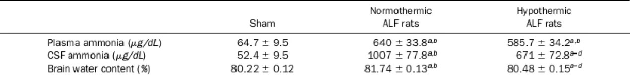
Following hepatic devascularization, normothermic ALF rats (n = 8) developed progressive encephalopathy consisting of loss of activity, loss of righting ability (precoma stage), and loss of corneal reflex (coma stage at 13.4 ± 1.1 hours). At times when the normothermic ALF rats were comatose, none of the hypothermic ALF (n = 8) rats had developed significant neurologic symptoms. Hypothermic ALF rats eventually became comatose at 18.3 ± 1.1 hours post-HAL, confirming a previous report.9 Plasma ammonia levels were not significantly different between the 2 groups at any time point studied (Table 1), whereas CSF ammonia concentrations were significantly less (by 34%, P < 0.01, n = 8) in the hypothermic than in the normothermic ALF group at coma stages of encephalopathy (672 ± 73 μg/dL vs. 1007 ± 78 μg/dL) (Table 1). Normothermic ALF rats manifested increased brain water content, whereas mild hypothermia led to a significant attenuation of this increase (sham, 80.22% ± 0.12%; normothermic ALF rats, 81.74% ± 0.13%; hypothermic ALF rats, 80.48% ± 0.15%; P < 0.01 compared with the normothermic ALF group, n = 8 in each experimental group) (Table 1).
Table 1. Plasma and CSF Ammonia and Brain Water Content in ALF Rats
Brain amino acid concentrations
The concentrations of amino acids related to the TCA cycle and neurotransmitter metabolism, i.e., glutamine,
glutamate, GABA, aspartate as well as the neuronal marker molecule N-acetyl-aspartate (NAA) in frontal cortex, were calculated from 1H-NMR spectra Figure 2 and Figure 3. At coma stages (10–12 hours after HAL), glutamine
concentrations were significantly increased both in the normothermic and the hypothermic rats compared with controls (to 374% ± 31.0%, P < 0.001 and 387% ± 49.0%, P < 0.001, respectively; n = 6). Glutamate, aspartate, and NAA concentrations in frontal cortex were decreased in normothermic rats to 60.9% ± 7.7% (P < 0.01), 42.2% ± 5.9% (P < 0.001), and 70.7% ± 7.0% (P < 0.01), respectively, but normalized to control values under hypothermic
conditions. GABA levels were unchanged in both normothermic and hypothermic ALF rats.
Synthesis of lactate and alanine
In normothermic rats, ALF resulted in an increase of lactate and alanine concentrations ([12C] + [13C]) to 169.2% ±
15.6% and 267.3% ± 34.0% (P < 0.001), respectively, which were both significantly attenuated by mild hypothermia (increase to 131.1% ± 16.6% and 154.2% ± 18.6%, respectively) (6 animals were used in each group) Figure 4 and Figure 5. Furthermore, the resonance attributed to 13C-labeled lactate (synthesized from [1-13C]glucose) can be distinguished from the unlabeled isotopomer (synthesized from substrates other than [1-13C]glucose) (Figure 4). During coma stages of encephalopathy, the concentration of 13C-labeled lactate increased to a much higher
concentration (to 446.5% ± 48.7%, compared with sham-operated controls, P < 0.001) compared to its total, resulting in 3-fold increased fractional 13C enrichments in lactate. In hypothermic ALF rats, de novo synthesized 13
C-labeled lactate was not significantly different from sham-operated control values. Thus, hypothermia led to a selective reduction of de novo synthesis of 13C-labeled lactate (P < 0.001). No significant changes in [3-13C]lactate
Figure 2. 1H-NMR spectra of brain extracts
in ALF: effect of hypothermia. Figure shows typical segments of 1H-NMR spectra of
extracts obtained from frontal cortex of a sham-operated control rat compared with rats with ALF maintained at either 37°C (ALF-coma) or 35°C (ALF-hypothermia). Peak assignments: GABA, γ-aminobutyric acid; Glu, glutamate; Gln, glutamine; NAA, N-acetyl-aspartate; Asp, aspartate.
Figure 3. Concentrations of brain amino acids in ALF: effect of hypothermia. Figure shows concentrations of amino acids, calculated from 1H-NMR spectra of extracts obtained
from frontal cortex of sham-operated control rats (n = 6) and of rats with ALF maintained at 37°C at coma stage (n = 6) and rats with ALF maintained at 35°C (n = 6). Values represent means ± SD for n = 6 animals per group (†significantly different from
sham-operated controls; ‡significantly different
between hypothermic and normothermic groups; ∗∗∗P < 0.001; ∗∗P < 0.01; ∗P < 0.05; ns, not significant by ANOVA).
Figure 4. Brain lactate synthesis in ALF: effect of hypothermia. Left: segments of 1H-NMR spectra of extracts
from frontal cortex of a sham-operated control rat compared with rats with ALF maintained at 37°C (ALF coma) or 35°C (ALF hypothermia). The percentage 13C enrichment in lactate (indicating de novo synthesis)
amounts of 13C-labeled lactate, and fractional 13C enrichments in lactate in rats with ALF compared with
sham-operated control rats. For statistical analysis, see legend to Figure 3.
Figure 5. 13C-NMR spectra of brain extracts from rats with ALF: effect of hypothermia. Figures shows 13
C-NMR spectra of extracts from frontal cortex of a sham-operated control rat compared with rats with ALF maintained at 37°C (ALF coma) or 35°C (ALF-hypothermia) 15 minutes after injection of [1-13C]glucose.
Peak assignments: Ala, alanine; GABA, γ-aminobutyric acid; Glu, glutamate; Gln, glutamine; Lac, lactate; NAA, N-acetyl-aspartate (signals assigned NAA are due to naturally abundant 13C-labelled NAA).
13C-labeled alanine was quantified from 13C-NMR spectra (Figure 5). As observed for lactate, ALF resulted in a
marked increase of de novo synthesis of [3-13C]alanine to 707.9 ± 65.7 (P < 0.001; n = 6) at coma stages, which was
significantly reduced by hypothermia (increased to 171.1% ± 20.7%; n = 6). Interestingly, the fractional 13C
enrichment in [3-13C]alanine was increased under normothermic conditions but remained within normal limits in
hypothermic animals (n = 6). The preferential increase of 13C labeling and the selective protective effect of
hypothermia on elevated 13C label incorporation from [1-13C]glucose into alanine closely resembles the changes
observed for 13C-labeled lactate.
Labeling of tricarboxylic acid cycle-related metabolites from [1-
13C]glucose
Synthesis of glutamine, glutamate, and GABA 13C resonances were quantified from 13C-NMR spectra (Figure 6). Six
animals were used in each group (sham-operated controls, normothermic ALF rats, and hypothermic ALF rats) for 13C-NMR analysis. The total amounts of 13C label (μmol/g tissue) and the fractional 13C enrichments (%) in these amino acids are presented in Figure 7. The increase in de novo synthesis of glutamine from [1-13C]glucose is much higher than the increase of its pool size at coma stages. 13C isotopomer analysis of the individual carbon positions in glutamine revealed a greater increase of C-2-labeled glutamine (to 718.5% ± 16.7% of control, P < 0.001) compared with C-4 (to 500.8% ± 80.3% of control, P < 0.001). Unlike the selective prevention of abnormal lactate synthesis
from [1-13C]glucose by mild hypothermia, the increase of 13C-labeled glutamine did not differ significantly between
normothermic and hypothermic ALF groups. In normothermic ALF rats, a relatively higher decrease of
[4-13C]glutamate (through the PDH pathway) compared with its pool size resulted in a decreased relative 13C
enrichment in C-4 of glutamate to 62.3% ± 10.7% of control (P < 0.001). Although hypothermia failed to prevent the increased glutamine synthesis, it led to a complete recovery selectively of PDH-mediated glutamate synthesis. The prevention of impaired flux of glucose through PDH by hypothermia is reflected also by the prevention of the decrease in the ratio of C-4/C-2 in glutamate (but without significant changes of C-4/C-2 in glutamine in
hypothermic compared with normothermic rats), which was calculated as a measure of the relative glucose oxidative metabolism (vs. the anaplerotic pathway) (Figure 8). No changes were observed for de novo synthesized GABA in either normothermic or hypothermic groups of rats with ALF (data not shown).
Figure 6. Brain alanine synthesis in ALF: effect of hypothermia. Figure shows changes in the concentration, amount of 13C-labeled alanine, and
fractional 13C enrichments in alanine in frontal
cortex of rats with ALF maintained at 37°C (ALF-coma) or 35°C (ALF-hypothermia) compared with sham-operated controls. For statistical analysis, see legend to Figure 3.
Figure 7. Synthesis of brain glutamate and glutamine in ALF: effect of hypothermia. Figure shows changes in the absolute amounts of 13C (μmol/g tissue) (left) and in the percentage of 13C enrichments (right) in
individual carbon positions (via the PC- and PDH pathways) of glutamate and glutamine labeled 15 minutes after injection of [1-13C]glucose, calculated from 13C-NMR spectra of extracts from frontal cortex of rats with
ALF maintained at 37°C (ALF coma) or 35°C (ALF hypothermia) compared with sham-operated controls. For statistical analysis, see legend to Figure 3.
Figure 8. Relative contribution of carbon flux through PDH and PC. Ratios of C-4/C-2 of glutamate and glutamine from 13C-NMR spectra of extracts from frontal cortex of sham-operated control rats compared
with rats with ALF maintained at 37°C (ALF coma) or 35°C (ALF hypothermia). For statistical analysis, see legend to Figure 3.
D
ISCUSSION
Results of the present study reveal that mild hypothermia delays the onset of encephalopathy and brain edema and selectively prevents alterations in energy and glucose metabolism in frontal cortex of rats with experimental ALF. In particular, hypothermia prevents the increase in brain lactate. These findings add to an accumulating body of evidence to suggest that increased brain lactate production is causally related to the encephalopathy and brain edema in ALF. Other evidence in support of this possibility includes the report of a significant correlation between brain lactate concentrations and both encephalopathy grade18 and EEG changes19 in experimental ALF. Results of in vitro studies support a key role of lactate in the pathogenesis of astrocyte swelling. For example, it was previously shown that exposure of primary cultures of rat astrocytes (the cell type shown to selectively exhibit swelling in the brain in ALF) to pathophysiologically relevant concentrations of lactate leads to significant swelling.20 A recent report advocated the use of a noninvasive technique to measure brain lactate in vivo as an evaluation of advanced encephalopathy in ALF.21 Results of the present study now show that the selective attenuation of ALF-related increases of lactate and alanine in frontal cortex, concomitant with the prevention of encephalopathy and brain edema, resides in the ability of hypothermia to prevent the de novo synthesis of lactate from glucose.
It should be borne in mind, however, that, although there is convincing evidence, both from the present and previous studies, for decreased glucose oxidation and lactate/alanine accumulation in the brain in ALF, there is no evidence to suggest that either encephalopathy or brain edema are consequences of diminished ATP synthesis.22, 23 and 24 The present 13C-NMR spectroscopic study is novel by virtue of its ability to differentiate between metabolite
concentrations and metabolic fluxes. In particular, the present findings suggest that one important mechanism of action of hypothermia in ALF is the facilitation of pyruvate oxidation as a consequence of decreased blood-brain transfer of ammonia, which has been shown to inhibit the tricarboxylic acid cycle (TCA) enzyme α-ketoglutarate dehydrogenase.25 Increased glycolytic activity has then compensated to maintain ATP production with consequent increases of brain lactate and alanine.
Decreased brain tissue concentrations of glutamate and aspartate have consistently been reported in both experimental13, 18, 26, 27 and 28 and human29 ALF. Results of the present study show that mild hypothermia prevents the reduction of brain glutamate and aspartate associated with experimental ALF. Furthermore, analysis of isotopomer patterns from the 13C-NMR spectra reveals that hypothermia leads to normalization of the de novo
synthesis of glutamate via pyruvate dehydrogenase, a key enzyme for oxidative and energy metabolism in the brain, which is expressed in both astrocytes and neurons. Glutamate and aspartate are not only potent excitatory
neurotransmitters in that both are involved in brain energy metabolism, serving as substrates/intermediates in the malate-aspartate shuttle (responsible for the transfer of reducing equivalents from the cytosol to mitochondria). Reduced availability of glutamate or aspartate in either cell type, therefore, could result in mitochondrial energy compromise. In the case of glutamate, studies using in vivo cerebral microdialysis have shown that increased release of glutamate in experimental ALF8 is sensitive to hypothermia.10 Furthermore, the aspartate derivative
N-acetylaspartate (NAA), present in high concentrations in the brain, is decreased in frontal cortex at coma stages of encephalopathy under normothermic conditions but is prevented by hypothermia. NAA is a neuronal marker molecule and is involved in brain energy metabolism.30 Decreased brain concentrations of NAA, therefore, are consistent with neuronal mitochondrial dysfunction in ALF.
In contrast to glutamate and aspartate, ALF because of hepatic devascularization does not result in alterations in either absolute concentrations or synthesis of the inhibitory amino acid GABA. These findings add to a convincing body of evidence that the neural inhibition in both human and experimental ALF13, 18, 26 and 29 is not the consequence of altered GABA synthesis in the brain.
It was previously suggested that brain edema in hyperammonemic syndromes such as ALF is the consequence of increased intracellular accumulation of glutamine by the astrocyte.4 In favor of this hypothesis were reports of increased intracranial pressure in primates infused with ammonia26 and of increased brain glutamine
concentrations in both human29 and experimental27 and 28 ALF. Moreover, brain edema because of ammonia infusions was partially prevented by pretreatment with methionine sulfoximine (MSO), a potent inhibitor of glutamine synthetase.4 and 31 However, MSO treatment may exert multiple effects. For example, MSO also induces massive efflux of glutamine from astrocytes in cultures.32 In addition, brain glutamine accumulation also occurs in chronic liver failure, a condition not normally associated with brain edema or its clinical consequences.4 and 33 Therefore, despite several experimental data supporting glutamine as accumulating osmolyte in HE, whether or not glutamine accumulation is a major cause for the development of brain edema in ALF is uncertain.33 Results of the present study confirm an increase in brain glutamine in ALF rats. However, mild hypothermia sufficient to abolish brain edema in these animals did not prevent either the increase in brain glutamine concentration or its de novo synthesis from glucose in astrocytes (i.e., de novo synthesis via the astrocytic enzymes pyruvate carboxylase and glutamine synthetase). A limited capacity of glutamine synthetase was also concluded by Deutz et al.,19 who demonstrated a faster increase of brain ammonia compared with glutamine and by Bosman et al.,34 who observed, after an initial increase of extracellular glutamine during mild HE, a subsequent decrease during severe HE. Direct demonstration of a lack of correlation between the grade of hepatic encephalopathy and glutamine synthetase activity was initially reported by Kanamori et al.35 using in vivo 1H MRS in a hyperammonemic rat model. Moreover,
a previous 13C-NMR study could find no significant correlation between brain de novo glutamine synthesis from
glucose and grade of encephalopathy or brain edema at coma stages in the hepatic devascularized animal model of ALF.13
In light of the apparently limited capacity of glutamine synthetase, which is thought to be the main detoxification process of ammonia, the question remains of how the brain cells regulate the disposal of excess ammonia in late stages of ALF. In this regard, it has been shown that ammonia can be incorporated into alanine after transamination of glutamate by alanine aminotransferase (ALAT) in both astrocytes and neurons. Consistent with this possibility, alanine has been found to be elevated in the hyperammonemic brain.18, 27 and 36 Results of the present study show for the first time that de novo alanine synthesis from glucose is increased as a function of the degree of
encephalopathy/brain edema in ALF, which is partly prevented by mild hypothermia. Like lactate formation,
synthesis of alanine via ALAT may supplement glycolytic activity, enabling cytosolic ATP production by regeneration of NAD+ via subsequent reamination of α-ketoglutarate by glutamate dehydrogenase (GDH). It is noteworthy that, in
the honeybee retina, which lacks lactate dehydrogenase activity, alanine synthesis in glia fixes ammonia at a rate exceeding glutamine formation,37 suggesting that ALAT and GDH may operate in a complementary manner to
oxidize NADH and fix ammonia. Like glutamine, alanine accumulation may increase the cellular content of osmotically active substances, leading to water uptake, cell swelling, and brain edema in ALF. Furthermore, it has been shown that both alanine and glutamine induce hepatocyte swelling,38 and, in acute HE, a close correlation between CSF alanine with both the degree of neurologic dysfunction18 and edema27 was demonstrated, suggesting that increased alanine synthesis may be related to the pathogenesis of brain edema in ALF.
Together, these findings do not support a major role for the accumulation of intracellular glutamine in the astrocyte as a cause of brain edema in ALF. The imperfect correlation of glutamine with brain edema and severity of
encephalopathy points to additional mechanisms contributing to brain edema at coma stages. Elevated
concentration and synthesis of alanine, which is selectively prevented by mild hypothermia, may play a role in the development of brain edema in ALF. In addition, the correlation between increased glucose flux through the glycolytic pathway (alanine and lactate de novo synthesis) and the severity of encephalopathy and brain edema suggest that a deficit in glucose oxidation and energy metabolism is a major contributor to the pathogenesis of these neurologic complications of ALF and provide a rational argument for the further use of hypothermia in its
management.
R
EFERENCES
1 A.T. Blei Cerebral edema and intracranial hypertension in acute liver failuredistinct aspects of the same problem Hepatology, 13 (1991), pp. 376–379
2 J.O. Clemmensen, F.S. Larsen, J. Kondrup, B.A. Hansen, P. Ott Cerebral herniation in patients with acute liver failure is correlated with arterial ammonia concentration Hepatology, 29 (1999), pp. 648–653
3 J.C. Szerb, R.F. Butterworth Effect of ammonium ions on synaptic transmission in the mammalian central nervous system Prog Neurobiol, 39 (1992), pp. 135–153
4 J. Cordoba, A.T. Blei Brain edema and hepatic encephalopathy Semin Liver Dis, 3 (1996), pp. 271–280
5 K.V. Rao, M.D. Norenberg Cerebral energy metabolism in hepatic encephalopathy and hyperammonemia Metab Brain Dis, 16 (2001), pp. 67–78
6 K. Knecht, A. Michalak, C. Rose, J.D. Rothstein, R.F. Butterworth Decreased glutamate transporter (GLT-1) expression in frontal cortex of rats with acute liver failure Neurosci Lett, 25 (1997), pp. 1032–1034
7 E. Kosenko, Y. Kaminsky, E. Grau, M.D. Minana, G. Marcaida, S. Grisolia, V. Felipo Brain ATP depletion induced by acute ammonia intoxication in rats is mediated by activation of the NMDA receptor and Na+,K(+)-ATPase J Neurochem, 63 (1994), pp. 2172–2178
8 A. Michalak, C. Rose, J. Butterworth, R.F. Butterworth Neuroactive amino acids and glutamate (NMDA) receptors in frontal cortex of rats with experimental acute liver failure Hepatology, 24 (1996), pp. 908–913
9 P. Traber, M. DalCanto, D. Ganger, A.T. Blei Effect of body temperature on brain edema and encephalopathy in the rat after hepatic devascularization Gastroenterology, 96 (1989), pp. 885–891
10 C. Rose, A. Michalak, M. Pannunzio, N. Chatauret, A. Rambaldi, R.F. Butterworth Mild hypothermia delays the onset of coma and prevents brain edema and extracellular brain glutamate accumulation in rats with acute liver
failure Hepatology, 31 (2000), pp. 872–877
11 N. Chatauret, C. Rose, G. Therrien, M. Pannunzio, R.F. Butterworth Mild hypothermia prevents cerebral edema and CSF lactate accumulation in acute liver failure Metab Brain Dis, 16 (2001), pp. 95–102
12 R. Jalan, S.W.M. Dalmink, A. Olde Lee, P.C. Hayes Moderate hypothermia for uncontrolled intracranial hypertension in acute liver failure Lancet, 354 (1999), pp. 1164–1168
13 C. Zwingmann, N. Chatauret, D. Leibfritz, R.F. Butterworth Selective increase of brain lactate synthesis in experimental acute liver failureresults of a [1H-13C]nuclear magnetic resonance study Hepatology, 37 (2003), pp.
420–428
14 S.H. Lee, B. Fischer Portacaval shunt in the rat Surgery, 50 (1961), pp. 668–672
15 A. Marmarou, W. Poll, H. Bhagavan A simple gravimetric technique for measurement of cerebral edema J. Neurosurg, 49 (1978), pp. 530–537
16 S.G. Dienst An ion exchange method for plasma ammonia concentration J Lab Clin Med, 58 (1961), pp. 149–155 17 C. Zwingmann, C. Richter-Landsberg, A. Brand, D. Leibfritz NMR spectroscopic study on the metabolic fate of [3-(13)C]alanine in astrocytes, neurons, and coculturesimplications for glia-neuron interactions in neurotransmitter metabolism Glia, 32 (2000), pp. 286–303
18 A.M. Mans, M.R. DeJoseph, R.A. Hawkins Metabolic abnormalities and grade of encephalopathy in acute hepatic failure J Neurochem, 63 (1994), pp. 1829–1838
19 N.E.P. Deutz, A.A. De Graaf, J.G. De Haan, W.M.M.J. Bovée, R.A.F.M. Chamuleau In vivo brain 1H-NMR spectroscopy
(1H-NMRS) during acute hepatic encephalopathy (HE) P.B. Soeters, J.H.P. Wilson, A.J. Meijer, E. Holm (Eds.), Advances
in ammonia metabolism and hepatic encephalopathy, chapter 57, Excerpta Medica, Amsterdam (1988), pp. 439–446 20 F. Staub, A. Baethmann, J. Peters, H. Weigt, O. Kempski Effects of lactacidosis on glial cell volume and viability J Cereb Blood Flow Metab, 10 (1990), pp. 866–876
21 S.L. Nyberg, F.B. Cerra, R. Gruetter Brain lactate by magnetic resonance spectroscopy during fulminant hepatic failure in the dog Liver Transpl Surg, 4 (1998), pp. 158–165
22 D.W. McCandless, S. Schenker Effect of acute ammonia intoxication on energy stores in the cerebral reticular activating system Exp Brain Res, 44 (1981), pp. 325–330
23 M. Matsuoka, H. Igisu, K. Kohriyama, N. Inoue Effects of ammonia on brain energy metabolites-dose-dependent alterations J Neurochem, 55 (1990), pp. 354–355
24 T.E. Bates, S.R. Williams, A.L. Busza, D.G. Gadian, E. Proctor A 31P nuclear magnetic resonance study in vivo of
metabolic abnormalities in rats with acute liver failure NMR Biomed, 1 (1988), pp. 67–73
25 J.C.K. Lai, A.J. Cooper Neurotoxicity of ammonia and fatty acidsdifferential inhibition of mitochondrial dehydrogenases by ammonia and fatty acyl coenzyme A derivatives Neurochem Res, 16 (1991), pp. 795–803 26 T.M. Voorhies, M.E. Ehrlich, T.E. Duffy, C.K. Petito, F. Plum Acute hyperammonemia in the young
primatephysiologic and neuropathologic correlates Pediatr Res, 17 (1983), pp. 970–975
27 M. Swain, R.F. Butterworth, A.T. Blei Ammonia and related amino acids in the pathogenesis of brain edema in acute ischemic liver failure in rats Hepatology, 15 (1992), pp. 449–453
28 D.K. Bosman, N.E. Deutz, A.A. De Graaf, R.W. vd Hulst, H.M. Van Eijk, W.M. Bovee, M.A. Maas, G.G. Jorning, R.A. Chamuleau Changes in brain metabolism during hyperammonemia and acute liver failureresults of a comparative
29 C.O. Record, B. Buxton, R.A. Chase, G. Curzon, I.M. Murray-Lyon, R. Williams Plasma and brain amino acids in fulminant hepatic failure and their relationship to hepatic encephalopathy Eur J Clin Invest, 6 (1976), pp. 387–394 30 J.B. Clark N-acetyl aspartatea marker for neuronal loss or mitochondrial dysfunction Dev Neurosci, 20 (1998), pp. 271–276
31 H. Takahashi, R.C. Koehler, S.W. Brusilow, R.J. Traysman Inhibition of brain glutamine accumulation prevents cerebral edema in hyperammonemic rats Am J Physiol, 261 (1991), pp. H825–H829
32 J. Albrecht, M.D. Norenberg L-methionine-DL-sulfoximine induces massive efflux of glutamine from cortical astrocytes in primary culture Eur J Pharmacol, 182 (1990), pp. 587–589
33 J. Albrecht, M. Dolinska Glutamine as a pathogenic factor in hepatic encephalopathy J Neurosci Res, 65 (2001), pp. 1–5
34 D.K. Bosman, N.E. Deutz, M.A. Maas, H.M. van Eijk, J.J. Smit, J.G. de Haan, R.A. Chamuleau Amino acid release from cerebral cortex in experimental acute liver failure, studied by in vivo cerebral cortex microdialysis J Neurochem, 59 (1992), pp. 591–599
35 K. Kanamori, B.D. Ross, J.C. Chung, E.L. Kuo Severity of hyperammonemic encephalopathy correlates with brain ammonia level and saturation of glutamine synthetase in vivo J Neurochem, 67 (1996), pp. 1584–1594
36 W. Hilgier, M. Zielinska, H.D. Borkowska, R. Gadamski, M. Walski, S.S. Oja, P. Saransaari, J. Albrecht Changes in the extracellular profiles of neuroactive amino acids in the rat striatum at the asymptomatic stage of hepatic failure J Neurosci Res, 56 (1999), pp. 76–84
37 M. Tsacopoulos, C.L. Poitry-Yamate, S. Poitry, P. Perrottet, A.L. Veuthey The nutritive function of glia is regulated by signals released by neurons Glia, 1 (1997), pp. 84–91
38 D. Haussinger Regulation and functional significance of liver cell volume Prog Liver Dis, 14 (1996), pp. 29–53
![Figure 1. Labeling of metabolites from [1- 13 C]glucose: label distribution in glycolytic and TCA cycle intermediates during metabolism of [1- 13 C]glucose](https://thumb-eu.123doks.com/thumbv2/123doknet/7712985.247594/4.918.232.779.99.596/figure-labeling-metabolites-glucose-distribution-glycolytic-intermediates-metabolism.webp)