Université de Montréal
Electrospinning and characterization of supramolecular
poly(4-vinyl pyridine)-small molecule complexes
par Xiaoxiao Wang
Département de chimie Faculté des arts et des sciences
Thèse présentée à la Faculté des études supérieures et postdoctorales en vue de l’obtention du grade de Philosophiae Doctor (Ph. D.)
en chimie
Décembre 2014
Résumé
La chimie supramoléculaire est basée sur l'assemblage non covalent de blocs simples, des petites molécules aux polymères, pour synthétiser des matériaux fonctionnels ou complexes. La poly(4-vinylpyridine) (P4VP) est l'une des composantes supramoléculaires les plus utilisées en raison de sa chaîne latérale composée d’une pyridine pouvant interagir avec de nombreuses espèces, telles que les petites molécules monofonctionnelles et bifonctionnelles, grâce à divers types d'interactions. Dans cette thèse, des assemblages supramoléculaires de P4VP interagissant par liaisons hydrogène avec de petites molécules sont étudiés, en ayant comme objectifs de faciliter l'électrofilage de polymères et de mieux comprendre et d'optimiser la photoréponse des matériaux contenant des dérivés d'azobenzène.
Une nouvelle approche est proposée afin d'élargir l'applicabilité de l'électrofilage, une technique courante pour produire des nanofibres. À cet effet, un complexe entre la P4VP et un agent de réticulation bifonctionnel capable de former deux liaisons hydrogène, le 4,4'-biphénol (BiOH), a été préparé pour faciliter le processus d’électrofilage des solutions de P4VP. Pour mieux comprendre ce complexe, une nouvelle méthode de spectroscopie infrarouge (IR) a d'abord été développée pour quantifier l'étendue de la complexation. Elle permet de déterminer un paramètre clé, le rapport du coefficient d'absorption d'une paire de bandes attribuées aux groupements pyridines libres et liées par liaisons hydrogène, en utilisant la 4-éthylpyridine comme composé modèle à l’état liquide. Cette méthode a été appliquée à de nombreux complexes de P4VP impliquant des liaisons hydrogène et devrait être généralement applicable à d'autres complexes polymères.
La microscopie électronique à balayage (SEM) a révélé l'effet significatif du BiOH sur la facilité du processus d’électrofilage de P4VP de masses molaires élevées et faibles. La concentration minimale pour former des fibres présentant des perles diminue dans le N, N'-diméthylformamide (DMF) et diminue encore plus lorsque le nitrométhane, un mauvais solvant pour la P4VP et un non-solvant pour le BiOH, est ajouté pour diminuer l'effet de rupture des liaisons hydrogène causé par le DMF. Les liaisons hydrogène dans les solutions et
les fibres de P4VP-BiOH ont été quantifiées par spectroscopie IR et les résultats de rhéologie ont démontré la capacité de points de réticulation effectifs, analogues aux enchevêtrements physiques, à augmenter la viscoélasticité de solutions de P4VP pour mieux résister à la formation de gouttelettes. Cette réticulation effective fonctionne en raison d'interactions entre le BiOH bifonctionnel et deux chaînes de P4VP, et entre les groupements hydroxyles du BiOH complexé de manière monofonctionnelle. Des études sur d’autres agents de réticulation de faible masse molaire ont montré que la plus forte réticulation effective est introduite par des groupes d’acide carboxylique et des ions de zinc (II) qui facilitent le processus d’électrofilage par rapport aux groupements hydroxyles du BiOH. De plus, la sublimation est efficace pour éliminer le BiOH contenu dans les fibres sans affecter leur morphologie, fournissant ainsi une méthode élégante pour préparer des fibres de polymères purs dont le processus d’électrofilage est habituellement difficile.
Deux complexes entre la P4VP et des azobenzènes photoactifs portant le même groupement tête hydroxyle et différents groupes queue, soit cyano (ACN) ou hydrogène (AH),
ont été étudiés par spectroscopie infrarouge d’absorbance structurale par modulation de la polarisation (PM-IRSAS) pour évaluer l'impact des groupements queue sur leur performance lors de l'irradiation avec de la lumière polarisée linéairement. Nous avons constaté que ACN
mène à la photo-orientation des chaînes latérales de la P4VP et des azobenzènes, tandis que AH mène seulement à une orientation plus faible des chromophores. La photo-orientation des
azobenzènes diminue pour les complexes avec une teneur croissante en chromophore, mais l'orientation de la P4VP augmente. D'autre part, l'orientation résiduelle après la relaxation thermique augmente avec la teneur en ACN, à la fois pour le ACN et la P4VP, mais la tendance
opposée est constatée pour AH. Ces différences suggèrent que le moment dipolaire a un impact
sur la diffusion rotationnelle des chromophores. Ces résultats contribueront à orienter la conception de matériaux polymères contenant des azobenzène efficaces.
Mots-clés: Chimie supramoléculaire, poly(4-vinylpyridine), électrofilage, azobenzène,
Abstract
Supramolecular chemistry is based on the non-covalent assembly of simple building blocks, from small molecules to polymers, to synthesize functional or complex materials. Poly(4-vinyl pyridine) (P4VP) is one of the most used supramolecular components because its side-chain pyridine rings can interact with many species, such as monofunctional and bifunctional small molecules, through various types of interactions. In this thesis, supramolecular assemblies of P4VP hydrogen-bonded with various small molecules are studied with the objectives of facilitating the electrospinning of polymers and to better understand and optimize the photoresponse of azobenzene-containing materials.
A new approach is proposed to widen the applicability of electrospinning, a common technique to produce thin nanofibers. To this end, a complex between P4VP and a bifunctional hydrogen bond crosslinker, 4,4’-biphenol (BiOH), is prepared to increase the electrospinnability of P4VP solutions. To better understand this complex, a new infrared (IR) spectroscopy method is first developed to quantify the extent of complexation. The method allows determining a key parameter, the absorption coefficient ratio of a pair of bands due to free and hydrogen-bonded pyridine rings, by using 4-ethylpyridine as a liquid model compound. This method is applied to many hydrogen-bonded P4VP complexes and should be generally applicable to other polymer complexes.
Scanning electron microscopy (SEM) reveals the significant effect of BiOH on the electrospinnability of P4VP with high and low molecular weights. The minimum concentration for the formation of beaded fibers decreases in N,N’-dimethylformamide (DMF) and to a greater extent when nitromethane, a poor solvent for P4VP and a non-solvent for BiOH, is added to decrease the hydrogen bond breaking effect of DMF. Hydrogen bonding in P4VP-BiOH solutions and fibers is quantified by IR spectroscopy and rheology results demonstrate the capability of the effective crosslinks, as analogs to physical entanglements, of increasing the viscoelasticity of P4VP solutions to better resist the formation of droplets. This effective crosslinking works due to bifunctional interactions of BiOH with two P4VP chains
and between the hydroxyl groups of monofunctionally complexed BiOH. Studies of other small crosslinkers show that the stronger effective crosslinking introduced by carboxylic acid groups and zinc (II) ions leads to better electrospinnability than the hydroxyl groups of BiOH. Additionally, sublimation is found to be effective to remove BiOH from fibers without affecting their morphology, providing a smart method for preparing fibers of pure polymers with limited electrospinnability.
Two complexes between P4VP and photoactive azobenzenes bearing the same hydroxyl head group and different tail groups, either cyano (ACN) or hydrogen (AH), are studied by
polarization modulation infrared structural absorbance spectroscopy (PM-IRSAS) to investigate the impact of the tail groups on their performance upon irradiation with linearly polarized light. We find that ACN leads to photo-orientation of both P4VP side-chains and
azobenzenes, while AH only leads to a weaker orientation of the chromophores.
Photo-orientation of the azobenzenes decreases for both complexes with increasing chromophore content, but the orientation of P4VP increases. On the other hand, the residual orientation after thermal relaxation increases with increasing ACN content, for both ACN and P4VP, but the
opposite trend is found for AH. Such differences suggest the impact of the dipole moment on
the rotational diffusion of chromophores. These findings will contribute to directing the design of efficient azobenzene-containing polymer materials.
Keywords: Supramolecular chemistry, poly(4-vinyl pyridine), electrospinning, azobenzene,
Table of contents
Résumé ...i
Abstract ... iii
Table of contents...v
List of tables... viii
List of schemes ...ix
List of figures...x
List of abbreviations... xvii
Chapter 1: Introduction ...1
1.1 Supramolecular polymers ...1
1.1.1 P4VP ...5
1.1.2 Side-chain P4VP-small molecule complexes...5
1.1.3 Crosslinked P4VP-small molecule complexes...12
1.2 Electrospinning and electrospinnability...13
1.2.1 Electrospinning ...14
1.2.2 Electrospinnability and effective crosslinking...16
1.2.3 Electrospinning of P4VP ...22
1.3 Photoinduced orientation of P4VP-azobenzene complexes ...22
1.3.1 Photoinduced orientation of azobenzene-containing polymers measured by polarized Fourier transform infrared (FT-IR) spectroscopy...26
1.3.2 Photoinduced orientation of azobenzene-containing polymers measured by polarization modulation infrared linear dichroism (PM-IRLD)...29
1.3.3 Polarization modulation infrared structural absorbance spectroscopy (PM-IRSAS) ...31
1.4 Objectives of the thesis ...35
1.5 Content of the thesis...37
1.6 References ...38
Chapter 2: Quantitative analysis of hydrogen bonding in electrospun fibers of poly(4-vinyl pyridine)/(4,4’-biphenol) complexes by ATR using liquid blends as models...48
2.1 Résumé...48
2.2 Abstract...49
2.3 Introduction ...50
2.4 Experimental...52
2.5 Results and discussion ...53
2.6 Conclusion ...63
2.7 Acknowledgements...64
2.8 Supplementary information ...64
2.9 References...69
Chapter 3: Effect of small molecule hydrogen-bond crosslinker and solvent power on the electrospinnability of poly(4-vinyl pyridine)...73
3.1 Résumé...73
3.2 Abstract...74
3.3 Introduction ...74
3.4 Experimental...77
3.5 Results and discussion ...79
3.5.1 Morphology of electrospun products...79
3.5.2 Solution properties...85
3.6 Conclusion ...91
3.7 Acknowledgment ...92
3.8 Supplementary information ...92
3.9 References...96
Chapter 4: Effect of small effective crosslinkers on the electrospinnability of low molecular weight poly(4-vinyl pyridine)...99
4.1 Résumé...99
4.2 Abstract... 100
4.3 Introduction ... 100
4.4 Experimental... 102
4.5 Results and discussion ... 104
4.5.1 Solution characteristics... 104
4.5.3 Effect of stronger small molecule crosslinkers ... 113
4.6 Conclusion ... 117
4.7 Acknowledgment ... 118
4.8 Supplementary information ... 119
4.9 References... 130
Chapter 5: Molecular level study of the photo-orientation of hydrogen-bonded poly(4-vinyl pyridine)-azobenzene complexes... 133
5.1 Résumé... 133
5.2 Abstract... 134
5.3 Introduction ... 134
5.4 Experimental... 137
5.4.1 Materials and samples ... 137
5.4.2 Measurements ... 138
5.5 Results and discussion ... 141
5.6 Conclusion ... 157 5.7 Supplementary information ... 159 5.8 References... 166 Chapter 6: Conclusion ... 170 6.1 Conclusions ... 170 6.2 Perspectives ... 175
6.2.1 Effect of small crosslinkers to increase the electrospinnability of P4VP (5 kg/mol) ... 175
6.2.2 Photo-orientation of other P4VP/azobenzene complexes ... 179
List of tables
Table 2.S1. Tentative band assignments for P4VP, BiOH and P4VP/BiOH(92%). ...66
Table 2.S2. Tentative band assignment for EtPy, BiOH and EtPy/BiOH(60%)...68
Table 3.1. Critical concentrations for the formation of beaded fibers (cf) and uniform fibers (cp) electrospun from P4VP-based solutions...79
Table 3.2. Rheological properties of the electrospinning solutions ...89
Table 3.S1. Ratio of fiber length to droplet size of electrospun fibers...92
Table 4.1. Rheological properties of P4VP solutions ... 108
Table 4.2. Fraction of hydrogen-bonded pyridine rings (fb) and specific viscosity (sp) in 20 wt% P4VP solutions... 115
List of schemes
Scheme 3.1. Idealized hydrogen-bonded complex between poly(4-vinyl pyridine) and
4,4’-biphenol (P4VP-BiOH) leading to effective crosslinking. ...77
Scheme 4.1. Hydrogen-bonded crosslinkers: (a) phenol; (b) 4,4’-biphenol (BiOH); (c) 4-hydroxy-4’-biphenyl carboxylic acid (HBCA)... 102
Scheme 5.1. Supramolecular complex between poly(vinyl pyridine) (P4VP) and
4-phenylazophenol (AH) and 4-hydroxy-4'-cyanoazobenzene (ACN). ... 137
Scheme 5.2. Experimental setup for the polarization modulation infrared structural
absorbance spectroscopy (PM-IRSAS) in situ study of the photo-orientation of azobenzene-containing supramolecular complexes. ... 140
List of figures
Figure 1.1. The winners of the 1987 Nobel Prize in Chemistry. ...1
Figure 1.2. (a) Hydrogels formed by nanofibers self-assembled from amphiphilic
hydrogelators, and (b) enzymatic conversion from hydrogelator precursors and the corresponding transition from solution to gel.. ...2
Figure 1.3. (a) Layer-by-layer assembly based on hydrogen bonding. (b) Mechanism of detection of oxyreductase biosensor.. ...3
Figure 1.4. (a) A multi-responsive micellar system based on poly(p-methoxyphenacyl
methacrylate)-block-poly((oligo ethylene glycol)methacrylate) diblock copolymers. (b) photocontrolled release of an encapsulated agent as a result of the photoinduced dissociation of the polymer micelle...4
Figure 1.5. Chemical structure of P4VP. ...5
Figure 1.6. Chemical structures of P4VP protonated by (a) simple inorganic acids, (b) mesogenic sulfonic acids, (c) P4VP quaternized with alkyl halides and (d) quaternized P4VP complexed with mesogenic sulfonic acid salts...6
Figure 1.7. (a) Chemical structure of P4VP complexes with wedge-shaped sulfonic acid
(P4VP(C12-H)DN). Proposed packing model (b) in the lamellar phase and (c)
in the hexyagonal cylinderical phase of P4VP(C12-H)DN.. ...7
Figure 1.8. (a) Schematic of a P4VP-CholHS complex, and (b) Polarized optical
micrograph of P4VP-CholHS at 140 °C.. ...8
Figure 1.9. (a) TEM micrograph of a P4VP-(4-nonadecylphenol) equimolar complex. (b)
Schematic of the P4VP-PDP complex.. ...10
Figure 1.10. PS-b-P4VP/PDP complex and AFM height images showing the dependence
of the morphology on the P4VP block content: (a) planar aggregates, (b) nanostrands, and (c) nanodots...11
Figure 1.11. Schematic of the fabrication of nanotemplate from
PS-b-P4VP(4’-hydroxyazobenzene-2-carboxylic acid).. ...12
Figure 1.12. (a) Photo of a Chinese silkworm spinning silk. (b) Mechanism of the spinning
Figure 1.13. (a) Schematic representation of electrospinning setup (b) Electrospinning setup used in our lab...15
Figure 1.14. Scheme of the formation of beads,beaded fiber and fiber. ...16
Figure 1.15. Physical representation of three solution regimes: (a) Dilute regime, (b) Semidilute unentangled regime, and (c) Semidilute entangled regime...17
Figure 1.16. (a) Unsubstituted azobenzene, (b) aminoazobenzene, (c) pseudo-stilbene, and
(d) typical absorbance spectra for trans-azobenzenes of unsubstituted azobenzene, aminoazobenzene and pseudo-stilbene...23
Figure 1.17. Photoisomerization of azobenezene between trans and cis forms.. ...24
Figure 1.18. Scheme of the photoisomerization of azobenezenes upon irradiation of
linearly-polarized light.. ...25
Figure 1.19. Scheme of the PM-IRLD setup.. ...29
Figure 1.20. Comparison of (A) Ap, (B) As, and (C) A0 recorded by static polarized FT-IR
and by PM-IRSAS for a uniaxially drawn poly(ethylene terephthalate) film.. ...35
Figure 2.1. Absorbance spectra of pure P4VP powder, pure BiOH powder and
electrospun P4VP/BiOH (92%) fibers. ...54
Figure 2.2. Second derivative spectra of pure EtPy liquid, pure BiOH powder and a EtPy/BiOH(60%) liquid blend and of P4VP powder and electrospun P4VP/BiOH(60%) fibers...57
Figure 2.3. The intensity, If, of the free pyridine ring band at 994 cm-1 versus the intensity,
Ib, of the hydrogen-bonded pyridine ring band at 1009 cm-1...58
Figure 2.4. The ratio of the intensity of free pyridine rings vs. the intensity of hydrogen-bonded pyridine rings (If/Ib) against FOH (mol% OH) obtained from the IR
spectra of the EtPy/phenol and EtPy/BiOH liquid blends. ...60
Figure 2.5. Percentage of hydrogen-bonded pyridine rings, fb (%), as a function of BiOH
content, FOH (%),for EtPy/BiOH liquid blends...61
Figure 2.6. Percentage of hydrogen-bonded pyridine rings, fb (%), as a function of BiOH
Figure 2.S1. (a) Absorbance spectra and (b) second derivative spectra of pure P4VP powder, pure BiOH powder and electrospun P4VP/BiOH(92%) fibers...65
Figure 2.S2. (a) Absorbance spectra and (b) second derivative spectra of pure EtPy liquid,
pure BiOH powder and an EtPy-BiOH(60%) liquid blend. ...67
Figure 2.S3. Absorbance spectra of pure BiOH powder, pure EtPy liquid, and
EtPy/BiOH(30%) and EtPy-BiOH(60%) liquid blends.. ...69
Figure 3.1. SEM images of the electrospun products from P4VP/DMF solutions
ofdifferent concentrations. ...80
Figure 3.2. SEM images of the electrospun products from P4VP-BiOH/DMF solutions of
different concentrations...81
Figure 3.3. SEM images of electrospun products from P4VP-BiOH/MIX solutions of
different concentrations...82
Figure 3.4. SEM images of electrospun products from P4VP/MIX solutions of different concentrations...83
Figure 3.5. Fiber diameters as a function of (a) P4VP concentration and (b) zero shear rate viscosity for solutions of P4VP-BiOH/MIX, P4VP-BiOH/DMF, P4VP/DMF and P4VP/MIX. ...84
Figure 3.6. (a) Infrared spectra of BiOH/DMF, P4VP-BiOH/DMF, and dried fibers
electrospun from P4VP-BiOH/DMF and P4VP/DMF. (b) Fraction of hydrogen-bonded pyridine rings (fb) as a function of P4VP concentration...87
Figure 3.7. Specific viscosity as a function of P4VP concentration in (a) DMF solutions and (b) MIX solutions. ...88
Figure 3.S1. Linear regression of the fiber diameter vs. zero shear rate viscosity for the solutions: P4VP-BiOH/MIX; P4VP-BiOH/DMF; P4VP/DMF; and P4VP/MIX. ...93
Figure 3.S2. Calculated percentage of hydrogen-bonded pyridine rings (fb) as a function of
BiOH content. ...93
Figure 3.S3. Specific viscosity as a function of P4VP concentration for (a) pure P4VP solutions and (b) P4VP-BiOH solutions. ...95
Figure 4.1. (a) ATR spectra of 10 wt% BiOH/DMF, P4VP-BiOH/DMF, and P4VP/DMF. (b) Percentage of hydrogen-bonded pyridine rings (fb) as a function of P4VP
concentration in P4VP-BiOH/DMF and P4VP-BiOH/MIX solutions... 105
Figure 4.2. Specific viscosity as a function of P4VP concentration for (a) DMF solutions
and (b) MIX solutions with and without BiOH... 107
Figure 4.3. SEM images of electrospun products from P4VP/DMF with different P4VP
concentrations... 110
Figure 4.4. SEM images of electrospun products from P4VP-BiOH/DMF with different
P4VP concentrations. ... 111
Figure 4.5. SEM images of electrospun products from P4VP-BiOH/MIX with different
P4VP concentrations. ... 112
Figure 4.6. SEM images of electrospun products from 20 wt% P4VP solutions in DMF
and its complexes with phenol, BiOH, HBCA and ZnCl2... 114 Figure 4.7. Infrared spectra of P4VP powder and fibers electrospun from solutions of
P4VP-BiOH before and after sublimation and of P4VP-HBCA fibers after sublimation in a vacuum oven at 120 ºC for 2 months. ... 116
Figure 4.8. (a) Plot of BiOH content in electrospun fibers of P4VP-BiOH/MIX, expressed
as the normalized ratio of the intensities of BiOH and P4VP bands at 1168 and 1068 cm-1, respectively, during sublimation at 140 °C... 117
Figure 4.S1. ATR spectra of 10 wt% BiOH/MIX, P4VP-BiOH/MIX, and P4VP/MIX. .. 119
Figure 4.S2. Specific viscosity as a function of P4VP concentration for (a) pure P4VP solutions and (b) P4VP-BiOH solutions. ... 120
Figure 4.S3. SEM image at low magnification of electrospun products from a 30 wt% P4VP/DMF solution... 121
Figure 4.S4. SEM images of electrospun products from P4VP/MIX with different P4VP
concentrations... 121
Figure 4.S5. (a) Second-derivative spectra of EtPy, phenol, phenol(60%) and EtPy-phenol(100%). (b) Percentage of hydrogen-bonded pyridine rings, fb, as a
function of BiOH content, FOH,for EtPy-phenol liquid blends... 123
Figure 4.S6. (a) Second-derivative spectra of EtPy, HBCA and the EtPy-HBCA(10.8%)
versus the intensity, Ib, of the hydrogen-bonded pyridine ring band at 1014
cm-1, as obtained from the second-derivative spectra of the EtPy-HBCA(0.97-15.4%) liquid blends. (c) Percentage of hydrogen-bonded pyridine rings, fb, as
a function of HBCA content, FOH,for EtPy-HBCA liquid blends. ... 127
Figure 4.S7. Second-derivative ATR spectra of P4VP/DMF; P4VP-HBCA/DMF, FOH =
100%; HBCA/DMF;... 128
Figure 4.S8. ATR spectra of HBCA powder and of electrospun fibers of P4VP-HBCA
before and after sublimation in a vacuum oven at 120 °C for 2 months... 129
Figure 4.S9. Molar fraction of hydrogen-bonded pyridine rings (fb) and content in BiOH in
electrospun fibers of P4VP-BiOH/DMF during sublimation at 120 °C. ... 130
Figure 5.1. Infrared spectra of a pure P4VP film, of the pure azobenzene powder, and of
drop-casted films of the P4VP complexes with (a) ACN and (b) AH powder
with azobenzene molar percentage with respect to the P4VP repeat units (FA)
of = 30%, 70% and 100%... 142
Figure 5.2. Percentage of hydrogen-bonded pyridine rings, fb (%), as a function of
azobenzene content, FA,for P4VP/ACN and P4VP/AH complexes. ... 143
Figure 5.3. Normalized UV-Vis spectra of (a) P4VP/ACN and (b) P4VP/AH with different
nominal FA, before irradiation (INS) and under irradiation at the
photostationary state (PSS). ... 144
Figure 5.4. Infrared structural absorbance spectra (A0) and dichroic difference spectra (A)
of (a) P4VP/ACN(100%) and (b) P4VP/AH(100%) after 30 min of irradiation.
... 146
Figure 5.5. Photoinduced orientation (<P2>) as a function of time for (a) different bands
of P4VP/ACN(100%) and (b) the1139 cm-1 AH band in P4VP/AH with different
FA. ... 149
Figure 5.6. Evolution of the maximum <P2> as a function of FA for (a) the azobenzene
and (b) P4VP in the P4VP/ACN and P4VP/AH complexes after 30 min of
irradiation. ... 150
Figure 5.7. (a) Residual orientation, after 1600 s of thermal relaxation, as a function of FA
complex. (b) Residual birefringence, after 300 s of thermal relaxation, as a function of the nominal FA for the P4VP/ACN and P4VP/AH complexes. .... 153
Figure 5.8. Evolution of the normalized IR absorbance of the azobenzene C-N band
parallel (Ap) and perpendicular (As) to the polarization direction of laser,
during the photo-orientation and thermal relaxation processes, when FA is (a)
80% and (b) 10%. ... 155
Figure 5.S1. Photoinduced birefringence as a function of nominal azobenzene content, FA,
for P4VP/AH and P4VP/ACN... 159 Figure 5.S2. Percentage of trans-isomer in the photostationary state as a function of
nominal azobenzene content, FA, for P4VP/AH and P4VP/ACN... 159 Figure 5.S3. Infrared spectra of pure P4VP powder, pure azobenzene powder and drop-casted P4VP/Azobenzene complexes with nominal FA of 30%, 70% and 100%
for (a) AH and (b) ACN systems... 161 Figure 5.S4. Calibration curves for determining the molar fraction of azobenze in (a)
P4VP/AH and (b) P4VP/ACN complexes... 162 Figure 5.S5. Estimated photoinduced birefringence for P4VP/ACN as a function of FA. .. 163
Figure 5.S6. Second derivative spectra of pure EtPy liquid, pure AH powder and a EtPy/AH
(50%) liquid blend and of P4VP powder and P4VP/AH (50%) film... 164 Figure 5.S7. The intensity, If, of the free pyridine ring band at 994 cm−1 versus the
intensity, Ib, of the hydrogen-bonded pyridine ring band at 1011 cm−1, as
obtained from the second derivative spectra of the EtPy/AH liquid blends.. 165 Figure 5.S8. Percentage of hydrogen-bonded pyridine rings, fb (%), as a function of AH
content, FA, for EtPy/AH liquid blends... 166 Figure 6.1. (a) Structures of ,and -cyclodextrin (CD). (b) geometric dimensions of CDs... 177
Figure 6.2. Representation of P4VP crosslinked by CD. ... 178
Figure 6.3. SEM images of electrospun products from P4VP-ZnCl2/DMF solutions with
different ZnCl2 contents based on preliminary results. ... 179
Figure 6.4. Chemical structures of (a) 4-phenylazophenol, (b) 4-(phenylazo)benzoic acid,
(c) 4-cyano-4’-hydroxyl azobenzene and (d) 4-cyanoazobenzene-4’-carboxylic acid... 180
Figure 6.5. Chemical structures of (a) 4,4’-dihydroxyazobenzene (HOAOH) and (b) 4-hydoxyazobenzene-4’-carboxylic acid (HOACOOH). ... 181
List of abbreviations
Angle between a transition dipole moment and a molecular axis of interest
A Dichroism
n Birefringence
b Absorption coefficient of hydrogen-bonded pyriding ring band
f Absorption coefficient of free pyriding ring band
Viscosity
[ Intrinsic viscosity
0 Zero shear rate viscosity
sp Specific viscosity
Wavelength
Density
a Ratio of band absorption coefficients
A Absorbance
A0 Structural absorbance
ACN 4-hydroxy-4’-cyanoazobenzene
AH 4-phenylazophenol
Ap Absorbance parallel to the reference direction
As Absorbance perpendicular to the reference direction
ATR Attenuated total reflection BiOH 4,4'-biphenol
c Concentration
c* Critical overlap concentration cD Critical concentrated concentration
CD Cyclodextrin
ce Critical entanglement concentration
CH3NO2 Nitromethane
CHCl3 Chloroform
CN Cyano
cp Critical uniform fiber formation concentration
D Diameter of fiber
DMF N,N'-dimethylformamide E trans isomer
EtPy 4-ethylpridine
FA Mole percent of azobenzenes relative to the pyridine rings
fb Percentage of the hydrogen-bonded pyridine rings
FOH Mole percent of hydroxyl groups relative to the pyridine rings
FTIR Fourier transform infrared
HBCA 4-hydroxy-4’-biphenyl carboxylic acid HOACOOH 4-hydoxyazobenzene-4’-carboxylic acid HOAOH 4,4’-Dihydroxyazobenzene
I Intensity
Ib Intensity of hydrogen-bonded pyridine ring
If Intensity of free pyridine ring
IR Infrared
Iref Intensity of the reference band
k Slope
m Mass
MeOH Methanol
MIX Mixed solvent of N,N'-dimethylformamide and nitromethane Mw Weight-average molecular weight
n Molar amount
Na Avogadro number
NM Nitromethane
OH Hydroxyl group
pKa Acid dissociation constant at logarithmic scale <P2> Orientation parameter
PM-IRLD Polarization modulation infrared linear dichroism
PM-IRSAS Polarization modulation infrared structural absorbance spectroscopy Rg Radius of gyration
Rh Hydrodynamic radius
s Perpendicular
SCLCP Side chain liquid crystalline polymer SEM Scanning electron microscopy SMs Small molecules
SRG Surface relief grating
T Temperature
t Time
Tg glass transition temperature
UV Ultraviolet
UV-vis Ultraviolet-visible spectroscopy V Volume of the liquid blend
W Weight fraction
wt % Weight percent
X Molar ratio of hydroxyl groups to pyridine Z cis isomer
Acknowledgements
I attribute my opportunity of pursuing the research on supramolecular chemistry to my two supervisors: Prof. Christian Pellerin and Prof. C.Geraldine Bazuin. I would like to thank sinerely for their patient discussions about my projects and their detailed corrections for my manuiscripts and thesis. I must also be grateful to all jury members of my thesis committee. I deeply appreciate the efforts from all these professors, which led to the final success of my thesis defense.
Many acknowledgements go to my office colleagues in Prof. Pellerin’s group and all group members in Prof. Bazuin’s lab. Hearfelt thanks go to Mr. Damien Mauran from Prof. Pellerin’s group, who trained me for using all the infrared instruments, and Dr. Qian Zhang from Prof. Bazuin’s group, who supervised my research during the semester of my Ph. D study. I thank both of them for their generous help.
I would also like to thank Sylvain Essiembre for his valuable support for using DSC, TGA and XRD and Pierre Ménard-Tremblay for using microscopy. Very special thanks go to Prof. Nanci Antonio and his group for SEM characterization. I also extend another gratitude to Prof. Xiaoxia Zhu for his selfless help to the access to using Rheometer and Zetasizer in his lab.
Far too numerous to list, but grateful thanks must go to all my friends in Pavillon J. A. Bombardier for your genuine support and encouragement. Thank you for all the good memories there.
Finally, I must say a huge thank you to my family from the bottom of my heart for all they have done for me.
Chapter 1: Introduction
1.1 Supramolecular polymers
“Supramolecular chemistry”, defined as a field focusing on the directed assembly of multiple components through non-covalent interactions, such as van der Waals’ forces, hydrophobic interactions, -stacking, hydrogen bonding, electrostatic forces, and metal coordination, has received persistent attention since it was developed by Donald J. Cram, Jean-Marie Lehn and Charles J. Pedersen, who shared the Nobel Prize in Chemistry in 1987 (Fig. 1.1).1 Pedersen first synthesized crown ethers, which can complex with metal salts.2-3 This work was expanded by Cram to the synthesis of crown ethers with various three dimensional structures, which allows selective interactions with different metal ions.4-7 Jean-Marie Lehn also synthesized cage-like molecules which can trap other molecules inside and further defined the concept of “supramolecular chemistry”.8
Figure 1.1. The winners of the 1987 Nobel Prize in Chemistry: Donald J. Cram (left),
Jean-Marie Lehn (middle) and Charles J. Pedersen (right).1
Due to the relatively weak and very often reversible characteristics of non-covalent interactions, supramolecular assemblies with dynamic structure and smart stimuli sensitivity
can easily be prepared, enabling the preparation of large groups of new materials, with shapes from fibers to films to spheres and with numerous potential applications. For example, as shown in Fig. 1.2a, a molecular hydrogel in the form of a network of nanofibers can be self-assembled from small molecular hydrogelators with amphiphilic structures.9 When these hydrogelators are modified with a more hydrophilic group, like a phosphate group, as precursors, the correct balance of amphiphilic interactions is broken and no gelation takes place. As shown in Fig. 1.2b, these precursors can convert back to hydrogelators by removing the phosphate group through enzymatic catalysis, which allows many potential applications like detecting enzymes, screening enzyme inhibitors and drug delivery.9 These hydrogels can also be used for maintaining high stability and activity of enzymes in organic solvents by embedding them inside a network of amino acid derivatives, and for the mimicry of enzymes by using enclosed artificial active centers in the peptide network.10
(a) (b)
Figure 1.2. (a) Hydrogels formed by nanofibers self-assembled from amphiphilic
hydrogelators, and (b) enzymatic conversion (indicated by the arrow) from hydrogelator precursors (left) and the corresponding transition from solution to gel (right). Adapted with permission from Ref. 9, © 2008 American Chemical Society.
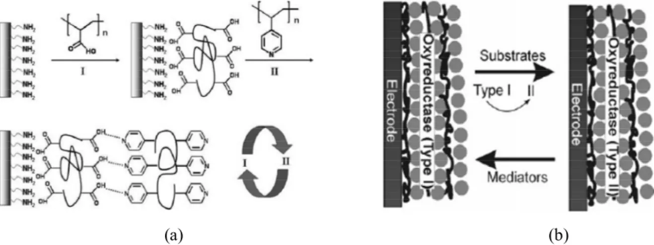
Multiple-layered thin films can be fabricated, as shown in Fig. 1.3, through layer-by-layer assembly, which is a technique of alternately depositing a sequence of monolayer-by-layered films on a substrate, where the layers are held together by non-covalent interactions like hydrogen bonding, hydrophobic interactions, electrostatic interactions and metal coordination.11 If these films are loaded with enzymes, antibodies, proteins, DNA, etc,
biosensors to probe bio-related species or processes can be prepared.12 One example is the use of electrodes fabricated with thin films of oxyreductase enzymes to detect substrates that are specifically responsive to these enzymes in solution. As shown in Fig. 1.3b, due to the catalysis of oxyreductases, substrates are transformed from type I (reduced or oxidized state) to type II (oxidized or reduced state), while oxyreductases also change from type I (oxidized or reduced state) to type II (reduced or oxidized state). Type II oxyreductases can then react with the electronic mediators to be converted back to type I, allowing the redox changes of the mediators to be detected and enabling the calculation of the substrate concentration.12
(a) (b)
Figure 1.3. (a) Layer-by-layer assembly based on hydrogen bonding. Reproduced with
permission from Ref. 11, © 2007 The Royal Society of Chemistry. (b) Mechanism of detection of oxyreductase biosensor. Reproduced with permission from Ref. 12,© 2006 Wiley-VCH Verlag GmbH & Co. KGaA, Weinheim.
Self-assembled micelles have been prepared from many amphiphilic block copolymers in aqueous solution and have been shown to be responsive to various environmental stimuli like light, temperature, pH, and specific cations.13 For example, as shown in Fig. 1.4a, poly(p-methoxyphenacyl methacrylate)-b-poly((oligoethylene glycol)methacrylate) (PMPMA-b-POEGMA) has a hydrophilic POEGMA block and a hydrophobic PMPMA block, so that in water it forms micelles with a PMPMA core and a POEGMA corona. These micelles disaggregate under light irradiation, which changes PMPMA to a hydrophilic poly(methacrylic acid) (PMAA). The obtained PMAA-b-POEGMA can be converted to a new
micellar structure with a PMAA core and a POEGMA corona by reducing the pH or introducing Ca2+, a process that is reversible by increasing the pH or adding ethylenediaminetetraacetic acid sodium salt (EDTA). PMAA-b-POEGMA can also form micelles with a POEGMA core and a PMAA corona by increasing the temperature above the LCST of POEGMA or by adding PO43-. The process is reversible by decreasing temperature
or using dialysis to eliminate the PO43-. This phenomenon has potential application in
photoresponsive drug delivery, as illustrated in Fig. 1.4b.14
(a) (b)
Figure 1.4. (a) A multi-responsive micellar system based on poly(p-methoxyphenacyl
methacrylate)-block-poly((oligo ethylene glycol)methacrylate) diblock copolymers. Reproduced with permission from Ref. 15, © 2012 American Chemical Society. (b) photocontrolled release of an encapsulated agent as a result of the photoinduced dissociation of the polymer micelle. Reproduced with permission from Ref. 14, © 2006 American Chemical Society.
Apart from the examples described above, which are all based on building blocks of a single type of low molar mass molecule or of polymer, a large number of supramolecular materials have been assembled from a combination of small molecules and polymers. Such materials have advantages of high stability, high strength, large flexibility and easy machinability, due to their polymer component. For instance, poly(ethylene oxide) has been used to form inclusion compounds with urea,16-19 thiourea19-20 and cyclodextrin.20-21 Similar
inclusion complexes have been prepared from the assembly between cyclodextrin and other linear main-chain polymers, such as poly(propylene oxide), poly(oxytetramethylene), poly(oxytrimethylene), polyesters, and polyamides.22 Another extensively investigated class is based on the assembly between polymers with side-chain functional groups and appropriately functionalized small molecules, which allows for a much wider variety of supramolecular compositions compared with linear main chain-polymers.23-24 Among them, poly(4-vinyl pyridine) (P4VP) is often used as the polymer component.
1.1.1 P4VP
According to a paper by Fitzgerald and Fuoss published in 1950, which is also the earliest report, to my knowledge, that describes the synthesis of P4VP, polymerization of vinyl pyridine began with its use as a comonomer with butadiene in synthetic rubber research during World War II.25 Various methods, like bulk,25 emulsion,25 living anionic26-27 and living radical28-29 polymerizations could be used to synthesize P4VP. As shown in Fig. 1.5, the pyridine ring of P4VP contains two free electrons on the N atom. Because the lone electron pair does not participate in the conjugation of the pyridine ring, it is weakly basic (pKa = 5.25±0.01)30 and can complex with many other species through ionic complexation, metal coordination, hydrogen bonding and halogen bonding.
Figure 1.5. Chemical structure of P4VP.
1.1.2 Side-chain P4VP-small molecule complexes
Ionic complexation with P4VP can be achieved by its conversion into a polycation. This occurs upon acid-base interactions with inorganic acids (Fig. 1.6a)31-32 and organic sulfonic acids (Fig. 1.6b),33-34 or upon quaternization with alkyl halogen compounds (Fig. 1.6c).35-38
X = Cl, Br, I, HSO4, H2PO4... X = Cl, Br, I; R = (CH2)nCH3 (a) (c) R = (CH2)nCH3 (b) (d)
Figure 1.6. Chemical structures of P4VP protonated by (a) simple inorganic acids, (b)
mesogenic sulfonic acids, (c) P4VP quaternized with alkyl halides and (d) quaternized P4VP complexed with mesogenic sulfonic acid salts. The blue bar in (b) and (d) indicates a rigid rod-like mesogenic part.
Polycations of P4VP protonated by HCl, H2SO4, H3PO4, etc., can be used as
immobilized catalysts for organic reaction,31 and as polycationic coatings in humidity sensors due to their water solubility.32 Such humidity sensors were also prepared from quaternized P4VP.35-36 Quaternized P4VP can be further modified into photosensitive side chain liquid crystalline polymers (SCLCPs) by ion exchange procedures with molecules containing a rigid rod-like azobenzene-based mesogenic part and a sodium sulfonate group (Fig. 1.6d). One of the simplest of these complexes has been made by Zhang et al. with a commercially available
dye, methyl orange, which has no alkyl spacer in its structure, leading to excellent photoactive properties.37-38 Zhu et al. prepared complexes between P4VP and wedge-shaped mesogenic sulfonic acids that form a supermolecular material with cylindrical morphology, as shown in Fig. 1.7.33 Ikkala et al. investigated an ionic complex with ordered lamellar structure between P4VP and p-dodecylbenzenesulfonic acid.34
Figure 1.7. (a) Chemical structure of P4VP complexes with wedge-shaped sulfonic acid
(P4VP(C12-H)DN), m = 12, where DN refers to the degree of neutralization. Proposed packing
model (b) in the lamellar phase (0.8 ≥ DN ≥ 0.5) and (c) in the hexagonal cylindrical phase (DN ≥ 0.8) of P4VP(C12-H)DN. Adapted with permission from Ref. 33, © 2006 American
Chemical Society.
P4VP can coordinate with metal cations, like Zn2+, Ca2+, Cu2+, Co2+ and Ni2+,39-40 making it useful for removing heavy metals from waste water.41 Metal coordination allows the preparation of metallosupramolecular P4VP systems with, for example, various liquid crystalline structures. For instance, a mesomorphic liquid crystal was obtained in a complex of P4VP and zinc dodecyl benzene sulfonate,42 and a hexagonal structured multicomb polymeric supramolecules was achieved by using 2,6-bis(octylaminomethyl)pyridine as an intermediate to link each P4VP repeating unit and two dodecylbenzenesulfonate counterions.43 Besides, comblike complexes were also formed between P4VP and zinc salts of carboxylic acid-functionalized alkoxybiphenyl mesogens whose structure changes from amorphous form to liquid crystalline as spacer length increases.44
The most often used supramolecular interaction with P4VP is hydrogen bonding. Compared with other interactions, hydrogen bonding can facilitate tuning the properties and the internal structure of complexes in a more controllable and dynamic fashion, for example to form supramolecular SCLCPs. An early attempt was made by Bazuin and Brandys, who blended a mesogenic dialkoxy biphenyl derivative functionalized by carboxylic acid with P4VP to obtain an ordered smectic phase, but they found that phase separation and crystallization of the mesogen occurred when the complex contained over 20 mol% mesogen and was heated above its glass transition temperature.45-46 Kato et al. reported that a copolymer of poly(4-vinylpyridine-co-styrene) with 10 mol% styrene can form a homogeneous complex with a stoichiometric amount of a carboxylic acid-functionalized mesogen, 6-[(4-octylpheylazo)phenoxyl]hexanoic acid, and that this complex exhibits an ordered smectic phase over a wider temperature range (93-132 °C) than that of the pure mesogen (130-139 °C).47 Ikkala and coworkers reported a complex between a carboxylic acid-functionalized cholesterol (CholHS) and P4VP (Fig. 1.8) that has a smectic A structure over a full range of degree of complexation, and stable liquid crystallinity at low degree of complexation.48
(a) (b)
Figure 1.8. (a) Schematic of a P4VP-CholHS complex, and (b) Polarized optical micrograph
of P4VP-CholHS (at a molar amount of CholHS to P4VP repeat unit of 0.5) at 140 °C showing bâtonnets during cooling from the isotropic state. Reproduced with permission from Ref. 48, © 2010 American Chemical Society.
Supramolecular P4VP complexes have been used recently in designing photosensitive materials. Examples based on ionic interactions between protonated or quaternized P4VP and
ionic azobenzene-containing small molecules have been given above. There are also numerous examples based on hydrogen bonding with photosensitive molecules. A film of P4VP complexed with 9-anthracene carboxylic acid can undergo reversible photo-controlled deformation upon UV irradiation. This can be enhanced by the addition of LC molecules.49 Photoinduced birefringence was achieved and investigated in P4VP complexes with hydroxyl-functionalized azobenzene chromophores.50-51 Surface relief gratings (SRGs) were achieved and investigated with P4VP complexes with azobenzene functionalized with hydroxyl52 and carboxylic acid53 groups. Priimagi and coworkers further showed that SRGs can also be obtained from complexes between P4VP and hydroxyl-functionalized bisazobenzene.54-55 Further discussion about the photoreactivities of P4VP/azobenzene complexes is given in section 1.3.
Very recently, halogen bonding, which is more directional than hydrogen bonding,56 has been shown to be effective for forming supramolecular complexes. In one of the earliest reports, Bertani et al. prepared comb-like crystals from the complexes between P4VP and iodoperfluorohexanes with various lengths.57 Priimagi et al. found that films of P4VP complexed with halogen-functionalized azobenezene molecules can be inscribed with SRGs under irradiation more efficiently than analogous hydrogen-bonded complexes.58
Other applications of supramolecular complexes involve the development of tunable internal structures. For example, the complex of P4VP and 3,4,5-tris(dodecyloxy)benzoic acid (TDBA) contains a layered internal structure at low TDBA content that enlarges and then changes to a cylindrical structure with increasing TDBA content.59 However, as the interaction between the pyridine ring and the carboxylic acid group competes with the interactions between carboxylic acid groups, the complexation extent is usually low, especially when the mesogen content is high, though never quantified, limiting the applications. ten Brinke, Ikkala and coworkers first noticed that the complexation between P4VP and nonmesogenic alkylphenols, like 1-dodecyl 3,4,5-trihydroxybenzoate and 4-nonadecylphenol, can lead to a homogeneous blend with mesomorphic structures (see Fig. 1.9a).60-61 Similar comb-like structures based on nanophase separation were found in complexes between P4VP and 4-(4’-alkylphenyl)azophenols,62 4-(4’-alkoxylphenyl)azophenols,62 and 3-pentadecylphenol (PDP)
(see Fig. 1.9b).63 PDP in particular was studied in detail and used subsequently by many research groups. Hydrogen bonding between PDP and P4VP allowed the tuning of many properties; for instance, the order of the lamellar structure and the thermal properties by changing the PDP content, and the level of the structural hierarchy by changing the P4VP block fraction in poly(styrene-block-(4-vinylpyridine)) (PS-b-P4VP), as will be discussed below.64-65
(a) (b)
Figure 1.9. (a) TEM micrograph of a P4VP-(4-nonadecylphenol) equimolar complex.
Reproduced with permission from Ref. 61, © 1998 American Chemical Society. (b) Schematic of the P4VP-PDP complex. Reproduced with permission from Ref. 63, © 1996 American Chemical Society.
Hierarchical structures – i.e. different structures at different length scales – can be formed in block copolymers of P4VP and a second block of a nonpolar (noninteracting) polymer such as polystyrene (PS). For example, in the complexes of CholHS with PS-b-P4VP, morphologies of smectic P4VP-CholHS structures form inside a larger scaled cylindrical or layered PS-b-P4VP structure, when the volume fraction of the two blocks varies.48 Complexes of PS-P4VP with PDP were investigated in different applications by various research groups. For example, Bazuin and coworkers used the Langmuir-Blodgett (LB) technique to fabricate monolayered films of PS-b-P4VP(PDP). They showed that, in general, the morphologies evolve from planar structure to nanostrands and to nanodots (see Fig. 1.10) with increasing polar block content, the precise block content depending on the nature of the blocks and, for PS-b-P4VP the presence or not of PDP.66-67 The nanostrand pattern itself was dependent on
the spreading solution concentration and the spreading solvent.68-69 Moreover, the dot morphology order shows a transition from quasi-hexagonal to quasi-square as a function of compression pressure.70 Such monolayered films with nanopatterns have potential as the templates for gold deposition,71 leading to ordered gold nanoparticle films.
Figure 1.10. Left: PS-b-P4VP/PDP complex. Right: AFM height images showing the
dependence of the morphology on the P4VP block content: (a) planar aggregates, (b) nanostrands, and (c) nanodots. Reproduced with permission from Ref. 69, © 2010 American Chemical Society.
Stamm and coworkers showed that internal structures of PS-b-P4VP could also be obtained from the complexes with much smaller molecules like 2-(4’-hydroxy-phenylazo)benzoic acid (HABA). They used the dip-coating method to obtain films exhibiting cylindrical morphology, the axis of which selectively orients either in horizontal or vertical direction relative to the substrate depending on the solvent used.72-73 One advantage of small molecules is that they can be very easily removed, so that ordered porous structures can be obtained for further use (see Fig. 1.11). A similar strategy was applied by Bazuin and coworkers, based on the complexes of PS-b-P4VP with various small molecules like 1,5-dihydroxynaphthalene (DHN),74 naphthol75-77 and naphthoic acid,75-77 to obtain films with different morphologies from nanocylinders to nanodots.
Figure 1.11. Schematic of the fabrication of nanotemplate from
PS-b-P4VP(4’-hydroxyazobenzene-2-carboxylic acid). Reproduced with permission from Ref. 73, © 2003 American Chemical Society.
1.1.3 Crosslinked P4VP-small molecule complexes
Compared with small molecules with monofunctional groups, P4VP complexed with small molecules with bifunctional groups have been reported less often.78-80 However, since they potentially can physically crosslink P4VP chains directly, bifunctional small molecules can facilitate the formation of polymer networks. Shibata et al. reported that, by using 1,5-naphthalenedisulfonic acid (NDS) and 1,3-propanedisulfonic acid (PDS) as hydrogen-bond crosslinkers, complexes of P4VP/NDS and P4VP/PDS show both higher glass transition temperatures and decomposition temperatures, which increase with increasing crosslinking density.79 Craig et al. investigated P4VP networks formed in DMSO using metal coordination with bisfunctional Pd(II) and Pt(II) organometallic crosslinkers, and found that, due to the different dissociation rates of pyridine-metal coordination controlled by the crosslinker structure, both the dynamic viscosity and the elastic storage modulus of the solution could be tailored quantitatively.78, 80
1.2 Electrospinning and electrospinnability
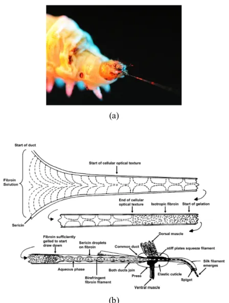
Since its development in ancient China, as early as 3500 BC based on the earliest finding of fragments of silk fabric, cloth woven from silk have been popular in high class societies of the ancient world. This has led to an international trade route called the “silk road”. As great profit was gained from the silk trade, its secret was carefully kept by Chinese emperors until around 550 BC, when Europeans finally discovered that silk was produced by a domesticated worm, called bombyx mori or silk worm, as shown in Fig. 1.12a.
(a)
(b)
Figure 1.12. (a) Photo of a Chinese silkworm spinning silk.81 (b) Mechanism of the spinning of spider silk. Reproduced with permission from Ref. 82, © 2007 American Chemical Society.
The process of silk production by silk worms, by spiders, and by other insects like bees, flies and ants, always involves the extrusion of the proteins from the silk glands through a spinneret, as shown in Fig. 1.12b. Based on the great need for silk in the fabric industry, artificial “silks” were created based on techniques like wet spinning, dry spinning, melt spinning, and gel spinning, which also involve extrusion of polymer solutions through a spinneret with a tiny opening, thus resembling the production of spider silk. Compared with all these techniques based on the extrusion and drawing of fibers by mechanical forces, a new method, called electrospinning, rather applies an electrical field as a driving force for fiber formation.
1.2.1 Electrospinning
Electrospinning is a technique enabling drawing micro- or nano-scaled fibers from polymer solutions or melts under the application of an electrical field. The history of electrospinning before 1990 was summarized by Tucker et al.83 A milestone in the early 1900s was the submission of patents by Cooley84-85 and Morton86 that opened the gate to the industrial use of electrospinning. Another breakthrough in the industrial practice happened in 1938 when Fuchs, Petryanov-Sokolov and Rosenblum used electrospun fibers of cellulose acetate as filter materials, which were called “Petryanov filters” and led to the manufacture of gas masks.83 A booming period of research about electrospinning started in the early 1990s, based on the pioneering work in Reneker’s group.87
A typical electrospinning setup used in the lab is shown in Fig. 1.13a. A semidilute or concentrated polymer solution is stored in a syringe equipped with a needle. The solution is pumped through the needle and forms a drop at the tip. When an electrical field is applied between the needle and the collector, the drop deforms and takes the shape of a cone, called the Taylor Cone,88 and then ejects as a straight jet. Since the jet is charged, the electrostatic repulsion forces on the surface stretch the jet, leading to a large surface/volume ratio which enables fast evaporation of the solvent. Due to this, the density of the charges on the jet surface increases quickly, leading to a much stronger electrostatic repulsion effect which causes bending instabilities. This leads to the bending of the straight jet which now adopts a
large scale whipping motion. During the propagation, the jet becomes thinner with the evaporation of solvent until reaching the collector as fibers. Fig. 1.13b shows the setup used in our lab.
(a)
(b)
Figure 1.13. (a) Schematic representation of electrospinning setup. Reproduced with
permission from Ref. 89, © 2004 WILEY-VCH Verlag GmbH & Co. KGaA, Weinheim. (b) Electrospinning setup used in our lab.
1.2.2 Electrospinnability and effective crosslinking
Polymer solutions can be electrospun into fibers with the precondition that they possess “electrospinnability”, the lack of which leading to the production of droplets via electrospray. As shown in Fig. 1.14, the formation of different morphologies depends on the competition between three forces acting on the jet: electrostatic repulsive stretching, surface tension, and a viscoelastic force which acts in response to the former two. Electrostatic repulsive stretching tends to thin the jet. In contrast, surface tension initiates the shrinkage of the jet, causing the formation of beads and the thinning of jet between beads. Both forces can lead to the breakage of jet, while the viscoelastic force always resists the effect of the other two forces. A polymer system with high inner viscoelasticity is therefore an advantage to avoid electrospray. Many factors like entanglements, hydrogen bonding, hydrophobic interactions and electrostatic interactions facilitate the formation of a network in the solution, thus increasing the viscoelastic forces; then we call the resulting entity as “effective crosslinking” (also known as “physical crosslinking” or “temporary crosslinking”).
Figure 1.14. Scheme of the formation of beads, beaded fiber and fiber.
1.2.2.1 Entanglement
It is well known that polymer solutions of high concentration and high molecular weight facilitate the production of fibers, demonstrating that the entanglement of polymer chains is a key factor leading to the electrospinnability. The entanglement level of the polymer solution is related to its rheological properties. It is well known that the viscosity of polymer solutions increases with increasing concentration as shown by the Huggins Equation in Eq. 1.1:
'
2 2 ''
3 3...
sp c k c k c
(1.1) where sp, [ and c are the specific viscosity, the intrinsic viscosity and the polymer
concentration, respectively, and k’ and k” are constants. sp represents the contribution of the
polymer to the solution viscosity, which is calculated as: s sp s (1.2) where and s are the viscosities of the solution and of the pure solvent, respectively. Eq. 1.1
indicates that the viscosity increases with concentration in a non-linear fashion (sp ~ cn, n > 1)
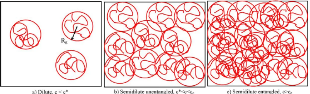
Figure 1.15. Physical representation of three solution regimes: (a) Dilute regime, (b)
Semidilute unentangled regime, and (c) Semidilute entangled regime. Adapted with permission from Ref. 90, © 2005 Elsevier Ltd.
Colby and coworkers have classified polymer solutions into three regimes with the increase of c: dilute regime, semidilute unentangled regime and semidilute entangled regime (see Fig. 1.15).91 A concentrated regime has also been reported.92-94 These regimes are separated by three critical concentrations: c*, ce and cD.
c* is defined as the critical concentration at which the overlap of the individual polymer coils starts to happen. Several methods can be applied to estimate c*. When c < c*, the polymer solution is in the dilute regime, where no overlap among polymer coils exists and for which the concentration dependence of the specific viscosity is given by sp ~ c1. According to
Frisch and Simha, c[, also called the Simha parameter or Berry number,95is a measure of the degree of coil overlap in solutions.96 It is usually accepted that the critical overlap point is approached when c[≈ 1,97 such that
1 1 * a c KM (1.3) where K and a are the Mark-Houwink-Sakurada constants and M is the viscosity-average molecular weight. Eq. 1.3 can be applied to estimate the value of c*. Another way to calculate c* is using Eq. 1.4, based on the definition that c* is the concentration at which the polymer concentration inside an individual polymer coil is equal to the solution concentration:90
3 3 * 4 g a M c R N (1.4)where Na is the Avogadro number and Rg is the radius of gyration.
ce is defined as the critical concentration over which entanglements start to form between
the polymer coils. When c* < c < ce, the polymer solution is in the semidilute unentangled
regime in which overlap among coils increases but is not high enough for the formation of an entanglement network in the solution. According to Graessley, ce canalso be calculated with
Eq. 1.5, c e w M c M (1.5) in which Mc and Mw represent the density of the pure polymer, the critical entanglement
molecular weight and the molecular weight of polymer, respectively.98 Mc is defined as the
minimum molecular weight to form entanglements for polymers in the melt state and can be determined from the turning point in a logarithmic plot of viscosity vs. molecular weight: ~ M1 when Mw < Mc and ~M3.4 when Mw > Mc.
When ce < c < cD, the polymer solution is in the semidilute entangled regime, in which an
entanglement network forms. Finally, when c > cD, the polymer solution is in the concentrated
regime, in which the chain dimensions become independent of concentration.98 According to Graessley, cD can be estimated by Eq. 1.6:
4 2 0 2 * D R c c R (1.6)
in which R20 is the mean-square end-to-end distance at zero concentration and R2is the
unperturbed dimensions of the polymer coil.
The link between electrospinnability and rheological properties was first illustrated in a pioneering work from Koski et al.,: fiber formation from poly(vinyl alcohol) (PVA)/water solutions was only observed when c > 5c*.99 McKee, Long and coworkers have tried to establish the relationship between the rheological properties and the electrospinnability. They concluded that fibers of neutral polymers could only be electrospun from semidilute entangled solutions (c > ce).94, 100
Shenoy et al. attempted to predict the formation of fibers based on the solution entanglement number (Ne(solu)), which could be calculated with Eq. 1.7:
( ) p w e solu e
M
N
M
(1.7) where p is the polymer volume fraction in the solution and Me is the apparent averagemolecular weight between two entanglement points of the polymer in the melt state.101 Me can
be determined using Eq. 1.8:
0 e N RT M G (1.8) in which ,R, T, and G0N are the density of the pure polymer, the ideal gas constant,
temperature, and the plateau modulus, which can be obtained from creep compliance measurements.97 Shenoy et al. further pointed outthat the number of entanglements per chain is equal to Ne(solu) – 1 and that, for linear polymers without specific polymer-polymer
interactions in a good solvent, beaded fibers can only be electrospun when the number of entanglements per chain is over 1 (Ne(solu) > 2) while uniform fibers can be produced when the
entanglement number per chain is over 2.5 (Ne(solu) > 3.5) in the solution.
Eqs 1.5 and 1.7 indicate that when Mw > Mc, increasing the molecular weight of the
polymer increases the number of entanglements and facilitates fiber formation. Eq. 1.7 also indicates that increasing the polymer concentration leads to entangled solutions, thus leading to the formation of fibers. This is consistent with the experimental observation that, as mentioned before, either increasing polymer concentration or polymer molecular weight can improve the electrospinnability.
1.2.2.2 Hydrogen bonding
Long and coworkers have reported a strategy for improving the electrospinning through the effect of self-complementary multiple hydrogen bonds. One example is by introducing 5 mol% of 2-ureido-4[1H]-pyrimidone pendant groups on the poly(methyl methacrylate) (PMMA) backbone, ce is lowered by 1-2 wt% as compared with that of pure PMMA with a
similar molecular weight.100 Another example: by introducing 35 mol% of 9-vinlybenzyladenine as comonomer with protonated 2-(dimethylamino)ethyl methacrylate (DMAEMA·HCl), the hydrogen bonding among adenine pendant groups leads to the decrease of ce by 1.9 wt% and of cD by 7.3 wt% in water, as compared with pure PDMAEMA·HCl.102
Tan et al. reported that the addition of 0.5 wt% of graphene oxide, which has many hydroxyl, carbonyl and carboxylic acid groups that can hydrogen-bond in a PVA/water solution, decreases the minimum concentration for fiber formation from 8.5 wt% to 5 wt%.103 Hermida-Merino et al. showed that, in tetrahydrofuran (THF), 42 wt% polyurethane linked with hydroxyl end-groups acts like a high molecular weight polymer and thus leads to electrospun fibers.104 In contrast, only droplets were obtained from 47 wt% polyurethane (with a similar molecular weight) terminated with morpholine groups, whose self-binding constant is only 10% of that of hydroxyl groups.104
1.2.2.3 Non-polar interactions
Talwar et al. prepared fibers, rather than droplets, from 3 wt% poly(ethylene oxide) (PEO) solutions in water, by blending them with a 0.2 wt% alkali-soluble emulsion polymer with hydrophobic side chains (C22 alkyl chains) that leads to the formation of a network based
on hydrophobic interactions in aqueous solutions.105-106 Wu et al. also reported the positive effect of non-polar interchain interactions. They showed that adding 10 wt% of small molecule liquid crystals (4’-cyanobiphenyl-4-yl 4’-propylbi(cyclohexane)-4-carboxylate) enables to form electrospun fibers from a solution of 33 wt% poly((methyl-(4-propoxybenzoic acid cholesterol ester)siloxane)-co-(methyl-(4-acetoxyphenyl-4-propoxybenzoate)siloxane)) in CHCl3, which originally only led to thin beaded fibers.107
1.2.2.4 Electrostatic interactions
Long et al. first reported that the effect of zwitterionic aggregation resembles the effect of entanglements and facilitates the electrospinning of poly(n-butyl acrylate-co-sulfobetaine methacrylamide) (PnBA-co-PSBMAm) in CHCl3/ethanol.108 They found that, with the same
total number of repeat units, the ce of PnBA-co-PSBMAm with 10 mol% of zwitterionic
SBMAm units is 1.5 wt% while ce is 5 wt% for pure PnBA.108 According to Shi et al., plasma
treatment on poly(ethylene oxide) (PEO) solutions can lead to charged PEO in the solution and improve the electrospinning. As an example, 4 wt% plasma-treated PEO in water leads to much more beadless fibers than a 4 wt% untreated PEO solution.109 An attempt to use Ca2+ for facilitating the fiber formation of alginate, although it only transited from droplets into more elongated shapes, was reported by Fang et al.110
1.2.2.5 Electrospinning of low molecular weight species
Based on these various effective crosslinking strategies, many low molecular weight species have been electrospun. For example, phospholipids are small molecules with a hydrophilic head and a hydrophobic tail, so their amphiphilic interactions drive the formation of spherical micelles above the critical micelle concentration (CMC, 0.1-0.5 wt%) in the solution. When increasing the concentration over 43 wt%, these micelles can further aggregate