Longitudinal study on ocular manifestations
in a cohort of patients with Fabry disease
Langis MichaudID*E´ cole d’optome´trie, Universite´ de Montre´al, Montre´al, Que´bec, Canada
Abstract
Purpose
This study aims to assess the evolution of ocular manifestations in a cohort of Fabry patients.
Methods
This is a prospective observational study conducted from 2013 to 2017 (5 consecutive exams). All subjects underwent a comprehensive ocular examination including oriented case history, refraction, corneal topography, biomechanical corneal properties and pacho-metry assessments, aberropacho-metry, anterior segment evaluation, double-frequency visual field (FDT), intra-ocular pressure, and ocular fundus. At baseline, 41 subjects enrolled but 9 dropped-out and 4 files were not kept for analysis (missing data). Remaining 28 subjects were classified into: Group 1 -hemizygotes (HMZ), all on enzyme replacement therapy (ERT) (N = 10); Group 2 -heterozygotes (HTZ) actively ERT-treated (N = 8), and Group 3 -HTZ not treated (N = 10).
Results
There is a high intra and inter-subjects variability. At baseline, prevalence of the ocular man-ifestations found is similar to published data: cornea verticillata (89.2%), conjunctival ves-sels tortuosity (85.7%), corneal haze (67.8%), retinal vesves-sels tortuosity (64.2%), anterior cataract (39.2%) and posterior cataract (28.5%). Prevalence for new elements are found: upper lid vessels toricity (96.4%) and micro-aneurysms (42.8%). At the end, micro-aneu-rysms (+82%), posterior cataract (+75%) corneal haze (+21%) anterior cataract (+17%) and retinal vessels tortuosities (+4%) evolved in prevalence and severity despite the fact that 68% of the patients were on ERT. Treated heterozygotes evolved more than other groups (p>0.05).
Conclusion
ERT does not seem to halt the clinical evolution of several ocular manifestations. Longer observational time and objective grading systems may be required to fully confirm these findings. a1111111111 a1111111111 a1111111111 a1111111111 a1111111111 OPEN ACCESS
Citation: Michaud L (2019) Longitudinal study on
ocular manifestations in a cohort of patients with Fabry disease. PLoS ONE 14(6): e0213329.https:// doi.org/10.1371/journal.pone.0213329
Editor: Gianni Virgili, Universita degli Studi di
Firenze, ITALY
Received: February 17, 2019 Accepted: May 26, 2019 Published: June 27, 2019
Copyright:© 2019 Langis Michaud. This is an open access article distributed under the terms of the Creative Commons Attribution License, which permits unrestricted use, distribution, and reproduction in any medium, provided the original author and source are credited.
Data Availability Statement: Supplementary file
within this submission.
Funding: This investigational study was funded
with a non-restricted grant received from Genzyme Canada. The funder had no role in study design, data collection and analysis, and decision to publish. Research agreement with Genzyme stipulated that their scientific committee may provide comments on the initial draft of any publication. In this case, a few modifications were suggested and freely accepted by author but nothing interfered with, or could reasonably be
Introduction
Fabry is qualified as a pan ethnic X-linked inherited condition and is considered a rare disease [1]; however, it represents the second most prevalent of the 50 known lysosomal storage disor-ders[2], which are all characterized by a cell deposit of a substrate, within lysosomes, as a result of abnormal enzymatic activity. With Fabry, the absence or deficient activity ofα-galactosidase A leads to the accumulation of globotriaosylceramide (Gb-3) in a variety of cells (renal, endo-thelial, cardiac, dorsal root ganglion neural)[3].
As the disorder evolves, and the substrate continues to build-up, cellular dysfunction will trigger organ impairment and eventually system damages, leading to substantial morbidity and reduced life expectancy[4], especially for patients left untreated[5].
The manifestations of the disease are highly variable, depending on genetic mutation and the sex of the patient. Based on mutations, patients can be categorized into 4 groups[6], vary-ing from an absent or non-functional enzyme protein (null alleles), with severe manifestations, to a group of subjects characterized with some enzymatic residual activities, and associated with an attenuated phenotype. In the past, asymptomatic subjects were only considered carri-ers of the disease[7], but must be considered true Fabry patients[8].
Sex divides patients into two distinct groups. Men are defined as hemizygotes, which are individuals having only one allele for a specific characteristic[9]. Hemizygotes can only inherit the deficiency (X) from their mother and will automatically transmit the disease to their daughters (boys receiving the normal Y). Females are considered heterozygotes, having differ-ent alleles at a particular gene locus on homologous chromosomes[9]. They can inherit their defective X from both parents. They have a 50% chance of transmitting the disease to any of their children, regardless of sex.
The clinical portrait becomes more complex when both classifications are combined. This means that hemizygotes and heterozygotes can present little or no residual enzymatic activity, which obviously impacts the severity of the disease and its clinical manifestations.As of 2018, 840 possible mutations were compiled[10], which explains the high variability of clinical find-ings among patients diagnosed with Fabry.
Clinical manifestations
In the case of Fabry, the presence of substrate accumulation can be traced back to the prenatal period[11] but generally, symptoms do not develop before early childhood (4–6 years old)[2], boys being affected earlier than girls[12]. From then on, affected children may suffer from acute and chronic neuropathic pain, which manifests itself as a burning sensation and tingling in the hands and feet (acroparaesthesiae). They may also show reduced or absent sweating (hypohidrosis), which eventually makes it difficult to exercise[13] and perform physical activi-ties[14]. Finally, abdominal pain and postprandial diarrhoea may be reported. Fabry children, mostly boys, exhibit greater weight and height variations, most of them remaining under the normal development curve[15]. During adolescence, gastro-intestinal problems may increase in severity and frequency, and proteinuria can become more elevated[2]. Delayed puberty and abnormal sexual functions were reported[16]. Fabry crisis, manifested as severe pain radiating from the hands and feet, may occur spontaneously as a response to various environmental conditions (heat, cold, stress, fatigue, illness, intense exercise, etc.)[5]. Swelling of the affected members (lymphedema) and redness may be present and are predictors of future heart and renal dysfunctions[2]. Vascular skin lesions (angiokeratomas) can appear, clustering around the umbilicus and swim trunk areas[17]. Later in life, respiratory problems, cardiac and cere-brovascular manifestations may be manifested. Hearing loss or tinnitus are not uncommon at perceived as interfering with, the full and objective
presentation of the results.
Competing interests: This investigational study
was funded with a non-restricted grant received from Genzyme Canada. Author received speaker fees from Shire on other topics (dry eye management). There are no patents, products in development or marketed products to declare. This does not alter our adherence to all the PLOS ONE policies on sharing data and materials.
this strage[18]. Late disease will be characterized by life-threatening renal failure (more the case for hemizygotes), stroke and severe cardiac problems (more often for heterozygotes)[19].
Ocular manifestations
Ocular manifestations are among the first observable signs of the presence of the disease, even before birth[20], and are easily identified through a regular slit lamp examination made by a trained eyecare professional. One prevalent feature is the presence of deposits in the cornea, with a whorl-shaped pattern. This keratopathy is known as cornea verticillata[21]. By diffusion from the conjunctival blood vessels, the cornea not being vascularized, substrates accumulate in a linear fashion at the epithelial basement membrane level and the adjacent anterior stroma, primarily on the lower third of the cornea[22]. Over time, it may evolve to reach the upper quadrants. Over 90% of hemizygotes show this type of deposition by age of 4, while heterozy-gotes present this clinical sign later, around the age of 10 on average[23]. The level of deposits is then associated with the progression and severity of the disease[24].
Less specific, a diffuse corneal haze can also be found in conjunction with corneal deposits[6]. Both manifestations are not habitually associated with any loss of visual acuity. Differential diag-nosis implies keratopathy related to the intake of medication such as amiodarone, tamoxifen, chlorpromazine, indomethacin and gold salts, just to name a few[25]. Clear understanding of the medical condition of the patient is obviously essential to establish the differential diagnosis.
Lens opacification is less pathognomonic than corneal verticillata and its prevalence is also lower[26]. Fabry’s cataract appears as a sub-capsular opacity along the posterior lens suture lines, very similar to the cortisone-induced cataract or those following head trauma. This type of cataract becomes visually disturbing soon after its onset and patients develop high light sen-sitivity and reduced visual acuity[27]. Hemizygotes can show early signs of cataract by age 20. This type of lens opacification may also be seen in patients with mannossidosis [28] and other lysosomal storage disorders. A second cataract manifestation relates to the presence of a white, wedge-shaped, linear deposit on the anterior sub-capsular area of the lens[21], usually covering all quadrants. They are less associated with visual disturbance and may not be visible under slit lamp examination without pupil mydriasis.
Blood vessel tortuosity appears as a consequence of the alteration of the vessels’ natural architecture by substrate accumulation in the vascular endothelium[29]. Hemizygotes are almost all affected by this clinical sign after age 30, while at least 50% of heterozygotes show similar manifestation[30]. Tortuosity can be seen in the bulbar conjunctiva, in the retina, and as reported earlier, on the external surface of the upper eyelid, [31]. Vessel tortuosity is not associated with functional loss affecting vision; however, Fabry-related vasculopathies (artery or veinule occlusions, choroidal neovascularization) are rarely seen [32,33], except among patients with more severe systemic involvement. Differential diagnosis should include the presence of fucosidosis[34], or gangliosidosis[35], where similar blood vessel alterations were reported.
Alteration of vascular insufficiency may lead to the development of optic nerve ischemia. Enlargement of the blind spot, and central defects in visual field testing, is related to optic neu-ropathy probably second to this phenomenon[36]. Finally, substrate accumulation in the lach-rymal gland fosters eye dryness, experienced by 50% of Fabry patients[37]. Recently, bulbar conjunctival lymphangiectasia, a sign of ocular surface irritation, was identified in a cohort of Fabry patients particularly symptomatic of eye dryness[38].
All of these ocular manifestations were reported as single events, on several cohorts of patients, with variable methods of data collection. To our knowledge, no publication evaluated the progression of ocular manifestations over time.
Treatments
Fortunately, since 2001 in Europe and 2003 in the US, enzyme replacement therapy (ERT) is available to manage the patient’s condition, in addition to other medication needed to stabilize or improve cardiac, renal, gastro-intestinal and other systemic implications[39]. In Canada, Fabry patients can access ERT if they meet nationally accepted criteria based mostly on cardiac and renal involvements at the time of diagnosis[40]. Once approved, patients are randomly assigned to one of the two drugs available. This targeted use of ERT seems to reduce the risk of adverse outcomes related to Fabry[41]. Use of supportive therapies such as aspirin, renin-angiotensin blockade and statins are also considered in ERT-treated subjects[41].
In general, ERT was shown to reduce the GL-3 level in the blood and lessen gastro-intesti-nal symptoms[42]. In some patients treated with ERT, renal function decline is slowed[43] and pain crisis is less severe[44]. It is also proven that ERT may significantly improve cardiac disease associated with Fabry, lowering the risk of cardiac death[45]; however, it does not seem to lower the risk of stroke[46]. It also provides better patient outcomes by preventing serious complications like left ventricular hypertrophy (LVH)[47] or by preserving pulmonary function if the treatment is initiated early[48]. This confirms that ERT initiation at a younger age results in a better biochemical response, especially in men with classical Fabry[49].
Two synthetic enzymes (ERT) are currently prescribed: agalsidaseα (Replagal; Shire Human Genetic Therapies, Inc. Cambridge, MA; 0.2 mg/kg per infusion) and agalsidaseβ (Fabrazyme; Genzyme Corporation, Cambridge, MA, 1.0 mg/kg body weight infused every 2 weeks as intravenous infusion). Generally, both drugs are considered equivalent. [50]. Both aim to stabilize the patient’s condition knowing that the damages made on cells, organs and systems are mostly irreversible.
It was reported recently that a greater biochemical response and an improved ventricular mass was observed with agalsidaseβ[51]. An improved estimated glomerular filtration rate (eGFR) is also associated with the use of agalsidaseβ after switching drug from agalsidase α formulation [52] A recent Cochrane report confirmed that agalsidaseβ may be associated to a lower incidence of cerebrovascular events than agalsidaseα[53].
Oral medication (chaperone) may be prescribed for a subgroup of patients affected by ame-nable mutations[54]. Migalastat increases alpha-galactosidase-A activity, stabilizes related serum biomarkers, improves cardiac integrity[55], renal function, LVMi, plasma lyso-Gb3, and diarrhea symptoms, especially in susceptible classic Fabry patients[56]. Other molecules are under development, like PRX-102 (PEGunigalsidase alfa), a novel PEGylated enzyme expressed in a plant production system[57], and are promising as future alternatives to injections[44].
Impact on the quality of life
There is a strong relationship between the severity of the symptoms and the patient’s quality of life. During childhood, class attendance may be negatively affected[13]. Adults may not be attentive to the nonspecific nature of the symptoms or may not believe the children’s com-plaints about chronic pain, in the absence of external visible signs. This denial of the condition delays the diagnosis and may have a significant psychological impact, children being perceived as cheaters or liars. Not surprisingly, Fabry patients are eventually more inclined to depression [58], alcoholism and drug dependency. Some will even consider committing suicide during their adolescence[59]. Higher severity of the symptoms, especially for men, may limit sports and social activities, often resulting in a sedentary lifestyle, low self-esteem and exclusion[60]. This is made even more complicated by chronic fatigue (misdiagnosed as depression), intoler-ance to physical activities and poor self-perception of health, particularly for women[61]. Employment opportunities become limited, making economic stress constant.
There are also multiple side effects associated with the treatment, that is demanding for the patient. Consequently, the positive outcomes from enzyme replacement therapy are weighted by the burden of the treatment. This is why it is not so obvious that the quality of life may be improved with ERT[62]. With that in mind, it is important to reiterate the usefulness of slit lamp screening by eyecare practitioners, knowing that ocular manifestations related to Fabry occur early in life. Longevity and quality of life may be improved with a timely diagnosis of the patient’s condition[37].
Objective
This study aims to assess the longitudinal evolution of ocular manifestations in a cohort of Fabry patients.
Methods
This is a prospective observational study, conducted in adherence to the tenets of the Declara-tion of Helsinki. It was approved by the Universite´ de Montreal (UM) review board for experi-mentation on humans. A majority of participants (80%) were recruited after referral from the only metabolic center managing Fabry patients in the province of Quebec at that time (Hoˆpital Sacre´-Coeur de Montre´al). Other patients (20%) were referred from optometric practices in Montreal area. Written consent was obtained for all participants, at enrollment, after they had been fully informed of the goals and procedures of the longitudinal study. The only entrance criteria was to having been diagnosed with Fabry disease by a metabolic disease specialist, after molecular genetic analysis and identification of the mutation All participants accepted to be seen every year from 2013 (baseline) up to 2017 (5 exams) for a comprehensive eye assessment, subject to their health condition.
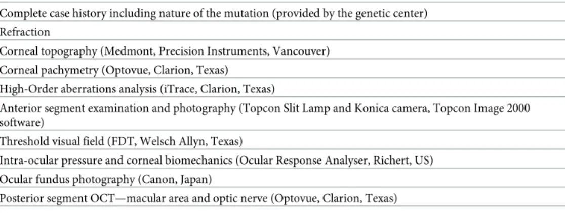
A complete description of the clinical procedures was described elsewhere[63]. To summa-rize, a list of clinical testing, made at each annual visit, is described inTable 1. All the testing were done in order. Among these procedures, the following ones were defined as essential components of a standard Fabry ocular assessment: a complete case history screening for signs and symptoms related to the disease, ocular anterior segment evaluation under slit lamp, with emphasis on the cornea and crystalline lens, evaluation of the visual field using double-fre-quency strategy (FDT), and ocular fundus assessment under pupil dilation. Pupil mydriasis was achieved after instillation of 1 drop of tropicamide 1% and 1 drop of phenylephrine 2.5% in each eye, subject to patient’s contraindications.
The threshold visual field (VF) was tested using frequency doubling technology (FDT). This is a screening strategy based on a flicker illusion that showed high sensitivity and
Table 1. List of testing- annual exam.
Complete case history including nature of the mutation (provided by the genetic center) Refraction
Corneal topography (Medmont, Precision Instruments, Vancouver) Corneal pachymetry (Optovue, Clarion, Texas)
High-Order aberrations analysis (iTrace, Clarion, Texas)
Anterior segment examination and photography (Topcon Slit Lamp and Konica camera, Topcon Image 2000 software)
Threshold visual field (FDT, Welsch Allyn, Texas)
Intra-ocular pressure and corneal biomechanics (Ocular Response Analyser, Richert, US) Ocular fundus photography (Canon, Japan)
Posterior segment OCT—macular area and optic nerve (Optovue, Clarion, Texas) https://doi.org/10.1371/journal.pone.0213329.t001
specificity to detect ganglion cells abnormality (leading to glaucoma), but less so for other retinal diseases. In this study, results were classified as normal (no VF defect) or abnormal (VF defect in at least one quadrant). A defect was considered present when a tested point (area) showed a threshold sensitivity (in decibels) reduced by 5% compared to a normal database. [64] Results can be altered by the presence of corneal scars or crystalline lens clinically relevant opacities.
Other testing listed inTable 1may be considered optional in practice but necessary from a research perspective. One example is the adjunct testing which evaluated the biomechanical properties of the corneas with the use of an Ocular Response Analyzer (ORA; Reichert Oph-thalmic Instruments, Depew, NY), a validated technology to screen eye disease [65,66] or to assess patient with lysosomal storage disorders[67] The measurements were made by applying a force on the cornea, via a jet of air. Results are based on the ability of the cornea to regain its shape and translates the corneal tissue behaviour. Three parameters were evaluated in this study population: the Corneal Resistance Factor (CRF), the Corneal Hysteresis (CH) and the non-contact corneal-compensated Intra-Ocular Pressure (IOPcc).
Optical aberrations can be defined as distortion in the image formed by an optical system. They can be divided in low-order aberrations, related to the refractive status of the eye, or in high-order aberrations (HOAs), above the previous ones. It is known that these HOAs vary with refractive status, pupil size, presence of corneal or lens opacities, etc[68]. HOAs optical impact in the eye differs from one type to another one and is patient dependent[69]. Spherical aberration, coma and trefoil are the only ones of clinical interest. Subjects affected with higher refractive errors, and/or those with larger pupils are habitually showing higher levels of aberrations.
HOAs presence in a Fabry’s patient cohort was rarely assessed. In this study, a wavefront aberrometer (iTrace, Tracey Technologies, US) was used. It allows to measure the quality of vision and the visual function using a fundamental thin beam principle of optical ray tracing. This aberrometer sequentially projects 256 near-infrared laser beams into the eye to measure forward aberrations, processing data point-by-point. Whenever a significant finding occurs, it is possible to identify which clinically relevant aberration is most prominent (coma, trefoil, secondary astigmatism or spherical aberrations).
The threshold visual field (VF) was also tested using frequency doubling technology (FDT). This is a screening strategy based on a flicker illusion that showed high sensitivity and specific-ity to detect ganglion cells abnormalspecific-ity (leading to glaucoma), but less so for other retinal dis-eases. In this study, results were classified as normal (no VF defect) or abnormal (VF defect in at least one quadrant). A defect was considered present when a tested point (area) showed a threshold sensitivity (in decibels) reduced by 5% compared to a normal database. [64] Results can be altered by the presence of corneal scars or crystalline lens clinically relevant opacities.
All the procedures listed were conducted by the same examiner (author), at the same loca-tion (UM clinic) for all participants. The same testing procedure was repeated periodically over five annual consultation and results were compared from baseline to the final visit results.
Statistical analysis
Both eyes were considered separately because of the high variability found between them dur-ing clinical observations. All calculations were made by an experienced statistician from the UM’s Statistics Department, using version 9.4 of the SAS software, with the 5% significance level.
Each measure was analyzed using a repeated-measure variance analysis with a mixed 4-fac-tor model; these fac4-fac-tors include intra-subject fac4-fac-tors (time and eye) and inter-subject fac4-fac-tors (group and mutation). For the sake of parsimony, given the number of participants and factors
studied, the analyses were first run by incorporating all possible terms of interaction. When the triple (3 factors) and quadruple (4 factors) interactions were found to be insignificant, the analyses were redone keeping order 2 interactions. Where no interaction was significant in this last analysis, the latter was redone focusing only on the main effects. For significant inter-actions, post-hoc analysis were controlled for one factor at a time in order to identify the source of the interactions.
For the categorical variables, the evolution of the initial time at 5 years was categorized according to 3 categories (deterioration, stability and improvement). The association between the evolution categories and the three groups was tested using the exact p-value of the chi-square test.All results were corrected for multiple comparisons.
Results
The enrollment began in 2013, defined as baseline, and closed in February 2015. The study ended in March 2017 after the fifth annual exam.
At baseline (2013), 41 subjects composed the study population. In 2017, 9 were lost for fol-low-up: 2 deceased, 2 moved out of the area and were not reachable, 2 voluntary redrawn, and 3 were not able to travel/be seen because of a poor health condition. Because of partial, unreli-able or missing data (< 5 exams completed), 4 other files were not considered for analysis. Consequently, data presented in this report relates to 28 subjects with full dataset.
Subjects are classified into 3 different groups for analysis. Group 1 is composed with hemi-zygotes (HMZ), all on enzyme replacement therapy ERT (N = 10). Group 2 is composed with heterozygotes (HTZ) actively ERT-treated (N = 8). Finally, Group 3 is composed with HTZ not treated with enzymes (N = 10), because they did not met the criteria set by the CFDI com-mittee. Among those on ERT, agalsidaseα and β was given to 8 and 10 subjects respectively at baseline. During the study period, agalsidaseβ became unavailable for approximatively 2 years and patients were switched over the other formulation. When the medication became again available, four subjects were switched back to their original formulation. At the conclusion of the study, 11/18 (6 HMZ, 5 HTZ) remained on agalsidaseα. Analysis of the result is made on the evolution of several parameters, by comparing baseline results with the same ones assessed after 5 annual visits. It was not possible, for administrative reasons, to access to the medical file and to track down the exact time of exposure to each medication for every patient during that time. Statistical analysis will not consider the 2 products used differently.
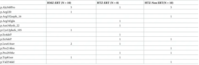
The most common mutation affecting subjects (11/28) wasp.Ala348Pro, a rare missense
one occurring at position c.1042G>C.[70] Other mutations, and their frequency, are listed in Table 2. For statistical analysis, those single mutations will be considered as a single group (“other mutations”).
Age of the subjects are reported inTable 3. There is a statistical difference based on the sex, men being younger than females in general (F = 6.049; p<0.05), but there is no sex�treatment effect (F = 1.440; p>0.05).
Ocular related parameters
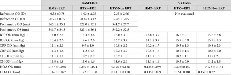
Refraction, pachymetry and ocular biomechanical parameters are reported inTable 4.
Refraction. For refraction, the three groups showed low myopia (spherical equivalent) at
baseline. The right eye showed no difference based on groups (F = 1.779; p>0.05) or mutation (F = 1.718; p>0.05). Similar results were observed for the left eye, with no notable differences based on groups (F = 1.773; p>0.05) or mutation (F = 2.080; p>0.05). This parameter was not assessed every year, refractive error variation being not characteristic of the disease evolution over time.
Corneal thickness. Corneal thickness for the right eye was found similar among the 3
groups at baseline (F = 1.363; p>0.05). There was no difference based on mutation (F = 0.499; p>0.05). Similar findings applied for the left eye in which no difference among groups (F = 1.332; p>0.05) or based on mutation (F = 0.385; p>0.05). These results are comparable to the average corneal thickness observed in a Caucasian population (550 um)[71].
Biomechanical: Corneal hysteresis (CH). Corneal hysteresis was found highly variable
among participants. It varied from a minimum of 10.3 (Group HTZ-ERT, OD and OS at 5 years) to a maximum of 11.8 (Group HMZ-ERT, OS, @ baseline). There was no significant dif-ference among groups at baseline (OD: F = 1.79; p>0.05; OS: F = 0.4615; p>0.05), and the same finding applied at the end of the study (OD: F = 1.41; p>0.05; OS F = 7.91; p>0.05). These values were considered comparable to those measured on a non-Fabry population (NFP- 10.3 to 11.8 vs 9.8 mm Hg to 10.8 mmHg)[72].
Biomechanical: Corneal resistance factor (CRF). In this study, CRF varied from 9.9
+ 1.0 mm Hg to 12.2 + 3.9 mm Hg (Group HTZ-ERT OD and Group HTZ-non ERT OS at baseline). There was a significant CRF difference among the groups at the beginning of the study, considering the right eye (F = 4.12; p<0.05 OD; F = 0.03; p>0.05 OS), Group HTZ-ERT showing lower values vs Group HMZ-ERT and Group HTZ-non ERT. Also, this group (2) showed increased values more than Group HMZ-ERT over time (p<0.05; 95%CI [-2.496, -0.145]), for the right eye again, but those results were not duplicated for the left eye (p>0.05; 95%CI [-1.412, 1.362]). There was also a time�eye�mutation effect (F = 4.91; p<0.05), which implies that one eye (OD) of patients with the pAla348Pro mutation showed systematically higher values (stiffer corneas) if compared with those carrying other mutations, and that this difference was confirmed over time. Despite high inter-subject variability, average values reported here about Fabry patient’s CRF were found similar to those of a non-Fabry popula-tion (NFP = 10.5 mmHg)[73,74].
Table 2. List of mutations affecting at least 1 subject of the study population.
HMZ-ERT (N = 10) HTZ-ERT (N = 8) HTZ-Non ERT(N = 10)
p.Ala348Pro 5 1 5 p.Arg220 1 p.Arg332aspfs_16 1 p.Arg342gln 1 p.Asn34lysfs_22 1 p.Cys12phefs_105 1 p.Ex4del? 1 p.Ex5del? 1 1 p.Leu414ser 2 1 p.Pro214leu 1 p.Pro293thr 1 1 p.Trp81ser 1 1 p.Val254del 1 https://doi.org/10.1371/journal.pone.0213329.t002
Table 3. Average age of the study population.
Average Age (+/- SD) (years old)
Range
Group 1 (HMZ-ERT) 37.5± 9.7 23.0 to 52.0
Group 2 (HTZ-ERT) 56.7± 9.1 43.0 to 70.0
Group 3 (HTZ- not treated) 52.3± 15.2 35.0 to 77.0
High Order Aberrations (HOA). At baseline, total HOA varied from 0.116± 0.036 um (Group HMZ-ERT, OS) to 0.230± 0.094 um (Group HTZ-ERT, OD) with no significant dif-ference among groups (OD F = 0.396, p>0.05; OS F = 0.138, p>0.05). The same findings are made at the end of the study (OD F = 0.326, p>0.05; OS F = 0.025, p>0.05). There was no sig-nificant evolution (increased or decreased values) over time (OD F = 0.426, p>0.05; F = 0.336, p>0.05). For reference, values of individual HOAs in the normal eye are randomly scattered around zero and the total RMS average value is 0.330 um[75]. This cohort of patients showed lower values, then not generating visual symptoms. Higher HOAs can also be found in the presence of nuclear cataracts[76]. In this study, based on the results found, anterior or poste-rior lens opacities were not developed enough to increase HOAs to a significant level.
Intra-ocular pressure (IOP). Intra-ocular pressure adjusted for corneal biomechanical
parameters (IOPcc) varied from 13.4 + 2.6 mm Hg (Group HMZ-ERT OS, at baseline) to 16.7 + 2.1 mm hg (Group HTZ-ERT OD, after 5 years). There was no significant effect observed among the 3 groups at baseline and after 5 annual visits (F = 2.99; p>0.05 OD; F = 2.78; p>0.05 OS). In general, IOPcc remained stable over time (F = 1.86; p>0.05 OD; F = 0.46; p>0.05OS). IOPcc under 21mm Hg is not considered a risk factor to develop ocular pathology, specifically glaucoma[77].
Threshold visual fields–FDT. Findings for each group are reported inTable 5. At base-line, 5/10 Group HMZ-ERT subjects showed abnormal VF, which was also the case for 3/8 Group HTZ-ERT subjects and 5/10 Group HTZ-Non ERT subjects. At the end of the study, there was almost no change in these findings, prevalence remaining the same: 5/10 in Group HMZ-ERT, 4/8 in Group HTZ-ERT and 7/10 in Group HTZ-Non ERT showed abnormal VF findings. Visual fields defects remained stable for OD on 24/28 subjects and 27/28 for OS. For those who varied, the location and intensity of the threshold defect differed from one exam to
Table 4. Refraction, pachymetry and corneal biomechanics.
BASELINE 5 YEARS
HMZ- ERT HTZ—ERT HTZ-Non ERT HMZ- ERT HTZ—ERT HTZ-Non ERT
Refraction OD (D) -0.33±0.78 -1.03± 2.95 -2.33± 2.96 Not evaluated
Refraction OS (D) -0.53± 0.85 -0.34± 3.42 -2.48± 3.05 Pachymetry OD (um) 546.1± 35.1 522.0± 52.1 541.7± 27.7 Pachymetry OS (um) 546.7± 34.3 523.1± 56.4 542.2± 32.5 IOP OD (mm Hg) 14.0± 2.4 14.4± 3.6 16.0± 3.0 13.8± 3.7 16.7± 2.1 15.7± 3.8 IOP OS (mm Hg) 13.4± 2.6 14.6± 2.6 16.2± 3.7 14.1± 3.7 15.9± 3.9 15.5± 2.3 CRF OD (mmHg) 11.1± 2.1 9.9± 1.0 10.8± 2.2 10.2± 1.7 10.5± 1.3 10.8± 2.3 CRF OS (mmHg) 11.3± 1.6 11.2± 1.5 12.2± 3.9 10.5± 1.6 10.5± 1.4 10.8± 2.0 CH OD (mmHg) 11.1± 1.1 10.7± 0.9 10.7± 1.8 11.1± 1.5 10.3± 1.1 11.2± 2.0 CH OS (mmHg) 11.8± 1.8 11.0± 1.6 11.6± 2.6 11.1± 1.4 10.3± 0.9 11.2± 1.8 HOA OD (um) 0.167± 0.036 0.230± 0.094 0.195± 0.128 0.135±0.089 0.202±0.152 0.173± 0.144 HOA OS (um) 0.116± 0.077 0.172± 0.100 0.141± 0.110 0.135±0.089 0.164±0.101 0.157± 0.223 https://doi.org/10.1371/journal.pone.0213329.t004
Table 5. Visual field findings.
HMZ-ERT HTZ-ERT HTZ-NonERT TOTAL
OD OS OD OS OD OS OD OS
Baseline (abnormal/normal) 6/4 6/4 3 /5 4/4 5/5 6/4 14 / 14 16 / 12
5 years 8/2 7/3 4/4 4/4 6/4 6/4 18 / 10 17/ 11
another. There was no statistical difference in the evolution among the groups (χ2 = 3.7048, p>0.05 OD;χ2 = 1.6027, p>0.05 OS). Obviously, a normal population would not show any abnormal field defect.
Ocular manifestations
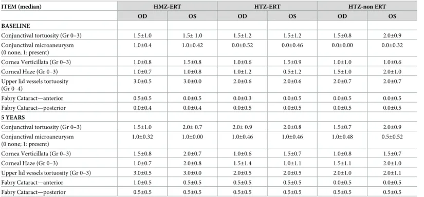
Anterior segment ocular manifestation findings are summarized inTable 6. All findings were graded according to the Fabry Outcome Survey[78] grading scale[19].
Conjunctival tortuosity. Bulbar conjunctival tortuosity was found in almost every subject
at baseline (24/28), except for 1/10 in Group HMZ-ERT, 2/8 in Group HTZ-ERT and 1/10 in Group HTZ-Non ERT. At the end, only one subject (Group HTZ-ERT) did not show this find-ing. On average, tortuosities were evaluated as moderate, varying from grade 1.5 to 2.0 on a scale of 3 (FOS grading scale). Only Group HTZ-ERT evolved in severity over time, with a pro-gression from 1.5 to 2.0, but this evolution was not considered statistically significant (p>0.05; 95%CI [-0.136, 0.615] OD p>0.05 95%CI [-0.122, 0.651] OS), most likely because of the low number of subjects. Other variations affecting Group HMZ-ERT and HTZ-Non ERT were not found significant as well (F = 0.51; p>0.05 OD; F = 0.91; p>0.05 OS); however, there was a time�group�mutation effect (F = 5.45; p<0.05) at baseline but not at the end.
Upper lid vessel tortuosity and microaneurysms. Upper lid vessel tortuosity is a new
clinical finding that has been recently identified[31]. Again here, almost every subject showed this manifestation, with the exception of one person in Group HTZ-Non ERT at the begin-ning. The same finding applied after 5 annual visits. This makes this ocular manifestation the most prevalent one. Subjects in Group HMZ-ERT showed more severe vessel tortuosity associ-ated with the presence of microaneurysms (grade 3 out of 4) at baseline (F = 9.226; p = <0.05) and at the end (F = 3.419; p<0.05), in both eyes, compared to other groups. There was no dif-ference in the evolution in prevalence or severity among groups over time as all remained sta-ble from baseline (F = 0.34; p>0.05 OD; F = 2.31; p>0.05 OS).
Table 6. Anterior segment findings.
ITEM (median) HMZ-ERT HTZ-ERT HTZ-non ERT
OD OS OD OS OD OS BASELINE Conjunctival tortuosity (Gr 0–3) 1.5±1.0 1.5± 1.0 1.5±1.2 1.5±1.2 1.5±0.8 2.0±0.9 Conjunctival microaneurysm (0 none; 1: present) 1.0±0.4 1.0±0.42 0.0±0.52 0.0±0.46 0.0±0.00 0.0±0.32 Cornea Verticillata (Gr 0–3) 1.0±0.8 1.5±0.8 1.0±0.6 1.5±0.9 1.0±1.0 1.0±0.6 Corneal Haze (Gr 0–3) 1.0±0.7 1.0±0.8 1.0±1.2 0.5±1.2 1.5±1.0 2.0±1.0
Upper lid vessels tortuosity (Gr 0–4) 3.0±0.5 3.0±0.0 2.0±0.6 2.0±0.6 2.0±0.7 2.0±0.7 Fabry Cataract—anterior 0.5±0.5 0.0±0.5 0.0±0.3 0.0±0.5 0.0±0.5 0.0±0.5 Fabry Cataract—posterior 0.0±0.4 0.0±0.4 0.0±0.5 0.0±0.5 0.0±0.5 0.0±0.5 5 YEARS Conjunctival tortuosity (Gr 0–3) 1.5±1.0 2.0± 0.7 2.0± 0.9 2.0±0.8 1.5±0.7 2.0±0.9 Conjunctival microaneurysm (0 none; 1: present) 1.0±0.32 1.0±0.00 1.0±0.46 1.0±0.46 1.0±0.48 0.5±0.52 Cornea Verticillata (Gr 0–3) 1.5±0.8 2.0±0.7 1.0±0.6 1.5±0.7 1.0±0.8 1.5±0.7 Corneal Haze (Gr 0–3) 1.0±0.7 2.0±0.8 1.5±1.4 1.0±1.1 1.5±1.1 2.0±1.0
Upper lid vessels tortuosity (Gr 0–3) 3.0±0.5 3.0±0.0 2.0±0.5 2.0±0.5 2.0±1.0 2.0±1.1
Fabry Cataract—anterior 1.0±0.5 0.5±0.5 0.5±0.5 0.5±0.5 0.0±0.5 0.0±0.5
Fabry Cataract—posterior 0.5±0.5 0.5±0.5 0.5±0.5 0.5±0.5 0.5±0.5 0.5±0.5
Conjunctival micro-aneurysms. Micro-aneurysms (MA) were detected mainly in the
bulbar conjunctiva, but also in the inner canthus, the lower palpebral conjunctiva, near the lid margin and on the superior external eyelid as well (see 5.2.2.). At baseline, 8/10 subjects of Group HMZ-ERT showed at least one MA in at least one eye, which was the case for 3/8 sub-jects in Group HTZ-ERT and 1/10 in Group HTZ-Non ERT. This difference was found to be significant (F = 3.88; p<0.05 OD; F = 3.761; p<0.05 OS), especially considering OD between Groups HMZ-ERT and HTZ-Non ERT (p<0.05; 95%CI [0.14, 0.91] OD); p<0.05; 95%CI [-0.02, 0.85] OS); and at a lesser extent between Groups HTZ-ERT and HTZ-Non ERT (p<0.05; 95%CI [-0.95, -0.14] OD); p<0.05; 95%CI [-0.82, 0.06]OS). At the end, Group HMZ-ERT remained stable with 9/10 subjects affected, which contrasts with a marked tion in prevalence for Group HTZ-ERT (6/8) and Group HTZ-Non ERT (7/10). This evolu-tion was found to be significant for the right eye (χ2 = 7.5616, p<0.05) but not for the left eye (χ2 = 1.8667; p>0.05), confirming the high intra-subject variability between both eyes.
Once a MA was found in the bulbar conjunctiva, it was usually found also on the superior external eyelid. This association is present in Group HMZ-ERT at baseline (5/10) increasing in prevalence with time (7/10), a similar situation occurring at a lesser extent in Group HTZ-ERT (4/8 at baseline and 5/8 after 5 years). There was almost no association found in Group HTZ-Non ERT at baseline (0/10), but this group became more aligned with others at the end of the study, with a higher rate (4/10). This difference among groups was found to be signifi-cant (F = 3.492; p<0.05 OD; F = 4.846; p<0.05 OS); however, the evolution in the numbers of positive association over time was considered the same among the groups (χ2 = 2.3407; p>0.05 OD;χ2 = 6.7183; p>0.05 OS).
Cornea verticillata and haze. Corneal verticillata is certainly considered an important
hallmark of ocular manifestations related to Fabry. Cornealdeposition was found in 10/10 Group HMZ-ERT subjects at baseline, and at the end. Similar data was observed in Group HTZ-Non ERT (9/10 at baseline and after 5 years). In GroupHTZ-ERT, subjects slightly evolved in prevalence from 5/8 to 6/8.
As reported inTable 6, Group HMZ-ERT showed more severe verticillata grading than the other groups at baseline, but this was not considered a significant difference (F = 0.562, p>0.05 OD; F = 0.633, p>0.05 OS). This was still the case at the end of the study. All groups in this study evolved in intensity by 0.5 deg (out of 4) over 5 years, which was considered a signif-icant progression if compared to baseline (F = 92.153, p<0.05 OD; F = 5.817, p<0.05 OS).
Verticillata can be accompanied by corneal haze, which is described as a loss of corneal transparency spreading habitually along the vertical meridian. At baseline, 7/10 subjects in Group HMZ-ERT showed low level (Grade 1 to 2) of corneal haze, much like 4/8 subjects in Group HTZ-ERT. For Group HTZ-Non ERT, 8/10 subjects were identified with moderate corneal haze (grade 2 to 3) on average. At the end, all Group HMZ-ERT subjects displayed cor-neal haze. Groups HTZ-ERT and HTZ-Non ERT remained stable with 5/8 and 8/10 subjects affected respectively. Overall, groups were found to be statistically similar at baseline (F = 0.745; p>0.05, OD; F = 1.670; p>0.05 OS). Their evolution varied over time, at least in terms of OS (F = 0.08; p>0.05 OD; F = 4.14; p< 0.05 OS). More specifically, there was a significant difference in the evolution in prevalence of Group HMZ-ERT vs Group HTZ-ERT (p<0.05; 95%CI [0.0624, 1.1207] OS), male patients treated with ERT (HMZ–ERT) showed more severe manifestations with time than female patients on ERT treatments. It is then not surprising to find a time�eye�group�mutation effect (F = 3.48; p<0.05).
Cataracts. Fabry’s cataracts can develop in the anterior portion of the crystalline lens, or
can be seen as a sub-cortical posterior lens opacity[37]. At baseline, anterior cataract was seen in 5/10 subjects of Group HMZ-ERT, 2/8 of Group HTZ-ERT, and 4/10 of Group HTZ-Non ERT. Subjects were considered as showing light to moderate opacities (grade 0.5 to 1). This
condition remained stable over time except for Group HTZ-ERT, showing a higher prevalence with 4/8 subjects affected at the end of the study. This represents twice the frequency of HTZ-ERT subjects, from baseline, especially marked on the right eye with a significant statisti-cal difference (χ2 = 5.3375; p<0.05 OD; χ2 = 2.9120; p>0.05 OS).
Posterior sub-capsular cataract was identifiable, at baseline, on 2/10 subjects of Group HMZ-ERT, 2/8 of Group HTZ-ERT, and 3/10 of Group HTZ-Non ERT. Grading reveals very early cataract onset. There was no significant difference among the groups (F = 0.510; p>0.05 OD; F = 0.938; p>0.05 OS). ERT-managed subjects (Group HMZ-ERT and Group HTZ-ERT) evolved significantly in prevalence over time, ending the study with a doubled number of affected subjects (Group HMZ-ERT: 5/10; Group HTZ-ERT: 4/10). Group HTZ-Non ERT, females untreated, did not evolve significantly in prevalence, with 4/10 subjects with lens opa-cification at the end of the study. Severity increased with time, especially for Group HMZ-ER individuals where 2 patients were referred for cataract surgery, second to disturbing light sen-sitivity. None of the other groups underwent surgery; however, most of the patients affected with some degree of sub-capsular posterior opacification reported symptoms of high light sen-sitivity, making it difficult for them to function in bright light conditions without wearing sun-glasses or a hat to generate more shade.
Retinal vessel tortuosity. Blood vessel tortuosity can be seen in arterioles and veinules. If
present, such modifications are graded from 0 (none) to 3 (severe), based on FOS scale[19]. Results can be found inTable 7. There were differences among the groups. At baseline, 9/10 subjects showed vessel tortuosity in Group HMZ-ERT, compared to 5/8 in Group HMZ-ERT and 4/10 in Group HMZ-Non ERT. The difference was found to be statistically significant (F = 3.781; p = 0.0481 OD; F = 4.634; p = 0.0401 OS). More specifically, HTZ-ERT subjects seemed to be more affected (prevalence) than HTZ-Non ERT subjects (p = 0.0438; 95%CI [-1.97, -0.41]). There was no significant evolution in prevalence and severity of the condition over study time.
It is interesting to note that blood vessels silver wiring was noticed in 2 patients (HMZ-advanced cases in severity), an occurrence that was not reported before.
Asymmetry. It is important to note that, in a vast majority of patients related to this
cohort, ocular manifestations were found highly variable between individuals, as already reported[21], but most interestingly, intra-subjects. This implies that clinical manifestations were not symmetrical between both eyes, which may help eyecare practitioners establishing their differential diagnosis for Fabry.
Discussion
This study tried to establish the prevalence and the natural course in intensity of ocular mani-festations, over 5 annual visits, in Fabry patients. Prevalence of the ocular manifestations found in this study (seeTable 8) can be easily compared to the results from a review published earlier[30]. All results are in keeping with known prevalence, although variations may occur with different demographic features, subject genotypes, technology used to evaluate clinical
Table 7. Posterior segment findings.
HMZ-ERT HTZ-ERT HTZ-Non ERT
OD OS OD OS OD OS Baseline (Grade 0–3) 2.0±0.8 2.0±0.8 1.5± 1.4 1.5± 1.4 0.0± 0.8 0.5± 0.8 5 years (Grade 0–3) 2.0±0.8 2.0± 1.1 2.0± 1.2 2.0± 1.2 0.5± 0.9 1.0± 0.8 https://doi.org/10.1371/journal.pone.0213329.t007
manifestations and the subjectivity of the observers. Based on this study results, it is possible to identify the stage of the disease evolution, as another factor to consider. A patient on ERT-treatment is expected to show more signs, with increased severity. Consequently, it is not pos-sible to exclude totally the age effect on the results presented here, all 3 groups being consid-ered older Fabry patients. Another cohort, composed of younger Fabry patients, would show a different clinical picture.
One important finding coming from the analysis of our data (Table 8) is the fact that ERT does not seem to halt the clinical evolution of several ocular manifestations as it does influence positively body’s functions. We did see an increased prevalence in MA (+35%), Corneal Haze (+14%), especially for heterzogygotes treated, anterior cataract (+26%) for hemizygotes treated; and finally posterior cataracts (+22%), in treated patients overall. Evolution in the prevalence of these signs over the study time was confirmed, despite the regular use of enzyme therapy.
In particular, vascular tortuosities prevalence and evolution did not seem to be altered with the use of ERT because synthetic enzyme does not enter the vessel wall beyond the vascular endothelial cell layer and does not enter the epithelial cell layer in the cornea. Then, vessels changes can be considered as irreversible once developed.
Some would argue that switching drugs during the study may have influenced these results. This was not the case for cardiac function.[79] Looking specifically at the same population studied here, it was reported that switching from alphagal-beta to alfa form, both in standard dose, was associated with a transient increase in clinical events for 6 months but thereafter the clinical event rate returned to baseline for the next 3 years[80]. Consequently, it is not likely that the switch of enzyme had influenced the evolution of ocular manifestations observed here.
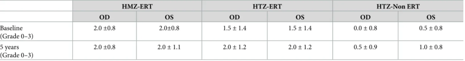
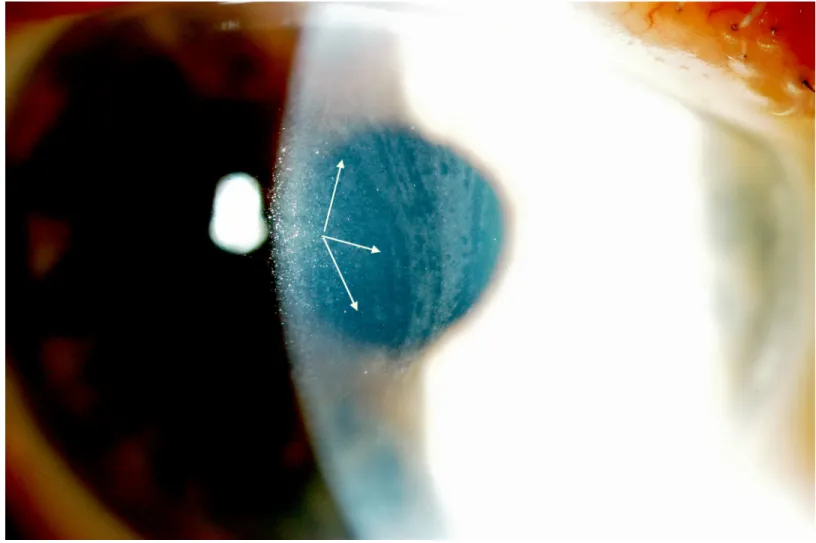
This study confirmed that cornea verticillata (Fig 1) represents a proeminent hallmark fea-ture of Fabry disease, being present in almost every subject (89.2%). Those not showing verti-cillata were all heterozygotes. Two of them were family related, aged 16 and 48, and affected by p.Arg332Aspfs16 mutation. Another one (58 y.o.) was showing p.Val254del mutation. None of them were treated (ERT) and their condition was considered less severe, associated with no systemic symptoms. Cornea verticillata, already well developed at baseline in other patients, did not evolve in intensity over time. For not treated females, corneal signs remained stable over time, as their systemic condition.
Table 8. Compared prevalence of ocular manifestations.
This study- Baseline(%) This study-5 years (%) Comments
(Prevalence–Evolution)
Litteraturereview (%)�
Conjuctival tortuosity 85.7 Not evolved P:>HTZ-ERT 41.6 to 97
Upper lid tortuosity 96.4 Not evolved P:HMZ> HTZ Not reported
Micro-Aneurysms 42.8 78 E: HMZ Not reported
Cornea Verticillata 89.2 Not evolved P :HMZ> HTZ ERT> Non -ERT 43.5 to 94.5
Corneal Haze 67.8 82.1 E: HMZ-ERT 10 to 84.3
Cataract- Anterior 39.2 46.4 E: HMZ-ERT 9 to 42
Cataract- Sub-capsular Posterior 28.5 50 P: HMZ >HTZ
ERT>Non- ERT
11.8 to 50
Retinal vessels abnormalities 64.2 67.8 P: ERT>Non-ERT 18.8 to 90
HMZ: HEmizygotes; HTZ: Heterozygotes; ERT: Enzyme Replacement Therapy; Non-ERT: not treated with ERT
�from Sodi A, Ioannidis AS, Mehta A, Davey C, Beck M, Pitz S. Ocular manifestations of Fabry’s disease: data from the Fabry Outcome Survey. Br J Ophthalmol.
2007;91(2):210–4.
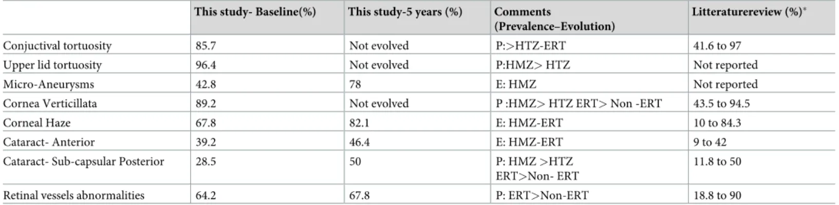
Corneal haze (Figs2and3) is another remarkable feature of Fabry. In this study, some cor-neas showed haze as the prominent corneal sign over the verticillata. In general, this finding was found in 68% of the patients at baseline, evolving in prevalence to 82% at the study com-pletion. Looking in details, Group HTZ-ERT evolve more in prevalence than the others. Put-ting this in perspective, Group HMZ-ERTwas already advanced, and this confirms that female patients under treatment seemed to tend matching with time the clinical findings of males under treatment.
This clinical portrait may be different in the next years. It was reported that the corneal ver-ticilatta is reversed with migalastat[81]. The latter is a small molecule that is widely distributed in the body contrary to the large protein that is alpha-gal.
Corneal deposits and haze did not affect the level of high-order aberrations nor the bio-mechanical properties of the cornea. Corneal hysteresis represents the energyabsorption dur-ing the stress applied on the cornea and is lowered when the cornea is softer or altered by a dystrophy or a structural disease[73]. Lower CH values are associated with a higher risk to develop ocular pathology like glaucoma[82]. This parameter was never assessed in a Fabry population before. As a reference, CH values measured in a normal cornea population varies from 10.2 to 11.1 mm, depending on the sex, ethnical origin and intra-ocular pressure[83]. The study Fabry population showed very similar results. Variations observed may be attributed
Fig 1. Typical Cornea Verticillata with haze.
to ageing instead of being related to the metabolic disorder. It is then possible to conclude that substrate accumulation within the cornea does not modify its biomechanical property.
To the same extent, the corneal resistance factor (CRF) represents the corneal elastic prop-erty and is related to the corneal thickness[84]. In this study, the CRF varied from 9.9 to 12.2 mm Hg, similar to a non-Fabry Caucasian population, as already mentioned. Again here, vari-ations found are explainable by ageing process instead of being linked with the metabolic dis-order itself or any change to the corneal structure second to substrate accumulation.
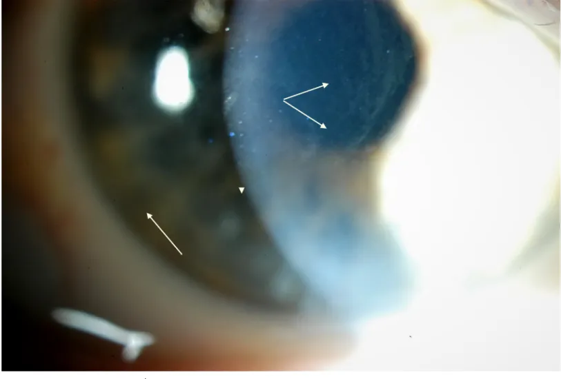
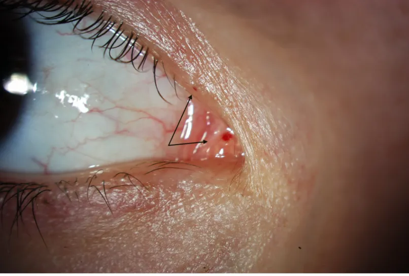
The presence of micro-aneurysm evolved in prevalence over time. It may suggest a deterio-ration of the patient systemic condition, more specifically an increased level of substrate accu-mulation within the blood vessel walls. Usually found in the lower bulbar conjunctiva, MA, in this study, was also found in the inner canthus fornix (Fig 4), at the lid margin (Fig 5), on the external inferior eyelid or superiorly. The variation of the location confirms the fact that ocular manifestation occurs where the substrate accumulates, which is not predictable.
Upper lid vessels tortuosity (Fig 6) was also highly prevalent (96.4%). This is not surprising that the small vessels of the upper lid are showing abnormalities, as they are very similar in structure to those of the bulbar conjunctiva where tortuosities are well documented. Similarity between these 2 areas extend also to the presence of micro-aneurysms on both locations espe-cially among patients ERT-treated. This means that if at least one micro-aneurysm is found
Fig 2. Corneal Haze as seen at baseline (1stvisit). https://doi.org/10.1371/journal.pone.0213329.g002
either on the bulbar conjunctiva, the inner canthus or the lid margin, it is very likely to find one micro-aneurysm along the vessels or in the area of the external upper lid. The strong asso-ciation between the occurence of MA on both locations may be used, in the future, as one of the potential validation element for Fabry diagnosis.
It may be surprising that posterior cataract did evolve in prevalence, while anterior cataracts did not change significantly. First of all, posterior cataracts are more prevalent than the ante-rior one, occurring in up to 70% of the male patients[37], and are known as “classic” Fabry cat-aracts. Anterior opacities are described as “propeller” and are less frequently seen, depending on where the substrate will accumulate within the body[21]. Fabry patients may report visual symptoms including eye dryness, blurry vision, difficulty in seeing under dim or reduced illu-mination, and soreness/tiredness[78]. These symptoms, including light sensitivity, may all be related to lens opacities or corneal deposits and should be assessed at every visit. Whenever present, they may prompt to consider a lens extraction on highly symptomatic patients. Evolu-tion of lens opacities may be caused by increased disease severity or simply with ageing. This study was not powered enough to be able to make specific analysis, in that regard, but we may suspect that chances are higher to develop disturbing symptoms as the patient’s condition becomes more severe and the patient is getting older.
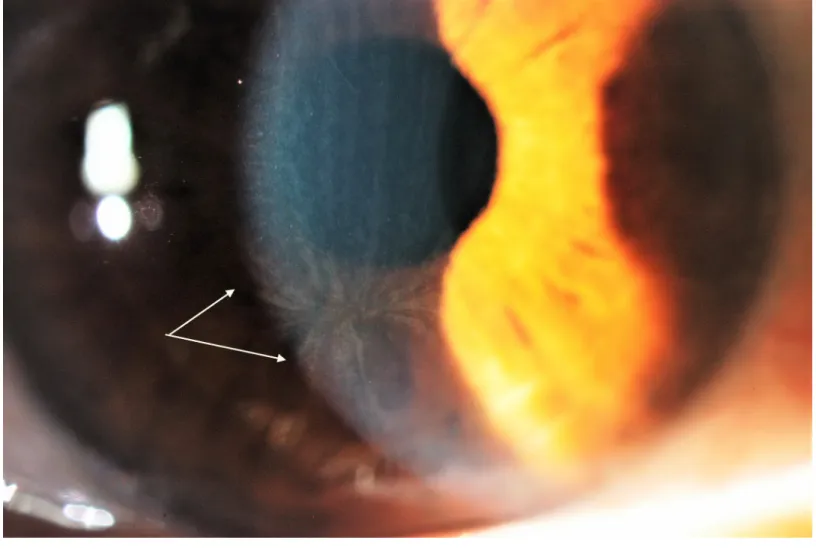
Fig 3. Cornal Haze of the same patient (photo 2) 5 years later, showing increased severity.
Retinal vessel tortuosity (Fig 7) brings another evidence that ERT subjects are more affected than non-ERT subjects. There was a significant difference in prevalence and intensity among the groups, at baseline, which did not vary over time. It is interesting to report that, in 2 indi-viduals (HMZ-ERT) retinal arteries displayed sliver wiring (Fig 8), which was never reported before for Fabry patients. This rare phenomenon is described as a thickening of walls of retinal arterioles leading to the loss of vessels visibility and reflection. This ocular manifestation is habitually related to abnormal systemic blood hypertension but we cannot exclude that, in the case of Fabry, thickening of the walls may be related to high substrate accumulation as a sec-ond contributor mechanism. Both subjects were highly symptomatic for Fabry, including a severe cardiac condition, and one died during the course of the study from a cardiac failure.
Visual field defects are manifested as a loss of sensitivity in a point or an area. Approxi-mately 50% of the study population showed such a variation during the study; however, sur-prisingly, the area where the defect was located at baseline, moved during the following years in a few cases. In order to understand visual field findings, it is important to remember that FDT technology targets magnocellular cells, which are the first ones to be affected by a change in the optic nerve perfusion, which may be altered[85] in the case of Fabry patients, second to GL-3 deposits causing dilatation and instability of the vascular lumen in Fabry patients [21]. This result about visual fields cannot be compared to previous findings, where 37% of the
Fig 4. Microaneurysm located in the inner canthus (fornix).
patients showed defects manifested as an enlarged blind spot[27]. In that case, the Goldman perimetry was used, which implies a seen to unseen strategy for any target presented in the field. Lens opacities (cataracts) may play a role, altering light perception and decreasing sensi-tivity. However, only 2 patients in this study developed cataracts significant enough to require surgery. Consequently, their influence on the overall results may be considered marginal. Future studies will be needed to confirm these hypotheses.
As a final note, it is important to indicate that, in general, one eye evolved more in preva-lence or in intensity of the manifestations, than the fellow eye. This confirms the high asymme-try between the clinical picture found is a vast majority of Fabry patients. If there is one rule about Fabry ocular manifestations, coming from this study, it is that what is found on one eye never mirrors what is seen on the other one.
This study may be limited by the number of drop-outs, expected by the nature of the disease and its evolution over time. However, most of these drop-outs were considered more severe cases. As we saw, the ocular manifestations are more numerous and more intense on such patients. Consequently, our results could have been modified if those subjects would had been able to remain involved and included in the final analysis. To the same extent, the recruitment of a younger population would have modified the findings, assuming that younger patients
Fig 5. Microaneurysm as seen near the inferior lid margin.
may be considered less advanced cases, and consequently, showing reduced ocular manifesta-tions in prevalence and intensity.
A second bias is the fact that all males recruited for the study were under treatment. In a perfect world, a group of non-treated hemizygotes would had been followed and compared to treated counterparts, as we were able to do for females. With the same rationale, establishing a correlation between ocular manifestations evolution and other outcomes i.e., kidney, heart, biomarkers that have been reported to respond to ERTs would had been interesting. This study was not designed for this purpose and limited access to the complete file of every patients, for administrative and legal reasons, did not allow to proceed. On the same note, it was not possible to access reliable data on the systemic condition of each participants, due to ethics limitations and privacy regulations. It would had been interesting to evaluate the influ-ence of the degree of systemic involvement, comorbidities on the results of the study, and to confirm the direct link between severity of the disease and intensity of ocular manifestations already identified by others.
A third element to consider is the time of follow-up. For renal outcomes, the CFDI reported a difference after 10 years but not after 5 years[41]. Limited funding and drop-out rate over the years reduced the possibility of extending this study.
Fig 6. Upper lid tortuosity–grade 4, with micro-aneurysm.
A fourth bias is the fact that images and clinical data were acquired and analyzed by the same individual. In a perfect scenario, images are taken by one individual and analyzed by a masked observer. Another possibility is to rely on 2 readers to grade clinical findings and to average their score. This was not possible, considering the longitudinal nature of the study and the resources available. Grading in this study was based on a validated scale (FOS). However, author acknowledges that alterations may be very subtle and poorly quantifiable in some patients. Reader must keep in mind that possible variability of data analyzed here may attenu-ate overall results and conclusions about ERT effect on ocular manifestations over time.
Many efforts have been done to obtain quantitative data but at present reliable measurable parameters are not available yet. Then at present even referring to possible existing grading systems, the evaluation of ocular signs in FD remain mostly subjective. Consequently it may be challenging to establish definitive conclusions on possible longitudinal ocular changes.
A fifth element is that, obviously, the results and the conclusion driven from this study can be applied only to a similar population of patients. Extrapolation to other Fabry’s patient cohorts, different in ethnical origin or disease severity, may seems to be logical, but may be hazardous. However, author is confident that this study provides a new understanding on ERT outcome on ocular manifestations and sheds a new lightning on the natural evolution of them over time.
Fig 7. Low retinal vessels tortuosities (arterioles and veinules).
Conclusion
Based on a subjective observation, it was possible to establish that five of the known ocular clinical signs related to Fabry did significantly evolve in prevalence and/or in intensity over 5 annual visits, despite enzyme replacement treatment. Micro-aneurysms (+82%) was the clini-cal manifestation that evolved the most in prevalence, followed by classic (posterior) Fabry’s cataract (+75%) corneal haze (+21%) and propeller (anterior) cataract (+17%). Three other manifestations remain stable over time. At the end, ocular manifestations of ERT-Heterozy-gotes were comparable with treated hemizyERT-Heterozy-gotes (HMZ). A more objective way to assess data and longer observational time may be required to fully confirm these findings.
Supporting information
S1 File. Overall results. This is the complete data set of elements evaluated in this study.
(SAV)
Acknowledgments
Dr Daniel Bichet M.D.–UM professor–metabolic disorder clinic Hopital Sacre Cœur.
Fig 8. Silver wiring of retinal arteries.
Author Contributions
Conceptualization: Langis Michaud. Formal analysis: Langis Michaud. Funding acquisition: Langis Michaud. Investigation: Langis Michaud. Methodology: Langis Michaud.
Project administration: Langis Michaud. Resources: Langis Michaud.
Supervision: Langis Michaud. Validation: Langis Michaud.
Writing – original draft: Langis Michaud. Writing – review & editing: Langis Michaud.
References
1. Fuller M, Meikle PJ, Hopwood JJ. Epidemiology of lysosomal storage diseases: an overview. In: Mehta A, Beck M, Sunder-Plassmann G, editors. Fabry Disease: Perspectives from 5 Years of FOS. Oxford: Oxford PharmaGenesis; 2006.
2. Zarate YA, Hopkin RJ. Fabry’s disease. Lancet. 2008; 372(9647):1427–35.https://doi.org/10.1016/ S0140-6736(08)61589-5PMID:18940466
3. Chan B, Adam DN. A Review of Fabry Disease. Skin Therapy Lett. 2018; 23(2):4–6. PMID:29562089 4. Vedder AC, Linthorst GE, van Breemen MJ, Groener JE, Bemelman FJ, Strijland A, et al. The Dutch
Fabry cohort: diversity of clinical manifestations and Gb3 levels. J Inherit Metab Dis. 2007; 30(1):68–78.
https://doi.org/10.1007/s10545-006-0484-8PMID:17206462
5. MacDermot KD, Holmes A, Miners AH. Anderson-Fabry disease: clinical manifestations and impact of disease in a cohort of 98 hemizygous males. J Med Genet. 2001; 38(11):750–60.https://doi.org/10. 1136/jmg.38.11.750PMID:11694547
6. Pitz S, Kalkum G, Arash L, Karabul N, Sodi A, Larroque S, et al. Ocular signs correlate well with disease severity and genotype in Fabry disease. PLoS One. 2015; 10(3):e0120814.https://doi.org/10.1371/ journal.pone.0120814PMID:25781336
7. MacDermot KD, Holmes A, Miners AH. Anderson-Fabry disease: clinical manifestations and impact of disease in a cohort of 60 obligate carrier females. J Med Genet. 2001; 38(11):769–75.https://doi.org/ 10.1136/jmg.38.11.769PMID:11732485
8. Wang RY, Lelis A, Mirocha J, Wilcox WR. Heterozygous Fabry women are not just carriers, but have a significant burden of disease and impaired quality of life. Genet Med. 2007; 9(1):34–45.https://doi.org/ 10.1097/GIM.0b013e31802d8321PMID:17224688
9. Miller-Keane. Encyclopedia and Dictionary of Medicine, Nursing, and Allied Health, Seventh Edition. 2003.
10. Ortiz A, Germain DP, Desnick RJ, Politei J, Mauer M, Burlina A, et al. Fabry disease revisited: Manage-ment and treatManage-ment recommendations for adult patients. Mol Genet Metab. 2018; 123(4):416–27.
https://doi.org/10.1016/j.ymgme.2018.02.014PMID:29530533
11. Vedder AC, Strijland A, vd Bergh Weerman MA, Florquin S, Aerts JM, Hollak CE. Manifestations of Fabry disease in placental tissue. J Inherit Metab Dis. 2006; 29(1):106–11.https://doi.org/10.1007/ s10545-006-0196-0PMID:16601876
12. El-Abassi R, Singhal D, England JD. Fabry’s disease. J Neurol Sci. 2014; 344(1–2):5–19.https://doi. org/10.1016/j.jns.2014.06.029PMID:25106696
13. Ries M, Gupta S, Moore DF, Sachdev V, Quirk JM, Murray GJ, et al. Pediatric Fabry disease. Pediat-rics. 2005; 115(3):e344–55.https://doi.org/10.1542/peds.2004-1678PMID:15713906
14. Ramaswami U, Parini R, Pintos-Morell G. Natural history and effects of enzyme replacement therapy in children and adolescents with Fabry disease. In: Mehta A, Beck M, Sunder-Plassmann G, editors. Fabry Disease: Perspectives from 5 Years of FOS. Oxford: Oxford PharmaGenesis; 2006.
15. Hopkin RJ, Bissler J, Banikazemi M, Clarke L, Eng CM, Germain DP, et al. Characterization of Fabry disease in 352 pediatric patients in the Fabry Registry. Pediatr Res. 2008; 64(5):550–5.https://doi.org/ 10.1203/PDR.0b013e318183f132PMID:18596579
16. Foda MM, Mahmood K, Rasuli P, Dunlap H, Kiruluta G, Schillinger JF. High-flow priapism associated with Fabry’s disease in a child: a case report and review of the literature. Urology. 1996; 48(6):949–52. PMID:8973687
17. Orteu CH, Jansen T, Lidove O, Jaussaud R, Hughes DA, Pintos-Morell G, et al. Fabry disease and the skin: data from FOS, the Fabry outcome survey. Br J Dermatol. 2007; 157(2):331–7.https://doi.org/10. 1111/j.1365-2133.2007.08002.xPMID:17573884
18. Hegemann S, Hajioff D, Conti G, Beck M, Sunder-Plassmann G, Widmer U, et al. Hearing loss in Fabry disease: data from the Fabry Outcome Survey. Eur J Clin Invest. 2006; 36(9):654–62.https://doi.org/ 10.1111/j.1365-2362.2006.01702.xPMID:16919049
19. Mehta A, Ricci R, Widmer U, Dehout F, Garcia de Lorenzo A, Kampmann C, et al. Fabry disease defined: baseline clinical manifestations of 366 patients in the Fabry Outcome Survey. Eur J Clin Invest. 2004; 34(3):236–42.https://doi.org/10.1111/j.1365-2362.2004.01309.xPMID:15025684
20. Tsutsumi A, Uchida Y, Kanai T, Tsutsumi O, Satoh K, Sakamoto S. Corneal findings in a foetus with Fabry’s disease. Acta Ophthalmol (Copenh). 1984; 62(6):923–31.
21. Sher NA, Letson RD, Desnick RJ. The ocular manifestations in Fabry’s disease. Arch Ophthalmol. 1979; 97(4):671–6. PMID:106811
22. Mastropasqua L, Nubile M, Lanzini M, Carpineto P, Toto L, Ciancaglini M. Corneal and conjunctival manifestations in Fabry disease: in vivo confocal microscopy study. Am J Ophthalmol. 2006; 141 (4):709–18.https://doi.org/10.1016/j.ajo.2005.11.053PMID:16564807
23. Nguyen TT, Gin T, Nicholls K, Low M, Galanos J, Crawford A. Ophthalmological manifestations of Fabry disease: a survey of patients at the Royal Melbourne Fabry Disease Treatment Centre. Clin Exp Ophthalmol. 2005; 33(2):164–8.https://doi.org/10.1111/j.1442-9071.2005.00990.xPMID:15807825 24. Kalkum G, Pitz S, Karabul N, Beck M, Pintos-Morell G, Parini R, et al. Paediatric Fabry disease: prog-nostic significance of ocular changes for disease severity. BMC Ophthalmol. 2016; 16(1):202.https:// doi.org/10.1186/s12886-016-0374-2PMID:27852300
25. Bartlett JD. Systemic Drugs affecting the Eye. In: Bartlett JD, editor. Ophthalmic Drugs Facts. 21st ed. St-Louis, MO: Wolters Kluwer Health; 2010. p. 335–42.
26. Sodi A, Ioannidis A, Pitz S. Ophthalmological manifestations of Fabry Disease. In: Mehta A, Beck M, Sunder-Plassman G, editors. Fabry Disease Perspective from 5 years of FOS. Oxford, UK: Oxford PharmaGenesis Ltd.; 2006. p. 249–61.
27. Orssaud C, Dufier J, Germain D. Ocular manifestations in Fabry disease: a survey of 32 hemizygous male patients. Ophthalmic Genet. 2003; 24(3):129–39. PMID:12868031
28. Arbisser AI, Murphree AL, Garcia CA, Howell RR. Ocular findings in mannosidosis. Am J Ophthalmol. 1976; 82(3):465–71. PMID:961797
29. Riegel EM, Pokorny KS, Friedman AH, Suhan J, Ritch RH, Desnick RJ. Ocular pathology of Fabry’s dis-ease in a hemizygous male following renal transplantation. Surv Ophthalmol. 1982; 26(5):247–52. PMID:6806927
30. Sodi A, Ioannidis AS, Mehta A, Davey C, Beck M, Pitz S. Ocular manifestations of Fabry’s disease: data from the Fabry Outcome Survey. Br J Ophthalmol. 2007; 91(2):210–4.https://doi.org/10.1136/bjo. 2006.100602PMID:16973664
31. Michaud L. Vascular tortuosities of the upper eyelid: a new clinical finding in fabry patient screening. J Ophthalmol. 2013; 2013:207573.https://doi.org/10.1155/2013/207573PMID:24223300
32. Sher NA, Reiff W, Letson RD, Desnick RJ. Central retinal artery occlusion complicating Fabry’s disease. Arch Ophthalmol. 1978; 96(5):815–7. PMID:207248
33. Sodi A, Bini A, Mignani R, Minuti B, Menchini U. Subfoveal choroidal neovascularization in a patient with Fabry’s disease. Int Ophthalmol. 2009; 29(5):435–7.https://doi.org/10.1007/s10792-008-9252-0
PMID:18784903
34. Snodgrass MB. Ocular findings in a case of fucosidosis. Br J Ophthalmol. 1976; 60(7):508–11.https:// doi.org/10.1136/bjo.60.7.508PMID:952824
35. O’Brien J. Generalized gangliosidosis. J Pediatr. 1969; 75(2):167–86. PMID:4240224
36. Pitz S, Grube-Einwald K, Renieri G, Reinke J. Subclinical optic neuropathy in Fabry disease. Ophthal-mic Genet. 2009; 30(4):165–71.https://doi.org/10.3109/13816810903148004PMID:19852573 37. Sivley MD. Fabry disease: a review of ophthalmic and systemic manifestations. Optom Vis Sci. 2013;