HAL Id: hal-02893181
https://hal-univ-lemans.archives-ouvertes.fr/hal-02893181
Submitted on 8 Jul 2020
HAL is a multi-disciplinary open access
archive for the deposit and dissemination of
sci-entific research documents, whether they are
pub-lished or not. The documents may come from
teaching and research institutions in France or
abroad, or from public or private research centers.
L’archive ouverte pluridisciplinaire HAL, est
destinée au dépôt et à la diffusion de documents
scientifiques de niveau recherche, publiés ou non,
émanant des établissements d’enseignement et de
recherche français ou étrangers, des laboratoires
publics ou privés.
Distributed under a Creative Commons Attribution| 4.0 International License
on Palmitate-Treated HepG2 Cells
Claire Mayer, Martine Côme, Vincent Blanckaert, Graziella Zittelli, Cecilia
Faraloni, Hassan Nazih, Khadija Ouguerram, Virginie Mimouni, Benoît
Chénais
To cite this version:
Claire Mayer, Martine Côme, Vincent Blanckaert, Graziella Zittelli, Cecilia Faraloni, et al.. Effect of
Carotenoids from Phaeodactylum tricornutum on Palmitate-Treated HepG2 Cells. Molecules, MDPI,
2020, 25 (12), pp.1-14. �10.3390/molecules25122845�. �hal-02893181�
Article
E↵ect of Carotenoids from
Phaeodactylum tricornutum on
Palmitate-Treated HepG2 Cells
Claire Mayer1 , Martine Côme1, Vincent Blanckaert1, Graziella Chini Zittelli2 ,
Cecilia Faraloni2, Hassan Nazih3 , Khadija Ouguerram4, Virginie Mimouni1
and Benoît Chénais5,*
1 EA 2160 MMS, Mer Molécules Santé, IUML FR 3473 CNRS, Institut Universitaire Technologique,
Le Mans Université, F-53020 Laval CEDEX 9, France; claire.mayer@univ-lemans.fr (C.M.); martine.come@univ-lemans.fr (M.C.); vincent.blanckaert@univ-lemans.fr (V.B.);
virginie.mimouni@univ-lemans.fr (V.M.)
2 National Research Council, Department of Biology, Agriculture and Food Sciences, Institute for BioEconomy,
I-50019 Sesto Fiorentino (Florence), Italy; graziella.chinizittelli@cnr.it (G.C.Z.); faraloni@cnr.it (C.F.)
3 EA 2160 MMS, Mer Molécules Santé, IUML FR 3473 CNRS, UFR Pharmacie, Université de Nantes,
F-44035 Nantes CEDEX 1, France; el-hassane.nazih@univ-nantes.fr
4 UMR1280 PhAN, Physiopathology of Nutritional Adaptations, INRAe, University of Nantes,
CHU Hôtel Dieu, IMAD, CRNH Ouest, F-44000 Nantes, France; khadija.ouguerram@univ-nantes.fr
5 EA 2160 MMS, Mer Molécules Santé, IUML FR 3473 CNRS, UFR Sciences et Techniques, Le Mans Université,
F-72085 Le Mans CEDEX 9, France
* Correspondence: bchenais@univ-lemans.fr; Tel.: +33-243-833-251 Academic Editor: Jesus Simal-Gandara
Received: 9 June 2020; Accepted: 17 June 2020; Published: 19 June 2020
!"#!$%&'(!
!"#$%&' Abstract: Non-alcoholic fatty liver disease represents the most common liver disease and is characterized by an excess of lipid accumulation in hepatocytes, mainly stored as triglycerides. Phaeodactylum tricornutum is a marine microalga, which is rich in bioactive molecules known to be hepatoprotective, such as n-3 long-chain polyunsaturated fatty acids and fucoxanthin. The aim of this study was to investigate the e↵ects of a carotenoid extract from P. tricornutum in a cellular model of non-alcoholic fatty liver disease induced by palmitate treatment. The combined e↵ects of carotenoids and lipids, especially n-3 long-chain polyunsaturated fatty acids, were also investigated by using a total lipophilic extract. HepG2 cells were exposed for 24 h to 250 µM palmitate with or without the addition of carotenoid extract (6 µg/mL) or total lipophilic extract (100 µg/mL). The addition of carotenoid extract or total lipophilic extract prevented the accumulation of triglycerides, total cholesterol and cholesterol esters. The carotenoid extract and total lipophilic extract also decreased the mRNA expression levels of genes involved in lipogenesis (ACACA, FASN, SCD and DGAT1) and cholesterol esterification (ACAT1/SOAT1). In addition, the total lipophilic extract also downregulated the LXR/NR1H3 and SREBF1 genes, which are involved in lipogenesis regulation. By contrast, the carotenoid extract increased the mRNA level of CPT1A, a -oxidation related gene, and reduced the lipid droplet accumulation. In conclusion, this study highlights the preventive e↵ects against non-alcoholic fatty liver disease of the two microalga extracts.
Keywords: Phaeodactylum tricornutum; total lipophilic extract; carotenoid extract; n-3 LC-PUFA; lipid metabolism; gene expression; HepG2 cells; palmitate; non-alcoholic fatty liver disease
1. Introduction
The changing lifestyle of our society, marked by an over-consumption of saturated fatty acids and sugars and a decrease in physical activity, is the main cause of the increase in the number of people with non-alcoholic fatty liver disease (NAFLD) [1,2]. NAFLD is currently the most common chronic liver disease in Western countries and a↵ects more than 30% of the general population [2,3]. NAFLD is characterized by an excessive accumulation of lipids in the parenchymal cells of the liver in the absence of excessive alcohol consumption [3]. Based on the analyses of the Dallas Heart Study cohort, NAFLD was determined by a triglyceride level above 5.5% in the liver [4]. NAFLD is a consequence of obesity and metabolic syndrome [3–5]. Moreover, NAFLD can progress to a more serious stage, namely non-alcoholic steatohepatitis, which can lead to hepatic fibrosis [3].
De novo lipogenesis plays a major role in fatty acid metabolism and, in particular, contributes significantly to triglyceride accumulation in the hepatocytes of NAFLD patients [6]. Acetyl-CoA carboxylase (ACACA) converts acetyl-CoA to malonyl CoA, and diacylglycerol o-acyltransferase (DGAT) enzymes use acyl-CoA as a substrate as well as diacylglycerols to form triglycerides [6,7]. During lipid droplet formation, in addition to triglycerides, cholesterol esters are generated by the enzymes acyl-CoA cholesterol acyltransferases (ACATs), also called sterol o-acyltransferase (SOAT) [7]. The stearyl-CoA desaturase (SCD) is also an enzyme of de novo lipogenesis that catalyzes the synthesis of monounsaturated fatty acids from saturated fatty acids and that is involved in the development of insulin resistance [8]. In NAFLD patients, an increase in SCD activity was observed in the liver [9]. The de novo synthesis of triglycerides is promoted by the activation of the sterol regulatory element-binding transcription factor 1 (SREBF1), which is a target of nuclear liver X receptor (LXR/NR1H3) [10,11]. In order to limit the accumulation of fatty acids, liver cells increase mitochondrial -oxidation by increasing the input of fatty acids through carnitine palmitoyltransferase-1 (CPT1) and CPT2 [12]. However, an increase in mitochondrial -oxidation decreases the antioxidant capacity and causes increased oxidative stress [12].
Several reports have highlighted that n-3 long-chain polyunsaturated fatty acids (n-3 LC-PUFA), particularly eicosapentaenoic acid (EPA) and docosahexaenoic acid (DHA), reduce NAFLD progression by decreasing hepatic contents in triglycerides and cholesterol esters [13,14]. These beneficial e↵ects could be explained by the ability of these n-3 LC-PUFA to control the activity and/or expression of transcription factors that regulate the expression of genes encoding proteins involved in de novo lipogenesis, fatty acid oxidation, fat uptake from the circulation and its assimilation into lipids, and very low density lipoprotein assembly and secretion [15]. Moreover, it was demonstrated that n-3 LC-PUFA exert preventive e↵ects against dyslipidemia, inflammation, insulin-resistance, oxidative stress, and metabolic disturbances, all involved in the progression and establishment of NAFLD [16–21]. n-3 LC-PUFA are abundantly present in fish such as salmon, herring or cod and are mainly marketed as oil [22]. However, the decrease in fishery resources linked to marine pollution necessitates the search for new alternative sources of n-3 LC-PUFA [23].
Investigations of the potential e↵ects of biomolecules from marine sources other than fish oil on NAFLD are crucial for new prevention strategies. To remedy this problem, n-3 LC-PUFA derived from microalgae could represent an interesting alternative. Indeed, thanks to their first place in the food chain, microalgae are less sensitive to heavy metal contamination [24]. Moreover, microalgae are a potential source of highly bioactive secondary metabolites such as pigments, sterols and soluble fibers and o↵er better nutritional value than fish oils [25]. Pigments, especially fucoxanthin, have also shown preventive e↵ects against NAFLD, thanks to their high anti-obesogenic, anti-dyslipidemic, antioxidant and anti-inflammatory activities [26–29].
P. tricornutum is a ubiquitous marine diatom, located particularly in coastal areas subjected to salinity variations [30]. P. tricornutum is generally found in temperate or cold waters and can be pelagic or benthic [30]. Its appeal lies in its high level of EPA and fucoxanthin [31–33]. Some studies conducted in rodents have shown the hepatoprotective e↵ects of microalgal lipophilic extracts rich in n-3 LC-PUFA against NAFLD [34,35]. However, to the best of our knowledge, no in vitro studies have demonstrated
the protective e↵ect of a pigment extract of P. tricornutum against NAFLD, whereas a recent study showed a reduction in fat accumulation and liver triglycerides in C57BL/6J mice supplemented with a P. tricornutum extract rich in fucoxanthin [36]. In addition, in a previous study in Wistar rats fed a high-fat diet supplemented with P. tricornutum at a dose of 12%, we also observed preventive e↵ects with respect to NAFLD [37].
Thus, the main objective of this work was to study the efficiency of carotenoids from p. tricornutum, combined or not with lipids, in preventing the cellular features of NAFLD. The retained cellular model was the human hepatocarcinoma HepG2 cell line, which is commonly used to study lipid metabolism and metabolic disturbances in the liver, particularly those associated with NAFLD [38,39]. NAFLD was simulated by the treatment of cells with 250 µM palmitate for 24 h, as commonly used to induce lipotoxicity and hepatic steatosis in HepG2 cells [40–42].
2. Results
2.1. E↵ects of Carotenoid and Total Lipophilic Extracts on Cytotoxicity at 24 h of Treatment
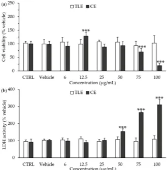
The cytotoxicity of P. tricornutum extracts towards HepG2 cells was assayed after 24 h of treatment with increasing amounts of carotenoid extract (CE) and total lipophilic extract (TLE) (Figure1a). The results showed the absence of significant cytotoxicity of the TLE for all the tested concentrations, ranging from 6 to 100 µg/mL, according to both the 3-(4,5-dimethylthiazol-2-yl)-2,5-diphenyltetrazolium bromide (MTT) assay (Figure1a) and the lactate dehydrogenase (LDH) activity assay (Figure1b). By contrast, CE induced an increase in LDH activity at the higher concentrations tested, i.e., 50–100 µg/mL (Figure1a), and a decrease in cell viability at 75 µg/mL (Figure1b). Therefore, further experiments were using 6 µg/mL of CE and 100 µg/mL of TLE.
Molecules 2020, 25, x FOR PEER REVIEW 3 of 14
cellular model was the human hepatocarcinoma HepG2 cell line, which is commonly used to study lipid metabolism and metabolic disturbances in the liver, particularly those associated with NAFLD [38,39]. NAFLD was simulated by the treatment of cells with 250 µM palmitate for 24 h, as commonly used to induce lipotoxicity and hepatic steatosis in HepG2 cells [40–42].
2. Results
2.1. Effects of Carotenoid and Total Lipophilic Extracts on Cytotoxicity at 24 h of Treatment
The cytotoxicity of P. tricornutum extracts towards HepG2 cells was assayed after 24 h of treatment with increasing amounts of carotenoid extract (CE) and total lipophilic extract (TLE) (Figure 1a). The results showed the absence of significant cytotoxicity of the TLE for all the tested concentrations, ranging from 6 to 100 µg/mL, according to both the 3-(4,5-dimethylthiazol-2-yl)-2,5-diphenyltetrazolium bromide (MTT) assay (Figure 1a) and the lactate dehydrogenase (LDH) activity assay (Figure 1b). By contrast, CE induced an increase in LDH activity at the higher concentrations tested, i.e., 50–100 µg/mL (Figure 1a), and a decrease in cell viability at 75 µg/mL (Figure 1b). Therefore, further experiments were using 6 µg/mL of CE and 100 µg/mL of TLE.
Figure 1. Effect of total lipophilic and carotenoid extracts from P. tricornutum on the cell viability and necrosis of HepG2 cells. The HepG2 cells were treated with the culture medium (control, CTRL), the vehicle (0.66% ethanol) or different concentrations of total lipophilic extract (TLE) and carotenoid extract (CE) from P. tricornutum (6–100 µg/mL) for 24 h, and cytotoxicity assays were performed. (a) Cell viability was evaluated by the MTT assay; (b) Necrosis of HepG2 cells was estimated by the measurement of extracellular lactate dehydrogenase (LDH) activity. Data are the means ± standard deviations from at least three independent experiments. Statistical significance was determined using ANOVA with post-hoc Fisher’s test and (an) asterisk(s) indicate significant difference compared to the vehicle with *** p < 0.001.
2.2. Carotenoid Extract Decreased the Palmitate (PAL)-Induced Lipid Droplet Accumulation
Figure 1. E↵ect of total lipophilic and carotenoid extracts fromP. tricornutum on the cell viability and necrosis of HepG2 cells. The HepG2 cells were treated with the culture medium (control, CTRL), the vehicle (0.66% ethanol) or di↵erent concentrations of total lipophilic extract (TLE) and carotenoid extract (CE) from P. tricornutum (6–100 µg/mL) for 24 h, and cytotoxicity assays were performed. (a) Cell viability was evaluated by the MTT assay; (b) Necrosis of HepG2 cells was estimated by the measurement of extracellular lactate dehydrogenase (LDH) activity. Data are the means ± standard deviations from at least three independent experiments. Statistical significance was determined using ANOVA with post-hoc Fisher’s test and (an) asterisk(s) indicate significant di↵erence compared to the vehicle with *** p < 0.001.
2.2. Carotenoid Extract Decreased the Palmitate (PAL)-Induced Lipid Droplet Accumulation
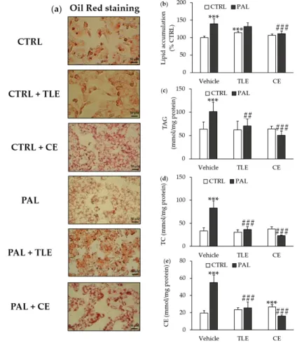
Lipid droplet accumulation was estimated by Oil Red staining (Figure2a) and quantified after the solubilization of lipid droplets (Figure2b), showing that PAL treatment increased lipid accumulation in the control cells. The addition of CE, which had no significant e↵ect in control cells, prevented the PAL-induced lipid droplet accumulation (Figure2b). By contrast, adding TLE induced an increase in the lipid droplet accumulation (Figure2b). Moreover, the addition of TLE did not prevent the PAL-induced lipid droplet accumulation (Figure2b).
Molecules 2020, 25, x FOR PEER REVIEW 4 of 14
Lipid droplet accumulation was estimated by Oil Red staining (Figure 2a) and quantified after the solubilization of lipid droplets (Figure 2b), showing that PAL treatment increased lipid accumulation in the control cells. The addition of CE, which had no significant effect in control cells, prevented the PAL-induced lipid droplet accumulation (Figure 2b). By contrast, adding TLE induced an increase in the lipid droplet accumulation (Figure 2b). Moreover, the addition of TLE did not prevent the PAL-induced lipid droplet accumulation (Figure 2b).
Figure 2. Effect of total lipophilic and carotenoid extracts from P. tricornutum on non-alcoholic fatty liver disease (NAFLD). The HepG2 cells were treated with the control vehicle (CTRL, 1.16% ethanol) alone or associated with total lipophilic extract (CTRL + TLE, 100 µg/mL) or carotenoid extract (CTRL + CE, 6 µg/mL) for 24 h. In parallel, the HepG2 cells were exposed to palmitate vehicle (PAL, 250 µM) alone or associated with the TLE (PAL +TLE) or the CE (PAL + CE) for 24 h. (a) Lipid droplets were detected by Oil Red staining, original magnification × 400; (b) Lipid accumulation was quantified by the solubilization of lipid droplets stained in Oil Red with isopropanol; (c) Intracellular triglyceride (TAG) values; (d) Intracellular total cholesterol (TC) values; (e) Intracellular cholesterol ester (CE) values. Data are the means ± standard deviations from at least three independent experiments. Statistical significance was determined using ANOVA with post-hoc Fisher‘s test, and asterisks indicate significant differences compared to the CTRL vehicle with *** p < 0.001. Hashtags indicate significant differences compared to the PAL vehicle with ## p < 0.01 or ### p < 0.001.
Figure 2. E↵ect of total lipophilic and carotenoid extracts fromP. tricornutum on non-alcoholic fatty liver disease (NAFLD). The HepG2 cells were treated with the control vehicle (CTRL, 1.16% ethanol) alone or associated with total lipophilic extract (CTRL + TLE, 100 µg/mL) or carotenoid extract (CTRL + CE, 6 µg/mL) for 24 h. In parallel, the HepG2 cells were exposed to palmitate vehicle (PAL, 250 µM) alone or associated with the TLE (PAL +TLE) or the CE (PAL + CE) for 24 h. (a) Lipid droplets were detected by Oil Red staining, original magnification ⇥ 400; (b) Lipid accumulation was quantified by the solubilization of lipid droplets stained in Oil Red with isopropanol; (c) Intracellular triglyceride (TAG) values; (d) Intracellular total cholesterol (TC) values; (e) Intracellular cholesterol ester (CE) values. Data are the means ± standard deviations from at least three independent experiments. Statistical significance was determined using ANOVA with post-hoc Fisher‘s test, and asterisks indicate significant di↵erences compared to the CTRL vehicle with *** p < 0.001. Hashtags indicate significant di↵erences compared to the PAL vehicle with ## p < 0.01 or ### p < 0.001.
2.3. Carotenoid and Total Lipophilic Extracts Decreased Cellular Levels of Triglycerides, Total Cholesterol and Cholesterol Esters
As expected, and in correlation with the above results, the PAL treatment of HepG2 cells resulted in increased cellular levels of triglycerides, total cholesterol and cholesterol esters (Figure 2c–e).
The addition of either CE or TLE to control cells had no significant impact on the lipid contents, with the exception of the cholesterol ester level, which increased in the presence of CE (Figure2e). However, both CE and TLE prevented the PAL-induced increase in triglycerides, total cholesterol and cholesterol esters from baseline levels in HepG2 cells (Figure2c–e).
2.4. Impact of Carotenoid and Total Lipophilic Extracts from P. tricornutum on mRNA Levels of Lipid Metabolism and -Oxidation Related Genes
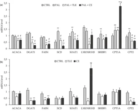
The mRNA levels for some lipogenic enzyme genes (i.e., ACACA, DGAT1 and FASN) were not a↵ected by the PAL treatment (Figure3a). By contrast, the mRNA levels of the SCD, ACAT1/SOAT1 and SREBF1 genes were increased in PAL-treated cells compared to control (CTRL) cells (Figure3a). The addition of CE or TLE to PAL-treated cells induced a decrease in the mRNA levels of lipogenic genes such as ACACA, DGAT1 and FASN (Figure3a). In the same way, the mRNA level of LXR/NR1H3 was decreased in PAL-treated cells by microalgae extracts and more markedly by TLE (Figure3a). Moreover, CE and TLE restored the mRNA level of the ACAT1/SOAT1 gene in PAL-treated HepG2 cells, and only PAL + TLE treatment restored the mRNA level of SREBF1 (Figure3a). Both CE and TLE prevented the PAL-induced increase in the mRNA level of SCD above the baseline level, and even a decrease was seen with CE (Figure3a). It should be noted that CE and TLE had an impact on CTRL cells, decreasing the DGAT1 and FASN mRNA levels, and only TLE decreased the ACACA mRNA level in CTRL cells (Figure3b). In CTRL cells, the LXR/NR1H3 mRNA level was decreased by TLE and, conversely, increased by CE (FigureMolecules 2020, 25, x FOR PEER REVIEW 3b). 6 of 14
Figure 3. Impact of total lipophilic and carotenoid extracts from P. tricornutum on mRNA levels of several genes involved in lipid metabolism regulation in palmitate (PAL)-treated HepG2 cells (a) and control (CTRL) cells (b). The HepG2 cells were treated with control medium and 1.16% ethanol (CTRL), PAL (250 µM) alone or associated with total lipophilic extract (PAL + TLE, 100 µg/mL) or carotenoid extract (PAL + CE, 6 µg/mL) for 24 h before RNA extraction. mRNA levels of genes were quantified using RT-qPCR. Data are the means ± standard deviations from at least three independent experiments. Statistical significance was determined using ANOVA with post-hoc Fisher’s test, and asterisk(s) indicate significant differences compared to the CTRL vehicle with *p < 0.05 and **p < 0.001. Hashtags indicate significant differences compared to the PAL vehicle with # p < 0.001.
3. Discussion
The aim of this study was to investigate the impact of CE from P. tricornutum on NAFLD and associated metabolic pathways, in human liver HepG2 cells. Twenty-four hours of PAL treatment at a concentration of 250 µM is well known to induce NAFLD without altering cell viability in HepG2 cells [40–43,44]. To assess the protective effects of microalga extracts on PAL-induced NAFLD, extracts have to be used at non-toxic concentrations. Here, CE showed some cytotoxic effects at high concentrations, increasing cell necrosis at concentrations between 50 µg/mL and 100 µg/mL. Accordingly, a recent study reported a decrease in cell metabolic activity associated with an increase in apoptosis in HepG2 cells treated with 50 µg/mL of fucoxanthin from P. tricornutum [45]. By contrast, TLE showed no toxicity toward HepG2 cells, suggesting that the presence of lipids and/or chlorophylls (chlorophyll a and derivatives such as pheophytin) counteracts the cytotoxicity of carotenoids. Indeed, according to the literature, chlorophylls have a chelating activity toward reactive oxygen species, which is used for pharmaceutical benefits, especially in liver recovery, and n-3 LC-PUFA have hepatoprotective effects, in particular through the boosting of the antioxidant system [46,47].
In agreement with several studies [40–42], PAL-treated HepG2 cells showed a significant increase in lipid droplet accumulation and the cellular levels of triglycerides, total cholesterol and
Figure 3. Impact of total lipophilic and carotenoid extracts fromP. tricornutum on mRNA levels of several genes involved in lipid metabolism regulation in palmitate (PAL)-treated HepG2 cells (a) and control (CTRL) cells (b). The HepG2 cells were treated with control medium and 1.16% ethanol (CTRL), PAL (250 µM) alone or associated with total lipophilic extract (PAL + TLE, 100 µg/mL) or carotenoid extract (PAL + CE, 6 µg/mL) for 24 h before RNA extraction. mRNA levels of genes were quantified using RT-qPCR. Data are the means ± standard deviations from at least three independent experiments. Statistical significance was determined using ANOVA with post-hoc Fisher’s test, and asterisk(s) indicate significant di↵erences compared to the CTRL vehicle with * p < 0.05 and ** p < 0.001. Hashtags indicate significant di↵erences compared to the PAL vehicle with # p < 0.001.
The mRNA levels of genes related to fatty acid catabolism were also studied and may be increased in PAL-treated cells, as suggested by the increase in CPT1A and CPT2 mRNA levels (Figure3a). The addition of CE strongly increased the mRNA level of CPT1A in PAL-treated cells, while TLE had no e↵ect. However, both CE and TLE decreased the mRNA level of CPT2 below the basal level in CTRL and PAL-treated cells (Figure3a,b).
3. Discussion
The aim of this study was to investigate the impact of CE from P. tricornutum on NAFLD and associated metabolic pathways, in human liver HepG2 cells. Twenty-four hours of PAL treatment at a concentration of 250 µM is well known to induce NAFLD without altering cell viability in HepG2 cells [40–44]. To assess the protective e↵ects of microalga extracts on PAL-induced NAFLD, extracts have to be used at non-toxic concentrations. Here, CE showed some cytotoxic e↵ects at high concentrations, increasing cell necrosis at concentrations between 50 µg/mL and 100 µg/mL. Accordingly, a recent study reported a decrease in cell metabolic activity associated with an increase in apoptosis in HepG2 cells treated with 50 µg/mL of fucoxanthin from P. tricornutum [45]. By contrast, TLE showed no toxicity toward HepG2 cells, suggesting that the presence of lipids and/or chlorophylls (chlorophyll a and derivatives such as pheophytin) counteracts the cytotoxicity of carotenoids. Indeed, according to the literature, chlorophylls have a chelating activity toward reactive oxygen species, which is used for pharmaceutical benefits, especially in liver recovery, and n-3 LC-PUFA have hepatoprotective e↵ects, in particular through the boosting of the antioxidant system [46,47].
In agreement with several studies [40–42], PAL-treated HepG2 cells showed a significant increase in lipid droplet accumulation and the cellular levels of triglycerides, total cholesterol and cholesterol esters. Both extracts prevented the elevation of triglyceride, total cholesterol and cholesterol ester levels in HepG2 cells. However, only CE reduced the lipid droplet accumulation induced by PAL treatment. These results suggest that microalga extracts may have the potential to prevent NAFLD. These biological e↵ects could be attributed to fucoxanthin, which is the main bioactive component of CE (Table S1), or fucoxanthin and EPA, which are present in the TLE (Table S2). Accordingly, EPA was reported to have a preventive e↵ect on lipid accumulation in HepG2 cells co-treated with PAL and EPA (50 µM) for 24 h, which could be mediated by a decrease in hepatic fatty acid synthesis [48]. In addition, Wan et al. have shown in a rodent study using a lipid extract from Chlorella pyrenoidosa, which is relatively rich in EPA (~2% dw) and DHA (~4% dw), at doses of 150 and 300 mg/kg of body weight that lipid extract supplementation prevented NAFLD by reducing the lipid contents in the livers of rats fed high-fat and high-sucrose diets for 8 weeks [33]. A preventive e↵ect against NAFLD has also been shown for fucoxanthin, originating from P. tricornutum (23.54 µg/mg of fucoxanthin by dry weight), which caused a reduction in fat accumulation and liver triglycerides in C57BL/6J mice [36]. Moreover, in our study, only the CE decreased the lipid droplet accumulation. Accordingly, fucoxanthin has previously been reported to inhibit lipogenic liver enzymes such as glucose-6-phosphate dehydrogenase, malic enzyme, FASN and phosphatidate phosphohydrolase, all involved in lipid droplet formation [27]. Besides, lipid droplet accumulation was not significantly a↵ected in HepG2 cells treated with PAL + TLE, probably due to the direct addition of lipids, since P. tricornutum is well known to be rich in neutral lipids (about 30% of the lipid fraction) [49].
The de novo synthesis of fatty acids and fatty acid oxidation in the mitochondria are crucial steps of fatty acid metabolism in the liver [50]. The modulation of the gene expression of enzymes involved in these pathways could explain the preventive e↵ect of microalga extracts against the NAFLD induced by PAL treatment. Firstly, PAL is known to increase lipid storage in the liver by a process mainly controlled by the SREBF1 transcription factor [51]. In accordance with the present study, previous studies using HepG2 cells treated for 24 h with palmitate at 250 µM or 300 µM showed an increase in the expression of enzymes involved in the oxidation and de novo synthesis of fatty acids such as CPT1A and SCD [41,52]. Although our analyses showed no significant change in the mRNA levels of
lipogenic genes (ACACA, DGAT1, FASN and LXR/NR1H3) in our cellular model of NAFLD, the mRNA levels of CPT2 and ACAT1/SOAT1 were increased in agreement with in vivo studies [53,54].
In the present study, the decrease in the cellular triglyceride level in the presence of CE or TLE in PAL-treated cells is correlated with a decrease in the mRNA levels of lipogenic enzyme genes such as ACACA, FASN, DGAT1 and SCD. In agreement with the literature, the fucoxanthin contained in both CE and TLE could regulate the gene expression of enzymes involved in lipid metabolism such as ACACA, FASN, DGAT1 and SCD [27,55–58]. Moreover, n-3 LC-PUFA (mainly EPA), which are present in TLE, have been reported to inhibit the hepatic activity of DGAT and SCD [59,60].
In addition, in cells co-treated with PAL and TLE, the decrease in the mRNA levels of lipogenic enzymes could be mediated upstream by the downregulation of the transcription factor SREBF1 [20]. The decrease in the SREBF1 mRNA level observed here in the presence of PAL + TLE could be mediated by the decrease in the LXR/NR1H3 transcription factor mRNA level. Indeed, n-3 LC-PUFA, especially EPA from the TLE, have already shown to have an inhibitory e↵ect on the mRNA levels of LXR/NR1H3 and genes involved in fatty acid synthesis such as FASN and ACACA [48,61]. Furthermore, n-3 LC-PUFA also have an inducing e↵ect on the gene expression of the farnesoid X receptor transcription factor, leading to a decrease in the SREBF1 mRNA level [61]. Although the mRNA level of LXR/NR1H3 was decreased in PAL + CE cells, the SREBF1 mRNA level was not reduced. These observations suggest that CE could act directly on the decrease in the mRNA level of lipogenic genes and not by inhibiting the expression of the SREBF1 gene.
The decrease in the cellular levels of total and esterified cholesterol (a major form of cholesterol in the lipid droplets) was observed in the presence of CE or TLE in PAL-treated cells and could be correlated with the decrease in the ACAT1/SOAT1 mRNA level, a gene involved in the esterification of cholesterol [7]. Moreover, the fucoxanthin present in both microalga extracts could decrease the mRNA level of ACAT1/SOAT1 in HepG2 cells [27].
Finally, only PAL + CE treatment increased the mRNA level of CPT1A, suggesting the ability of the fucoxanthin from CE to decrease lipid droplet accumulation and lipid synthesis, via the activation of the -oxidation pathway [62–64]. Surprisingly, the CPT1A mRNA level in PAL + TLE cells was unchanged compared to that in cells treated with PAL, suggesting that complex interactions between fucoxanthin and lipids from TLE may counteract the promoting e↵ects of fucoxanthin and n-3 LC-PUFA on the -oxidation pathway [27,35,62–65]. Thus, CE appears to have modulating e↵ects on lipid metabolism through decreasing the mRNA levels of lipogenesis genes as well as increasing the mRNA level of CPT1A, involved in fatty acid oxidation, suggesting that CE may be an e↵ective anti-NAFLD agent. The comparison of CE and TLE also highlights the combined action of lipids (mainly EPA) and pigments (carotenoid and chlorophylls), showing complex interactions.
4. Materials and Methods
4.1. Microalgal Extracts and Palmitate Solution
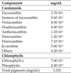
The TLE and CE from the microalga P. tricornutum were obtained from the CNR-IBE laboratory (Florence, Italy). TLE was prepared from freeze-dried biomass using three rounds of chloroform/methanol (2:1, v/v) extraction as described by Ma et al. [66] with slight modifications, and the dry extract was finally dissolved in 95% ethanol. Carotenoids were extracted from the biomass using 90% acetone, and chlorophyll was separated from other pigments by using 10 mM KOH as described previously by Li et al. [67]. In a separator funnel, 100 mL of petroleum ether was added to the raw extract in order to obtain two phases: the lower of bright green color, containing chlorophyll, and the upper of yellow-orange color, containing carotenoids. The upper phase was recovered, dried in a Rotavapor and dissolved in 95% ethanol. The concentrations of individual carotenoids were assessed by reverse-phase high-pressure liquid chromatography according to Van Heukelem and Thomas [68]. The composition of CE and TLE is reported in Tables S1 and S2, respectively. The CE from P. tricornutum contains 97% carotenoids (of which 27% is fucoxanthin) and 3% chlorophylls (Table S1). TLE is
a mixture of lipids (91.7%) and lipophilic pigments including carotenoids (2.3%, of which 72% is fucoxanthin) and chlorophylls (6%, mainly pheophytin) (Table S2). Both CE and TLE were stored in ethanol at 20 C under a nitrogen atmosphere and protected from light.
PAL was purchased from Sigma (St. Louis, MO, USA) and dissolved extemporaneously in ethanol at a final concentration of 50 mM. The purity of the PAL used in this study was more than 98%. The PAL solution was filtered and diluted in fetal bovine serum-free Dulbecco’s Modified Eagle’s Medium (DMEM) containing 5% of bovine serum albumin (BSA) and 1% of penicillin and streptomycin mix to reach a 2 mM concentration. The PAL solution was sonicated for 3 min, heated for 10 min at 55 C and further diluted to reach a 250 µM concentration. The BSA-enriched (5%) DMEM facilitated PAL incorporation into the HepG2 cells, and the PAL/BSA ratio was 3:1. BSA-bound PAL solution was warmed up at 37 C to ensure BSA binding before treatment.
4.2. Cell Culture
HepG2 human liver cells, purchased from the American Type Culture Collection (Manassas, VA, USA), were seeded in enriched DMEM (Sigma, St. Louis, MO, USA) containing 4.5 g/L of glucose, 10% fetal bovine serum, L-glutamine (2 mM) and 1% of penicillin (10,000 IU/mL) and streptomycin mix (10 mg/mL). The cells were kept in a humidified atmosphere of 5% CO2. The HepG2 cells were grown until reaching 70–80% confluence before cell treatment, as described below.
4.3. Cell Treatment
Human HepG2 cells were seeded at 1.102cells/cm2and maintained in a culture medium. One day later, the HepG2 cells reached 70% confluence and were washed with phosphate bu↵ered saline and treated with BSA-bound PAL solution at 250 µM, BSA-bound PAL solution and 6 µg/mL of CE (PAL + CE, corresponding to 120 µg/mL of biomass) or 100 µg/mL of TLE (PAL + TLE, equivalent to 600 µg/mL of biomass) for 24 h. In parallel, HepG2 cells were treated with culture medium alone (CTRL) or associated with TLE (CTRL + TLE, 100 µg/mL) or CE (CTRL + CE, 6 µg/mL) for 24 h. The CE and TLE were preserved in ethanol, and the CTRL cells received the same percentage of ethanol as the PAL + TLE cells (i.e., 1.16% ethanol), the experimental condition with the highest ethanol concentration. 4.4. Cell Viability and Mortality Assays
The cell viability was assessed by using the MTT assay. The cells were seeded in 96-well culture plates in a volume of 100 µL at a density of 4000 cells/well. After 24 h, the cells were incubated with di↵erent concentrations of CE and TLE from P. tricornutum (6 to 100 µg/mL) in a serum-free medium. After 24 h of incubation, 50 µL of MTT (Sigma, St. Louis, MO, USA) was added to each well at a concentration of 2.5 mg/mL for 4 h at 37 C. The cells were washed and lysed with DMSO at 200 µL/well. The absorbance was measured spectrophotometrically at 540 nm. In addition, cell necrosis was determined with the LDH release assay, using a commercial enzymatic kit from Roche (Basel, Switzerland).
4.5. Oil Red Staining
HepG2 cells were seeded at a density of 1000 cells/well in 6-well plates, and after 24 h of incubation, they were treated with PAL associated or not with microalgal extracts. After 24 h of treatment, lipid droplets in HepG2 cells were revealed with an Oil Red staining kit from ScienCell Research Laboratories (Carlsbad, CA, USA) according to the manufacturer’s protocols. The images were acquired by optical microscopy using ⇥400 magnification. Then, the lipid droplets previously stained with Oil Red were solubilized with 2 mL of isopropanol/well. The red coloration of the solution obtained was proportional to the lipid droplet accumulation in the HepG2 cells and was measured spectrophotometrically at 520 nm.
4.6. Triglyceride, Cholesterol and Cholesterol Ester Contents in HepG2 Cells
To determine the cellular levels of total and free cholesterol, HepG2 cells were seeded at a density of 1000 cells/well in 6-well plates, while 5000 cells/dish (size 92 ⇥ 17 mm) were seeded to measure cellular triglycerides. After 24 h, HepG2 cells were treated with PAL, alone or in combination with microalgal extracts for another 24 h. The cellular levels of total cholesterol and free cholesterol were measured by enzymatic methods using commercial enzymatic kits (Abnova, Taipei, Taiwan), and cellular triglycerides were quantified using a commercial enzymatic kit from Cohesion Biosciences (London, England). For each sample, the cell pellet was solubilized in 1 mL of 0.01 N NaOH at 37 C for 3 h in order to determine the protein concentrations by the Bio-Rad protein assay (Bio-Rad, Marnes-la-Coquette, France). The cellular levels of cholesterol esters were calculated from the di↵erence between the total cholesterol and free cholesterol values.
4.7. RNA Extraction and Real-Time Quantitative Polymerase Chain Reaction (RT-qPCR)
HepG2 cells were seeded at a density of 1000 cells/well in 6-well plates. After 24 h, the cells were treated with PAL (250 µM) associated or not with microalga extracts (6 µg/mL of CE or 100 µg/mL of TLE) for another 24 h at 37 C. The cells were lysed with TRIzol Reagent (Thermo Fisher, Waltham, MA, USA) and total RNA was isolated following the manufacturer’s instructions. The mRNAs (1 µg) were then reverse-transcribed into cDNA using iScript Reverse Transcriptase (Bio-Rad) according to the manufacturer’s instructions. An initial priming step of 5 min at 25 C was followed by reserve transcription for 30 min at 42 C and a reverse-transcriptase inactivation for 5 min at 85 C. Quantitative PCR was performed on a MyiQ2 Real-Time PCR Detection System (Bio-Rad) using iQ™ SYBR Green Supermix. PCR was carried out for 45 cycles of 95 C for 30 s and 60 C for 30 s. The relative expression levels of the studied genes were standardized to the -actin (ACTB) reference gene expression—except that of LXR/NR1H3, which was normalized to that of the 18S reference gene—using the DDCT method. The sequences of the primers used are listed in Table S3.
4.8. Data Analysis
Results were obtained from triplicate experiments, and the data presented are the means +/ standard deviations (SDs) obtained after at least three independent experiments. The analysis of variance by one-way ANOVA followed by a Fisher’s least significant di↵erence post hoc test (LSD) was performed. Statistical analyses were carried out with Statgraphics Plus 5.1 software (Manugistics Inc., Rockville, MD, USA), and the results were considered statistically di↵erent for values of p < 0.05, p < 0.01 or p < 0.001 as indicated in the figure legends.
5. Conclusions
This study highlights the preventive e↵ect of total lipophilic and carotenoid extracts of the marine microalga P. tricornutum on the hepatic accumulation of triglycerides and cholesterol. Moreover, only the carotenoid extract reduced the accumulation of lipid droplets in PAL-treated HepG2 cells. This observation could be explained by an additional inhibitory e↵ect of CE on lipolysis in parallel with the stimulation of the de novo lipogenesis pathway, whereas TLE acts only on the latter. This suggests a greater protective e↵ect of carotenoids against NAFLD compared to the total lipophilic compounds of TLE. Therefore, carotenoid extracts of P. tricornutum might become a promising nutritional ingredient for a therapeutic strategy against hepatic disorders such as NAFLD associated with metabolic syndrome and obesity. Finally, by comparing CE’s and TLE’s e↵ects, this study also highlights the complex interactions between the bioactive molecules contained in TLE such as fucoxanthin, chlorophylls and n-3 LC-PUFA. Further studies should be conducted with a purified lipid extract, free of chlorophylls and carotenoids, in order to better elucidate the specific e↵ects of lipids and carotenoids of P. tricornutum.
Supplementary Materials: The following are available online, Table S1: Composition of carotenoid extract from
P. tricornutum biomass, Table S2: Composition of total lipophilic extract from P. tricornutum biomass, Table S3: RT-qPCR primer sequences.
Author Contributions: Conceptualization, C.M., V.B., H.N., K.O., V.M. and B.C.; methodology and investigations,
C.M., M.C., V.B., H.N., V.M. and B.C.; sample analyses and statistical analysis, C.M. and M.C.; G.C.Z. and C.F. provided us with total lipid and carotenoid extracts and carried out the analysis of their compositions; validation, V.B., H.N., K.O., V.M. and B.C.; writing the initial draft of the article, C.M. and B.C.; review and correction, C.M., V.B., G.C.Z., C.F., H.N., K.O., V.M. and B.C.; supervision, K.O, V.M. and B.C.; project administration, V.M.; funding acquisition, V.M. All authors have read and agreed to the published version of the manuscript.
Funding: This research was funded by financial support from the regional program “Food for Tomorrow-Cap
Aliment” Research, Formation and Innovation in Pays de la Loire including a co-financing at 50% of the grant to C.M. The thesis was also co-funded at 50% by Le Mans University, with joint financial support from Laval Agglomération and the Conseil Général de la Mayenne.
Acknowledgments: We are grateful to Jean-Michel Huvelin and Alexandra Burghelea-Kovshevnikov for their
technical advice in RT-qPCR operation.
Conflicts of Interest: The authors declare no conflict of interest.
Abbreviations
ACACA acetyl-CoA carboxylase
ACAT acyl-CoA cholesterol acyltransferase
ACTB -actin
BSA bovine serum albumin
CE carotenoid extract
CPT carnitine palmitoyltransferase
CTRL control
DGAT diacylglycerol O-acyltransferase
DHA docosahexaenoic acid
DMEM Dulbecco’s Modified Eagle’s Medium EPA eicosapentanenoic acid
LDH lactate dehydrogenase
MTT 3-(4,5-dimethylthiazol-2-yl)-2,5-diphenyltetrazolium bromide n-3 LC-PUFA n-3 long-chain polyunsaturated fatty acids
LXR/NR1H3 liver X receptor
NAFLD non-alcoholic fatty liver disease
PAL palmitate
RT-qPCR reverse-transcriptase quantitative polymerase chain reaction SCD stearyl-CoA desaturase
SOAT sterol O-acyltransferase
SREBF1 sterol regulatory element-binding transcription factor 1 TLE total lipophilic extract
References
1. Kuipers, R.S.; de Graaf, D.J.; Luxwolda, M.F.; Muskiet, M.H.A.; Dijck-Brouwer, D.A.J.; Muskiet, F.A.J. Saturated fat, carbohydrates and cardiovascular disease. Neth. J. Med. 2011, 69, 372–378. [PubMed] 2. Byrne, C.D.; Targher, G. NAFLD: A multisystem disease. J. Hepatol. 2015, 62, S47–S64. [CrossRef] [PubMed] 3. Luyendyk, J.P.; Guo, G.L. Steatosis DeLIVERs high-sensitivity C-reactive protein. Arterioscler. Thromb. Vasc.
Biol. 2011, 31, 1714–1715. [CrossRef] [PubMed]
4. Browning, J.D.; Szczepaniak, L.S.; Dobbins, R.; Nuremberg, P.; Horton, J.D.; Cohen, J.C.; Grundy, S.M.; Hobbs, H.H. Prevalence of hepatic steatosis in an urban population in the United States: Impact of ethnicity. Hepatology 2004, 40, 1387–1395. [CrossRef]
5. Lonardo, A.; Ballestri, S.; Marchesini, G.; Angulo, P.; Loria, P. Nonalcoholic fatty liver disease: A precursor of the metabolic syndrome. Dig. Liver Dis. 2015, 47, 181–190. [CrossRef] [PubMed]
6. Alkhouri, N.; Lawitz, E.; Noureddin, M.; DeFronzo, R.; Shulman, G.I. GS-0976 (Firsocostat): An investigational liver-directed Acetyl-CoA Carboxylase (ACC) inhibitor for the treatment of non-alcoholic steatohepatitis (NASH). Expert Opin. Inv. Drug. 2020, 29, 135–141. [CrossRef]
7. Wilfling, F.; Haas, J.T.; Walther, T.C.; Jr, R.V.F. Lipid droplet biogenesis. COCEBI 2014, 29, 39–45. [CrossRef] 8. Gutierrez-Juarez, R. Critical role of stearoyl-CoA desaturase-1 (SCD1) in the onset of diet-induced hepatic
insulin resistance. J. Clin. Invest. 2006, 116, 1686–1695. [CrossRef]
9. Kotronen, A.; Seppanen-Laakso, T.; Westerbacka, J.; Kiviluoto, T.; Arola, J.; Ruskeepaa, A.-L.; Oresic, M.; Yki-Jarvinen, H. Hepatic Stearoyl-CoA Desaturase (SCD)-1 activity and diacylglycerol but not ceramide concentrations are increased in the nonalcoholic human fatty liver. Diabetes 2009, 58, 203–208. [CrossRef] 10. Tilg, H.; Moschen, A.R.; Roden, M. NAFLD and diabetes mellitus. Nat. Rev. Gastroenterol. Hepatol. 2017, 14,
32–42. [CrossRef]
11. Pawar, A.; Botolin, D.; Mangelsdorf, D.J.; Jump, D.B. The role of liver X receptor-↵ in the fatty acid regulation of hepatic gene expression. J. Biol. Chem. 2003, 278, 40736–40743. [CrossRef] [PubMed]
12. Spahis, S.; Delvin, E.; Borys, J.-M.; Levy, E. Oxidative stress as a critical factor in nonalcoholic fatty liver disease pathogenesis. Antioxid. Redox Signal. 2017, 26, 519–541. [CrossRef] [PubMed]
13. Spooner, M.H.; Jump, D.B. Omega-3 fatty acids and nonalcoholic fatty liver disease in adults and children: Where do we stand? Curr. Opin. Clin. Nutr. 2019, 22, 103–110. [CrossRef] [PubMed]
14. Tobin, D.; Brevik-Andersen, M.; Qin, Y.; Innes, J.K.; Calder, P.C. Evaluation of a high Concentrate Omega-3 for correcting the Omega-3 fatty acid Nutritional Deficiency In Non-alcoholic fatty liver disease (CONDIN). Nutrients 2018, 10, 1126. [CrossRef]
15. Jump, D.B.; Lytle, K.A.; Depner, C.M.; Tripathy, S. Omega-3 polyunsaturated fatty acids as a treatment strategy for nonalcoholic fatty liver disease. Pharmacol. Ther. 2018, 181, 108–125. [CrossRef]
16. Bertrand, C.; Pignalosa, A.; Wanecq, E.; Rancoule, C.; Batut, A.; Deleruyelle, S.; Lionetti, L.; Valet, P.; Castan-Laurell, I. E↵ects of dietary Eicosapentaenoic Acid (EPA) supplementation in high-fat fed mice on lipid metabolism and apelin/APJ system in skeletal muscle. PLoS ONE 2013, 8, e78874. [CrossRef]
17. Poudyal, H.; Panchal, S.K.; Ward, L.C.; Brown, L. E↵ects of ALA, EPA and DHA in high-carbohydrate, high-fat diet-induced metabolic syndrome in rats. J. Nutr. Biochem. 2013, 24, 1041–1052. [CrossRef] 18. Calder, P.C. Marine omega-3 fatty acids and inflammatory processes: E↵ects, mechanisms and clinical
relevance. BBA 2015, 1851, 469–484. [CrossRef]
19. Kunesová, M.; Braunerová, R.; Hlavat˛, P.; Tvrzická, E.; Stanková, B.; Skrha, J.; Hilgertová, J.; Hill, M.; Kopeck˛, J.; Wagenknecht, M.; et al. The influence of n-3 polyunsaturated fatty acids and very low calorie diet during a short-term weight reducing regimen on weight loss and serum fatty acid composition in severely obese women. Physiol. Res. 2006, 55, 63–72.
20. Albracht-Schulte, K.; Kalupahana, N.S.; Ramalingam, L.; Wang, S.; Rahman, S.M.; Robert-McComb, J.; Moustaid-Moussa, N. Omega-3 fatty acids in obesity and metabolic syndrome: A mechanistic update. J. Nutr. Biochem. 2018, 58, 1–16. [CrossRef]
21. Albracht-Schulte, K.; Gonzalez, S.; Jackson, A.; Wilson, S.; Ramalingam, L.; Kalupahana, N.; Moustaid-Moussa, N. Eicosapentaenoic acid improves hepatic metabolism and reduces inflammation independent of obesity in high-fat-fed mice and in HepG2 Cells. Nutrients 2019, 11, 599. [CrossRef] [PubMed] 22. Abedi, E.; Sahari, M.A. Long-chain polyunsaturated fatty acid sources and evaluation of their nutritional
and functional properties. Food Sci. Nutr. 2014, 2, 443–463. [CrossRef] [PubMed]
23. Martins, D.; Custódio, L.; Barreira, L.; Pereira, H.; Ben-Hamadou, R.; Varela, J.; Abu-Salah, K. Alternative sources of n-3 long-chain polyunsaturated fatty acids in marine microalgae. Mar. Drugs 2013, 11, 2259–2281.
[CrossRef] [PubMed]
24. Lakra, N.; Mahmood, S.; Marwal, A.; Sudheep, N.M.; Anwar, K. Bioengineered plants can be an alternative source of omega-3 fatty acids for human health. In Plant and Human Health; Ozturk, M., Hakeem, K.R., Eds.; Springer International Publishing: Cham, Switzeland, 2019; Volume 2, pp. 361–382.
25. Ryckebosch, E.; Bruneel, C.; Termote-Verhalle, R.; Goiris, K.; Muylaert, K.; Foubert, I. Nutritional evaluation of microalgae oils rich in omega-3 long chain polyunsaturated fatty acids as an alternative for fish oil. Food Chem. 2014, 160, 393–400. [CrossRef] [PubMed]
26. Sakai, S.; Sugawara, T.; Matsubara, K.; Hirata, T. Inhibitory e↵ect of carotenoids on the degranulation of mast cells via suppression of antigen-induced aggregation of high affinity IgE receptors. J. Biol. Chem. 2009, 284, 28172–28179. [CrossRef] [PubMed]
27. Gammone, M.; D’Orazio, N. Anti-obesity activity of the marine carotenoid fucoxanthin. Mar. Drugs 2015, 13, 2196–2214. [CrossRef] [PubMed]
28. Sathasivam, R.; Ki, J.-S. A review of the biological activities of microalgal carotenoids and their potential use in healthcare and cosmetic industries. Mar. Drugs 2018, 16, 26. [CrossRef]
29. Sansone, C.; Brunet, C. Promises and challenges of microalgal antioxidant production. Antioxidants 2019, 8, 199. [CrossRef]
30. De Martino, A.; Meichenin, A.; Shi, J.; Pan, K.; Bowler, C. Genetic and phenotypic characterization of Phaeodactylum tricornutum (Bacillariophyceae) accessions. J. Phycol. 2007, 43, 992–1009. [CrossRef]
31. Rebolloso-Fuentes, M.M.; Navarro-Pérez, A.; Ramos-Miras, J.J.; Guerero, J.L. Biomass nutrient profiles of the microalga Phaeodactylum tricornutum. J. Food Biochem. 2007, 25, 57–76. [CrossRef]
32. Kim, S.M.; Jung, Y.-J.; Kwon, O.-N.; Cha, K.H.; Um, B.-H.; Chung, D.; Pan, C.-H. A potential commercial source of fucoxanthin extracted from the microalga Phaeodactylum tricornutum. Appl. Biochem. Biotechnol.
2012,166, 1843–1855. [CrossRef] [PubMed]
33. Koller, M.; Muhr, A.; Braunegg, G. Microalgae as versatile cellular factories for valued products. Algal Res.
2014,6, 52–63. [CrossRef]
34. Wan, X.; Li, T.; Liu, D.; Chen, Y.; Liu, Y.; Liu, B.; Zhang, H.; Zhao, C. E↵ect of marine microalga Chlorella pyrenoidosa ethanol extract on lipid metabolism and gut microbiota composition in high-fat diet-fed rats. Mar. Drugs 2018, 16, 498. [CrossRef] [PubMed]
35. Yu, J.; Ma, Y.; Sun, J.; Ran, L.; Li, Y.; Wang, N.; Yu, T.; Gao, W.; Jia, W.; Jiang, R.; et al. Microalgal oil from Schizochytrium sp. prevents HFD-induced abdominal fat accumulation in mice. J. Am. Coll. Nutr. 2017, 36, 347–356. [CrossRef]
36. Koo, S.Y.; Hwang, J.-H.; Yang, S.-H.; Um, J.-I.; Hong, K.W.; Kang, K.; Pan, C.-H.; Hwang, K.T.; Kim, S.M. Anti-obesity e↵ect of standardized extract of microalga Phaeodactylum tricornutum containing fucoxanthin. Mar. Drugs 2019, 17, 311. [CrossRef]
37. Mayer, C.; Côme, M.; Ulmann, L.; Chini Zittelli, G.; Faraloni, C.; Nazih, H.; Ouguerram, K.; Chénais, B.; Mimouni, V. Preventive e↵ects of the marine microalga Phaeodactylum tricornutum, used as a food supplement, on risk factors associated with metabolic syndrome in Wistar rats. Nutrients 2019, 11, 1069. [CrossRef] 38. Cui, W.; Chen, S.L.; Hu, K.-Q. Quantification and mechanisms of oleic acid-induced steatosis in HepG2 cells.
Am. J. Transl. Res. 2010, 2, 95–104.
39. Goto, T.; Takahashi, N.; Kato, S.; Egawa, K.; Ebisu, S.; Moriyama, T.; Fushiki, T.; Kawada, T. Phytol directly activates peroxisome proliferator-activated receptor ↵ (PPAR↵) and regulates gene expression involved in lipid metabolism in PPAR↵-expressing HepG2 hepatocytes. Biochem. Biophys. Res. Commun. 2005, 337, 440–445. [CrossRef]
40. Youn, Y.; Kim, Y.-S. Inhibitory e↵ects of Citrus unshiu pericarpium extracts on palmitate-induced lipotoxicity in HepG2 cells. Food Sci. Biotechnol. 2016, 25, 1709–1717. [CrossRef]
41. Rial, S.; Ravaut, G.; Malaret, T.; Bergeron, K.-F.; Mounier, C. Hexanoic, octanoic and decanoic acids promote basal and insulin-induced phosphorylation of the Akt-mTOR axis and a balanced lipid metabolism in the HepG2 hepatoma cell line. Molecules 2018, 23, 2315. [CrossRef]
42. Ma, Z.; Liu, H.; Wang, W.; Guan, S.; Yi, J.; Chu, L. Paeoniflorin suppresses lipid accumulation and alleviates insulin resistance by regulating the Rho kinase/IRS-1 pathway in palmitate-induced HepG2Cells. Biomed. Pharmacother. 2017, 90, 361–367. [CrossRef] [PubMed]
43. Zang, Y.; Fan, L.; Chen, J.; Huang, R.; Qin, H. Improvement of lipid and glucose metabolism by capsiate in palmitic acid-treated HepG2 cells via activation of the AMPK/SIRT1 signaling pathway. J. Agric. Food Chem.
2018,66, 6772–6781. [CrossRef] [PubMed]
44. Ishii, M.; Maeda, A.; Tani, S.; Akagawa, M. Palmitate induces insulin resistance in human HepG2 hepatocytes by enhancing ubiquitination and proteasomal degradation of key insulin signaling molecules. Arch. Biochem. Biophys. 2015, 566, 26–35. [CrossRef] [PubMed]
45. Neumann, U.; Derwenskus, F.; Flaiz Flister, V.; Schmid-Staiger, U.; Hirth, T.; Bischo↵, S. Fucoxanthin, a carotenoid derived from Phaeodactylum tricornutum exerts antiproliferative and antioxidant activities in vitro. Antioxidants 2019, 8, 183. [CrossRef]
46. Kang, Y.-R.; Park, J.; Jung, S.K.; Chang, Y.H. Synthesis, characterization, and functional properties of chlorophylls, pheophytins, and Zn-pheophytins. Food Chem. 2018, 245, 943–950. [CrossRef]
47. Adeyemi, W.J.; Olayaki, L.A. Diclofenac - induced hepatotoxicity: Low dose of omega-3 fatty acids have more protective e↵ects. Toxicol. Rep. 2018, 5, 90–95. [CrossRef]
48. Gonzalez, S.M.; Albracht-Schulte, K.M.; Ramalingam, L.; Kalupahana, N.S.; Moustaid-Moussa, N. Mechanisms mediating e↵ects of eicosapentaenoic acid in hepatic steatosis in high fat fed mice and in HepG2 hepatoma Cells. FASEB J. 2017, 31, 646.53–646.53.
49. Yang, Y.-H.; Du, L.; Hosokawa, M.; Miyashita, K.; Kokubun, Y.; Arai, H.; Taroda, H. Fatty acid and lipid class composition of the microalga Phaeodactylum tricornutum. J. Oleo. Sci. 2017, 66, 363–368. [CrossRef] 50. Kohjima, M.; Enjoji, M.; Higuchi, N.; Kato, M.; Kotoh, K.; Yoshimoto, T.; Fujino, T.; Yada, M.; Yada, R.;
Harada, N.; et al. Re-evaluation of fatty acid metabolism-related gene expression in nonalcoholic fatty liver disease. Int. J. Mol. Med. 2007, 20, 351–358. [CrossRef]
51. Strable, M.S.; Ntambi, J.M. Genetic control of de novo lipogenesis: Role in diet-induced obesity. Crit. Rev. Biochem. Mol. 2010, 45, 199–214. [CrossRef]
52. sik Um, E.; Kim, Y.C. E↵ect of Samhwangsasim-tang and Daehwanghwangryunsasim-tang on palmitate-induced lipogenesis in HepG2 cells. J. Korean Med. 2016, 37, 62–76. [CrossRef]
53. Lee, J.-Y.; Carr, T.P. Dietary fatty acids regulate acyl-CoA: Cholesterol acyltransferase and cytosolic cholesteryl ester hydrolase in hamsters. J. Nutr. 2004, 134, 3239–3244. [CrossRef] [PubMed]
54. Rom, O.; Xu, G.; Guo, Y.; Zhu, Y.; Wang, H.; Zhang, J.; Fan, Y.; Liang, W.; Lu, H.; Liu, Y.; et al. Nitro-fatty acids protect against steatosis and fibrosis during development of nonalcoholic fatty liver disease in mice. EBioMedicine 2019, 41, 62–72. [CrossRef] [PubMed]
55. Jang, W.S.; Choung, S.Y. Antiobesity e↵ects of the ethanol extract of Laminaria japonica Areshoung in high-fat-diet-induced obese rat. Evid. Based Complementary Altern. Med. 2013, 2013, 1–17.
56. Tong, L. Acetyl-coenzyme A carboxylase: Crucial metabolic enzyme and attractive target for drug discovery. CMLS 2005, 62, 1784–1803. [CrossRef]
57. de Vries, R.; Borggreve, S.E.; Dullaart, R.P.F. Role of lipases, lecithin: Cholesterol acyltransferase and cholesteryl ester transfer protein in abnormal high density lipoprotein metabolism in insulin resistance and type 2 diabetes mellitus. Clin. Lab. 2003, 49, 601–613.
58. Eberlé, D.; Hegarty, B.; Bossard, P.; Ferré, P.; Foufelle, F. SREBP transcription factors: Master regulators of lipid homeostasis. Biochimie 2004, 86, 839–848. [CrossRef]
59. Bays, H.E.; Tighe, A.P.; Sadovsky, R.; Davidson, M.H. Prescription omega-3 fatty acids and their lipid e↵ects: Physiologic mechanisms of action and clinical implications. Expert Rev. Cardiovasc. Ther. 2008, 6, 391–409.
[CrossRef]
60. Lamaziere, A.; Wolf, C.; Barbe, U.; Bausero, P.; Visioli, F. Lipidomics of hepatic lipogenesis inhibition by omega 3 fatty acids. Prostag. Leukotr. Ess. 2013, 88, 149–154. [CrossRef]
61. Masterton, G.S.; Plevris, J.N.; Hayes, P.C. Review article: Omega-3 fatty acids - a promising novel therapy for non-alcoholic fatty liver disease: Review: Omega-3 fatty acids - a novel therapy for NAFLD? Aliment. Pharm. Ther. 2009, 31, 679–692. [CrossRef]
62. Chang, Y.-H.; Chen, Y.-L.; Huang, W.-C.; Liou, C.-J. Fucoxanthin attenuates fatty acid-induced lipid accumulation in FL83B hepatocytes through regulated Sirt1/AMPK signaling pathway. Biochem. Biophys. Res. Commun. 2018, 495, 197–203. [CrossRef] [PubMed]
63. Park, H.J.; Lee, M.K.; Park, Y.B.; Shin, Y.C.; Choi, M.S. Beneficial e↵ects of Undaria pinnatifida ethanol extract on diet-induced-insulin resistance in C57BL/6J mice. Food Chem. Toxicol. 2011, 49, 727–733. [CrossRef]
[PubMed]
64. Maeda, H.; Hosokawa, M.; Sashima, T.; Funayama, K.; Miyashita, K. Fucoxanthin from edible seaweed, Undaria pinnatifida, shows antiobesity e↵ect through UCP1 expression in white adipose tissues. BBRC 2005, 332, 392–397. [CrossRef] [PubMed]
65. Gille, A.; Stojnic, B.; Derwenskus, F.; Trautmann, A.; Schmid-Staiger, U.; Posten, C.; Briviba, K.; Palou, A.; Bonet, M.; Ribot, J. A lipophilic fucoxanthin-rich Phaeodactylum tricornutum extract ameliorates e↵ects of diet-induced obesity in C57BL/6J mice. Nutrients 2019, 11, 796. [CrossRef] [PubMed]
66. Ma, Y.; Wang, Z.; Zhu, M.; Yu, C.; Cao, Y.; Zhang, D.; Zhou, G. Increased lipid productivity and TAG content in Nannochloropsis by heavy-ion irradiation mutagenesis. Bioresour. Technol. 2013, 136, 360–367. [CrossRef] 67. Li, T.; Xu, J.; Wu, H.; Wang, G.; Dai, S.; Fan, J.; He, H.; Xiang, W. A saponification method for chlorophyll
68. Van Heukelem, L.; Thomas, C.S. Computer-assisted high-performance liquid chromatography method development with applications to the isolation and analysis of phytoplankton pigments. J. Chromatogr. A
2001,910, 31–49. [CrossRef]
Sample Availability: Not available.
© 2020 by the authors. Licensee MDPI, Basel, Switzerland. This article is an open access article distributed under the terms and conditions of the Creative Commons Attribution (CC BY) license (http://creativecommons.org/licenses/by/4.0/).
Supplementary materials
Table S1. Composition of carotenoid extract from P. tricornutum biomass Components mg/mL Carotenoids Fucoxanthin 2.70 10-1 Isomers of fucoxanthin 2.60 10-1 Violaxanthin 8.50 10-2 Diadinoxanthin 8.00 10-2 Antheraxanthin 1.10 10-1 Dinoxanthin 1.20 10-2 Diatoxanthin 7.10 10-2 β-carotene 5.00 10-2 Others 6.20 10-2 Chlorophylls Chlorophyll a 7.00 10-3 Pheophytin 2.40 10-2 Total pigments (mg/mL) 1.031
Table S2. Composition of total lipophilic extract from P.
tricornutum biomass Components mg/mL Lipids 13.94 Carotenoids Fucoxanthin 2.58 10-1 4 keto-19'-hexanol-oxy-fucoxanthin 5.80 10-2 Diadinoxanthin 2.20 10-2 Other xanthophylls 4.70 10-4 β-carotene 1.16 10-2 Chlorophylls Chlorophyllid a 6.70 10-1 Pheophytin 9.20 10-2 Chlorophylls a + c 1.46 10-1 Total extract 15.20
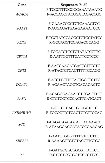
Table S3. Sequences of primers used for qRT-PCR
Gene Sequences (5’-3’)
ACACA F-TCGCTTTGGGGGAAATAAATG R-ACCACCTACGGATAGACCGC
SOAT1 R-AGGAGATGAAGAAAATCCC F-GAAACCGCTGTCAAAGTCC
ACTB F-TGCTATCCAGGCTGTGCTATCC R-GCCAGGTCCAGACGCAGG
CPT1A F-TGGATCTGCTGTATATCCTTC R-AATTGGTTTGATTCCTCCC
CPT2 F-AACCAACATGACTGTTTCTG R-ATAGTGTCACTTTTTGCAGG
DGAT1 F-ATCTTCTTCTACTGGCTCTTC R-AGAAGTAGGTGACAGACTC
FASN F-ACAGGGACAACCTGGAGTTCT R-CTGTGGTCCCACTTGATGAGT
LXR/NR1H3 R-TGCCCTTCTCAGTCTGTTCCAC F-GCTCCCACCGCTGCTCTC
SCD R-ATAAGGACGATATCCGAAGAG F-CAGAGGAGGTACTACAAACC
SREBF1 R-AAAAGTTGTGTACCTTGTGG F-AATCTGGGTTTTGTCTCTTC
18S F-GATGCGGCGGCGTTATTCC R-CTCCTGGTGGTGCCCTTCC
ACACA: acetyl-CoA carboxylase alpha, ACTB: actine beta, CPT: carnitine palmitoyltransferase, DGAT: diacylglycerol acyltransferase, FASN: fatty acid synthase, LXR: liver X receptor, NR1H3: nuclear receptor subfamily 1 group H member 3, SCD: stearoyl-CoA desaturase, SOAT: sterol O-acyltransferase, SREBF: sterol regulatory element-binding protein factor.