This manuscript has been reproduced from the microfilm master. UMI films the text directly from the original or copy submitted. Thus, some thesis and dissertation copies are in typewriter face, while others may be from any type of computer printer.
The quality of this reproduction is dependent upon the quality of the copy submitted. Broken or indistinct print, colored or poor quality illustrations and photographs, print bleedthrough, substandard margins, and improper alignment can adversely affect reproduction.
ln the unlikely event that the author did not send UMI a complete manuscript and there are missing pages, these will be noted. Aise, if unauthorized copyright material had to be removed, a note will indicate the deletion.
Oversize materials (e.g., maps, drawings, charts) are reproduced by sectioning the original, beginning at the upper left-hand corner and continuing from left to right in equal sections with small overlaps.
Photographs included in the original manuscript have been reproduced xerographically in this copy. Higher quality 6" x 9" black and white photographie prints are available for any photographs or illustrations appearing in this copy for an additional charge. Contact UMI directly ta order.
Bell & Howell Information and Leaming
300 North Zeeb Road, Ann Arbor, Ml 48106-1346 USA 800-521-0600
ETUDE PHARMACOLOGIQUE DE LA REACTION D' ARTHUS CHEZ LA
SOURIS DE TYPE SAUVAGE OU TRANSGENIQUE 82 KNOCKOUT
par
RANA SAMADFAM
Département de Pharmacologie
Mémoire présenté a la Faculté de Médecine en vue de l'obtention du grade de
maitre ès Sciences (M.Sc.)
l+I
of Canada Acquisitions andBibliographie Services Acquisitions et services bibliographiques
395 Wellington Street Ottawa ON K1A ON4 Canada
395, rue Wellington Ottawa ON K 1 A ON4 Canada
The author has granted a
non-exclusive licence allowing the
National Library of Canada to
reproduce, loan, distribute or sell
copies of this thesis
in
microfonn,
paper or electronic formats.
The author retains ownership of the
copyright in this thesis. Neither the
thesis nor substantial extracts from it
may be printed or otheiwise
reproduced without the author' s
perrruss1on.
Your file Votre rèferencs
Our ~le Notre retsrence
L'auteur a accordé une licence non
exclusive permettant à la
Bibliothèque nationale du Canada de
reproduire, prêter, distribuer ou
vendre des copies de cette thèse sous
la forme de microfiche/film, de
reproduction sur papier ou sur format
électronique.
L'auteur conserve la propriété du
droit d'auteur qui protège cette thèse.
Ni la thèse
ni
des extraits substantiels
de celle-ci ne doivent être imprimés
ou autrement reproduits sans son
autorisation.
0-612-56967 -5
TABLE T OF CONTENTS
TABLE T OF CONTENTS ... 111
LIST OF PUBLICATIONS ... VII LIST OF COMMUNICATIONS ... VIII LIST OF FIGURES ... IX LIST OF TABLES ... XI LIST OF ABREVIATIONS .•..•.••....••••...•....••....•••••••••••.••..••.••.••••.••.••.•••.••••••••.••• XII LIST OF ABREVIATIONS ... XII SUMMARY •••••••••.••••••••••••••••••••.•••••••••••••••••.••••••••••.••••••.•••••.••....•••••••.••••••••••••..• XIV INTRODUCTION ••..•••.•..••..••••.••....•.••.•••••.••••...•...••...•.•...•... 1
1. ARTHUS REACTION (HYPERSENSITMTY TYPE Hl) ... 2
1.1 Arthus Reaction ... 2
1.2 The mechanism of Arthus reaction ... 3
1.3 Active Arthus Reaction ... 5
1.4 Direct passive Arthus reaction ... 6
1.5 Reverse passive Arthus reaction ... 6
2. POSSIBLE ROLES OF CELL ADHESION MOLECULES (CAM) IN THE RP A ••••••••••••••••••••••••••••••••••••••••••••••••••••••••••••••••••••••••••••••••••••••••••••••••••••••••••••••••••••••••••••••••• 7 2.1 Selectins ... 7
2.2 Integrins ... 8
2.3 lg superfamily ... 9
3. THE COMPLEMENT SYSTEM .•••••••.•..••...•.•...•.••.•...••....•.•...•.•.••...•.•. 12
3 .1 The classical complement pathway ... 12
3.2 Biological activity of the complement component ... 14
3 .2.1 Anaphilatoxins ... 14
3 .2.2 Chemotaxins ... 14
3 .2.3
Immune adherence ... 153 .2.3 Opsonization ... 15
4. KIN'IN'S ... 15 4.1 History ... 16 4.2 Synthesis ... 18 4.2.1 Tissue Kallikrein ... 18 4.2.2 Plasma Kallikrein ... 20 4.2.3 Kininogens ... 20 4.3 Kinin Peptidase ... 22 4.3.1 Kininase I (KI) ... 22 4.3.2 Kininase II
(KID ...
23 4.4 Receptors ... 24 4.5 Antagonists ... 244.6 Mechanism of action ofkinins receptors ... 25
4. 7 Phannacological properties ... 26
4.7.1 Stimulation of nerve ending and production of pain ... 26
4.7.2 Effect ofkinin on renal function and blood pressure ... 27
4. 7.3 Extravascular smooth muscle cells ... 27
4. 7 .4 V ascular endothelium and smooth muscle cells ... 28
S. ROLE OF KALLIKREINS AND KININS IN INFLAMMATION ... 28
5.2 Role of tissue kallikrein in inflammation ... 29
5.3 Role of plasma kallikrein in inflammation ... 29
5.3 Role of B1 receptors in inflammatory responses ... 30
5.4 Role ofB2 receptors in inflammation ... 30
6.
Bi
RECEPTOR KN'OCKOUT MICE ... 31RA TIONALE OF THE STUDY ... 32
METHODS ... 33
1. ANIMALS USED (MICE) ... 33
2. QUANTITATIVE MEASUREMENT OF THE ENHANCED V ASCULAR PERM:EABILITY •••••••••••.•••••••••••..••••.••••••••••••••.••••••••••••••••••••••••••••••••••••••••.•.•••••.••••.••••• 33
3. PERITONEAL PLASMA EXTRAVASATION INDUCED BY BK RECEPTOR AGONISTS ... 35
4. PERITONEAL PLASMA EXTRAVASATION INDUCED BY RP A •••.••••••..•• 36
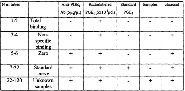
S. DETERMINATION OF PGE2 CONCENTRATION IN THE PERITONEAL CA VITY ... 36
N of tubes ... 37
7. ETmCS ... 38
RESUL TS ... 41
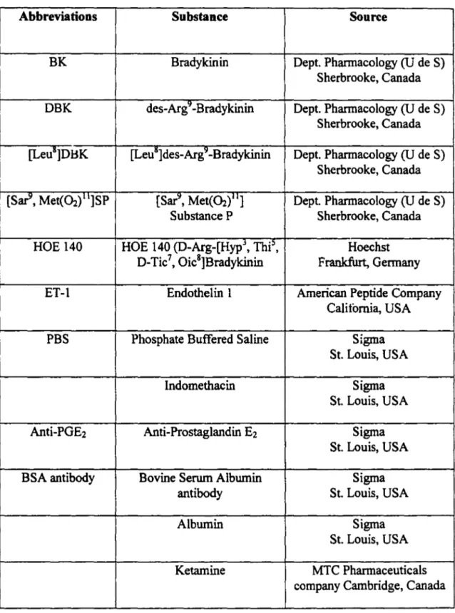
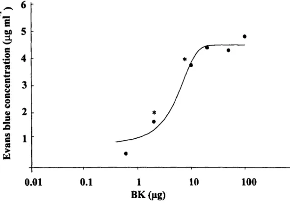
1. DOSE RESPONSE CURVE OF BK-INDUCED PLASMA EXTRAVASATION ... 41
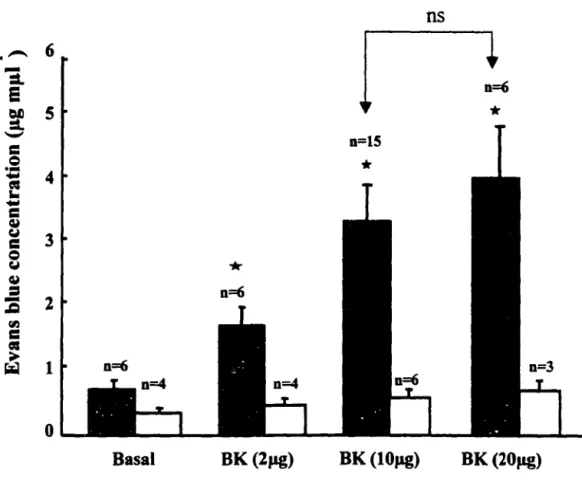
2. TIME COURSE OF BK-INDUCED PLASMA EXTRAVASATION ... 41
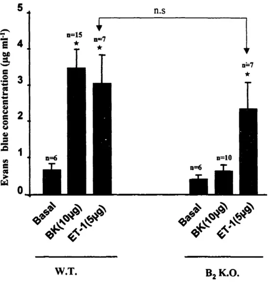
3. PLASMA EXTRAVASATION INDUCED BY ET-1 ... 45
4. PLASMA EXTRAVASATION INDUCED BY [Sar9,Met(02)11)SP ••.••••••••.•..••• 45
5. LACK OF CONTRIBUTION OF B1 RECEPTORS IN THE BK-INDUCED INCREASE IN PLASMA EXTRAVASATION ... 45
5.1 Effects of BK agonists ... 45
5.2 Effects of BK receptor antagonists ... 46
6. RPA-INDUCED PLASMA EXTRAVASATION IN WILD TYPE MICE ... 46
7. TIME COURSE OF RP A-INDUCED PLASMA EXTRA V ASA TI ON •...••..•••• 46
8. CONTRIBUTION OF
Bi
RECEPTORS IN RPA-INDUCED PLASMA EXTRAVASATION ... 538.1 Plasma extravasation induced by RPA in transgenic 82 K.O. mice ... 53
8.2 Effects of BK receptor antagonists ... 53
9. EFFECTS OF INDOMETHACIN ON PLASMA EXTRAVASATION ••••••••••• 53 9.1 Effect of indomethacin on BK-induced plasma extravasation ... 53
9.2 Effect of indomethacin on RP A-induced plasma extravasation ... 54
10. PGE2 RELEASE INTO PERITONEAL CA VITY ... 54
DISCUSSION ... 60
1. PLASMA EXTRAVASATION ... 60
1.1 Plasma extravasation induced by RP A ... 60
1.2 BK-induced plasma extravasation ... 62
2. RP A-INDUCED SYNTHESIS OF KININ ... 63
2.1 Immune complex-mediated kinin synthesis ... 63
2.2 Kinin synthesis during neutrophil migration and diapedesis ... 64
3. ROLE OF PGE2 IN RP A & BK-INDUCED PLASMA EXTRA V ASA TI ON .• 67 4. LACK OF B1 RECEPTOR CONTRIBUTION IN THE EARL Y STAGE OF RP A ...•...••...•.••.•.•....•.•.•...••.•••...••.•.•...•••...•••..••....••...•...•••••.•....••.. 69
5. STRENGHTS AND LIMITATIONS OF THE RPA MODEL USED IN T~ PRESENT ST'UDY •••••••..••••••••••••••••••.•••••••.•••••••.•••••••••••••.•••••.•••••.•••••••••••••••••••••••••••••••• 73 CONCLUSION ... ., ... 7 4 PERSPECTIVE ... 7 5 ACKNOWLEDGEMENT ... 77 REFERENCES ... 78
LIST OF PUBLICATIONS
SAMADFAM, R., TEIXEIRA, C., BKAILY, G., SIROIS, P., de BRUM-FERNANDES, A., AND •D'ORLÉANS-JUSTE, P. (1999). Significant contribution of the B2
receptor in Arthus reaction-induced extravasation in normal or B2 transgenic mice. British Journal of Pharmacology (in press).
LIST OF COMMUNICATIONS
R. SAMADF AM, A. BRUM-FERNANDES AND PEDRO D'ORLEANS-JUSTE (1999). Wild type and transgenic knockout models of passive Arthus reaction in the mouse. 4 rn Mini-symposium on the Endothelium and its vascular related diseases. 28/3/99 Montréal
LIST OF FIGURES
Figure 1 Schematic representation of the possible events occurring in the RP A
(ROSSI, 1992) ... 5
Figure 2 The sequence of intravascular- events for leukocyte migration and the relevant adhesion molecules for eacb step ... 11 Figure 3 Synthesis of kinins from their precurson (GILMAN, et al., 1990 &
BHOOLA, 1992) ···-··· 22
Figure 4 Standard curve of Evans Blue dye ... 34 Figure 5 BK dose response curve ... 42 Figure 6 BK-induced plasma extravasation wild-type and
Bi
knockout mice .••••• 43 Figure 7 Time course of plasma extravasation induced by BK in wild-type mice 44 Figure 8 ET-1-induced plasma extravasation in wild type and B1 K.O. mice ••.•••• 47 Figure 9 NKl agonist ([Sar9,Met(02)]SP(20µg)-induced plasma extravasation inwild type and 82 K.O. mice ... 48
Figure 10 Plasma extravasation induced by kinin receptorial agonists, des [Arg9]BK(DBK) and BK (lOµg), in wild-type and 8
2 K.O. mice •••.•...•.•••••• 49 Figure 11 Effect of Dl and 82 receptors antagonists ([Leu8] DBK, HOE 140 ,
lOµg/mouse) on BK-induced plasma extravasation in wild type mice ... 50 Figure 12 RPA-induced plasma extravasation at different concentration of immune
complexes ... SI
Figure 13 Plasma extravasation induced by RP A in wild-type mice at different time
points ... 52
Figure 14 RPA-induced plasma extravasation (Ab; 50 µg & Ag; 50 µg) .•.••.•.•••.•• SS Figure 15 Effect of 81 and
Bi
receptors antagonists ([Leu8] DBK; HOE140,lOµg/mouse) on RPA-induced plasma extravasation in wild type mice .•••••••• 56 Figure 16 Effect of indomethacin (IND, 200 µg) on BK (10 µg)-induced plasma
Figure 17 Effect of indomethacin (IND, 200 µg) on RP A-induced plasma
extravasation in wild type mice ... 58 Figure 18 Neutropbil-mediated kinin synthesis ... 72
LIST OF TABLES
Table 1 Amino acid sequences of the kinins ... 18
Table 2 RI.A ...•...•...•...•. 38 Table 3 Materials used ... 39
LIST OF ABREVIATIONS
AA Arachidonic Acid
Ab Antibody
Ag Antigen
AR Arthus Reaction
81 Bradykinin type I receptor
82 Bradykinin type II receptor
BK Bradykinin
Cs Complement Components
COX Cyclooxygenase
DAG Diacyl Glycerol
DBK des-Arg9 Bradykinin
EDRF Endothelial-Derived Relaxing Factor
ET-I Endothelin 1
t'MLP N-Fonnyl Methionine Leucyl-Phenylalanine
HAE Hereditary Angioneurotic Edema
HF Hageman Factor
HK High Molecular Weight Kininogens
KBP Kallikrein Binding Protein
KI Kininase I
KII Kininase II
LK Low Molecular Weight Kininogens
NK.1 Neurokinin Type I Receptor
NO Nitric Oxide
PAF Platelet Activating Factor
PGA1 Prostaglandin A1 PGA2 Prostaglandin A2 PGB1 Prostaglandin B 1 PGE2 Prostaglandin E2 PGE1 Prostaglandin E 1 PGF1a Prostaglandin 1a
PGFia Prostaglandin Fia
PGs Prostaglandins
PLA2 Phospholipase A2
PLC Phospholipase C
PMNs Polymorphonuclear Cells
SP Substance P
VLA Very Late Antigen
ICAM Intracellular Adhesion Molecule
VCAM V ascular Cell Adhesion Molecule
MAdCAM Mucosal Addressin Cell Adhesion Molecule
LFA Leukocyte Function-associated Antigen
Université de Sherbrooke
ETUDE PHARMACOLOGIQUE DE LA REACTION D' ARTHUS CHEZ LA SOURIS DE TYPE SAUVAGE OU TRANSGENIQUE
Bi
KNOCKOUTpar
RANA SAMADFAM
Département de Pharmacologie Mémoire présenté a la Faculté de Médecine
en vue de l'obtention du grade de maitre ès Sciences (M.Sc.)
Octobre 1999
La réaction passive inverse d'Arthus (RPA), un modèle d'hypersensibilité de type 3, implique des réactions inflammatoires et hémorragiques (Steil et al., 1995). La RPA consiste en une réponse aigüe impliquant l'activation d'un complexe immun et des mécanismes subséquents d'extravasation plasmatique suivis d'une réponse tardive caractérisée par une migration de cellules inflammatoires. Dans les dernières années, plusieurs agents ont été reconnus comme médiateurs inflammatoires, dont les prostaglandines, les leucotriènes, le facteur activateur des plaquettes, l' interleukine-1, le complément, les neutrophiles et le système kallicréine kininogène-kinine. De tous les médiateurs inflammatoires ci-haut mentionnés, la bradykinine semble jouer un rôle crucial dans la réponse inflammatoire induite par la réaction d'Arthus. Le but de la présente étude fut d'étudier la contribution à la fois de la bradykinine et des récepteurs 82 des kinines dans la réaction inverse passive d'Arthus chez des souris de type sauvage et des souris transgéniques 82 knockout. Nous avons aussi étudié la contribution de la prostaglandine E2 dans la réaction d'Arthus qui pourrait être impliquée à la suite de l'activation des récepteurs 82 des kinines. Nos résultats démontrent que la bradykinine induit une augmentation soutenue de l'extravasation plasmatique qui est renversée par un antagoniste B2, le HOE 140, mais non par un antagoniste Bi. le [Leu8,desArg9]BK. De façon identique, l'extravasation plasmatique induite par la RP A était complètement renversée par l'antagoniste 82 mais non B 1• Contrairement aux animaux de type sauvage, la bradykinine et la réaction d'Arthus n'ont pas induit une augmentation significative de l'extravasation plasmatique chez les souris 82 knockout. De plus, la réaction d'Arthus mais non la bradykinine induit une augmentation significative des concentrations péritonéales de prostaglandine E1. Un traitement avec l'indométhacine rédnit significativement l'extravasation plasmatique du bleu d'Evans induite par la RP A mais non par la bradykinine. Nos résultats suggèrent donc un rôle crucial pour les récepteurs 82 dans les mécanismes d'extravasation plasmatiques induits par la RP A. Nos résultats supportent de plus la contribution de la PGE2 dans les processus d'extravasation induite par la réaction d'Arthus alors que l'augmentation de l'extravasation induite par la bradykinine et les récepteurs 82 n'implique pas le même médiateur inflammatoire.
INTRODUCTION
Diseases such as rheumatoid arthritis, in which tissue injury and pathophysiologic abnormalities are due to immunologie mechanisms, may be initiated by immune responses to foreign or self-antigens (W ALPORT, et al., 1997). Such diseases in man are believed to be the clinical counterparts of hypersensitivity reactions including the acute and chronic Arthus reactions. The effector mechanisms by which antibodies and immune complexes induce tissue damage include the activation of the complement system and a variety of inflammatory cells (ROITT, 1991 ). Therefore, the study of inflammatory responses and the mechanisms involved in hypersensitivity reactions, particularly Arthus reactions, has some relevance to what occurs in such diseases as rheumatoid artrithis and may be useful in designing new drugs and new treatments for these conditions.
The reverse passive Arthus reaction (RPA), a model of type III hypersensitivity, involves inflammatory and haemorrhagic states (STEIL et al., 1995). The induction of RP A leads to a sequence of events including the activation of the classical pathway of the complement cascade, the migration of neutrophils and also plasma extravasation into the inflamed site (STEIL et al., 1995). Kinins are powerful proinflammatory autacoids (CAMBRIDGE et al., 1995; SHARMA, et al., 1990) which may play a crucial role in inflammatory responses.
In this chapter, more detailed accounts of Arthus reaction, cell adhesion molecules (CAM) and the complement component will be described. This is followed by sections describing the kinins, their metabolism, pharrnacological properties, and their
role in inflammatory reactions. A final section deals with the generation and the importance of bradykinin receptor type 2 (B2) knockout mice.
1. ARTHUS REACTION {HYPERSENSITIVITY TYPE III)
1.1 Arthus Reaction
Disorders that result from aberrant, excessive, or uncontrolled immune reactions are called hypersensitivity diseases. This term arises from the clinical definition of immunity as sensitivity. Failure to control physiologie immune responses against foreign antigens or to maintain self-tolerance leads to hypersensitivity diseases in which the primary pathogenic mechanism is immunologie. Hypersensitivity reactions are classified into four types. Arthus reaction is equated with type III or immune complex mediated. Type Ill hypersensitivity reactions are mediated by IgM and lgG antibodies which can bind to soluble antigens and lead to the generation of immune complexes. The deposition of immune complexes in certain tissue sites causes the pathology of type III hypersensitivity reactions. The pathogenic potential of immune complexes is determined, in part, by their size and by the physicochemical properties of the antigens and antibodies involved, such as charge and valence. Larger immune complexes are readily cleared from the circulation by the phagocytic system, while the very small ones are not deposited (ROITT, 1991).
Arthus reaction was described by MAURICE ARTHUS in 1903, as acute inflammatory and haemorrhagic reaction produced in the skin of rabbits when a local injection of horse serum was adrninistered to previously sensitized rabbits. Although much progress has been made in the elucidation of the mechanism involved in this
complex reaction, it still remains only partially understood. The Arthus reaction are characterized by edema formation, neutrophil and platelet accumulation, and haemorrhage and in severe cases, necrosis. Severa! membrane derived lipids (HELLER al., 1998), the complement protein fragment C5a (HÔPKEN, et al, 1997), nitric oxide, platelet activating factor (STEIL, et al, 1995), 1NFa and eicosanoid (ROCHA, et al., 1997) have been identified as important mediators of Arthus reaction (WlilTE, 1999). The cobra venom factor, which suppresses the complement system, depresses the edema in Arthus reaction. Depletion of neutrophils with nitrogen mustard or anti-neutrophil antiserum also suppresses the Arthus reaction (HUMPHREY, 1955, COCHRANE et al., 1970). Furthermore the blockade of cell adhesion molecule by antibody or their deficiency also significantly decreased the edema formation and neutrophil accumulation (BURCH, et al., 1999; BULLARD, et al., 1995). These observations indicate the crucial role of the complement component and neutrophils and cell adhesion molecules in the Arthus reaction.
1.2 The mecbanism of Arthus reaction
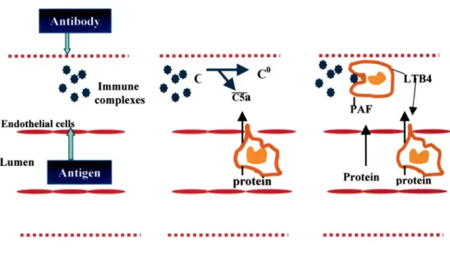
It is now known that the Arthus reactions, including peritoneal RP A, consist of an early-phase response involving the activation of complement components and mast cell degranulation (RAMOS et al., 1994) and a late-phase response characterized by the migration of inflammatory cells. It seems that plasma extravasation occurs at the early stage of the inflammatory responses. The mechanism of RP A has not been fully understood. Figure 1 represents the events that may occur in the RP A (ROSSI, et al., 1992). After the injection of the antigen (intravenously) into the individual who has
already been locally injected with its cognate IgG antibody, the antigen diffuses out of the lumen of the blood vessel and across the endothelial cells barrier where it meets the antibody. The formation of the immune complexes activates the classical pathway of the complement cascade resulting in the generation of C5a. The C5a is a potent chemoattractant for neutrophils (COLLINS, et al., 1991). C5a induces the adherence of neutrophils to the endothelial cells of the blood vessels and their migration via endothelial junctions. The interaction between cell adhesion molecules on neutrophils and endothelial cells leads to plasma extravasation (HUSTON, 1997). The plasma leakage into the interstitium provides more antigen resulting in a further fonnation and deposition of immune complexes. Neutrophils that have migrated into the interstitium begin the phagocytosis of the deposited immune complexes, which lead to activation of the ncutrophils and the release of platelet-activating factor (PAF) and Leukotriene 84 (L TB4). PAF could act directly on the endothelial cells resulting in
an
increase between endothelial junctions which then leads to further plasma leakage. L T84 is also a chemoattractant for neutrophils and may contribute in the edema fonnation (ROSSI, et al., 1992). These events may also occur in the mice peritoneal reverse passive Arthus reaction. Investigations in the peritonealRP A
have shown that, except for the activated complement components, mast cell mediators such as tumor necrosis factor also play a key role in the initiation of the inflammatory process and neutrophil accumulation(ZHANG et al., 1995).
Arthus reaction can be elicited by different ways, direct Arthus reaction (AR), direct passive Arthus reaction (PAR) and reverse passive Arthus reaction (RP A).
...
~
..
•••
• • Immune • complexes Endothelial ce Lumen...
,...
,••• c-V
co
•••
csa
protem ••••••••••••••••••• • • • • • • • • • • • • • • • • • • • • 1•••
••
Protein protem • • • • • • • • • • • • • • • • • • • 1Figure 1 Schematic representation of the possible events occurring in the RP A (ROSSI, 1992)
The circulating antigen passes across the blood vessel barrier into the interstitium where it forms immune complexes with cognate antibodies. Deposition of immune complexes leads to the activation of the classical pathway of the complement cascade resulting in the generation of C5a. C5a induces the recruitment of neutrophils into the interstitium, resulting in plasma leakage. The migrated neutrophils phagocyte the immune complexes resulting in the release of PAF and L TB4, which in turn lead to more
plasma extravasation.
1.3 Active Arthus Reaction
The Arthus reaction that Maurice Arthus described at the tum of this century was an example of active Arthus reaction (ARTHUS, 1903). The active Arthus reactions
occur when an individual bas produced IgM or IgG antibodies in response to an innocuous antigen and subsequently encounters the same antigen. The term active refers to immuniz.ation of an individual by administration of an antigen.
1.4 Direct passive Arthus reaction
This type of Arthus reaction can be elicited by the injection of an innocuous antigen to the skin of an individual followed by an injection of the antibody. The passive word refers that the antibody is being injected rather than being synthesized by the immune system of the individual.
l.S Reverse passive Arthus reaction
In reverse passive Arthus reaction bath antibody (Ab) and antigen (Ag) also are being injected and the reverse refers to that the injection of Ab is prior to the injection of Ag and passive refers to immunization through the transfer of specific antibody from an immunized individual to a non immunized individual. Experimentally, it is convenient to investigate the RP A, which is elicited by a local injection of Ab and an intravenous injection of its cognate Ag or by the formation of Ab-Ag complexes in the peritoneal cavity (intraperitoneal injection of bath Ab and its cognate Ag). In the present study, the formation of Ab-Ag complexes in the peritoneal cavity of mice bas been used to investigate the role of bradykinin and its receptors in RP A.
As it was mentioned previously, neutrophil migration is one of the important feature of the Arthus reaction. The adhesion of neutrophil to microvascular endothelium is essential for their migration into inflamed site and it is a highly regulated process
requiring the coordinated participation of chemokins (including complement component Sa, 3b) and cell adhesion molecules (CAM). The CAM and complement component will be discussed separately in the following sections.
2. POSSIBLE ROLES OF CELL ADHESION MOLECULES (CAM) IN THE
RPA
Three families of CAM which subserve the adhesion and migration of neutrophil are selectins, integrins, and lg superfamily.
2.1 Selectins
The three member of selectin families are selectin, E-selectin, and P-selectin. L-selectin expressed on leukocyte, whereas E and P L-selectin are expressed on activated endothelial cells, and P-selectin is expressed on the surface of activated platelets (LASKY, 1995). The blockade of L-selectin with L-selectin-lg chimera reduces neutrophil accumulation (WATSON et al., 1991 ). Theoglycolate-induced migration of neutrophil into the peritoneal cavity of P-selectin knockout mice was delayed (MA Y ADAS et al., 1993). In addition the acute migration of neutrophil was completely absent in the P-selectin/ICAM-1 double mutant mice (BULLARD, et al., 1995) after 6 hours of the induction of the inflammatory response. Others have suggested that neutrophil accumulation under these conditions requires a combination of both P-and E selectins (RAMOS et al., 1997). ln the recent publication, MIZGERD et al., (1999) showed that P-selectin is essential to ICAM-1 and E-selectin-independent acute peritoneal inflammation at early stage ( 4 heurs after induction). But after 24 h of
peritonitis, there were no differences between wild type and E-selectin/P-selectin/ICAM-1 mutant mice, demonstrating that these endothelial adhesion molecules are not essential to neutrophil emigration during later stages of peritonitis. Since migration into the peritoneum requires both P-selectin and ICAM-1 adhesion molecules while migration into the lung requires neither (BULLARD, et al., 1995), the implication of different CAMs in the inflammatory response may be organ specific.
2.2 Integrins
The integrins are involved in the cell-matrix interactions. Integrins are heterodimeric membrane glycoproteins and they consist of a a subunit and a common
p
subunit At least 8 different ~ subunits and more than 16 different a subunits have been reported(HUMPHRIES, 1996). The
p
subunit composition of the heterodimer determines to which subfamily the integrins belong. The integrins family includes the very late antigen (VLA 4, VLAs. VL~; they are belong toP1
integrin subfamily), leukocyte functionassociated antigen (LFA-1; CD1 la/CD18;
P2
integrin) and Mac-1 (CDl lb/CD18; ~iintearin). The fibronectin (an extracellular matrix protein), VCAM-1 (vascular cell adherence molecule), laminin, ICAM and fibrinogen (a soluble adhesion molecule) are biological ligands for integrins (WILLIAMS, et al., 1999; TA OO KA, et al., 1999).p
1 integrins are chemotaxic and they are implicated in the cell adherence wbereasp
2 integrins are implicated in the cell aggregation and diapedesis (WILLIAMS, et al., 1999). Neutrophil possess mainly integrins belonging to theP1
andP
2 integrin subfamilies,p
2 integrins predominating (WILLIAMS, et al., 1999). In the resting neutrophils, integrins are inactive and unable to bind their ligand. To confer theadhesiveness to counter-receptor they must undergo a confonnational change. The exposure to pro-inflammatory substances (such as interleukin-8 (IL-8), CSa, leukotriene 84 and platelet activating factor) leads to the activation of the integrins by two ways: a. increasing the density of membrane bound integrin b. inducing the confonnational changes of the integrins (ROSENBLOOM et al., 1999). Reverse passive Arthus reaction in rat was inhibited by NPC 15669. This compound inhibits the increase in expression of CD11/CD1s on activated neutrophils (BURCH et al., 1992). In addition it has been reporte<l that edema formation and hemorrhage associated with RP A reactions in responses to zymosan and ZAP are completely CD1s-dependent, and are mediated, at least in part, via ICAM-1. Responses to the neutrophil-independent edema forming mediators, PAF and BK are not dependent upon CD1s or ICAM-1 (NORMAN, et al.,
1994).
2.3 lg superfamily
Ig superfamily of cell adhesion molecules includes intracellular adhesion moleculc-1 (ICAM-1), platelet endothelial adhesion molecule-1 (PECAM-1), and vascular adhesion molecule-1 (VCAM-1) (MADDOX, et al., 1999). Inflammatory mediators greatly increase the synthesis and membrane density of ICAM-1 and VCAM-1 (RICE, et al., 1989)
Platelet-endothelial-cell adhesion molecule 1 (PECAM-1 ), expressed also on leukocytes, appears to have a role in the migration of leukocytes across endothelium. Other members of the immunoglobulin superfamily mediate heterophilic interactions with integrins. For example, vascular-cell adhesion molecule l (VCAM-1) found on stimulated endothelial
cells binds the <l.4~1 or <l.4~1 integrin on lymphocytes. Monoclonal antibodies against
ICAM-1 significantly inhibited leukocyte influx into pleural cavity of rats induced by reverse passive Arthus reaction. This effect was dependent on the dose of the antibody (FIELDING et al., 1995). It bas been suggested that neutrophil accumulation in the peritoneal cavity depend upon L-selectin and ~2 integrins (LAF-1 and Mac-1) on
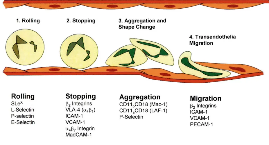
neutrophils, and E-selectin and ICAM-1 on the endothelium (MULLIGAN et al., 1998). Figure 2 summarizes the role of CAMs in the migration of leukocyte. There are four different steps involved in the migration of neutrophils and each step requires different CAM: l.Rolling 2. Stopping 3. Aggregation 4. Transendothelial migration. Figure 2 gives a general idea about the sequence of intravascular events and the relevant adhesion molecule for each step of the leukocyte migration (HUSTON, 1997). However, depending on the species or the type of the tissue, the implication of these molecules may vary (BULLARED, et al., 1995).
In the figure SLex indicates sialyl Lewis X; VLA, very late antigen; ICAM, intracellular adhesion molecule; VCAM, vascular cell adhesion molecule; MAdCAM, mucosal addressin cell adhesion molecule; LF A, leukocyte function-associated antigen; and PECAM, platelet endothelial cell adhesion molecule.
Rolling
Slex
L-Selectin P-selectin E-SelectinStopping
~2 lntegrins VLA-4 (a4~1) ICAM-1 VCAM-1 a4~7 lntegrin MadCAM-1 Shape ChangeAggregation
CD11bCD18 (Mac-1) CD11aCD18 (LAF-1) P-Selectin 4. Transendothelia MigrationMigration
~2 lntegrins ICAM-1 VCAM-1 PECAM-1Figure 2:
The sequence of intravascular events for leukocyte migration and the relevant
3. THE COMPLEMENT SYSTEM
In type Ill hypersensitivity reactions including Arthus reaction, the formation of immune complexes leads to the activation of the classical pathway of the complement cascade. Nature has devised two pathways for the activation of complement, the classical pathway and the alternative pathway. Although some components are common to both pathways, they differ in the ways by which they are activated. The alternative pathway arase in evolution earlier than the classical pathway and does not require immune complex for initiation. lt may be triggered by LPS (lipopolysaccharides) or endotoxin from cell wall. In the section that follows only the classical pathway of the complement component will be described.
3.1 The classical complement pathway
The classical complement pathway involves the activation, in an orderly fashion, of nine protein components termed Cl through C9. Usually in the activation process, the product is an enzyme that catalyzes the subsequent step.
Activation of Cl. Cl is the first component to become activated and consists of three proteins, Clq, Clr, and Cls. The complex is held together by calcium ions. Activated Clq activates Clr which, in turn, activates Cls. Activated Cls initiates the subsequent step by acting on C4.
Activation of C4. C4 is a glycoprotein which. is synthesized by macrophages. The proteolytic action of Cls on C4 cleaves the C4 resulting in the generation of activated C4. Interaction between activated C4 and Cls leads to the cleavage of C2, the next complement component to become activated.
Activation of C2. C2 is a glycoprotein and it is cleaved by the action of C4 and C 1 s. The activated C2 interacts with the activated C4 and generates a new complex. This
complex which is designated C42 is also called C3 convertase. The term C3 convertase refers that this complex acts on C3 and converts it to small fragments.
Activation of C3. C3 is a ~-globulin which is secreted by macrophages as
pro-C3. C3 convertase (C42) cleaves the C3 into a small fragment, C3a, and a larger fragment, C3b. Both C3a and C3b are biologically active. C3b attaches to the target cell membrane such as platelet, polymorphonuclear cells, and bacteria. C3b also interacts with C42 and forms C423 that is also called CS convertase.
Activation of CS, C6, C7. CS, C6 and C7 are globular proteins. CS by the action of CS convertase (C423), is cleaved into two fragments termed CSa and CSb. C5b, the bigger fragment, combines stoichiometrically with C6 and C7 to form C567 on the cell membrane This complex activates the next components, C8 and C9. CSa bas several important biological activities including leukocyte chemotaxis and activation, enhancement of neutrophil-endothelial cell adhesion, induction of granule secretion in phagocytes (HÔPKEN ET AL., 1997), and induction and release of several cytokines (i.e., IL-1, TNF-a, IL-6, and IL-8). ( OKUSAWA et al., 1987; OKUSAWA et al., 1988; SCHOLZ et al., 1990; EMBER et al., 1994). The immune complex-mediated inflammation was significantly depressed in CSa anaphylatoxin receptor knock out mice (HÔPKEN, et al; 1997).
Activation of CS and C9. CS67 interacts with C8 and C9 on the target cell membrane. This interaction results in conformational changes in these proteins. These proteins then can insert themselves into the cell membrane and produce transmembrane
channels. The passage of ions through these channels disturbs the osmotic equilibrium, resulting in a rapid influx of water into the target cell which, in tum, leads to the lysis of the target cell (BENJAMINI & LESKOWITZ, 1993). The cell lysis by the activation of complement components, as we shall later see, is important in kinin synthesis.
3.2 Biological activity of the complement component
3.2.1 Anapbilatoxins
C3a and C5a are potent anaphilatoxins. These activated complements induce the degranulation of mast cells and/or basophils causing the release of histamine. Histamine released from mast cells produces vascular effects such as local vasodilatation and increased vascular permeability, and also influences visceral smooth muscle and exocrine glands. These effects are associated with anaphylaxis and other allergie reactions. The increase
in
vascula!' permeability results in local edema formation.3.2.2 Cbemotaxins
Substances which recroit and attract inflammatory cells, mostly neutrophils and monocytes from an area of lesser concentration to an area of higher concentration are called chemotaxins. C5a is a potent chemoattractant for neutrophils and can be generated by the classical or alternative complement pathways (COLLINS, et al., 1991 ). Cytokines, such as tumor necrosis factor or interleukin-1 which are produced by activated mast cells, may also act as chemoattractant (UTSUNOMIYA et all, 1998; PICCOLO et al., 1999). Activated neutrophils in inflamed sites produce hydrolytic enzymes, reactive oxygen
species, lipid mediators, and nitric oxide, all of which can contribute to cell and tissue
damage.
3.2.3
Immune adherenceComplement components could coat the immune complexes and induce the adherence of these complexes to various surfaces, including the walls of blood vessels. Immune complexes which are attached to the blood vessels wall are phagocytosed more readily.
3.2.3 Opsoni7.ation
Opsonization refers to the coating of an antigen or an immune complex by the complement components that would render the antigen more "attractive" to phagocytic cells. C3b is a potent opsonin.
3.2.4 Complement mediated cell lysis.
As we discussed before the activation of complement leads to the generation of a membrane attack complex, which causes the osmotic lysis of cells.
4. KININS
A variety of factors including tissue damage, viral infections, and other inflammatory events activate a group of proteolytic enzymes resulting in the generation . of kinins. Kinins are patent proinflammatory peptides, which are synthesized de novo and act locally to produce their effects. Kinins are involved in the regulation of every
major physiological system including motility of the gastrointestinal system, fluid transport in the respiratory system, renal perfusion in the excretory system, and the reproductive system (BHOOLA et al., 1992). They also act as vasodilators and increase vascular penneability (BHOOLA et al., 1992). Of ail the inflammatory mediators, kinins are more likely involved in RPA (CRUWYS et al., 1994). ln this section kinins history, structure, synthesis, metabolism, and phannacological properties will be discussed.
4.1 History
In 1909 ABELOUS and BARDIER showed that an intravenous injection of an alcohol insoluble fraction of human urine causes hypotension in dog. PRIBRAM and HERNHEISER ( 1920) later reported a similar observation in rabbits. The compound responsible for the hypotensive property was isolated by FREY and KRAUT (1928). This high molecular weight substance was thermolabile anà nondialysable. Later .KRAUT and WERLE showed that similar substances could be nbtained from saliva, blood and pancreas and since the pancreas was a rich source for this hypotensive agent, they named it KALLIKREIN after an old Greek synonym for that organ, KALLIKREAS (KRAUT et al., l 930a). The pharmacological activity of kallikrein was established in 1937 by WERLE and his colleagues. Kallikrein acts on an inactive substance in plasma and converts it to an active substance which in turn leads to smooth muscle contraction. Later the active substance was named kallidin and its precursor was tenned as kallidinogen (WERLE, 1970). Interest in the field intensified when ROCHA SIL V A and his colleagues reported that venom extracts of B. jararaca or trypsin acted on plasma globulin to produce a hypotensive substance which could also cause a slow and delayed
contraction in the isolated guinea pig ileum. By derivation from the Greek words, bradys fer slow and kinein for movement, the new substance was termed bradykinin ( ROCHA SIL V A et al., 1949). Later the nonapeptide bradykinin was isolated and synthesized (ELLIOTI et al., 1960); BOISSONNAS et al., 1960). Shortly after, kallidin was found to be a decapeptide bradykinin with a lysine residue at the aminoterminus. A number of peptides with related chemical structures and pharmacological activity to bradykinin or kallidin have since been discovered and for the whole group the tenn kinin, and for their precursors the tenn kininogen, have been adopted.
Beside bradykinin and lys-bradykinin (kallidin), the birds kinin, insect kinin T-kinin, Met-T-T-kinin, and T-kinin Lue are other members of the kinin family. Table 1 (BHOOLA 1992) shows the chemical structure of the kinin family peptides. Mammalian kinins possess a highly conserved structure that shows a high degree of homology with insect, amphibian skin, reptile, and omitho kinins (BHOOLA, 1992). Eventually the peptide receptors were discovered and their antagonists were then developed, thus providing new tools to explore the function ofkinins.
Y..inin family Bradykinin Lys-bradykinin Met-Lys-bradykinin T-kinin Met-T-kinin Birds (omitho) kinin Vespa-kinin X Vespa-kinin M 1 2 3456789 Arg-Pro-Pro-Oly-Phe-Ser-Pro-Phe-Arg Lys-Arg-Pro-Pro-Oly-Phe-Ser-Pro-Phe-Arg Met-Lys-Arg-Pro-Pro-Oly-Phe-Ser-Pro-Phe-Arg Ile-Ser-Arg-Pro-Pro-Oly-Phe-Ser-Pro-Phe-Arg Met- Ile-Ser- Arg-Pro-Pro-Gly-Phe-Ser-Pro-Phe-Arg
Arg-Pro-Pro-0 ly-Phe-Thr-Pro-Leu-Arg Ala-Arg-Pro-Pro-Gly-Phe-Ser-Pro-Phe-Arg-lle-V al
Gly-Arg-Pro-Hyp-Oly-Phe-Ser-Pro-Phe-Arg-Ile-Asp
Table 1 Amino acid sequences of the kinins (BHOOLA, 1992).
4.2 Synthesis
A number of serine proteases will generate kinins from their precursors, high molecular weight (HK) and low molecular weight (LK) kininogens, but the highly specific proteases that release bradykinin and kallidin are termed kallikreins. The kallikreins circulate in plasma in an inactive state and must be activated by other proteases. Two different types of kallikrein exist, the tissue kallikrein and plasma kallikrein. These are two distinct enzymes and their precursors, tissue prekallikrein and plasma prekallikrein are activated by different mechanisms.
4.2.1 Tissue Kallikrein
The true tissue kallikrein should be able to release a kinin (kallidin I bradykinin) from kininogen. However in most species tissue kallikrein represents a unique class of enzymes that hydrolyze one arginyl and one methionyl bond in the kininogen molecule
to release kallidin (BHOOLA, 1992). The tissue kallikrein is encoded by different genes, tenned kallikrein multigene family (SWIFT et al., 1982; ASHL Y & MAC DONALD, 1985; DRINKWATER et al., 1988; MAC DONALD et al., 1988; CLEMENTS, 1989). Even though these genes are highly homologous, their products show distinct differences in substrate recognition and amino acid sequences. The kallikrein multigene family varies in number between the different mamrnalian species. So far, three genes have been identified in human (SCHEDLICH et al., 1987: EV ANS et al., 1988; QIN et al., 1991) and 24 in the mouse (RICHARDS et al., 1982; EV ANS et al., 1987). The true tissue kallikreins are acidic glycoproteins, with a molecular weight of 24000-45000 Daltons (PISANO, 1975). The position and number of glycolysation sites vary according to the species and the synthesizing cell (T AKAHASHI et al., 1988; LU et al., 1989; KELLERMANN et al., 1988). The enzymes are synthesized as prokallikreins and by the action of several proteases including trypsin, they are cleaved to kallikreins. The primary physiological substrate for tissue kallikreins is kininogen from which kinin:) are formed. The complete kinetics and sequence of cleavage by these enzymes are not yet clearly understood (FIELDER & HINZ, 1992).
The presence of tissue kallikrein in the intracellular granules in different tissues have been determined by the use of immunocytochemistry, in situ hybridization and specific antibodies (BHOOLA, 1992). The type and sites of sequestration granules of the enzyme vary from one cell type to another and may be related to the function of the enzyme in each tissue. Kallikrein binding protein (KBP) is an acidic protein which specifically binds to active tissue kallikrein and inhibits its activity (CHAO et al., 1990). Tissue kallikrein has been implicated in organ-specific disorders such as pancreatitis
(UEHARA et al., 1989). By the use of potent
kinin
anta.gonists or kallikrein inhibitors, new therapeutic drugs may be designed for these diseases.4.2.2 Plasma Kallikrein
The precursors of plasma kallikrein are termed prekallikrein. Plasma prekallikrein is an inactive protein which is encoded by a single gene and synthesized in the liver. Plasma prekallikrein is a single-chain glycoprotein which binds toits substrate (high molecular weight kininogen, HK) and forms a 1: 1 complex (MANDLE et al., 1976). It has two isoforms of 85 and 88 KD, both of which are present in hwnan plasma (MANDLE & KAPLAN, 1977; VELOSO & COLMAN, 1992) and which are cleaved and activated by Hageman factor (HFa). Beside human, the mature plasma kallikrein has been identified in several other species including porcine, rat, and guinea pig (YAMAMOTO et al., 1980; SEIDAH et al., 1988; PAQUIN et al., 1989). Although low molecular weight kininogen (LM) is a poor substrate for plasma kallikrein, by the synergistic action of hwnan PMN elastase, it will form BK in vitro (SATO &
NAGASA W A, 1988). First neutrophil elastase acts on LM kininogen and forms a kinin-containing fragment, from which BK is readily released by the action of plasma kallik:rein.
4.2.3 K.ininogens
Kininogens are kinin precursors which, by the action of kallikreins, cleave to kinins (WERLE ET AL., 1937). They are purified and isolated from different species including hwnans (WEBSTER & PIERCE, 1963; JACOBSEN, 1966; SPRAGG &
AUSTEN, 1971; SPRAGG & AUSTEN, 1974), bovine animais (SUZUKI & KATO, 1977; KATO et al., 1981), rat (OKAMOTO & GREENBAUM, 1983) and guinea pig (YAMAMOTO, 1987). A single K gene codes for H- and L-prekininogens in bovine animais and humans (KITAMURA et al., 1983; KITAMURA et al., 1985). Alternative splicing of the gene transcript produces the HK and LK molecules. In the rat, beside the K gene which codes for HK and LK, another gene termed T gene that codes for the T-kininogen has been identified (ENJYOn et al., 1988). The T-gene shows an extensive homology of about 90% with the K gene (KAKIZUKA et al., 1990).
The kininogens are single-chain glycoproteins. They posses an amino-terminal heavy chain and a carboxyl-terminal light chain. These two polypeptides are linked by a disulfide bridge and the kinin moiety is intervalled between the two polypeptides (IŒLLERMAN et al., 1987). Kininogens are synthesized by hepatocytes and they undergo posttranslational modification such as glycosylation prior to their secretion into the circulation (KIT AMURA et al., 1983; TAKAGAKI et al., 1985). The presence of a specific receptor for HK on neutrophils (GUSTAFSON et al., 1989), endothelial cells (V AN IW AARDEN et al., 1988a,b; SCHMAIER et al., 1988a,b) and plate lets (GUST AFSON et al., 1986) has been reported.
HF a
L....:Pre~K:.::a~ll~ikr::::::e,:::in:.._t-__ ..___~ Plasma Kallikrein
HK LK Tissue Kallikrein Release of Kininogenases BK Kallidin .___ Cell lysis
Amino
Peptidases Activation of immune complexes Figure 3 Synthesis of kinins from their precursors (GILMAN, et al., 1990 &BHOOLA, 1992) 4.3 Kinin Peptidase
The kinins half-life in plasma is only about 15 seconds and they are inactivated by the action of Kininases types 1, II and some additional peptidases as described below.
4.3.1 Kininase 1 (KI)
KI is a family of two carboxypeptidases, Kininase 1-carboxypeptidase N (KI-CPN) (ERDÔS & SLOANE, 1962) and Kininase 1-carboxypeptidase M (KI-CPM) (SKIDGEL et al., 1984; SKIDGEL et al., 1989; TAN et al., 1989). CPN and
KI-CPM remove the carboxyl-terminal arginine residue and produce des-Arg9 BK which is the agonist for B1 receptors. Beside desArg9aK., KI-CPM is also able to release des [Arg9]Lys-BK from LysBK.
4.3.2 Kininase II (Kii)
The family of Kininase II consists of two polypeptides tenned Angiotensin 1-converting enzyme (KII-ACE)(SKEGGS et al., 1956; YANG et al., 1970a,b; IGIC et al., 1972) and Neutra! endopeptidase (Kil-NEP). The KII family hydrolyses the Pr7-Phe8 bond on the carboxyl terminus of the kinin molecule. After cleavage of Pro7-Phe8 the remaining kinin molecule undergoes another cleavage reaction on Ser5 -Pro6 by the action of KII-ACE (ZACEST et al., 1974). Beside Kininase I and II, the Prolidase (aminopeptidase p), as well as kininase A and B may also play a role in kinin metabolism (RYAN ET AL., 1968; OLIVEIRA et al., 1976). They respectively cleave the Arg1-Pro2 bond, the Phe5-Ser6 bond and the Pro7-Phe8 bond.
KII-~P KJ-CPN
Arg
1
-Pro
2
-Pro
3
-Gly
4
-Phe
5
-Ser
6
-Pr~-Phe~Arg
9
f
'\
l
t
Prolidase Kil-ACE KI-CPM
4.4 Receptors
So far evidence shows die existence ofat least two types of receptors for kinins, 8 1 and B2 (V A VREK & STEWART, 1985; REGOLI et al., 1990). Kinin receptors are classified according to the relative potencies of
kinin
agonists on isolated smooth muscle preparations. B1 receptors are more sensitive to the carboxyl terminal desArg metabolites of BK and B2 receptors are more sensitive to intact peptides (REGOLI et al., 1996).The order of potency of agonists on the 8 1 receptor, namely:
LysdesArg
9BK > desArg
9BK > BK
And on the B2 receptor, narnely:
[Tyr(Me)
8]BK >BK> LysdesArg
9BK
>
desArg
9BK
Normally B1 receptors are absent in the cells, and their synthesis appears to be induced by trauma or pathological insults (REGOLI, 1981 ). In 1991, DE BLOIS and his colleagues have proposed IL-1 as the endogenous mediator responsible for the induction of B1 receptors. Bl and B2 receptors could coexist in the same cells. Most of the actions of BK and kallidin are mediated by B2 receptors including plasma extravasation, vasodilatation, muscle relaxation (REGOLI et al., 1986; WHALLEY et al., 1987a; NW ATOR & WHALLEY, 1989) and pain (STEWART et al., 1985; STERANKA et al., 1988).
4.S Antagonists
The structure-activity studies of BK by STEWART and V A VREK showed that the replacement of Pro7 by D-Phe gave the analogue an antagonist profile and they
presented [D-Phe7]BK as a first competitive bradykinin antagonist (STEWART and V A VREK, 1986). Of ail the B2 antagonists descnbed by STEWART and V A VREK., D-Arg[Hyp3 ,Thi5•8, D-Phe7], named NPC 567 was the most effective one (1991). The selective antagonist for B2 receptors is HOE 140 [D-Arg-Arg-Pro-Hyp-Gly-(j3-2-thienyl)-Ala-Ser-Dtic-Oic-Arg] which is developed by introducing several unnatural amino acids into the BK molecule (HOCK et al., 1991) and has been used in the present study. HOE 140 is much more potent than NPC 567 and bas not shown any nonspecific side effects yet. The most selective and potent antagonist for B 1 receptors is desArg9[Leu8]BK which bas also been used in the present study. The antagonists mentioned above and other peptidic BK antagonists have been reviewed by REGOLI and his colleagues in 1996.
4.6 Mechanism of action of kinins receptors
BK receptors have been cloned in different species (MENKE et al., 1994; EGGERICKX et al, 1992; HESS et al, 1992; MCEACHERN et al., 1991; Mclntyre et al., 1993; POWELL et al., 1993). Although it bas been demonstrated that the BK receptors are coupled to Gi, Gq/Gl l, and G13 proteins (depending on the cell types and also on the stimuli) (LIAO, et al., 1993; WILK-BLASZCZAK, et al., 1994; FABIAN, et al., 1998;HUANG, et al.,1996; PAN, et al., 1996), there is strong evidence that the main signaling pathway of bradykinin receptors involves activation of a Gq protein (REGOLI et al., 1996). Stimulation of BK receptors may lead to an increase in cytosolic free calcium by the activation of phospholipase C with the resultant formation of inositol triphosphate and diacyl glycerol (DAG) (REGOLI et al., 1996; QUITTERER, et al.,
1999), an increase in cGMP (GADD & BHOOLA, 1988), and the release of PGs by the activation of phospholipase A2 (BURCH & AXELROD, 1987).
Apart from provoking direct effects, BK receptor activation may also induce the release and/or activation of other mediators, such as substance P (FIGINI et al., 1995). In addition NO synthase, which leads to the synthesis of nitric oxide, can also be induced by BK. NO plays an important role in the vasodilatation (GOODMAN & GILLMAN, 1990).
4. 7 Pharmacological properties
K.inins are potent vasodilators. They increase vascular permeability and cause edema. They have algesic properties. In addition, they participate in the regulation and the maintenance of fluid and electrolyte balance by the kidney. However, the most important function of kinins appears to be their substantial role in the inflammatory responses (GOODMAN & GILMAN, 1991).
4.7.1 Stimulation of nerve ending and production of pain.
BK is a potent nociceptor that acts directly on nerve endings and evokes pain (CLARK, 1979). BK receptors in the nervous system are localized in the superficial layers of the spinal cord, thin unmyelinated fibers, and neurons of the dorsal root and trigeminal ganglia, and sites in subastantia gelatinosa (STERANKA et al., 1988). Application of BK in these sites which are involved in nociception elicits pain. Studies using the selective B1 or B2 receptor antagonists and more recently the B2 knock out transgenic mice suggest a differential role for B2 receptors in nociception and for B 1 in
chronic inflammatory hyperalgesia (DRAY & PERKINS, 1993; PERKINS et al., 1993; BOYCE et al., 1996; RUPNIAK et al., 1997). These studies also suggest that B1 but not
B1 receptor antagonists may be clinically useful as analgesic drugs.
4. 7 .2 Eff ect of kinin on renal fonction and blood pressure
Kinin fonnation could occur in the tubular fluid (exocrine) by the concomitant release of tissue kallikrein and kininogen in this fluid or in the extracellular tissue space (paracrine). Infusion of kinin into the renal tissue activates its receptors on the basolateral surface of tubule cells particularly in the cortical and outer medullary collecting tubules. Activation of these receptors results in the generation of nitric oxide which in turn increases renal blood flow, diuresis, and natriuresis without changing the glomerular filtration rate (WEBSTER & GILMORE 1964; STEIN et al., 1972; ORANGER
&
HALL, 1985).4.7.3 Extravascular smooth muscle cells
Kinins contract extravascular, isolated smooth muscle preparation but relax the rat duodenum (ROCHA SIL V A et al., 1949; SCHACHTER, 1956; ELLIOT et al., 1960). Depending on the concentration of applied bradykinin, isolated airway smooth muscle of most species, including humans and guinea pig, shows a biphasic response. The biphasic responses may be due to the existence of multiple mechanisms involved in the bronchoconstrictor action of bradykinin, namely the release of prostanoid, substance P and other neuropeptides, C-fiber-evoked axon reflexes, and direct stimulation (KAUFMAN et al., 1980).
4.7.4 Vascular endotbelium and smootb muscle cells
At molar level kinins are ten tÎines more potent than histamine in causing vasodilitation. The vascular system shows a multiple response to kinins: in most arteries and veins kinins produce contraction when applied, whereas, in arterioles and venules they produce relaxation (BARABE et al., 1979). Kinin-induced actions in the vascular system depend on a number of factors, including the presence of endothelium relaxing factor (EDRF; nitric oxide, NO), the type and potency of kininase present in the endothelial cell and type of kinin receptor (BHOOLA, 1992). Kinins increase the vascular permeability in small venules by separation of the junctions between the endothelial cells. This, together with the silnultaneous contraction of veins by the kinin peptides, causes an increase in plasma extravasation and edema (GOODMAN GILMAN, 1990). This action of kinins may have a crucial role in inflammatory responses and will be discussed in a separate section.
S. ROLE OF KALLIKREINS AND KININS IN INFLAMMATION
Kinins are implicated in a variety of inflammatory disorders such as rhinitis, asthma, inflammatory bowel disease and hereditary angioneurotic edema (HAE). Ail of the cardinal signs and symptoms of inflammation are observed when kinins are injected in human skin. Depending on the inflammatory model and the time F"int after the induction, 82 , 81 or both receptors could be involved in the inflamrnatory responses.
S.2 Role of tissue kallikrein in inflammation
Tissue kallikrein bas been identified in synovial fluid aspirated from swollen knee joints of patient with rheumatoid arthritis or osteoarthritis by RIA or amidase assay (WORTHY et al., 1990). In inflammatory exudates or synovial fluid of arthritis patients there is an amidase with kinin-forming activity 15% of which was inhibited by anti-tissue kalli.krein antibodies (THOMAS & ZEITLIN, 1981; AL-HABOUBI et al., 1986; WORTHY et al., 1990a). So, part of this activity can be ascribed to tissue kallikrein. Both tissue and plasma kallikrein could activate human granulocyte procollagenase without a further degradation of the enzyme (EECKHOUT & V AES, 1977; TSCHESCHE et al., 1989). The activated collagenase could, at least in part, be responsible for the extensive loss ofbone and cartilage in rheumatoid arthritis patients. 5.3 Role of plasma kallikrein in inflammation
Mature plasma kallikrein is a chemoattracting agent for neutrophils (KALP AN ET AL., 1972). In addition, it causes the aggregation of these cells (SCHAPIRA et al., 1982). These studies indicate the important role for plasma kallikrein in inflammatory events. The administration of a plasma kallikrein inhibitor has been shown to prevent PG-APS induction of acute arthritis (DELA CADENA et al., 1991). In fact, plasma kallikrein or complement system, or both, are believed to be responsible for the pathological states of diseases such as HAE or inflammatory joint disease (LANDERMAN et al., 1962; MELMON et al., 1967; KAPLAN, 1987). HAE is an
autosomal disorder arising from a decrease in both the amount and the efficiency of Cl inhibitor which is a endogenous plasma kallikrein inhibitor.
S.3 Role of B1 receptors in inflammatory responses
The synthesis of B 1 receptors can be induced by the exposure to inflammatory agents (REGOLI & 8ARABE, 1980). Local injection of B1 agonist exacerbated inflammatory hyperalgesia caused by intra-articular injection of Freund's adjuvant without affecting weight bearing by normal joint, indicating the local induction of B 1 receptors in the inflamed sites (DA VIS & PERKINS, 1994a). B1 receptors are implicated in the maintenance of chronic inflammation in antigen-induced arthritis (CRUWYS et al., 1994). In addition, B1 receptors are also involved in polymorphonuclear leukocyte accumulation in a murine model of acute inflammation induced by IL-1
p
(AHLUW ALIA & PERRETII, 1995). In PG-APS-induced arthritis, both 81 and B2 receptors are involved (BLAIS et al., 1997) in the paw edema and joint swelling of the Lewis rat.S.4 Role of
Bi
receptors in inflammationThe transgenic B2 knockout mice (BORKOWSKI et al., 1995) provides a unique opportunity to explore the relative contribution of the 81 and B2 receptors in acute and chronic inflammatory responses. 82 knockout mice have failed to develop paw edema or thermal hyperalgesia in response to an intraplantar injection of carrageenan (BOYCE et al., 1996), indicating that the activation of 82 receptors play a significant role in the initiation of the inflammatory response to this polysaccharide. This observation is in
agreement with the findings of DÉCARIE et al. (1996) in that 30 minutes after the induction of carrageenan-induced paw edema, the response was blocked by a B2, but not by a B 1 antagonist.
6.
Bi
RECEPTOR KNOCKOUT MICEThe recent advance in recombinant knockout technologies has allowed the development of transgenic B2 knockout mice. In these mice the entire sequence coding for the B2 receptor is replaced by a neomycin resistance gene (BORKOWSKI et al., 1995). The transgenic B2 knockout mice are fertile and indistinguishable from their littermates by visual inspection. Bradykinin receptor function was totally eliminated in both uterus and sympathetic superior cervical gangelia of 8 2 knockout mice (BORKOWSKI et al., 1995). Total repression of
B2
receptor will complete the pharmacological analysis to define the physiological role of this receptor, particularly in inflammatory responses, the cardiovascular system, renal function and reproduction. In addition, it provides an animal model for the exploration of the raie of B1 receptors in the absence of B2 receptor and also it allows the investigation of the existence of additional genes encoding B2 receptor subtypes. Previous studies showed that the total repression of the B2 receptors abolishes the BK-induced hypertension a~ well as theendothelium-dependent vasodilatation in vivo and in vitro in the murine models (ALFIE et al., 1996 & BERTHIAUME et al., 1997). By the use of B2 transgenic mice ALLOGHO and colleagues showed that the contractions in isolated stomachs evoked by BK and desArg9 BK were similar and followed the B1 receptor but not the B2 agonist pattern (ALLOGHO et al ., 1997).
RATIONALE OF THE STUDY
During the past few decades, the intensive effort of investigators have been focused on revealing and understanding the possible mechanisms involved in the pathophysiology of inflammatory diseases. Their findings resulted in a better diagnosis and an improvement in the therapeutic approaches. However, the treatment of inflammatory diseases like rheumatoid arthritis and psoriasis is still not satisfactory and represents an unresolved problem. Many factors contribute to the complex course of inflammatory reactions. Since the mechanisms of the pathology of inflammatory diseases are a close counterpart of RP A, in the present study we first developed the mouse model of RP A. Tuen, we investigated the contribution of kinins and their receptors (B 1 and Bi) in RP A induced plasma leakage at the early stage of inflammatory response by the use of BK receptor antagonists, Leu8 des (Arg9) BK and HOE 140. We also investigated
whether the release of prostaglandins in RP A is due to the activation of BK receptors in the inflammatory responses. The use of transgenic B2 K.O. mice in the present study helped to overcome any limitation or nonspecific side effects related to the use of the peptidic bradykinin antagonist investigated in the present study. The findings of this study may be helpful for the therapeutic approaches of inflammatory diseases.
METHODS
1. ANIMALS USED (MICE)
The C57bl/6 Bi receptor knockout mice were initially supplied by Dr. Howard Chen (Merk, Rahway, USA) and are now routinely bred in our institution (Université de Sherbrooke). These mice are kept under normal conditions as their wild-type littermates. In the present study, only male mice (22-23 gr.) have been used.
2. QUANTITATIVE MEASUREMENT OF THE ENHANCED VASCULAR PERMEABILITY
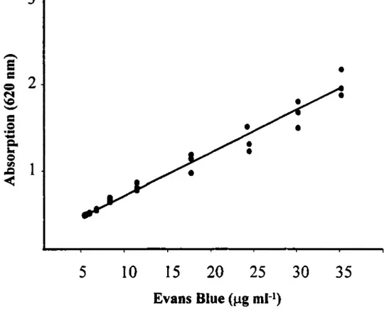
It has been generally accepted that some vital dyes such as Evans blue when, given intravenously, beccme bound to plasma proteins particularly alburnin. Therefore the accumulation of such a dye in inflamed sites could be used as an index of plasma extravasation. In the present study, the concentration of Evans Blue dye in the peritoneal cavity fluid (inflarned site) was determined by spectrophotometry (at a wavelength of 620 nm) and the data were then compared with a standard curve (figure 3) of Evans Blue dye at the sarne wavelength (JANCAR et
al,
1988)...-..
e
•
=
2
=
N \C-
=
=
·-
-
Q,,....
=
fil1
.c
<
5
10
15
20
25
30
35
Evans Blue (µg ml-
1)Figure 4: Standard curve of Evans Blue dye.
The equation
of the curve is Y=0.053X
+
0.25 in which Y represents the
absorption at 620nm and X represents the different
concentrations of Evans blue dye.
3. PERITONEAL PLASMA EXTRAVASATION INDUCED BY BK RECEPTOR AGONISTS
After anaesthetizing animals with ketamine and xylazine (2.25 & 0.28 mg/mouse, respectively; i.p.), Evans blue (O.lmg/mouse, 150 µl) was injected into the penile vein (with a sterile 3001/2 needle) concurrently with the intraperitoneal injection of BK. In some selccted experiments Evans blue was injected 20 or 70 minutes prior of BK injection (IOµg/mouse). ln another series of experiments, ET-1 (5µg/mouse; i.p.) or [Sar9, Met (02) 11] Substance P (20µg/mouse; i.p.) were injected by the same route as
BK and considered as positive controls of plasma extravasation in wild type and B1 K.O. mi ce.
The effect of HOE 140 or [Leu8]des-Arg9-BK {lOµg/mouse; i.p.; 30 minutes
prior to Evans blue and BK injection) was tested against the BK-induced plasma extravasation in wild-type animais. Thirty minutes after the injection of BK, mice were sacrificed by decapitation and exsanguination. The peritoneal cavity of each animal was then washed with phosphate-buffered saline (PBS, 1 ml, PH 7.4) and the concentration of Evans blue dye, as an index of vascular permeability was determined from a recovered volume of at least 800 µl. In some other experiments, inoomethacin (200µg/mouse i.p.) was administered 1 hour prior to BK injection.