MÉCANISMES DE CONTRÔLE DE LA PRISE
ALIMENTAIRE PAR LE SYSTÈME DE
L'HORMONE DE LA
MÉLANO-CONCENTRATION (MCH) :
IMPORTANCE DES RÉCEPTEURS OPIOÏDES
Mémoire
Carlos Andres Lopez
Maîtrise en Physiologie-Endocrinologie Maître ès sciences (M.Sc.)
Québec, Canada
iii Résumé
L’hormone de la mélano-concentration (Melanin-Concentrating Hormone - MCH) joue un rôle important dans la régulation du bilan d'énergie. Les projections des neurones à MCH vers l’enveloppe du noyau accumbens (Nucleus Accumbens Shell - NAcSh) induisent, lorsque stimulées, la prise alimentaire. Aussi, l'injection intra-NAcSh d’opiacés induit une augmentation de la prise alimentaire chez les rongeurs. Compte tenu de la présence de récepteurs du MCH de type 1 (MCHR1) sur les neurones dynorphine et enképhaline du NAcSh, le système opioïde du NAcSh pourrait jouer un rôle de médiateur de l'effet orexigène du MCH. Les sites et les mécanismes de l'action orexigène du MCH sont cependant encore incomplètement connus.. Connaissant la longue portée des fonctions attribuées au NAcSh dans la modulation de la récompense et du plaisir, nous avons supposé que l’agoniste du MCH dans le NAcSh augmente le plaisir associé à la prise alimentaire à travers des neurones opioïdes dans le NAcSh. Deux expériences chez des rats Wistar mâles ont été effectuées. Nous avons d’abord mesuré la capacité de 3 antagonistes des récepteurs opioïdes (µ: β-Funaltrexamine, β-FNA ; δ: Naltrindole, NTI ; κ: Nor-Binaltorphimine, Nor-BNI) de bloquer l'augmentation de la prise alimentaire induite par l'injection de MCH. Le Nor-BNI, le NTI ou le β-FNA ont été administrés à différentes doses dans le ventricule latéral 90 min ou 22 h avant l’injection de MCH ou de véhicule et la prise alimentaires des animaux a été mesurée pendant les 3 heures suivant le traitement. .. Nous avons constaté que le blocage des récepteurs opioïdes par des antagonistes spécifiques a diminué la prise alimentaire induite par le MCH. Nous avons aussi évalué le niveau de plaisir en réponse à un stimulus sucré (1 ml de sucre à 2%, intra-oral) en quantifiant les mimiques faciales, suivant le test de réactivité gustative décrit précédemment. Les réponses hédoniques ont été évaluées 15 min après l’injection dans le ventricule latéral de 4 µl de l’agoniste du MCH (2 nmol) par rapport au véhicule. Le même test de réactivité gustative a été répété après le traitement avec les antagonistes des différents récepteurs des opioïdes. La capacité des antagonistes des opioïdes de bloquer l'effet du MCH a été mesurée. Nous avons
constaté que les trois antagonistes des récepteurs opioïdes ont modifié l'augmentation de la réponse hédonique induite par le MCH en atténuant les propriétés hédoniques de la solution de sucre. Nos résultats indiquent que les effets orexigènes et hédoniques du MCH sont liés aux 3 récepteurs opioïdes. Le MCH peut activer un sous-type de récepteurs spécifiques aux opioïdes (κ, µ) pour exercer ses effets sur la consommation alimentaire. L’interaction des deux systèmes, MCH et opioïdes, semble jouer un rôle important dans la régulation hédonique du contrôle de l'appétit.
Abstract
The brain melanin-concentrating hormone (MCH) system plays an important role in the regulation of energy balance. The sites and mechanisms of the orexigenic action of the MCH are still uncertain. Knowing the function of the NAcSh in the regulation of reward and pleasure, we hypothesize that MCH agonism in the NAcSh increases the pleasure associated with food intake through the opioid neurons within NAcSh. The presence of MCH receptor (MCHR1) on dynorphin and enkephalin neurons of NAcSh supports this hypothesis. Two different experiments in Wistar male were conducted. Firstly, we measured the capacity of three different opioid antagonists injected in the lateral ventricle at doses of 10 nmol and 40 nmol (for κ and δ) or of 10 nmol and 50 nmol (for µ) to block the increase in food intake induced by MCH injection in the lateral ventricle. The rats pretreated with a microinjection of an opioid antagonist 90 min (for the κ and δ) or 22 h (for µ), received a MCH or vehicle injection. Food intake was monitored during one, two and three hours after MCH or vehicle injection. We found that the blockade of opioid receptors by selective antagonists decreased MCH-induced feeding. Secondly, we assessed the level of pleasure in response to sweet stimulus (one milliliter of a sucrose solution, intraoral) by quantifying facial mimics induced by the presence of sucrose in the mouth. The hedonic responses were monitored, 15 min after the injection of a MCH agonist in the lateral ventricle. The same taste reactivity test was repeated following the injection of MCH in the presence of κ, δ and opioid antagonists. We found that the three opioid antagonists were capable of modifying the increased hedonic response induced by MCH by attenuating the positive hedonic properties of sucrose solution. Our results indicate that the orexigenic and hedonic effects of MCH are linked to three opioid receptors. MCH might activate a specific opioid receptor subtype (κ, µ) for exerting its effects on food intake. The interaction of the MCH and opioids systems could represent an important link in the modulation of the hedonic appetite control.
vii Avant-propos
Ce mémoire est présenté à la Faculté des Études supérieures de l’Université Laval pour l’obtention du grade maître ès sciences (M.Sc.). Il est composé de deux parties.
La première partie comprend un résumé en français et anglais, une introduction en anglais sur l’hormone de la mélano-concentration et le système opioïde et la relation avec le noyau accumbens qui est reconnu comme centre de plaisir. Ensuite, la méthodologie et les objectifs du mémoire sont présentés aussi en anglais. Dans la deuxième partie du mémoire on retrouve un manuscrit présentant des résultats originaux. L’article scientifique est rédigé en anglais et il est précédé d’un résumé en français. L’article est publié dans la revue scientifique The American Journal of Physiology – Regulatory, Integrative and Comparative Physiology. (Am J Physiol Regul Integr Comp Physiol October 2011 301:(4) R1105-R1111).
La conclusion générale de ce mémoire résume en anglais les principaux résultats. Finalement, le dernier chapitre contient la bibliographie générale pour l’introduction, les objectifs, la méthodologie et la conclusion générale.
Remerciements
Je remercie à Denis pour me donner l’opportunité de développer ma maitrise dans son groupe de recherche et son soutien.
Au groupe de recherche en obésité pour tout ses aides et conseilles pendant mes études et travaux de laboratoires, spécialement à Dana, Julie, Benjamin et Marie-Claude.
Aux techniciens de l’animalerie pour son aide pendant mes protocoles avec les rats. À ma famille pour leur appui inconditionnelle et encouragement spirituel.
Table of Contents
Résumé ... iii
Abstract ... v
Avant-propos ... vii
Remerciements ... ix
Table of Contents ... xiii
List of Figures ... xv
List of Abbreviations ... xvii
1. GENERAL INTRODUCTION ... 1
1.1. The regulation of energy balance ... 2
1.2 The control of energy expenditure ... 3
1.3 Brain pathways involved in energy balance regulation ... 4
1.3.1 The ARC-PVH axis ... 6
1.3.2. The lateral hypothalamus ... 7
1.3.3. The brainstem ... 7
1.3.4. The nucleus accumbens ... 8
1.4. The melanin-concentrating hormone system ... 10
1.4.1 MCH system peptides ... 10
1.4.2. MCH receptors ... 11
1.4.3. The MCH system and the regulation of energy balance ... 13
1.5. The opioid system ... 15
1.5.1. The opioid peptides ... 15
1.5.2. The opioid receptors ... 17
1.5.2.1. Delta Receptor ... 17 1.5.2.2. Kappa Receptor ... 18 1.5.2.3. Mu Receptor ... 19 1.6. Interaction MCH-opioid ... 19 1.7. Objectives ... 20 1.8. Methodology ... 21
2. INVOLVEMENT OF THE OPIOID SYSTEM IN THE OREXIGENIC AND
HEDONIC EFFECTS OF MELANIN-CONCENTRATING HORMONE ... 23
2.1. Résumé... 24 2.2. Abstract ... 25 2.3. Introduction ... 26 2.4. Methods ... 28 2.4.1. Animals ... 28 2.4.2. Surgery ... 28
2.4.3. Drugs and Microinjections... 29
2.4.4. Food intake ... 30
2.4.5. Hedonic response to a sweet stimulus ... 31
2.4.6. Statistical analysis ... 32
2.5. Results ... 33
2.5.1. Food intake ... 33
2.5.2. Hedonic response to an intraoral sweet stimulus ... 34
2.6. Discussion ... 35
2.7. Perspectives and Significance ... 37
2.8. Acknowledgements ... 37 2.9. References ... 38 2.10. Figures ... 41 3. GENERAL CONCLUSIONS ... 47 4. PERSPECTIVES ... 49 5. GENERAL BIBLIOGRAPHY ... 51
List of Figures
Figure 1: Overview of the regulation of energy balance presenting the main brain regions and chemical mediators involved in the control of food intake and energy expenditure (from Richard and Timofeeva 2010). ... 3 Figure 2: Connectivity of the NAc with other brain regions such as prefrontal cortex,
ventral tegmental area (from Shirayama and Chaki 2006). ... 9 Figure 3: A, In situ hybridization showing wide distribution of MCH
mRNA-expressing neurons in the lateral hypothalamus and ZI of the mouse. B, immunohistochemical detection of MCH-expressing neurons reveals a similar distribution pattern throughout the lateral hypothalamus and the ZI (from Pissios et al 2006) . ... 11 Figure 4: MCHR1 activates multiple signaling pathways. Through coupling to
different intracellular effectors, MCHR1 decreases the activity of adenylate cyclase, increases the levels of intracellular calcium (Cai++), activates ERKs, and interacts with intracellular proteins such as periplakin and MIZIP (from Pissios et al 2006). ... 12 Figure 5: Regional distribution of SLC-1 mRNA A-D. Localization of MCHR1
transcripts in rat brain sections by in situ hybridization using a cRNA probe. (from Saito1999) ... 12 Figure 6: Brain sites where opioid agonists or antagonists modulate food intake. A:
brain sites where opioid receptor agonists injected locally either increase or decrease food intake. B: brain sites where microinjections of opioid receptor antagonists decrease feeding behavior (from Le Merrer et al 2009). ... 16 Figure 7: Brain hedonic hot spots and hedonic circuits. A sagital brain view of the
hedonic hot spots in the nucleus accumbens, Anatomical projections are indicated by lines that may create a hedonic circuit by connecting hot spots together (red circles) or incorporating hot spots into larger mesocorticolimbic loops (green) (from Pecina et al 2006). ... 20
List of Abbreviations
α-MSH: alpha-melanocyte-stimulating hormone -FNA: β-Funaltrexamine
aCSF: artificial cerebrospinal fluid AgRP: agouti-related peptide AP: area postrema
ARC: arcuate nucleus of the hypothalamus BAT: brown adipose tissue
CART: cocaine- and amphetamine-regulated transcript CB1: cannabinoid receptor 1
CCK: cholecystokinin
CRF: corticotropin-releasing factor
DAMGO: [D-Ala2, N-MePhe4, Gly-ol]-enkephalin DVC: dorsal vagal complex
GLP1: glucagon-like peptide 1 GPCR: G-protein-coupled receptors ICV: intracerebroventricular IML: intermediolateral column LC: locus coeruleus
LH: lateral hypothalamus
LPGi: lateral paragigantocellular nucleus LV: lateral ventricle
MC3R: melanocortin receptor 3 MC4R: melanocortin receptor 4 MCH: melanin-concentrating hormone MCHR1: MCH receptor 1
MSN: medium spiny neurons NAc: nucleus accumbens
NAcSh: nucleus accumbens shell NEI: neuropeptide E-I
NGE: neuropeptide G-E Nor-BNI: Norbinaltorphimine NPY: neuropeptide Y
NTI: naltrindole
NTS: nucleus of the tractus solitarius PAG: periaqueductal gray
Pdyn: preprodynorphin Penk: preproenkephalin POMC: proopiomelanocortin PRV: pseudorabies virus
PVH: paraventricular hypothalamic nucleus PYY: peptide tyrosine-tyrosine
SNS: sympathetic nervous system TRH: thyrotropin-releasing hormone UCP1: uncoupling protein 1
VTA: ventral tegmental area ZI: zona incerta
1. GENERAL INTRODUCTION
The unrelenting rise in obesity in most developed countries over the last 30 years is alarming. Data from the National Health and Nutrition Examination Survey (NHANES) revealed that, in 2003-2004, approximately two thirds of United States citizens (66.3%) were overweight [body mass index (BMI) between 25 and 30 kg/m2] or obese (BMI > 30 kg/m2), 32.42% were obese, and 4.8% were massively obese (BMI 40 kg/m2). The situation is all the more preoccupying in that the obesity epidemic has spread to non-industrialized countries and now involves children. The most recent NHANES data [1] found that obesity prevalence among children and adolescents (2-19) now reaches 16.3% in the United States (up from to 3-4% in the 1960s). Obesity and the ensuing abnormal fat deposition in the abdominal cavity, liver, heart, pancreas, and muscle tissue (ectopic fat), which mostly (but not uniquely) characterizes male obesity [2], lead to major metabolic impairments including diabetes, insulin resistance, dyslipidemia, and greatly increase the risk of cardiovascular and respiratory diseases, and cancer [3-5]. Because of all the health impairments it causes, obesity leads to enormous costs. The burden of obesity in United States stands at 9% of healthcare costs [6] and is widely predicted to increase for years to come, as the spreading of the obesity epidemics is still concrete [7].
The escalating prevalence of obesity together with the rising awareness of the detrimental impact of this condition on health and health costs have considerably stimulated research related to the etiology and complications of excess fat deposition. Investigations of the main factors causing obesity have led to appreciable progress in our understanding of the respective and integrated roles of the environment and genetics in the development of this condition. Virtually all experts now share the view that obesity largely results from complex gene-environment interactions [8-10]. The ‘obesogenic’ milieu in which we live, characterized by a sedentary lifestyle, excess ingestion of energy dense palatable food, stress and pollution, proves to be particularly obesity-inducing in individuals who are genetically predisposed to positive energy balance. Obesity-promoting genes might not only be
energy-conserving genes acquired through evolution to combat periods of food shortage[8] but all those genes (‘thrifty’ or not) that can be involved in the overall adaptation/inadaptation to the changes imposed by the ‘modern lifestyle’ and whose under- or overexpression potentially leads to overeating and other obesity-promoting behaviors.
1.1. The regulation of energy balance
Obesity is first and foremost an energy-balance-related disorder leading to excess fat deposition, which results from an imbalance between energy intake and energy expenditure in line with the etymology of the word obesity, derived from the latin word obedere, meaning ‘to eat in excess’. Notably, once acquired excess body fat becomes strongly ‘defended’ through energy balance regulation mechanisms opposing any attempts to lose weight through strategies implemented to increase appetite and to reduce energy expenditure.
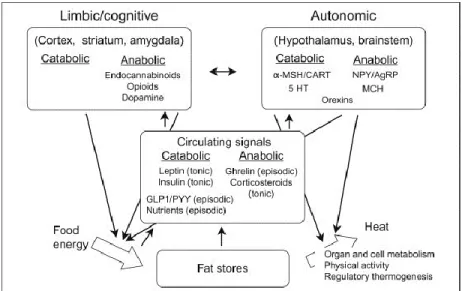
The regulation of energy balance is determined by controls exerted on both food intake and energy expenditure (figure 1) [11]. The brain and peripheral organs that are sensitive to variations in energy stores and to the nutritional status, as well as to changes in environmental hedonic cues are crucial structures in these controls [9, 12-24]. The term homeostatic has been discerningly coined by certain authors [14, 18, 25] to describe the metabolic and uncognitive aspects of energy balance regulation. The control of food intake and energy expenditure is referred to as being primarily homeostatic when it is mainly exerted in response to variations in the energy stores and nutritional status. The homeostatic control is largely autonomic or involuntary and likely applies to both food intake and energy expenditure. Contrastingly, the non-homeostatic control, which is rather specific to food intake, has been described as having cognitive and rewarding components as it is strongly influenced by the pleasurable aspects of eating [17-18, 25]. Even though not primarily dependent on energy stores, the non-homeostatic control of food intake is nonetheless influenced by the deficit or surfeit of energy; attractive food items are even more palatable in a
condition of energy deficit [17-18]. In the obesogenic environment in which we live, non-homeostatic controls often triumphs over homeostatic controls because of the strong pleasurable signaling associated with food ingestion.
Figure 1: Overview of the regulation of energy balance presenting the main brain regions and chemical mediators involved in the control of food intake and energy expenditure (from Richard and Timofeeva 2010).
1.2 The control of energy expenditure
Compared to the control of energy intake, that of energy expenditure has received a rather mitigated attention, considering that energy expenditure is as significant as energy intake in the energy balance equation. The impact of the expenditure component on energy balance is particularly important in laboratory rodents, in which a strong regulatory control is exerted through the sympathetic nervous system (SNS) on brown adipose tissue (BAT), a potent effector of thermogenesis [26-28]. In contrast to white adipocytes, brown fat cells are highly adapted to dissipate chemical energy in the form of heat. The thermogenic power of BAT is conferred by the presence of uncoupling protein-1 (UCP1) [29]. UCP1 is unique to brown adipocytes and generates heat by ‘uncoupling’ ATP synthesis from cellular respiration. Active UCP1 allows the dissipation of the electrochemical gradient, which is generated
across the inner mitochondrial membrane by the electron transport along the respiratory chain, and which is normally used to generate ATP. UCP1 activity is driven by the activation of the SNS, whose postganglionic neurons densely innervate brown adipocytes. SNS-mediated UCP1 activity is governed by neurons [30] found in the brain structures implicated in energy balance regulation and whose activity is modulated by fluctuations in energy stores and fluxes [31-32]. In rodents, BAT thermogenesis not only allows for cold adaptation, but also contributes to energy balance regulation; food deprivation reduces SNS-mediated UCP1 activity, whereas excess food ingestion may increase it.
Up until recently, there was the general consensus that BAT was not present in significant amount in adult humans. However, recent papers in cancer research, using positron emission tomography and computed tomography (PET/CT) to primarily detect tumors, demonstrated that adipose tissue sites can take significant amount of the PET tracer 18fluorodeoxyglucose that were not tumors but brown fat depots [33]. The demonstration that BAT can exist in humans is currently rejuvenating the interest for the role played by adaptative thermogenesis in humans [34-38]. It is as yet uncertain as to whether excess energy intake or excess fat stores triggers regulatory thermogenesis in humans, the literature on that subject being controversial [39-40]. In laboratory rodents fed palatable food (so-called cafeteria diets), excessive energy consumption has been reported to induce BAT thermogenesis to limit excess energy deposition [41-42].
1.3 Brain pathways involved in energy balance regulation
The brain is critically involved in the complex controls of food intake and energy expenditure [9, 12-22], which are achieved through harmonized crosstalk between the autonomic neurons of the hypothalamus and dorsal vagal complex (DVC), and the cognitive and limbic circuitries of the hippocampus, amygdala, striatum, and cortex. The limbic structures are known to support functions such as emotion, learning,
memory, hedonism, olfaction, vision and taste. The autonomic and cognitive/limbic brain systems work inseparably in regulating energy balance [43]. The strength of hedonic stimulus related to food is influenced by the deficit or surfeit of energy that exerts metabolic controls on food intake and energy expenditure; palatable food items are undeniably more appetizing in conditions of energy deficit. It also has to be pointed out that the pleasurable signaling associated with the ingestion of palatable foods inherent to the modern world is capable of distorting the controls exerted on food intake and energy expenditure [43].
The control of food intake and energy expenditure is insured by interconnected neurons expressing varied receptor types and producing diverse peptides or classic neurotransmitters that have been grouped into anabolic and catabolic mediators [12]. These mediators are found in nuclei such as the arcuate nucleus of the hypothalamus (ARC), paraventricular hypothalamic nucleus (PVH), lateral hypothalamus (LH), nucleus of the tractus solitarius (NTS), nucleus accumbens (NAc), ventral tegmental area (VTA), amygdala and the cortex. All these nuclei are tied to each other to form pathways that control the intake as well as the expenditure of energy. They comprise neurons that produce energy-balance-associated mediators such as neuropeptide Y (NPY), agouti-related peptide (AgRP), alpha-melanocyte-stimulating hormone (α-MSH), cocaine- and amphetamine-regulated transcript (CART), melanin-concentrating hormone (MCH), orexins, corticotropin-releasing factor (CRF), urocortins, thyrotropin-releasing hormone (TRH), endocannabinoids, opioids, dopamine and serotonin. The production of these diverse molecules is modulated by short- and long-term signals that inform the brain about the status of the energy stores and energy fluxes. Whereas leptin, insulin, glucocorticoids are recognized as the main long-term tonic signals, the gastrointestinal hormones ghrelin, peptide tyrosine-tyrosine (PYY), cholecystokinin (CCK), and glucagon-like peptide 1 (GLP1) are known as short-term or episodic regulatory signals [22, 25]. Circulating nutrients, including glucose, lipids and amino acids, are also sensed by the brain ‘catabolic’ and ‘anabolic’ neurons [44-46].
1.3.1 The ARC-PVH axis
The ARC forms with the PVH what has emerged to be one of the most important regulating hypothalamic duets in energy homeostasis [12, 15-16, 47-49]. The ARC is a tiny nucleus found in the basomedial hypothalamus just above the median eminence and adjacent to the third ventricle [49]. It comprises two populations of neurons strongly involved in the control of energy intake and energy expenditure. These neurons abundantly project to the neuroendocrine and metabolic divisions of the PVH. One population of neurons synthesizes proopiomelanocortin (POMC) and CART, whereas the other synthesizes NPY and AgRP [12]. POMC and CART are catabolic whereas NPY and AgRP are anabolic [50-53]. POMC/CART neurons exert their catabolic effect mainly via α-MSH, a peptidergic fragment ensuing from POMC cleavage. Within the brain, α-MSH binds to melanocortin 3 (MC3R) and 4 (MC4R) receptors with which it constitutes, together with AgRP, the metabolic melanocortin system [54-57]. The functional significance of the MC4R in energy homeostasis has been validated in Mc4r KO mice, which exhibit a massive and widespread body fat deposition [57-58]. In humans, MC4R deficiency represents the most commonly known monogenic form obesity [59]. MC4R is endogenously antagonized by AgRP, which is solely expressed in the ARC neurons where it is co-synthesized with NPY [60]. MC4R are found in abundance in the PVH, which also expresses NPY1[13] and NPY5 receptors [61]. The latter are recognized as the main receptors mediating the action of NPY on energy balance. NPY represents a potent orexigenic peptide [62], whose expression in the ARC is markedly stimulated in obese [63] and food-deprived animals [63-64]. The functional importance of NPY as well as that of AgRP in the regulation of energy balance has recently found a genetic endorsement when mice genetically engineered for a selective deletion of AgRP-NPY-expressing neurons were shown to exhibit at the adult stage an acute reduction in feeding [50-51].
1.3.2. The lateral hypothalamus
The LH has a long-established role in the regulation of energy balance [18, 65-67]. Neurons from the LH region send descending projections to the brainstem and spinal cord areas involved in autonomic functions associated with BAT [68-69]. Furthermore, this region is connected to the NAc and VTA, two key parts of the brain reward system. LH has been suggested to bridge the homeostatic and non-homeostatic neuronal pathways regulating energy balance [17-18]. It is involved in food intake as well as in energy expenditure; the loss in body weight following LH lesions is not only accompanied by hypophagia (reduced food intake) but also by hyperthermia and increased energy expenditure. The LH control on energy expenditure has recently been further supported by the work of Cerri and Morrison [70], who demonstrated the LH ability to stimulate BAT thermogenesis. The neural mechanisms and network involved in this thermogenesis remain to be unraveled. However, the role of LH peptides MCH and orexins A and B in this process seems highly probable and are anatomically supported. Transneuronal labeling experiments have indeed established a clear polysynaptic link between MCH and orexin neurons and BAT [69, 71].
1.3.3. The brainstem
The brainstem, which includes the hindbrain (cerebellum, pons and medulla oblongata) and the midbrain, also plays an important role in the regulation of energy balance. It comprises the NTS, which, together with the area postrema (AP) and the motor nucleus of the vagus nerve, form the DVC. The DVC integrates peripheral signals conveyed by gastrointestinal hormones such as cholecystokinin, PYY and GLP1, which influence energy balance regulation [19, 21, 72-73]. The actions of these hormones are exerted via the circulation and on vagal afferents via a paracrine mode of communication. The role of the DVC (and the hindbrain in general) in energy balance has been advocated by Grill and coll. [20, 74], who carried out
elegantly-designed experiments with decerebrated rats (rats subjected to the complete transection of the neuroaxis at the mesodiencephalic juncture) to decisively demonstrate the brainstem implication in the effects of leptin [75], melanocortins [76] and ghrelin [77].
In addition to integrating peripheral inputs, the brainstem comprises nuclei, such as the caudal raphe, periaqueductal gray (PAG), pontine reticular nuclei, lateral paragigantocellular nucleus (LPGi), and locus ceruleus (LC) that are likely involved in SNS-mediated thermogenesis [68, 78-79]. These nuclei have been described as structures capable of relaying information from the hypothalamus to the intermediolateral column (IML), from which originate SNS preganglionic neurons implicated in the control of BAT thermogenesis. LH MCH neurons are among the neurons projecting to the brainstem to potentially exert a control on energy expenditure through BAT thermogenesis [69, 80].
1.3.4. The nucleus accumbens
Given the prominent role it plays in reward and addiction, the NAc has emerged as a crucial structure in hedonic control of food intake [81]. Being part of the limbic system and located in the ventral striatal region, the NAc comprises two distinct sections, namely, the NAc core and the NAc shell (NAcSh) [82]. The NAcSh and NAc core differ in their anatomical projection patterns of connectivity [83-84], and in their intrinsic organization regarding cell morphology, gene expression, and distribution of neurotransmitters and receptors [85-86]. The NAc contains small populations of γ-aminobutyric acid (GABA)-containing and cholinergic interneurons, in addition to a large number of efferent GABAergic medium spiny projecting neurons [87]. The activity of projecting MSN is regulated by glutamatergic afferents arising from the prefrontal cortex, hippocampus and amygdala, by dopaminergic afferents from the VTA [88], by serotonergic afferents from the raphe nucleus, and by noradrenergic afferents from the LC (figure 2) [89].
Figure 2: Connectivity of the NAc with other brain regions such as prefrontal cortex, ventral tegmental area (from Shirayama and Chaki 2006).
The NAc forms with the VTA the mesolimbic pathway, also referred to as the mesolimbic reward pathway because of its ability to produce pleasurable feelings in response to drugs and food [90]. The mesolimbic reward pathway comprises dopaminergic neurons originating from the VTA that terminates in the NAc. These dopaminergic neurons play a major role in the motivational component of reward (referred to as ‘incentive salience’ or ‘wanting’) [91-92]. The dopaminergic system does not appear to be essential in sensing the hedonic value or the liking aspects of food [93]. Indeed, the destruction of the ascending dopamine fibers innervating the striatal area does not block the positive taste reactivity response elicited by a sweet cue [94]. The hedonic response of a sweet taste appears however to strongly depend on opioids and endocannabinoids [95]. Injection of the µ-opioid receptor agonist [D-Ala2, N-MePhe4, Gly-ol]-enkephalin (DAMGO) in the NAcSh markedly increases the hedonic value of sweetness [96]. The NAcSh abundantly expresses the µ-opioid receptor as well as the cannabinoid receptor 1 (CB1) [97-98]. It is noteworthy that anandamide, an endogenous ligand of the CB1, also enhances the sweet reward when injected in the NAcSh [99].
1.4. The melanin-concentrating hormone system
The MCH system consists of the peptides MCH, neuropeptide E-I (NEI) and neuropeptide G-E (NGE) and two receptors known as MCHR1 and MCHR2.
1.4.1 MCH system peptides
MCH is a 19-amino-acid cyclic peptide that was first discovered in teleost fish, in which it acts as a skin color-regulating hormone [100]. MCH induces melanosome aggregation within melanophores of teleost fish, and makes the skin color of fishes pallor [101]. MCH have a disulfide bond between the 7th and 16th cysteines, which is necessary for its biological functions. Mammalian MCH is the posttranslational product of prepro-MCH that is 165 amino acids in length, and its proteolytic cleavage results in MCH as well as two other bioactive peptides, NEI and NGE [102-103]. The role of the two other peptides encoded by prepro-MCH remains somewhat obscure. Limited experiments suggest that NEI is present in the same nerve terminals as MCH and injections of this neuropeptide produce behavioral changes in rats [104].
In rodents the MCH neurons are found in the brain and located in the lateral hypothalamic area and zona incerta (ZI) (figure 3) [104] and project widely and diffusely to forebrain and hindbrain [102]. Similar distributions have been reported in both bird [105] and monkey [106].
Figure 3: A, In situ hybridization showing wide distribution of MCH mRNA-expressing neurons in the lateral hypothalamus and ZI of the mouse. B, immunohistochemical detection of MCH-expressing neurons reveals a similar distribution pattern throughout the lateral hypothalamus and the ZI (from Pissios et al 2006).
1.4.2. MCH receptors
MCH produces its effects through two G-protein-coupled receptors, MCHR1 and MCHR2 [104]. MCHR2 is expressed in humans but a functional MCHR2 is not expressed in rats and mice and little is known about its physiological function [107-108].
MCHR1 contains three consensus N-glycosylation sites and several potential phosphorylation sites in its intracellular loops [109]. The actin- and intermediate filament-binding protein, periplakin, appears to be coexpressed with MCHR1 in mouse brain and may interact with the intracellular C terminal of MCHR1 to impede MCHR1-initiated signal transduction [110].
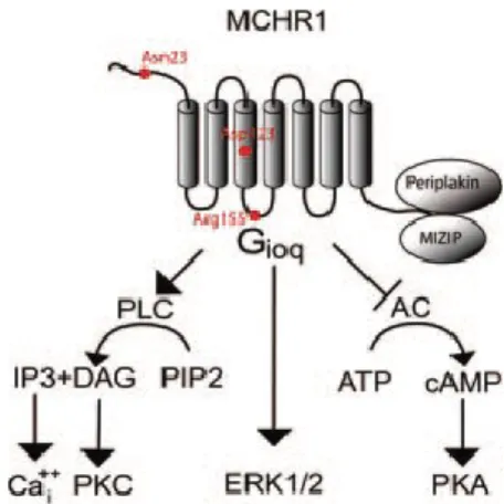
MCHR1 binds MCH with nanomolar affinity and couples to Gi, Go, and Gq proteins to activate multiple intracellular signaling pathways [111]. Activation of MCHR1 leads to an increase in intracellular free Ca2+ suppression of forskolin-stimulated cAMP, as well as stimulation of phosphoinositide metabolism and activation of ERKs (figure 4) [104, 111-112].
Figure 4: MCHR1 activates multiple signaling pathways. Through coupling to different intracellular effectors, MCHR1 decreases the activity of adenylate cyclase, increases the levels of intracellular calcium (Cai++), activates ERKs, and interacts with intracellular proteins
such as periplakin and MIZIP (from Pissios et al 2006).
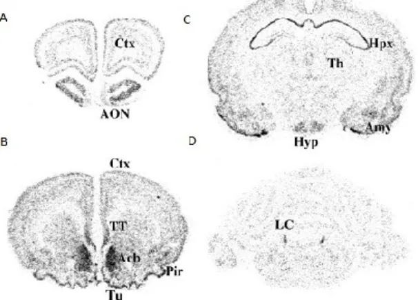
MCHR1 mRNA is expressed not only in brain, but also in peripheral tissues, such as adrenal glands, tumor tissues of pheochromocytomas and neuroblastomas, and various human tumor cell lines [113]. In the brain, MCHR1 is localized in different regions implicated in mood disorders, such as the LC, amygdala, and hippocampus. The highest densities of MCHR1 are probably found in the NAcSh and caudate putamen of rats and mice (figure 5) [114].
Figure 5: A-D. Localization of MCHR1 transcripts in rat brain sections by in situ hybridization using a cRNA probe (from Saito1999).
This distribution corresponds to the monosynaptic connections that MCH neurons make with several areas in the brain involved in integrating inputs related to taste and learning. MCHR1 localization in these areas shows a possible role for MCH in reward and reinforcement mechanisms, which are fundamental processes in the regulation of feeding behavior. The presence of MCHR1 in parts of the hypothalamus that regulate feeding and metabolism, such as the ventromedial nucleus of hypothalamus, further supports the idea of interaction in satiety. Moderate expression of MCHR1 mRNA was found in the substantia nigra, VTA and in the amygdala, indicating that MCH may modulate the dopaminergic system. Moderate expression in
the LC, indicates that MCH may participate in the control of various noradrenaline-modulated responses including vigilance, attention, memory and sleep [115].
1.4.3. The MCH system and the regulation of energy balance
There is strong evidence that the MCH system plays a genuine role in the regulation of energy balance [104, 116]. Whereas chronic treatment with MCH [117] and MCH overexpression [118] lead to obesity and to an increased susceptibility to high-fat feeding, antagonism of MCHR1 [119] and ablations of MCH [120], MCH neurons [121] and MCHR1 [122-123] promote leanness.
MCH-containing neurons are concentrated within the LH and the adjacent ZI from where they project to the rest of the brain in a pattern that generally conforms to metabolic effects [102]. There is also a good correspondence between the distribution of the MCH-immunoreactive fibers and that of the MCHR1 [124], which is the energy-balance-related MCH receptor in rats and mice. MCH+/CART+ cells have been described as MCH type B neurons and been argued to play a role in the control of food intake and locomotor activity as they contact forebrain structures involved in goal-oriented behaviors [116, 125-126]. These structures include the NAc, where the MCHR1 is abundantly expressed [127]. On the other hand, the MCH+/CART- cells, which have been referred to as MCH type A neurons and which extend to the caudal brainstem and the spinal cord, could logically be involved in the control of energy expenditure through BAT thermogenesis since these cells terminate in areas that have been demonstrated to be linked to BAT activity [79, 124]. There is a clear polysynaptic connection between MCH neurons and BAT [69] as more than 50% of the LH neurons that are infected following the injection in BAT of the transneuronal retrograde tracer pseudorabies virus (PRV, PRV is a marker used to map the SNS outflow to BAT) are MCH neurons found in LH caudal aspects surrounding the fornix [69]. MCH Type B neurons could also participate to the thermogenic function of MCH as they also connect the ARC-PVH axis and the dorsal brainstem [125].
The evidence for a role of MCH in the control on energy expenditure is not only based on anatomical considerations linking MCH neurons to BAT. Ablations of MCH [120], MCH neurons [121] and MCHR1 [122-123] have all been reported to promote fat loss by increasing energy expenditure. In the ob/ob mouse for instance, deletion of MCH induces a dramatic fat loss without any food intake reduction [128]. The importance of the MCH system in energy expenditure is further supported by the observation that MCH [129] and MCHR1 disruptions [122-123] lead to leanness despite hyperphagia. It is noteworthy that the increased energy expenditure led to by the lack of MCH in ob/ob mice is accompanied by increases in both metabolic rate and locomotor activity [128], suggesting that the absence of MCH could stimulate energy expenditure by increasing physical activity. However, the observation that the daily enhanced metabolic rate (seen throughout the day) of the MCH-/-ob/ob mice is not totally paralleled by an increase in locomotor activity (seen only during the night) suggests that the augmented expenditure led to by the lack of MCH [128] is not solely (perhaps not primarily) due to an increase in physical activity. Alternatively, BAT thermogenesis may contribute to a significant extent to the elevated metabolic rate seen in MCH-/-ob/ob mutants, which exhibit an increase in BAT expression of UCP1 [128]. The latter effect is likely due to the lack of MCH as the MCH treatment decreases UCP1 mRNA levels in mice [130].
In rats and mice, the effects of MCH, including those on energy metabolism, are mediated through the MCHR1, which appears to be the sole MCH receptor expressed in those species. However, species such as humans [131], monkeys [132], dogs and ferrets [133] also express the MCHR2. Not much is currently known about this receptor except that its highest mRNA levels are found in the frontal cortex, amygdala, and NAc [134], which suggests the MCHR2 could be involved in the cognitive, motivational and hedonic (non-homeostatic) aspects of feeding.
The mechanism whereby MCH exerts its effects on energy balance and on food intake, in particular, remains to be elucidated. The NAc appears to be a key target for
the action of MCH on energy balance. We [135] and others [136] have clearly demonstrated the stimulating effect of NAc MCH agonism on food intake in sated rats. The exact intra NAc site of this effect has not been firmly established although there are reasons to believe that MCH acts primarily in the NAcSh since this sub region markedly expresses the MCHR1 [127]. In addition, there is as yet indication as to whether the hyperphagic action of MCH in the NAc enhances the hedonic part of feeding. The NAcSh is recognized as a key structure in the liking aspect of reward, which is enhanced by µ-opioid receptor agonists [96, 137] and the endocannabinoid anandamide [99]. The MCH system could interact in the NAc with the endocannabinoid system but results from our laboratory tend to indicate that this is not the case (Lopez, C and Richard, D, unpublished). Alternatively, MCH could act through the opioid system; the MCHR1 receptor is expressed in enkephalin and dynorphin cells [136], supporting the interaction of MCH and opioids in the control appetite.
1.5. The opioid system
The opioid system is a complex system involving the peptides enkephalins, dynorphins, and β-endorphin and the κ, μ and δ opioid receptors.
1.5.1. The opioid peptides
The endogenous opioid peptides, enkephalins, dynorphins, and β-endorphin are produced by proteolytic cleavage of large protein precursors known as preproenkephalin (Penk), preprodynorphin (Pdyn), and POMC, respectively. All opioid peptides share a common NH2-terminal Tyr-Gly-Gly-Phe signature sequence, which interacts with opioid receptors [138].
The dynorphins are a family of opioid peptides that are derived from the prodynorphin precursor and that contain a leu-enkephalin sequence at their N-termini. The dynorphins have subsequently been shown to have a widespread distribution
throughout the central nervous system [139]. Although the major source of enkephalins within the central nervous system is not prodynorphin, but rather proenkephalin, which yields four met-enkephalins, one leu-enkephalin, and two C-terminal-extended met-enkephalins. One of these extended met-enkephalin sequences, met-enkephalin-Arg-Gly-Leu, is unique to the proenkephalin precursor and acts as an excellent marker for this system [139].
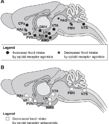
The opioid brain system is crucial to both eating behavior and reward, and activation of the opioid system can cause increases in food intake [140-142]. Opioid brain systems have been suggested to control food intake in part by mediating the palatability, or reward aspects, of the taste of food [137, 143]. Local microinjection of opioid agonists on food intake in many brain structures, including the NAc, amygdala, striatum, hypothalamus, tegmentum, and hindbrain, has been reported to cause increases in food intake (figure 6) [137-138].
Figure 6: Brain sites where opioid agonists or antagonists modulate food intake. A: brain sites where opioid receptor agonists injected locally either increase or decrease food intake. B:
brain sites where microinjections of opioid receptor antagonists decrease feeding behavior (from Le Merrer et al 2009).
1.5.2. The opioid receptors
The existence of opioid binding sites in the brain was established in 1973, and these were later referred to as κ, μ and δ opioid receptors [85]. The opioid receptor gene family includes four members encoding μ, δ, and κ, and the nonopioid orphaninFQ/nociceptin (Oprl1) receptors. Opioid receptors are membrane receptors with a seven-transmembrane topology. These receptors belong to the large G protein-coupled receptor super family, which comprises several hundred members within the mammalian genome [138].
The NAcSh is rich in μ, δ, and κ opioid receptor binding [144-145]. Neurobehavioral studies have shown that δ, κ, and especially μ opioid agonists and antagonists delivered to the NAc can alter food intake [137].
The anatomical distribution of both the µ and δ receptors in areas controlling olfaction, visual, auditory and nociceptive processing implies a role in sensory integration, whilst the predominance of κ receptors in the hypothalamus and median eminence point to a major role in neuroendocrine regulation [145].
1.5.2.1. Delta Receptor
A study reported that endogenous enkephalins modulate the basal hedonic state in pro-enkephalin knockout mice [146]. It has been reported that the enkephalin catabolism inhibitor exhibited antidepressant-like activity in learned helplessness and that this effect was reversed by the δ opioid receptor antagonist naltrindole [147]. Consistent with this hypothesis, δ opioid receptor agonists have been reported to display antidepressant-like activity in the forced swimming test [148], while KO mice
lacking δ-opioid receptor display a depressive-like phenotype in the forced swimming test [149].
The NAc contains δ opioid receptors, which are supposed to be located in the presynaptic of dopaminergic terminals because local administration of δ opioid receptor agonists increases dopamine release in the NAc [150]. Several lines of evidence have suggested the implication of enkephalin/δ opioid receptor in the NAc in depression. The activity of the enkephalinergic system in the NAc could be reduced in a stress-induced model of anhedonia. Indeed, δ opioid receptor agonists, when infused into the NAc, increase dopamine release in the NAc [151], which may explain the antidepressant-like action of δ opioid receptor agonists.
Quantitative analysis of δ receptors from coronal sections of fore- and midbrain in wild-type mice showed high levels of [3H]DELTI a selective opioid ligand, binding in olfactory bulbs, olfactory tubercle, caudate putamen, NAc, basolateral amygdala and throughout the cortex [152].
1.5.2.2. Kappa Receptor
The NAc is involved in aversive behaviors associated with dynorphin and its receptor (κ opioid receptor) agonists. Indeed, microinjection of κ opioid receptor agonists into the NAc causes conditioned place aversions that likely reflect dysphoric states [153]. It has also been reported that exposure to various stressors such as immobilization, forced swimming, and inescapable shock markedly increase dynorphin immunoreactivity in both the shell and core of NAc [154]. There are several lines of evidence showing that blockade of κ-opioid receptor produces antidepressant-like activity in animal models. Thus, intracerebroventricular (ICV) or intraaccumbal injection of norbinaltorphimine (nor-BNI), a κ-opioid receptor antagonist, decreases immobility in the forced swimming test [155] or reduces escape failure in the learned helplessness test [154, 156].
The κ-opioid receptors have been reported to mediate aversion by regulating mesolimbic dopaminergic activity in the NAc [157]. Indeed, both systemic and intraaccumbal injection of the κ opioid receptor agonist reduce dopamine release in the NAc, while both systemic and intraaccumbal injection of the κ-opioid receptor antagonist nor-BNI increase dopamine release [158-159].
Quantitative analysis of κ-receptors from coronal sections of fore- and midbrain in wild-type mice showed high levels of [3H]CI-977 a selective opioid ligand, binding in claustrum, NAc, endopiriform nucleus, ventral pallidum, preoptic area, fundus striati, hypothalamus and substantia nigra [152].
1.5.2.3. Mu Receptor
Opioid signaling, particularly through the µ receptor, has long been known to be involved in the expression of reward behaviors [160-161]. Injection of the selective µ-opioid receptor agonist DAMGO elicited voracious ingestion of high-fat diet and sucrose solution [162-163]. A small hedonic hot spot within the NAcSh was identified where DAMGO amplified positive orofacial hedonic reactions liking to the taste of sucrose [96].
Injection of β-funaltrexamine ( βFNA) into the cerebral ventricles leads to sustained suppression of food intake and body weight in rats [164-165]. Quantitative analysis of µ-receptors from coronal sections of fore- and midbrain in wild-type mice showed high levels of [3H]DAMGO a selective opioid ligand, binding in caudate putamen, NAc, endopiriform nucleus, amygdaloid nuclei, thalamus, hypothalamus, ZI, VTA, interpeduncular nucleus, central gray and substantia nigra [152].
1.6. Interaction MCH-opioid
The NAcSh is known to be an important center for reward and motivation (figure 7) [95].
Figure 7: Brain hedonic hot spots and hedonic circuits. A sagital brain view of the hedonic hot spots in the nucleus accumbens. Anatomical projections are indicated by lines that may create a hedonic circuit by connecting hot spots together (red circles) or incorporating hot spots into larger mesocorticolimbic loops (green) (from Pecina et al 2006).
The NAcSh receives inputs from different brain areas, in particular, from the LH. The importance of the LH in reward and motivation has been shown by Olds, who discovered that electrical activation of the LH induced an extraordinarily intense self-stimulation response in rats [166]. The actions of MCHR1 agonist on both food intake and energy expenditure after ICV and intra NAcSh injections are well known [135]. A number of studies chiefly by Kelley and co-workers [142] have indicated differences between the roles of NAc shell and the core in eating behavior. Several neurotransmitter manipulations seem clearly able to alter food intake more powerfully when directed to the NAc shell than to the core. An injection of MCH into the NAcSh or in the third ventricle of the brain caused an increase in food intake [135].
Microinjections of opioid agonists and antagonists into the NAc are also sufficient to alter food intake. Other groups have indicated at least that reward properties of opioid agonists may be more significant in the shell than in the core of the NAc [137].
1.7. Objectives
The orexigenic effect of MCH has previously been demonstrated by our group [135] and others [136]. The NAcSh is recognized as hedonic hotspot, therefore the effect of MCH in this area become important [96], where we can found others hormones with similar effect in food intake like the opioid system. However the relationship between
MCH and opioid system is not well understood. This research demonstrating that the effect of MCH in food intake by increasing rewarding properties is mediated by the opioid system. Our hypothesis was that acute blockade of opioids receptor signaling reduced palatable food intake by decreasing hedonic evaluation.
The objective of this study was to clarify the mechanisms whereby the MCH system controls feeding in the brain. In addition, we investigated the hedonic and opioid-mediated component of the orexigenic effect of MCH.
1.8. Methodology
We designed two series of experiments to study the implication of MCHR1 and opioids receptors in the feeding behavior. Firstly, we wanted to block the orexigenic effect of MCH by blocking opioid receptors. Secondly, we tested the influence of antagonist opioid receptors in MCH effect on hedonic responses using a taste reactivity test.
2. INVOLVEMENT OF THE OPIOID SYSTEM IN THE OREXIGENIC AND HEDONIC EFFECTS OF MELANIN-CONCENTRATING HORMONE
Carlos Andres Lopez, Benjamin Guesdon, Elena-Dana Baraboi, Boris Monge Roffarello, Marylène Hétu, Denis Richard
Centre de recherche de l’Institut universitaire de cardiologie et de pneumologie de Québec, Université Laval, Québec, Canada;
Short title: Opioids and MCH in the food intake behavior
Address all correspondence to:
Dr Denis Richard, Directeur de la recherche
Centre de recherche de l’institut universitaire de cardiologie et de pneumologie de Québec
Pavillon Y (Marguerite d'Youville) 2725, Chemin Sainte-Foy
Québec, (Québec), G1V 4G5, Canada Tel: 418 656-8711 ext 5390
2.1. Résumé
L’hormone de mélano-concentration (MCH) exerce un effet orexigène semblable aux opioïdes, ce qui suggère que MCH et le système opioïde pourraient interagir dans le contrôle du comportement alimentaire. Trois séries d’expériences ont été effectuées chez des rats male Wistar (i) pour tester la capacité des antagonistes des récepteurs opioïdes Binaltorphimine (Nor-BNI — κ), β-Funaltrexamine (β-FNA — µ), et Naltrindole (NTI — δ), à bloquer les effets stimulateurs de MCH sur la prise alimentaire, (ii) pour évaluer la capacité de MCH a induire une réponse positive hédonique par un stimulus sucré, lorsqu’il est injecté dans l’enveloppe du noyau accumbens (NAcSh) ou dans le ventricule latéral du cerveau, et (iii) pour évaluer l’habilité de Nor-BNI, β-FNA et NTI à bloquer les effets hédoniques de MCH suite à un stimulus sucré. Nor-BNI et NTI (0, 10 and 40 nmol) et β-FNA (0, 10 and 50 nmol) ont été administrés dans le ventricule latéral avant l’injection de MCH (2nmol). Afin d’évaluer la réponse hédonique, les rats ont été implantés avec une canul intra oral que permettant l’infusion d’une solution sucrée dans la cavité orale. La prise alimentaire a été évaluée chez les rats rassasiés pendant les trois premières heures après l’injection de MCH ou du véhicule (aCSF). La réponse hédonique a été évaluée par l’analyse des réactions faciales, suite à l’administration d’une solution sucrée. Le blocage des trois récepteurs opioïdes par des antagonistes sélectifs prévient la prise alimentaire induite par MCH. En plus l’injection de MCH dans le NAcSh ou dans le ventricule latéral droit produit une augmentation des réponses hédoniques. Finalement, le blocage des trois récepteurs opioïdes réduit les expressions faciales induites par un stimulus sucré intra oral, alors qu’elles sont augmentées par l’injection de MCH dans le ventricule latéral. En résumé, cette étude met en évidence le lien entre MCH, le système opioïdes et le comportement alimentaire.
Mots clés: comportement alimentaire, la réactivité gustative, antagoniste des récepteurs opioïdes, enveloppe du noyau accumbens, hypothalamus latéral.
2.2. Abstract
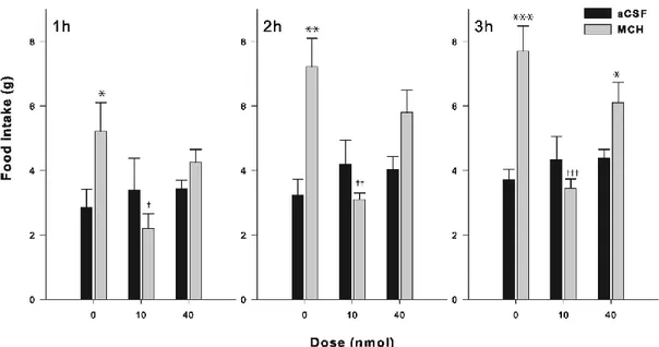
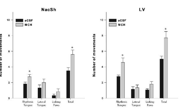
Melanin-concentrating hormone (MCH) exerts an orexigenic effect that resembles that of opioids, suggesting that the MCH and opioid systems could interact in controlling the food intake behavior. Three series of experiments were conducted in male Wistar rats (i) to test the ability of the κ, µ and δ opioid receptor antagonists NorBinaltorphimine (Nor-BNI — κ), β-Funaltrexamine (β-FNA — µ), and Naltrindole (NTI — δ), respectively, to block the stimulating effects of MCH on food intake, (ii) to verify the ability of MCH to induce a positive hedonic response to a sweet stimulus, when injected into the nucleus accumbens shell (NAcSh) or right lateral ventricle (LV) of the brain, and (iii) to assess the ability of Nor-BNI, β-FNA and NTI to block the effects of MCH on the hedonic response to a sweet stimulus. Nor-BNI and NTI (0, 10 and 40 nmol) and β-FNA (0, 10 and 50 nmol) were administered into the LV prior to injecting MCH (2.0 nmol). In order to assess the hedonic response, rats were implanted with an intraoral cannula allowing for the infusion of a sweet solution into the oral cavity. Food intake was assessed in sated rats during the first three hours following the MCH or vehicle (i.e. aCSF) injection. The hedonic response to a sweet stimulus was assessed by examining facial mimics, following the intraoral administration of a sucrose solution. Blockade of each of the three opioid receptors by selective antagonists prevented MCH-induced feeding. Furthermore, MCH-injections into the NAcSh and right LV resulted in enhanced hedonic responses. Finally, antagonism of the three opioid receptors blunted the LV-injected, MCH-induced facial-liking expressions, in response to an intra-oral sweet stimulus. Overall, the current study provides evidence to link the MCH and opioid systems in the food intake behavior.
Key words: Food intake behavior, taste reactivity, opioid receptor antagonism, nucleus accumbens shell, lateral hypothalamus.
2.3. Introduction
Energy balance regulation depends on complex controls exerted on both energy intake and energy expenditure. Those controls are essentially ensured by the hypothalamus, brainstem, and corticolimbic structures such as the insula, orbitofrontal cortex and ventral striatum (4, 6, 12). All the regions involved in energy balance produce chemical mediators capable of influencing food intake and energy expenditure (34), which includes neuropeptide Y, proopiomelanocortin, endocannabinoids, opioids and melanin-concentrating hormone (MCH).
MCH has been described as an anabolic (promoting energy /fat gain) peptide (28, 31). It is prominently expressed in the lateral hypothalamus (LH) and zone incerta (5). Both chronic MCH treatment (15) and MCH over expression (24) resulted in obesity and an increased susceptibility to high-fat feeding, whereas ablation of either MCH (39) or MCH neurons (1) promoted fat loss. In rodents, MCH produces its metabolic effects through a G protein-coupled receptor, the MCH receptor 1 (MCHR1) (31). Antagonism (38) and deletion of the MCHR1 (8, 27) led to leanness. There is clear evidence that the effects of MCH on energy balance are mediated through a stimulating action on energy intake and an inhibitory effect on energy expenditure (31).
The sites and mechanisms of the orexigenic action of the MCH are still uncertain. Recently, we demonstrated that bilateral injections of MCH into the nucleus accumbens shell (NAcSh) caused an increase in food intake, which was comparable to that induced by the injection of MCH in the third ventricle of the brain, suggesting the importance of the NAcSh as a site of the orexigenic action of MCH (19). The NAcSh represents the brain area with the most noticeable expression of the MCHR1 (35). Since it is also rich in κ, μ, and δ opioid receptors (25-26) and recognized as a prominent site of the orexigenic and hedonic response to opioids (16), one may hypothesize that there is a connection between the MCH and opioid systems in modulating the ingestive behavior.
The goal of the present study was to investigate the involvement of the opioid system in the effects of MCH on the ingestive behavior in rats. Firstly, we tested the ability of the κ, µ, and δ opioid receptor antagonists NorBinaltorphimine (Nor-BNI — κ), β-Funaltrexamine (β-FNA — µ), and Naltrindole (NTI — δ) to block the stimulating effects of MCH on food intake. Secondly, we verified the ability of MCH to enhance the hedonic response to a sweet stimulus, when injected into the NAcSh and right lateral ventricle (LV). Thirdly, we assessed the ability of Nor-BNI, β-FNA and NTI to block the stimulating effects of MCH on the hedonic response to a sweet stimulus. The hedonic response to a sweet stimulus was measured using the taste reactivity paradigm developed by Grill and Norgren (18).
2.4. Methods 2.4.1. Animals
Rats (250–275 g) were housed individually in plastic cages and kept on a 12:12 light /dark cycle with lights turned on at 07:00 h. They were fed a standard chow diet (Rodent Laboratory chow Purina # 5075, Charles River, St-Constant, QC, Canada). Rats were cared for and handled in conformance with the Canadian Guide for the Care and Use of Laboratory Animals. The protocols were approved by Université Laval’s animal care and use committee.
Three series of experiments were carried out in line with the objectives presented above. In the first series, we tested the ability of Nor-BNI (κ antagonist), β-FNA (µ antagonist), and NTI (δ antagonist) to block the stimulating effects of MCH on food intake by injecting the antagonists followed by MCH into the right LV of the brain. In the second series, we verified the ability of MCH to enhance the hedonic response to a sweet stimulus, when injected into the NAcSh or right LV. In the third series, we assessed the ability of Nor-BNI, β-FNA and NTI to block the stimulating effects of MCH on the hedonic response to a sweet stimulus. (18).The antagonists and MCH were injected into the right LV during this third series.
2.4.2. Surgery
One hundred and forty (140) rats were anesthetized with ketamine-xylazine (ketamine 37.5 mg/ml, xylazine 5 mg/ml) and implanted with the guide-cannula (Plastics One, Roanoke, VA) above the right LV using the following stereotaxic coordinates: 0.9 mm anterior to bregma, 1.2 mm lateral from midline and 3.5 mm ventral to the brain surface (29). Additional rats (for the purpose of the second series of experiments only) were implanted with a guide-cannula aimed at the NAcSh (n= 12) using the following stereotaxic coordinates: 1.7 mm anterior to bregma, 0.75 mm from midline, and 5.5 mm ventral to the brain surface (19). The cannula placement in the LV was confirmed by evaluating the dipsogenic response to angiotensin II (50
ng). The guide-cannula was anchored to the skull with sterile stainless-steel screws and acrylic cement. Stylets were inserted into the guide-cannula to prevent occlusion. The cannula placement into the LV was further established by visual inspection of brain slices under microscope at the end of the study. To facilitate that, rats were injected with methylene blue (into the LV) prior to the sacrifice. The placement of the cannula into the NAcSh was histologically verified under microscope on brains slices stained with thionine. The chronic intra oral cannula was secured as described by Grill and Norgren (18) and Cabanac (7). The cannula (heat-flared PE 100 tubing) entered the mouth just lateral to the first maxillary molar, ascended laterally to the skull, and exited over the dorsal part of the skull, where it was attached to acrylic cement. This cannula allowed the direct infusion of solutions into the mouth for taste-reactivity tests and did not interfere with the normal eating behavior (18).
2.4.3. Drugs and Microinjections
Rat MCH (Sigma-Aldrich, Oakville, ON, Canada) was used at a dose of 2.0 nmol. β-FNA was used at doses of 10 and 50 nmol, whereas both nor-BNI and NTI were administered at doses of 10 and 40 nmol. The three antagonists were bought from Tocris Bioscience (Ellisville, Missouri, USA). The doses of MCH and opioid antagonists were selected based on previous studies (21, 40). All drugs were dissolved in artificial cerebrospinal fluid (aCSF — Harvard Apparatus, Holliston, MA, USA), which served as the vehicle.
Prior to every series of experiments, rats were handled and familiarized with the testing procedures for at least three days. Microinjections were carried out using a 50 µl Hamilton syringe connected to a syringe pump (4 µl/min for LV and 1 µl/min for NAcSh). While animals were gently handheld, the injection cannula (30 gauge) was inserted into the guide-cannula. The injection-cannula projected into either the LV or the NAcSh. For each structure, the solution was carefully infused, for LV over 1 minute and for NAcSh over 30 seconds. The antagonists or aCSF were first injected
in the LV in a volume of 4 µl, followed by the injection of MCH (or aCSF) also in a volume of 4 µl. For the taste reactivity test in NAcSh, MCH (or aCSF) was injected bilaterally in a volume of 1 µl (0.5µl per side) (19). In taste reactivity tests, each rat received tested drugs (MCH and opioid antagonists) or aCSF. The microinjections were performed every 4 days (for tests with opioid antagonists) or every 2 days (for tests with MCH alone). The choice of a 4-day interval was based on previous in vitro and in vivo studies, investigating the duration of the opioid receptor antagonist activity (19, 23).
2.4.4. Food intake
Food intake measurements were conducted in rats chronically implanted with a stainless-steel, 22-gauge guide-cannula, aimed at the right LV of the brain. Three distinct groups of animals were used to test each of the three opioid receptor antagonists. Each group received a unique dose of antagonist with injections of MCH or aCSF (4 µl).
Food intake was measured over three hours between 11:00 and 15:00. Prior to the food intake measurements, rats were trained for 8 days to eat their daily ration of food (standard chow diet) between 17:00 to 08:00. This feeding schedule was designed to reduce appetite during the early light phase (41). Laboratory chow was used in feeding tests, which began 1 week after surgery. The tests were performed as following. All rats received a microinjection of the opioid receptor antagonist 90 min (for the κ and δ antagonists) or 22 h (for the µ antagonist) prior to the MCH or aCSF injection. After the MCH or aCSF injections, which were performed between 10:30 and 11:50, rats were allowed to eat and drink water ad libitum. Food intake (g) was recorded at 1, 2, and 3 h following the MCH or aCSF injections. The time interval between the administration of the antagonist and MCH (or aCSF) was chosen based on previous investigations (2, 40).
2.4.5. Hedonic response to a sweet stimulus
The hedonic response to a sweet stimulus was assessed in rats implanted with an intraoral cannula, in addition to the guide-cannula aimed at either the LV or NAcSh. Two distinct groups of rats were used to verify the ability of MCH (injected in the LV; n= 14 or NAcSh; n= 12) to induce a positive hedonic response to a sweet stimulus. Additionally, four groups of rats were used to further confirm the ability of MCH to induce a positive hedonic response to a sweet stimulus, and to assess the ability of µ, κ and δ opioid receptor antagonists to block the stimulating effects of MCH on the hedonic responses.
MCH-induced hedonic response to a sweet stimulus was determined using the methodology developed by Grill and Norgren (18), for assessing the changes in palatability produced by neural or pharmacological manipulations. This technique has been extensively used by Berridge (3) and others (33).
The hedonic response tests began two weeks after the surgery. To reduce the stress response during the test, animals were manipulated daily and habituated to the test chamber for 8 days and to the specific taste-reactivity procedure for 4 days. The tests were performed by injecting the opioid receptor antagonists at a dose of 10 nmol, prior to injecting MCH or the vehicle, as described above. The dose of 10 nmol for each antagonist was chosen based on our food intake results and the order of MCH and vehicle administration was counterbalanced. Ten minutes after the MCH or aCSF injection, the oral cannula was connected to a delivery tube (PE 50 tubing attached to a PE 10 nozzle), and the rat was placed in a Plexiglas test chamber. A mirror positioned beneath the transparent floor of the chamber reflected upon the face and mouth of the rat in the lens of the video camera. One ml of a 2% sucrose solution was infused manually into the mouth of the rat, through the oral cannula using a 1-ml syringe during 1 min. The hedonic reactions elicited by the sucrose taste were videotaped for subsequent analyses.
Hedonic reaction patterns were scored during a slow-motion (1/3 speed), double-blind and frame by frame video analysis. Positive hedonic reactions included rhythmic midline tongue protrusions, lateral tongue movements, and paw licks (18). Neutral reactions such as rhythmic mouth movements and passive drip of the solution were not scored. Aversive reactions such as like gaps, headshakes, limb flails and chin rubs were scored to probe that tested drugs did not alter the mobility of rats (data not shown). Separate occurrences were scored for each lateral tongue movements. Time bin scoring procedures were used to score behaviors that were produced in longer continuous bouts such as tongue protrusions (5 second bins) and paw licks (10 second bins). Use of these procedures allows multiple components to be combined into an overall hedonic reaction score (18). The double-blind video analyses were performed by two experienced individuals and the scores of each individual were averaged.
The LV-cannulated rats subjected to the taste reactivity tests were also implanted with an intraoral cannula, and distinct groups of rats were used for each of the tested drugs (Control, n= 9; MCH + Nor-BNI 10 nmol, n= 10; MCH + β-FNA 10 nmol, n= 14; MCH + NTI 10 nmol, n= 10).
2.4.6. Statistical analysis
Data obtained from rats with improper placement of cannula into the LV or NAcSh were excluded from the statistical analyses. Values are reported as means ± SEMs. In the first series of experiments two-way ANOVAs followed by Tukey-Kramer post hoc tests (when appropriate) were used to assess the differences between various conditions. In the second and third series of experiments paired t-tests were used to assess the difference between two treatments (aCSF vs. MCH) during the taste reactivity tests. Effects were considered to be significant with P values < 0.05. Data were analyzed using the SAS v 9.1.3 software (SAS Institute, Cary, NC, USA).