Science Arts & Métiers (SAM)
is an open access repository that collects the work of Arts et Métiers Institute of Technology researchers and makes it freely available over the web where possible.
This is an author-deposited version published in: https://sam.ensam.eu Handle ID: .http://hdl.handle.net/10985/14977
To cite this version :
Houssam BOULOUSSA, Claudio VERGARI, Thomas-Xavier HAEN, Wafa SKALLI, Raphael VIALLE - Biplanar stereoradiography predicts pulmonary function tests in adolescent idiopathic scoliosis: a cross-sectional study - European Spine Journal p.1-8 - 2019
Any correspondence concerning this service should be sent to the repository Administrator : archiveouverte@ensam.eu
1
Biplanar Stereoradiography Predicts Pulmonary Function Tests
in Adolescent Idiopathic Scoliosis: a Cross-Sectional Study
H. Bouloussa 1 MD MSc, R. Pietton 1,2 MD, C. Vergari 3 PhD,
T.X. Haen 1,2 MD MSc, W. Skalli 2 PhD, R. Vialle 1 MD PhD
ABSTRACT
Purpose: Various spinal and rib cage parameters measured from complex exams were found to be correlated with preoperative pulmonary function tests (PFT). The aim was to investigate the relationship between preoperative rib cage parameters and PFT using biplanar stereoradiography in patients with severe adolescent idiopathic scoliosis (AIS). Methods: Fifty-four patients, 45 girls and nine boys, aged 13.8 ± 1.2 years, with Lenke 1 or 2 thoracic scoliosis (>50°) requiring surgical correction were prospectively included. All patients underwent preoperative PFT and low-dose biplanar X-rays. The following data were collected: forced vital capacity (FVC), forced expiratory volume in 1s (FEV1), FEV1/FVC ratio, residual volume (RV), slow vital capacity (SVC), total lung capacity (TLC), rib cage volume (RCV), maximum rib hump (MRH), maximum width, mean thoracic index, spinal penetration index (SPI), apical vertebral rotation (AVR), main curve Cobb angle (MCCA), T4-T12 kyphosis. The primary outcome was the relationship between rib cage parameters and PFT. The secondary outcome was the relationship between rib cage parameters and spine parameters. Data were analyzed using Spearman’s rank test. A multivariable regression analysis was performed to compare PFTs and structural parameters. Significance was set at α = 0.05.
Results: The mean MCCA was 68.7 ± 16.7°. RCV was highly correlated with all pulmonary capacities: TLC (r=0.76, p < 0.0001), SVC (r=0.78, p < 0.0001) and FVC (r=0.77, p < 0.0001). RCV had a low correlation with FEV1/FVC (r= -0.34, p=0.014). SPI was not correlated with any pulmonary parameters.
Conclusion: Rib cage volume measured by biplanar stereoradiography may represent a prediction tool for PFTs.
Keywords: pulmonary function tests, AIS, three dimensional reconstructions, rib cage, scoliosis.
Level of evidence: Non-randomized cross-sectional study amoung consecutive patients, Level 2
INTRODUCTION
Scoliosis has a direct effect on the rib cage in the presence of a thoracic deformity. In the absence of underlying disorders, mild, moderate or even medium-severe scoliosis is rarely responsible for respiratory impairment. However, severe thoracic scoliosis is usually associated with the appearance of restrictive lung disease, resulting from a decrease in lung volumes and therefore the total lung capacity measured by pulmonary function tests (PFTs) (1)(2)(3)(4). The decrease in lung volumes is multifactorial. The pathophysiology differs depending on patients’ age at the onset of scoliosis and the chronicity of disorders. The main cause of these disorders is a mechanical alteration of diaphragm and chest wall function due to the three-dimensional chest deformity. Respiratory involvement is mainly due to scoliosis severity (Cobb angle), main curvature location and loss of thoracic kyphosis (5).
PFTs are now routinely used in the preoperative assessment of adolescent idiopathic scoliosis (AIS) before correction and spinal fusion both for the early prediction of immediate postoperative pulmonary complications (prolonged intubation or atelectasis) and as an indicator of baseline respiratory status (6). However, these tests may be poorly performed by children or disabled patients.
Correlation studies between spinal structural parameters and pulmonary function were described in the literature using chest CT-scan, dynamic MRI (7) or plain X-rays (8) with the main disadvantage of being performed in the supine position and respectively irradiating, poorly available, or involving long 3D reconstruction time. The need for long-term follow-up during growth and the repetition of X-rays have raised concern regarding radiation exposure (9). This issue was recently addressed with the development and validation of biplanar stereoradiography. This technology provides digital 2D high quality views with reduced distortion while being 8 to 10 times less irradiating than standard X-rays and 800 times less than traditional three-dimensional imaging (10).
Therefore, it could potentially become the gold standard for both diagnosis and monitoring of
scoliosis (11). Biplanar stereoradiography can provide three-dimensional images of the spine in a functional position (standing position) unlike computed tomography (supine position). 3D spinal reconstruction using this system is already a validated process (12) and is currently used in clinical practice, while validated methods for rib cage 3D reconstructions are more recent and not fully established yet. Also, Pasha et al. showed that the standing position with knuckles on clavicles position replicates the functional standing balance in AIS (13). Only one study by Ilharreborde et al. (14) investigated the relationships between structural spinal and rib cage parameters in AIS and pulmonary function using 3D reconstructions from biplanar stereoradiography (three groups: Cobb angle 20 to 40 degrees, 40 to 65 degrees and over 65 degrees).
In fact, rib cage volume has been progressively appearing as the main structural predictor of pulmonary function. Arguably, data providing evidence for the relationship between biplanar rib cage reconstructions and pulmonary function are still lacking. These reconstructions have the potential to predict pulmonary function in patients with AIS and detect restrictive lung disease, even at an early stage. Moreover, a clarification regarding which ribcage parameters are most representative of lung capacity is desirable. A preliminary work demonstrated that 3D reconstructions of the rib cage using biplanar stereoradiography was feasible and reliable to estimate thoracic parameters in patients with AIS (15).
The aim of this study was to determine the relationship between rib cage parameters and pulmonary function using low-dose biplanar stereoradiography in AIS patients with severe thoracic curves (Cobb angle > 50°).
MATERIAL AND METHODS
Patients
Fifty-four consecutive patients (45 girls, 9 boys) aged 13.8 ± 1.2 years with AIS (Lenke 1 or 2, main thoracic curve > 50°) were prospectively included by a single surgeon in
our institution before surgical correction and spinal fusion between January 2014 and June 2016, following institutional review board approval (CPP-Paris VI- 16-06). Patients were included according to the following criteria: male or female, Lenke 1 or Lenke 2 AIS, no other scoliosis etiology found, main curve Cobb angle >50°, planned surgical correction and spinal fusion by the same surgeon within the following 12 months, with or without previous bracing, signed parental consent. The following patients were excluded: patients with secondary scoliosis (neuromuscular, congenital, syndromic or post-traumatic), history of previous spinal surgery, history of spinal infection, history of chronic and debilitating lung disease preceding the diagnosis of AIS, inability to perform PFT, severe intellectual disability. All patients underwent full preoperative assessment including PFT and biplanar X-rays in our institution.
Imaging protocol and 3D modeling
Patients were in erect standing position, hands on clavicles to avoid any superimposition with the spine. Anteroposterior (AP) and lateral X-ray exposure parameters were respectively 83 kV, 200 mA and 102 kV, 200 mA. All images included at least both the last cervical vertebra (C7) and the pelvis. AP and lateral X-ray dose area product respectively averaged 411mGy·cm2 and 656 mGy·cm2. Spine and rib cages were reconstructed using validated methods(16)(12). Also, the reproducibility of rib cage reconstructions (including rib hump) in AIS patients with a main thoracic curve > 50° was demonstrated by Pietton et al. (15). A first estimation of spine and rib cage morphology was provided by the software, based on manually positioned anatomical landmarks and rib contouring. Consequently, a 3D model was projected on the anteroposterior and lateral X-rays. Finally, the operator manually adjusted the model until it perfectly fit the X-rays. Reconstructions were performed by a single trained operator.
Rib cage parameters
Structural spinal and rib cage parameters were automatically measured using model-based 3D reconstructions: Cobb angle (degrees), apical vertebral rotation (AVR, degrees), thoracic kyphosis (T4-T12, degrees), lumbar lordosis (L1-S1, degrees), rib hump (RH, degrees), rib cage volume (RCV, liters), spinal penetration index (SPI, %), mean thoracic index (%), maximum anteroposterior diameter (AP diameter, mm), maximum width (mm). The spinal penetration index was defined by Dubousset et al.(17) as the penetration rate of the spine into the rib cage due to deformity. The operator was blinded from patients’ pulmonary status during the reconstruction process. Patients were divided into two groups based on prior treatment (bracing or no bracing).
Pulmonary function tests
All patients were assessed with standard spirometry and plethysmography. PFT parameters were: total lung capacity (TLC, liters), forced vital capacity (FVC, liters), slow vital capacity (SVC, liters), forced expiratory volume in one second (FEV1) and FEV1/FVC ratio (Tiffeneau-Pinelli index).
Statistical analysis
Correlations were analyzed using Spearman’s rank test; limits of agreement were illustrated with Bland-Altman graphs. Standard error of the estimate (SEE) was calculated to quantify the spread of the correlations. Significance was set at α = 0.05. 1. Non-parametric tests were used because not all variables had a normal distribution (Lilliefors test, p > 0.05).
RESULTS
Demographic data
Seventeen patients had a prior history of asthma (31.5 %). Nineteen patients out of 54 (35.2 %) had normal PFTs. The most prevalent PFT finding was isolated restrictive lung disease (39 patients, 72.2%). Twenty-nine patients had prior brace treatment. There were 44 patients with Lenke 1 scoliosis and 10 Lenke 2.
Spinal and rib cage parameters
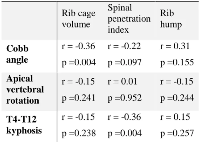
The mean main curve Cobb angle was 68.7 ± 16.7°. Mean T4-T12 kyphosis was 20.5 ± 13°. Pelvic parameters were within normal limits. There was no global sagittal malalignment due to lumbar or lumbosacral angular anomalies. The mean Risser stage was 3.4 ± 1.1. There were seven patients staged Risser 5, 20 patients Risser 4, 19 patients Risser 3, five patients Risser 2, one patient Risser 1 and two patients Risser 0. The mean rib cage volume (RCV) was 5.0 ± 1.2L while mean AP diameter was 147 ± 17 mm and maximum width averaged 232 ± 21 mm. Spinal and rib cage parameters are summarized in Table 1. Cobb angle had a low correlation with rib cage parameters and the same observation was valid for T4-T12 kyphosis.
Correlations between spinal and rib cage parameters are summarized in Table 2.
Pulmonary function
The mean total lung capacity was 4.0 ± 0.9L. The mean slow vital capacity and forced vital capacity were respectively 2.9 ± 0.7L and 2.9 ± 0.7L. Mean PFT values are summarized in Table 3.
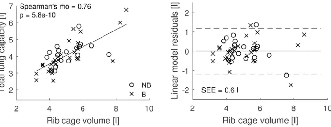
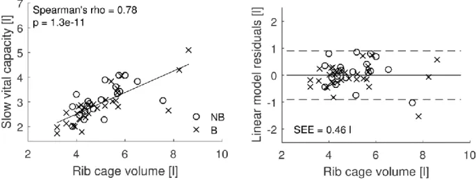
The majority of rib cage parameters showed a mild to high correlation with PFT parameters. Rib cage volume was correlated with TLC (rho = 0.76, p<0.0001) and SVC (rho = 0.78, p < 0.0001) with a standard error of the estimate of 0.61 L and 0.46 L respectively. Figures 1 and 2 illustrate the agreement between rib cage volume and TLC or SVC, respectively. The rib cage volume was also correlated with FEV1/FVC (rho = -0.34, p=0.014) and FVC
Figure 1. Agreement between rib cage volume and TLC.
Table 1. Main spinal and rib cage parameters
Parameter Mean ± SD Range
Cobb angle (°) 68.2 ± 16.4 (25.8-110.7)
T4-T12 kyphosis (°) 20.5 ± 13.1 (2.3-59.6)
L1-S1 lordosis (°) 52.7 ± 20.8 (-76.8-77.6)
AVR (°) 19.6 ± 9.9 (1.2-48.6)
SPI (%) 5.9 ± 1.6 (2.3-9.4)
Rib cage volume (l) 5.01 ±1.28 (3.19-9.49)
Thoracic index 0.6 ± 0.1 (0.5-0.8)
(rho = 0.77, p < 0.0001). Maximum rib hump was correlated to SVC (r= -0.36, p<0.01) and FVC (r= -0.28, p<0.05). Maximum rib cage width demonstrated a mild correlation with TLC (r= 0.56, p<0.0001), FVC (r= 0.62, p<0.0001), SVC (r= 0.60, p<0.0001), FEV1/FVC (r= -0.32, p<0.05), FEV1 (r=0.52, p<0.0001). Thoracic depth or rib cage anteroposterior (AP) diameter was correlated with TLC (r=0.26, p<0.05), SVC (r=0.23, p<0.05) and FVC (r=0.30, p<0.001). However, it was not correlated with FEV1/FVC (r=-0.09, p=0.51). No correlations were found between SPI and pulmonary parameters. Correlations between rib cage parameters and PFT values are summarized in Table 4.
As for spinal structural parameters, T4-T12 kyphosis was not correlated with any PFT parameter, neither with TLC (rho = -0.16, p = 0.27) nor with SVC (rho = -0.15, p = 0.29). The Cobb angle had a mild correlation with pulmonary function parameters: TLC (rho = -0.47, p = 0.0001), SVC (r = -0.44, p = 0.001) and FVC (r = -0.37, p = 0.007).
Group analysis
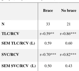
Rib cage volume remained significantly correlated with TLC in the brace group (rho = 0.86, p < 0.0001) and “no brace” group (rho = 0.59, p = 0.01). In addition, the standard error of the estimate was almost identical for these two groups (SEE = 0.60 L)
DISCUSSION
Contribution of stereoradiographic 3D analysis
This study reports data from a homogeneous cohort of AIS patients that were prospectively enrolled preoperatively and suggested that biplanar X-rays may also become a reliable tool to obtain fast screening for restrictive lung disease.
The use of CT-scans, while being a reliable procedure, does not allow proper estimation of rib cage 3D parameters in standing position. Moreover, high irradiation on children raises concern about a hypothetical risk of
developing neoplasia during adulthood (18), which prevents CT from becoming a routinely used tool for the preoperative respiratory assessment of AIS children. Yet, Wen et al. studied correlations between total lung volume calculated from CT and pulmonary function in patients with AIS. They found numerous correlations between measured total lung volume with vital capacity, forced vital capacity, FEV1, TLC, maximum ventilator volume, as well as the predicted values of forced vital capacity, FEV1 and TLC (19). In reality, 3D reconstructions from CT-scans require long reconstruction time and are technically difficult (8). They may not be suitable for routine clinical practice. Dynamic MRI has the advantage of providing direct access to lung volumes. Nevertheless, it remains a technically demanding and poorly available method that does not allow precise measurements of rib cage bony components. To this date, few studies have reported results from low-dose natural standing position biplanar stereoradiographic imaging with 3D rib cage analysis (20)(21)(11)(14). Its semi-automated reconstruction technique was validated and can now be routinely used to quantify most rib cage parameters (16). Another advantage of stereoradiography is its capacity to provide data from the true 3D deformity, given that two-dimensional analysis of curves may prevent clinicians from recognizing kyphosis or lordosis due to rotational abnormalities. This was detailed and studied extensively by Yaszay et al. who showed that the best spinal parameter to
Table 2. Spinal and rib cage parameters main
correlations. Rib cage volume Spinal penetration index Rib hump Cobb angle r = -0.36 p =0.004 r = -0.22 p =0.097 r = 0.31 p =0.155 Apical vertebral rotation r = -0.15 p =0.241 r = 0.01 p =0.952 r = -0.15 p =0.244 T4-T12 kyphosis r = -0.15 p =0.238 r = -0.36 p =0.004 r = 0.15 p =0.257
predict FEV was the 3D thoracic kyphosis. Our findings do not fundamentally differ from that since 3D thoracic kyphosis obviously acts on respiratory function via a more direct parameter, which is the rib cage volume (22).
Pulmonary function and spinal parameters The correlation between the Cobb angle and respiratory parameters is still controversial. Redding and Mayer (23) found weak correlations between Cobb angle measures and PFT while Johnston et al. reported stronger correlations (2). Even though the Cobb angle measurement is at the center of the decision-making process and surgical planning, it is not certain that it should still bear a fundamental role in the benefit-risk balance explained to patients and their parents concerning their respiratory condition before spinal correction and fusion. However, to this date, there is no method to predict respiratory function deterioration and indicate surgical treatment accordingly.
Johnston et al. and Ilharreborde et al. (2)(14) reported a correlation between pulmonary impairment with the Cobb angle and with hypokyphotic patients. In the current study, only the Cobb angle was shown to be mildly correlated with PFT parameters. Our mean T4-T12 kyphosis being slightly higher with no patients suffering from thoracic lordosis, it may be hypothesized that the absence of correlation between kyphosis and pulmonary
function can be mostly due to the population selection (no neuromuscular cases and higher kyphosis).
The choice of a Cobb angle > 50° was based on surgical cut-offs. Since it is a common belief that there is no respiratory impairment at this degree of scoliosis, we showed that PFTs are modified early in the natural history of scoliosis, in accordance with Abdelaal et al., who reported from an observational cohort study that even adolescents with mild idiopathic scoliosis had mild pulmonary and functional restriction (24).
Rib cage parameters
Rib cage volume was identified as the most important structural factor affecting lung function in a pediatric population with AIS and a severe thoracic curve magnitude (Cobb angle > 50°). The best correlation was found with TLC, SVC, FVC, and FEV1 suggesting that a poor rib cage volume was a predictor for the diagnosis of restrictive lung syndrome on PFTs. However, the agreement between rib
Figure 2. Agreement between rib cage volume and SVC.
Table 3. Pulmonary function test values
Mean ± SD Range
Total lung capacity (L) 4.1 ± 1 (2.6-8.2)
Slow vital capacity (L) 3 ± 0.8 (1.7-6.6)
Forced vital capacity (L)
3 ± 0.9 (1.8-6.7)
cage volume and pulmonary function does not allow accurate regression between the two values, i.e., rib cage volume cannot be used to estimate TLC and SVC with an uncertainty below 0.6 L. In contrast, the relationship between rib cage volume, TLC and SVC gets more accurate for volumes below seven litters. Indeed, the standard errors of estimate below seven liters of rib cage volume were respectively of 0.49 L for TLC (0.61 L for all volumes) and 0.37 L for SVC (0.49 L for all volumes). Besides, rib cage volume poorly correlated with FEV1/FVC, which is the diagnostic criterion for obstructive lung disease. Obviously, this finding suggested that structural rib cage or spinal parameters did not match respiratory parameters that depended mostly on bronchial diameter.
Ilharreborde et al. (14) described a correlation between the rib cage volume and PFT (FVC and TLC) using biplanar stereoradiography in AIS of progressive severity. We report similar correlations to these parameters in our AIS cohort with severe Cobb angles only.
Interestingly, maximum rib cage width, which could potentially be much faster to measure than rib cage volume, was shown in this study to be correlated with most PFT parameters. Even though it was correlated with PFTs to a lesser extent than rib cage volume, its measurement could have an interest in clinical practice on plain X-rays if biplanar stereoradiography is not available or 3D reconstructions are not completed.
Another interesting approach to assess thoracic parameters is the use of body surface light
band projection techniques used for brace manufacturing (25)(26). This has the advantage of being fast and non-invasive. Since it is part of standard care in numerous institutions, its use can provide a reliable estimation of various external thoracic geometric parameters such as thoracic volume, perimeter, anterior-posterior and transverse diameters, T1-T12 length, and sternal length. Charles et al. (27) used this system and found that mild and moderate scoliosis did not affect thoracic diameters and volume at any stage of growth. Their estimated thoracic volumes for healthy boys and boys with moderate scoliosis were respectively 7.17 ± 2.15 L (2.8–10.0) and 8.12 ± 3.02 L (4.4–11.8). Healthy girls and girls with moderate scoliosis had the respective thoracic volumes: 8.47 ± 3.16 L (2.9–20.7) Table 4. Correlations between rib cage and respiratory parameters (* = p < 0.05; ** p < 0.01; *** p < 0.001). TLC (L) SVC (L) FVC (L) FEV1/FVC RCV (L) r= 0.78*** r= 0.81*** r= 0.78*** r= -0.32* SPI (%) r= -0.19, p=0.18 r= -0.17, p=0.21 r= -0.21, p=0.11 r= -0.07, p=0.61 Rib hump (°) r= -0.28* r= -0.39** r= -0.28* r= 0.06, p=0.65 AP diameter (mm) r= 0.30* r= 0.28* r= 0.92*** r= 0.39, p=0.38 Max width (mm) r= 0.59*** r= 0.62*** r= 0.63*** r= -0.31*
Table 5. Correlations and standard errors of the
estimate (SEM) between total lung capacity (TLC) and slow vital capacity (SVC) with rib cage volume (RCV) according to the different groups (*** p < 0.001, * p < 0.01) Brace No brace N 33 21 TLC/RCV r=0.59** r=0.86*** SEM TLC/RCV (L) 0.59 0.60 SVC/RCV r=0.70*** r=0.82*** SEM SVC/RCV (L) 0.50 0.43
and 8.64 ± 1.75 L(3.2–12.3). In our study, the mean thoracic volume was 5.0 ± 1.2L. The difference could be explained by a different population mean age and distribution (3.0– 24.9 years for girls and 4.1–25 years for boys in their study). While we only included patients with severe scoliosis, Charles et al. included patients with mild and moderate scoliosis (27). Not surprisingly, our volumes were lower. Also, using an optical scanner estimates rib cage parameters using models. Soft-tissue juxtaposition and various body mass indices may not give an estimation as accurate as computed tomography for example. We showed a strong correlation between rib cage volume and PFT. However, this was an estimation of PFT using a near-direct measurement of rib cage volume. We believe that optical scanners may not be ideal for an estimation of PFT as the technique would only provide a second degree estimation (PFT) of a first degree estimation (rib cage volume).
Another way to conceptualize the importance of assessing thoracic and rib cage deformities was elegantly shown by Akazawa et al. (28) From a cohort of 194 patients who underwent spinal fusion with Harrington instrumentation and a minimum 27-year follow-up, they reported restrictive ventilation defects in 27.7% of their cohort. Interestingly, they also found that the actors aggravating %FVC were large rib humps and large vertebral rotations. Arguably, we hope that stressing the importance of correcting these parameters, independently of the classic coronal Cobb angle, will provide better outcomes for the future generations of patients with AIS. Limitations of the study
Seventeen patients out of 54 (31.4%) had a history of asthma with typical findings of obstructive lung disease. However, in all cases, the asthma was controlled, with or without long-term control medications. This may have biased our correlation study between PFT and structural parameters due to the role of bronchoconstriction in asthma. Although several authors reported the increased incidence of lung obstructive disease in patients with AIS, its pathophysiology is still
uncertain (29)(6). Extrinsic mechanisms such as compression (indirectly caused by rib humps or apical rotation), gas trapping or even intrinsic mechanisms including hypersecretion were suggested (30). Nonetheless, excluding those patients would have altered the external validity of this study regarding the AIS population.
Validation of a screening tool for restrictive syndrome in AIS populations requires a larger cohort. Though preliminary, our findings would benefit from a multi-center study. Moreover, the clinical relevance of our results has yet to be confirmed by a wider approach comparing preoperative and postoperative analyses with a long-term follow-up. Furthermore, 3D modeling could be improved by allowing the direct measurement of total lung capacity in stereoradiographic images instead of rib cage volume. A simplification of the reconstruction process would allow its routine use in clinical practice. While being useful for patients unable to properly perform PFTs such as patients with autism, we remind that most patients in that situation suffer from neuromuscular scoliosis and not AIS.
Patients included in this study presented various stages of growth. Most of them were included in or close to their period of peak growth velocity. Though this was not a prospective study including follow-up, this may have biased correlations since rib cages were therefore not geometrically homogeneous. Indeed, Dimeglio et al. (31) described a thoracic peak growth velocity between age 10 and skeletal maturity. The rib cage volume doubles during that period and its growth pattern varies tremendously between children and adolescents due to various ages of puberty (32). Furthermore, Canavese et al. (33) described the evolution of the rib cage shape from ovoid at birth to elliptical at skeletal maturity. This shows that external shape parameters such as width, AP diameter and circumference vary during growth. These authors also described a discrepancy between thoracic volume (30% of its definitive volume at five years of age) and thoracic external shape parameters (trunk height is already 66% of its final size at five years of age) (32). However, we believe that this cohort is
representative of patients with AIS for whom surgical correction and fusion is indicated. Although growth was reported to affect the relationship between volume and shape of the rib cage, this was not the main focus of our study. Indeed, the main interest was the relationship between PFT and rib cage parameters at a given time. To our knowledge, no study has reported an altered ratio between PFT and rib cage volume affected by growth.
CONCLUSION
Low-dose biplanar X-rays have the potential to predict total lung capacity and slow vital capacity in patients with AIS, provided that a
more accurate estimation of lung volumes can be achieved. These results may help develop fast and non-invasive tools for the preoperative structural and respiratory assessment of AIS children. Substituting PFTs with stereoradiography may also benefit physically and mentally disabled patients who cannot perform PFTs. Low-dose biplanar X-rays could provide exhaustive and fast musculoskeletal and respiratory preoperative assessment for scoliosis surgery, providing insight into patients’ natural spinal balance, deformity, and respiratory status with minimal irradiation.
REFERENCES
1. Takahashi S, Suzuki N, Asazuma T, Kono K, Ono T, Toyama Y. Factors of thoracic cage deformity that affect pulmonary function in adolescent idiopathic thoracic scoliosis. Spine. 2007 Jan 1;32(1):106–12.
2. Xue X, Shen J, Zhang J, Zhao H, Li S, Wang Y, et al. An analysis of thoracic cage deformities and pulmonary function tests in congenital scoliosis. Eur Spine J Off Publ Eur Spine Soc Eur Spinal Deform Soc Eur Sect Cerv Spine Res Soc. 2015 Jul;24(7):1415–21.
3. Johnston CE, Richards BS, Sucato DJ, Bridwell KH, Lenke LG, Erickson M, et al. Correlation of preoperative deformity magnitude and pulmonary function tests in adolescent idiopathic scoliosis. Spine. 2011 Jun 15;36(14):1096– 102.
4. Dreimann M, Hoffmann M, Kossow K, Hitzl W, Meier O, Koller H. Scoliosis and chest cage deformity measures predicting impairments in pulmonary function: a cross-sectional study of 492 patients with scoliosis to improve the early identification of patients at risk. Spine. 2014 Nov 15;39(24):2024–33.
5. Newton PO, Faro FD, Gollogly S, Betz RR, Lenke LG, Lowe TG. Results of preoperative pulmonary function testing of adolescents with idiopathic scoliosis. A study of six hundred and thirty-one patients. J Bone Joint Surg Am. 2005 Sep;87(9):1937–46.
6. Vedantam R, Crawford AH. The role of preoperative pulmonary function tests in patients with adolescent idiopathic scoliosis undergoing posterior spinal fusion. Spine. 1997 Dec 1;22(23):2731–4.
7. Chu WC, Ng BK, Li AM, Lam T-P, Lam WW, Cheng JC. Dynamic magnetic resonance imaging in assessing lung function in adolescent idiopathic scoliosis: a pilot study of comparison before and after posterior spinal fusion. J Orthop Surg. 2007;2:20.
8. Ledonio CGT, Rosenstein BE, Polly DW, Johnston CE, Regelmann WE, Nuckley DJ. Pulmonary function tests correlated with thoracic volumes in adolescent idiopathic scoliosis. J Orthop Res Off Publ Orthop Res Soc. 2016 May 21; 9. Hoffman DA, Lonstein JE, Morin MM, Visscher W, Harris BS, Boice JD. Breast cancer in women with scoliosis exposed to multiple diagnostic x rays. J Natl Cancer Inst. 1989 Sep 6;81(17):1307–12.
10. Dubousset J, Charpak G, Skalli W, Kalifa G, Lazennec J-Y. [EOS stereo-radiography system: whole-body simultaneous anteroposterior and lateral radiographs with very low radiation dose]. Rev Chir Orthop Reparatrice Appar Mot. 2007 Oct;93(6 Suppl):141–3.
11. Ilharreborde B, Dubousset J, Le Huec J-C. Use of EOS imaging for the assessment of scoliosis deformities: application to postoperative 3D quantitative analysis of the trunk. Eur Spine J Off Publ Eur Spine Soc Eur Spinal Deform Soc Eur Sect Cerv Spine Res Soc. 2014 Jul;23 Suppl 4:S397–405.
12. Humbert L, De Guise JA, Aubert B, Godbout B, Skalli W. 3D reconstruction of the spine from biplanar X-rays using parametric models based on transversal and longitudinal inferences. Med Eng Phys. 2009 Jul;31(6):681–7. 13. Pasha S, Capraro A, Cahill PJ, Dormans JP, Flynn JM. Bi-planar spinal stereoradiography of adolescent idiopathic scoliosis: considerations in 3D alignment and functional balance. Eur Spine J. 2016 Oct;25(10):3234-3241
14. Ilharreborde B, Dubousset J, Skalli W, Mazda K. Spinal penetration index assessment in adolescent idiopathic scoliosis using EOS low-dose biplanar stereoradiography. Eur Spine J Off Publ Eur Spine Soc Eur Spinal Deform Soc Eur Sect Cerv Spine Res Soc. 2013 Nov;22(11):2438–44.
15. Pietton R, Bouloussa H, Vergari C, Skalli W, Vialle R. Rib Cage Measurement Reproducibility Using Biplanar Stereoradiographic 3D Reconstructions in Adolescent Idiopathic Scoliosis. J Pediatr Orthop. 2017 Nov 16;
16. Aubert B, Vergari C, Ilharreborde B, Courvoisier A, Skalli W. 3D reconstruction of rib cage geometry from biplanar radiographs using a statistical parametric model approach. Comput Methods Biomech Biomed Eng Imaging Vis. 2016 Sep 2;4(5):281–95.
17. Dubousset J, Wicart P, Pomero V, Barois A, Estournet B. Spinal penetration index: new three-dimensional quantified reference for lordoscoliosis and other spinal deformities. J Orthop Sci Off J Jpn Orthop Assoc. 2003;8(1):41– 9.
18. Mathews JD, Forsythe AV, Brady Z, Butler MW, Goergen SK, Byrnes GB, et al. Cancer risk in 680,000 people exposed to computed tomography scans in childhood or adolescence: data linkage study of 11 million Australians. BMJ. 2013;346:f2360.
19. Wen Y, Kai S, Yong-Gang Z, Guo-Quan Z, Tian-Xiang D. Relationship between Lung Volume and Pulmonary Function in Patients With Adolescent Idiopathic Scoliosis: Computed Tomographic-based 3-Dimensional Volumetric Reconstruction of Lung Parenchyma. Clin Spine Surg. 2016 Oct;29(8):E396-400
20. Courvoisier A, Vialle R, Skalli W. EOS 3D Imaging: assessing the impact of brace treatment in adolescent idiopathic scoliosis. Expert Rev Med Devices. 2014 Jan;11(1):1–3.
21. Sabourin M, Jolivet E, Miladi L, Wicart P, Rampal V, Skalli W. Three-dimensional stereoradiographic modeling of rib cage before and after spinal growing rod procedures in early-onset scoliosis. Clin Biomech Bristol Avon. 2010 May;25(4):284–91.
22. Yaszay B, Bastrom TP, Bartley CE, Parent S, Newton PO. The effects of the three-dimensional deformity of adolescent idiopathic scoliosis on pulmonary function. Eur Spine J. 2017 Jun;26(6):1658-1664
23. Redding GJ, Mayer OH. Structure-Respiration Function Relationships Before and After Surgical Treatment of Early-onset Scoliosis. Clin Orthop. 2011 May;469(5):1330–4.
24. Abdelaal AAM, Abd El Kafy EMAES, Elayat MSEM, Sabbahi M, Badghish MSS. Changes in pulmonary function and functional capacity in adolescents with mild idiopathic scoliosis: observational cohort study. J Int Med Res. 2018 Jan;46(1):381-391.
25. Cottalorda J, Kohler R, Garin C, Lecante P. [Orthopedic treatment of scoliosis: new technique using impression by optic procedure]. Arch Pediatr Organe Off Soc Francaise Pediatr. 1997 May;4(5):464–7.
26. Cottalorda J, Kohler R, Garin C, Genevois P, Lecante C, Berge B. Orthoses for mild scoliosis: a prospective study comparing traditional plaster mold manufacturing with fast, noncontact, 3-dimensional acquisition. Spine. 2005 Feb 15;30(4):399–405.23.
27. Charles YP, Marcoul A, Schaeffer M, Canavese F, Diméglio A. Three-dimensional and volumetric thoracic growth in children with moderate idiopathic scoliosis compared with normal. J Pediatr Orthop Part B. 2017 May;26(3):227–32.
28. Akazawa T, Kuroya S, Iinuma M, Asano K, Torii Y, Umehara T et al. Pulmonary function and thoracic deformities in adolescent idiopathic scoliosis 27 years or longer after spinal fusion with Harrington instrument. J Orthop Sci. 2018 Jan;23(1):45-50
29. Boyer J, Amin N, Taddonio R, Dozor AJ. Evidence of airway obstruction in children with idiopathic scoliosis. Chest. 1996 Jun;109(6):1532–5.
30. Hale K, Rasp F. Pulmonary function testing. Rev. ed. of: Moe’s textbook of scoliosis and other spinal deformities.
31. Dimeglio A, Canavese F. The growing spine: how spinal deformities influence normal spine and thoracic cage growth. Eur Spine J Off Publ Eur Spine Soc Eur Spinal Deform Soc Eur Sect Cerv Spine Res Soc. 2012 Jan;21(1):64– 70.28.
32. Charles YP, Diméglio A, Marcoul M, Bourgin J-F, Marcoul A, Bozonnat M-C. Influence of idiopathic scoliosis on three-dimensional thoracic growth. Spine. 2008 May 15;33(11):1209–18.
33. Canavese F, Dimeglio A. Normal and abnormal spine and thoracic cage development. World J Orthop. 2013 Oct 18;4(4):167–74.