Marc-Gilbert Lagny
Francine Blaffart
ECCP
Department of Nursing and
Department of Cardiovascular and Thoracic Surgery
Prof. JO Defraigne
University Hospital of Liège, Belgium.
CPB AND AORTIC SURGERY
The State of the Art
From a Theoretical to a Practical
Approach
INTRODUCTION
§
Prevention
Michel JB, et al.
Novel aspects of the pathogenesis of aneurysms oh the abdominal aorta in humans.
Cardiovasc Res. 2011 Aprl 1;90(1):18-27
Golledge J, Norman PE,
§
Medical treatment
Current status of medical management for abdominal aortic aneurysm.Atherosclerosis,
2011. 217 (1):p.57-63.
§
Endovascular aortic repair
Nienaber CA, et al.
Randomized comparison of strategies for type B aortic dissection: the INvestigation of STEnt
Grafts in Aortic Dissection (INSTEAD) trial. Circulation, 2009. 120(25):p.2519-28.
Hao Z, et al.
Endovascular stent-graft placement or open surgery for the treatment of acute type B aortic
dissection: a meta-analysis. Ann Vasc Surg, 2012. 26(4):p.454-61.
AIM OF THE TOPIC
M-G LAGNY, F. BLAFFART, ECCP CHU de Liège
STATE-OF-THE-ART PAPER
Evidence, Lack of Evidence, Controversy,
and Debate in the Provision and Performance
of the Surgery of Acute Type A Aortic Dissection
Robert S. Bonser, MD,*†‡ Aaron M. Ranasinghe, MD,*† Mahmoud Loubani, MD,‡§
Jonathan D. Evans, BM
EDS
CI,† Nassir M. A. Thalji, MB C
HB,† Jean E. Bachet, MD,!
Thierry P. Carrel, MD,† Martin Czerny, MD,‡¶ Roberto Di Bartolomeo, MD,‡#
Martin Grabenwöger, MD,‡** Lars Lonn, MD, P
HD,†† Carlos A. Mestres, MD, P
HD,‡‡‡
Marc A. A. M. Schepens, MD,त Ernst Weigang, MD, P
HD‡!!
Birmingham and Hull, United Kingdom; Abu Dhabi, United Arab Emirates; Berne, Switzerland;
Bologna, Italy; Vienna, Austria; Copenhagen, Denmark; Barcelona, Spain; Brugge, Belgium; and Mainz, Germany
Acute type A aortic dissection is a lethal condition requiring emergency surgery. It has diverse presentations, and the diagnosis can be missed or delayed. Once diagnosed, decisions with regard to initial management, transfer, appropriateness of surgery, timing of operation, and intervention for malperfusion complications are necessary. The goals of surgery are to save life by prevention of pericardial tamponade or intra-pericardial aortic rupture, to resect the primary entry tear, to correct or prevent any malperfusion and aortic valve regurgitation, and if possi-ble to prevent late dissection-related complications in the proximal and downstream aorta. No randomized trials of treatment or techniques have ever been performed, and novel therapies—particularly with regard to extent of surgery—are being devised and implemented, but their role needs to be defined. Overall, except in highly spe-cialized centers, surgical outcomes might be static, and there is abundant room for improvement. By highlight-ing difficulties and controversies in diagnosis, patient selection, and surgical therapy, our over-archhighlight-ing goal should be to enfranchise more patients for treatment and improve surgical outcomes. (J Am Coll Cardiol 2011;58:2455–74) © 2011 by the American College of Cardiology Foundation
Acute type A aortic dissection (ATAAD) is highly lethal
and might be increasing in incidence (
1
). Surgery is believed
to save and extend life, but despite apparent advances,
diagnosis is often delayed, evidence for improving outcomes
is modest (
2
), and optimal surgical management remains
unclear (
3–8
). Recent reviews (
6,7,9–12
) have directed
limited attention to the provision and performance of
surgery. This review specifically examines areas of
uncer-tainty and controversy in diagnosis, provision of care, and
surgical management of ATAAD and has the objectives of
promoting investigation to detect and treat the condition
earlier and to improve treatment outcomes.
Anatomy and Incidence
By definition, ATAAD involves the ascending aorta (AscAo) and
includes types I and II of the DeBakey classification. The
DeBakey classification categorizes dissections as type I
(involving the AscAo with distal extension), type II affecting
the AscAo only, and type III delineating disease of the
descending aorta (DescAo) and beyond. Modifications of
these classifications—which describe patho-anatomical
variants—exist, but for practical purposes, the original
classifications by specifying AscAo involvement and hence
risk of intra-pericardial rupture define those cases requiring
surgery (
Fig. 1
).
Neither classification dictates the site of the
originat-ing entry tear. In ATAAD a primary intimal tear is
usually present within the AscAo, sometimes
accompa-nied by secondary, more distal tears (
13
). In others,
involvement of the AscAo is due to retrograde
propaga-From the *University Hospitals Birmingham, National Health Service Foundation Trust, Birmingham, United Kingdom; †School of Clinical and Experimental Med-icine, Birmingham, United Kingdom; ‡Vascular Domain Committee of the Euro-pean Association of Cardiothoracic Surgery, Windsor, Berkshire, United Kingdom; §Castle Hill Hospital, Hull, United Kingdom;!Zayed Military Hospital, Abu Dhabi, United Arab Emirates; ¶University Hospital Berne, Berne, Switzerland; #Cardiac Department, Universita Di Bologna, Bologna, Italy; **Department of Cardiovascular Surgery, Hospital Hietzing, Vienna, Austria; ††Vascular Surgery and Cardiovascular Radiology, Rigshospital, Copenhagen University, Copenhagen, Denmark; ‡‡Hospi-tal Clinico, University of Barcelona, Barcelona, Spain; §§AZ St. Jan, Brugge, Belgium; and the!!Medical Center of the Johannes Gutenberg University, Mainz, Germany. Dr. Lonn is the medical director for Mentice Company; and has shares in the Le Maitre Vascular Company. All authors have reported that they have no relationships relevant to the contents of this paper to disclose.
Manuscript received March 30, 2011; accepted June 7, 2011.
Journal of the American College of Cardiology Vol. 58, No. 24, 2011 © 2011 by the American College of Cardiology Foundation ISSN 0735-1097/$36.00 Published by Elsevier Inc. doi:10.1016/j.jacc.2011.06.067
CPB for aortic surgery:
state of the art
§
Knowledge
§
Anatomical and pathophysiological pre-requests
§
Flexibility
§
Equipment
Ascending aortic surgery and arterial
cannulation:
Surgical
repairment
M-G LAGNY, F. BLAFFART, ECCP CHU de Liège
Ascending aorta
Left subclavian/
axillary
Femoral
The arterial cannulation
M-G LAGNY, F. BLAFFART, ECCP CHU de Liège
Bonser RS, et al.
Acute Aortic Dissection. JAAC Vol. 58, No. 24, 2011.
Arm numbness
Embolic event
Arterial cannulation complication and
perfusion: local dissection
M-G LAGNY, F. BLAFFART, ECCP CHU de Liège
§
Pressure monitoring
SWITCH TO ANOTHER CANNULATION SITE
Anticipation: Y Line
Arterial cannulation complication and
perfusion: malperfusion (FLAP)
M-G LAGNY, F. BLAFFART, ECCP CHU de Liège
§
Sudden increase of arterial line pressure
§
Inequate cerebral perfusion?: NIRS, TCD
§
Inadequate spinal cord perfusion (MEP)
§
Late diagnostic (lactates)
§
Left radial pressure
Pre and post arch arterial lines
(+ left femoral)
Swich to another cannulation site
RE-INSTORE ANTEGRADE FLOW
Arterial femoral cannulation complication
and perfusion:
false lumen
M-G LAGNY, F. BLAFFART, ECCP CHU de Liège
§
Pressure monitoring
§
Transesophagal echocardiography(TOE)
§
Arterial pressure (left radial)
CHECK THE CANNULATION
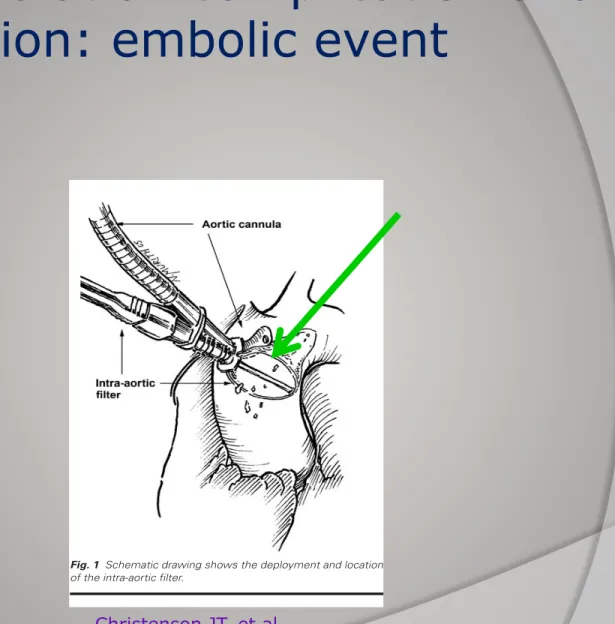
Arterial cannulation complication and
perfusion: embolic event
M-G LAGNY, F. BLAFFART, ECCP CHU de Liège
Christenson JT, et al.
Tex Heart Inst J. 2005;32(4):515-21.
§
Doppler
§
Specific cannula
Volume 32, Number 4, 2005 516 Aortic Cross-Clamping and Particulate Emboli
tion before aortic declamping would increase the num-ber of particles captured and thus further minimize the risk of systemic embolism.
Patients and Methods
Patients
From November 2002 through January 2003, 15 consecutive patients underwent elective CABG, valve surgery, or both, with intra-aortic filtration at 2 dif-ferent times, in accordance with our study protocol. The hospital ethics committee approved the study, and signed consent was obtained from all patients.
Three women and 12 men were enrolled in the study (mean age, 71.6 ± 11.5 years; range, 50–93 years); 9 of the 15 patients (60%) were older than 70 years. Table I presents preoperative characteristics of the 15 patients. Four patients underwent CABG alone for multivessel coronary artery disease, and the other 11 underwent left-sided intracardiac procedures alone or in combination with CABG: mitral valve annu-loplasty (MVA; 1 patient), aortic valve replacement (AVR; 7 patients), and AVR and CABG for single-vessel disease (3 patients).
All surgical interventions included cardiopulmo-nary bypass with complete systemic hemodilution and normothermia or mild hypothermia, according to standard techniques.
The Intra-Aortic Filter
The intra-aortic filter device (EMBOL-X®, Slim
Pro-tection System, Edwards Lifesciences; Irvine, Calif ) consists of a 24F metal-tipped aortic cannula that has been modified with a side port so that a collapsible filter can be inserted into and retracted from the aor-ta, in a position immediately distal to the aortic cross-clamp but proximal to the arterial return cannula and the brachiocephalic artery (Fig. 1). The filter itself consists of a heparin-coated polyester mesh that can capture particles larger than 120 µm in diameter. The filter is mounted on a flexible frame so that it can con-form to the interior diameter of the ascending aorta (Fig. 2). The risks associated with the use of this intra-aortic filter are iatrogenic injury and intra-aortic dissection.
Study Design
For each patient, the size of the filters to be used was estimated by measuring the diameter of the internal ascending aorta with use of transesophageal echocar-diography (TEE). The filter size was ultimately cho-sen by using an external sizer.
In each patient, before the aorta was clamped, the 1st filter (Filter A) was deployed into the aorta via the cannula’s side port and was extracted approximately 5 minutes later. The filter was then inspected macro-scopically and placed in a formalin solution. Before
the aorta was declamped, a 2nd filter (Filter B) was deployed and was kept in place until the heart was fully ejecting. Then this filter was also extracted, in-spected macroscopically, and placed in a formalin so-lution. All 30 filters were subsequently sent to an
Fig. 1 Schematic drawing shows the deployment and location
of the intra-aortic filter.
TABLE I. Preoperative Patient Characteristics (n=15
Patients)
Characteristics Number
Age (years), mean ± SD 71.6 ± 11.5
range 50–93 Male 12 (80%) BMI (kg/m2)* >30 3 (20%) 25–30 7 (47%) <25 5 (33%)
Pre-CPB serum creatinine 85.6 ± 24.2
(µmol/L), mean ± SD (58.0–141.0)
range Lipid index**
Pathologic (>5) 7 (47%)
Normal (5 or less) 8 (53%)
**Body mass index (BMI) = body weight in kilograms / height in meters2
**Lipid index = [Total serum cholesterol (mmol/L) / HDL cho-lesterol (mmol/L)] – [Serum triglycerides (mmol/L) / 2.2] CPB = cardiopulmonary bypass
Cerebral protection
Protecting the brain during aortic
surgery: an enduring debate with
unanswered questions.
Stein LH, Elefteriades JA,
Section of Cardiothoracic Surgery, Yale University School of
Medicine, New Haven, CT 06510, USA.
Cardiothorac Vasc Anesth. 2010 Apr;24(2):316-21. Epub 2009 Jul 30.
Surgery on the ascending aorta and the arch
cerebral protection
Surgery
M-G LAGNY, F. BLAFFART, ECCP CHU de Liège
DHCA+SCP
DHCA
Selective cerebral perfusion:
M-G LAGNY, F. BLAFFART, ECCP CHU de Liège
SCP
ANT
Retro
R+L carot
R axillary or
SS Clav.
SVC
JUG
Antegrade selective cerebral perfusion:
Complications and monitoring
M-G LAGNY, F. BLAFFART, ECCP CHU de Liège
Pro
Cons
Monitoring
Control of flow delivery
Local dissection
Downstream dissection
Pression
Nirs, Doppler
Embolic load
Doppler
Cerebral oedema in case
of overflow and or over
pressure
Flow and pressure control
Integrity of the circle of
Willis? in case of single
carotid perfusion
NIRS, transcranial
doppler, Left radial
arterial pressure
(60mmHg)
(JbSVO
2)
Retrograde cerebral perfusion:
complications and monitoring
M-G LAGNY, F. BLAFFART, ECCP CHU de Liège
Pro
Cons
monitoring
Easy of access
Poor control of flow
delivery,
Dispertion of the flow
through the Azygos vein
NIRS, transcranial
doppler
Retrograde flush of the
carotids
Cerebral oedema in case
of overflow
And or overpressure
Flow control and Venous
pressure (30 mmHg)
Deep hypothermia circulatory arrest:
state of the art
§
Respect of temperatures gradients (6-10°C max)
§
Normoxia
§
Hct level versus viscosity (25% Hct max)
§
Homogenization of temperatures (cerebral and systemic)
§
Hardware:
Heater cooler device
Efficient heat exchanger
Cooling helmet
Blanket
Deep hypothermia circulatory arrest:
state of the art:blood gases management
alpha-stat stategy (adult surgery)
J Thorac Cardiovasc Surg 2006 Aug;132(2):283-90
Cardiopulmonary
Support and
Physiology
An evidence-based review of the practice of cardiopulmonary
bypass in adults: A focus on neurologic injury, glycemic
control, hemodilution, and the inflammatory response
Kenneth G. Shann, CCP,aDonald S. Likosky, PhD,bJohn M. Murkin, MD,cRobert A. Baker, PhD CCP(Aust),dYvon R. Baribeau, MD,eGordon R. DeFoe, CCP,bTimothy A. Dickinson, MS,fTimothy J. Gardner, MD,gHilary P. Grocott, MD,h
Gerald T. O’Connor, PhD, DSc,bDavid J. Rosinski, CCP,iFrank W. Sellke, MD,jand Timothy W. Willcox, CCP(Aust)k
Supplemental material is available online.
C
ardiopulmonary bypass (CPB) can be used during cardiac surgery to oxy-genate and subsequently recirculate blood that has been diverted from the heart and lungs. The practice of CPB has changed—and continues to change— dramatically since its advent in the 1950s. Although structured reviews of the evidence supporting the practice of cardiac surgery have been in the literature for more than a decade and continue to be refined in the wake of new and emerging evidence,E1,E2additional targeted reviews, focusing on issues such as minimizingthe effect of the inflammatory response or minimizing neurologic injury, are warranted.E3-E5Previous attempts, by Edwards and colleaguesE6 and Bartels and
associates,E7at synthesizing the evidence base to support the principles of CPB have
selectively reviewed the cardiac surgery literature or focused on unique patient populations. Additionally, the development of these reviews has not involved all members of the clinical team, most notably the individuals tasked with operating the CPB circuit. This gap in knowledge is in stark contrast with the shared goal of the cardiac team, namely to improve the conduct of CPB to reduce the patient’s risk of adverse outcomes caused by cardiac surgery.
Despite a preponderance of evidence supporting key principles of managing safe and effective CPB practice, wide variation in the use of technology and techniques for conducting CPB persists regionally and nationally.E8,E9Variations in practice
have previously been shown to be associated with increased costs, lengths of stay, neurologic injury, and mortality.1-3,E5,E10,E11This variation might be attributed to
clinical uncertainty or institutional or local practice standards. To reduce this unwanted practice variation, we must provide our clinical colleagues with critically evaluated and evidence-based review for conducting CPB.
What follows is an evidence-based review for conducting safe, patient-centered, and effective CPB practice. The authors have graded the level of evidence and classified the findings listed below by using the criteria promulgated by the Amer-ican Heart Association and the AmerAmer-ican College of Cardiology Task Force on Practice Guidelines(Table 1). The development of these findings evolved from a structured MEDLINE search coupled with critical review of the peer-review liter-ature and debates stemming from presentations at regional and national conferences, including the Connecticut Society of Perfusion (2004), Outcomes 2004: The Key West Meeting, New York State Society of Perfusion (2004), 12th Annual Meeting on Optimization of Blood Management During Surgery (2004), Florida State Society of Perfusion (2004), Tennessee State Society of Perfusion (2004), American Academy of Cardiovascular Perfusion (2005, 2006), and Outcomes 2005: The Key West Meeting.
The authors, a multidisciplinary group of physicians, epidemiologists, and per-fusionists, seek to develop and share evidence-based reviews for conducting CPB through rigorous, structured, and expert-driven analysis of the peer-reviewed
liter-From the Department of Cardiothoracic Sur-gery,a Montefiore-Einstein Heart Center,
Bronx, NY; the Departments of Surgery, Medicine, and Community and Family Medi-cine,bDartmouth Medical School, Hanover,
NH; the Department of Anesthesiology and Perioperative Medicine,cLondon Health
Sci-ences Center, London, Ontario, Canada; Car-diac Surgical Research Group,d Flinders
Medical Centre, South Australia, Australia; the Department of Cardiothoracic Surgery,e
Catholic Medical Center, Manchester, NH; Fresenius Medical Care Extracorporeal Alli-ance,f San Diego, Calif; Christina Care
Health System,gWilmington, Del; the
De-partment of Anesthesiology,hDuke Medical
Center, Durham, NC; the Department of Per-fusion Services,i University of Connecticut
Health Center, Farmington, Conn; Beth Israel-Deaconess Medical Center,j Boston,
Mass; and the Green Lane Cardiothoracic Surgical Unit,k Auckland City Hospital,
Auckland, New Zealand.
Received for publication Dec 12, 2005; re-visions received Jan 10, 2006; accepted for publication March 13, 2006.
Address for reprints: Donald S. Likosky, PhD, Departments of Surgery and Commu-nity and Family Medicine, Dartmouth-Hitchcock Medical Center, One Medical Center Dr, Lebanon, NH 03756 (E-mail: donald.likosky@dartmouth.edu). J Thorac Cardiovasc Surg 2006;132:283-90 0022-5223/$32.00
Copyright © 2006 by The American Asso-ciation for Thoracic Surgery
doi:10.1016/j.jtcvs.2006.03.027
See related editorial on page 223. Earn CME credits at http://cme.
Deep hypothermia circulatory arrest
euglycemia
M-G LAGNY, F. BLAFFART, ECCP CHU de Liège
Seminars in Cardiothoracic and Vascular Anesthesia 14(2) 95 –101 © The Author(s) 2010
Reprints and permission: http://www. sagepub.com/journalsPermissions.nav DOI: 10.1177/1089253210370902 http://scv.sagepub.com
Background
The life-saving benefits of cardiac surgery are frequently accompanied by negative side effects, such as stroke, which is one of the most devastating and significant neurological complications. The incidence of this complication varies according to type of surgery, from 2% after coronaryartery bypass grafting (CABG) to up to 13% following aortic sur-gery.1,2 Stroke is associated with substantial increases in
mortality, length of hospitalization, and use of intermediate- or long-term care facilities, with estimated costs of $43 billion per year for the United States.3
Stroke presents either as early neurological injury imme-diately after surgery or, in more than half of the patients, as delayed stroke within the first few days postoperatively. Early strokes are most likely caused by embolic events, whereas delayed strokes may be related to atrial fibrillation and/or coagulopathy during the postoperative period.4
Although stroke presents commonly as a severe compli-cation after cardiac surgery, no guidelines for any routine
use of neuroprotective drugs or strategies have yet been established.5 Stroke during cardiac surgery may be avoided
potentially by 2 different protective approaches—nonphar-macological and pharapproaches—nonphar-macological.
Nonpharmacological Approach
Monitoring Strategies
Neuromonitoring during cardiac surgery assists in detecting injurious events and may minimize secondary cerebral dam-age. Near infrared spectroscopy (NIRS) is a noninvasive tool to measure regional tissue oxygen saturation and
Avoiding Stroke During
Cardiac Surgery
Kristine Kellermann, DVM1, and Bettina Jungwirth, MD1
Abstract
The life saving benefits of cardiac surgery are frequently accompanied by negative side effects such as stroke, that occurs with an incidence of 2%-13% dependent to type of surgery. The etiology is most likely multifactorial with embolic events considered as main contributor. Although stroke presents a common complication, no guidelines for any routine use of pharmacological substances or non-pharmacological strategies exist to date.
Non-pharmacological strategies include monitoring of brain oxygenation and perfusion with devices such as near infrared spectroscopy and Transcranial Doppler help. Epiaortic and transesophageal echocardiography visualize aorta pathology, enabling the surgeon to sidestep atheromatous segments. Additionally can the use of specially designed aortic cannulae and filters help to reduce embolization. Brain perfusion can be improved by using antero- or retrograde cerebral perfusion during deep hypothermic circulatory arrest, by tightly monitoring mean arterial blood pressure and hemodilu-tion. Controlling perioperative temperature and glucose levels may additionally help to ameliorate secondary damage.
Many pharmacological compounds have been shown to be neuroprotective in preclinical models, but clinical studies failed to confirm these results so far.
Remacemide, an NMDA-receptor-antagonist showed a significant drug-based neuroprotection during cardiac surgery. Other substances currently assessed in clinical trials whose results are still pending are acadesine, an adenosine-regulating substance, the free radical scavenger edaravone and the local anesthetic lidocaine.
Stroke remains as significant complication after cardiac surgery. Non-pharmacological strategies allow perioperative caregivers to detect injurious events and to ameliorate stroke and its sequelae. Considering the multi-factorial etiology though, stroke prevention will likely have to be addressed with an individualistic combination of different strategies and substances.
Keywords
approach, cardiac surgery, stroke
1Klinik für Anaesthesiologie, Klinikum rechts der Isar, Munich, Germany
Corresponding Author:
Bettina Jungwirth, Klinik für Anaesthesiologie, Klinikum rechts der Isar der Technischen Universität München, Ismaninger Str. 22,
81675 Munich, Germany Email: b.jungwirth@lrz.tum.de
at Universite de Liege on July 19, 2012
scv.sagepub.com
Deep hypothermia circulatory
arrest and reperfusion injury
§
Low pressure
§
Normoxia
§
Reperfusion solution?
J Thorac Cardiovasc Surg 2003; 125:625-32
§
Hyperkaliemia (?)
è
hemodiafiltration
M-G LAGNY, F. BLAFFART, ECCP CHU de Liège
Cardiopulmonary
Support and
Physiology
Deep hypothermic circulatory arrest and global reperfusion
injury: Avoidance by making a pump prime reperfusate—A
new concept
Bradley S. Allen, MD Jeffrey S. Veluz, MD Gerald D. Buckberg, MD Ernesto Aeberhard, MD, PhD Louis J. Ignarro, MDSee related editorial on page 460.
Objective: We sought to determine whether damage after deep hypothermic
circu-latory arrest can be diminished by changing pump prime components when rein-stituting cardiopulmonary bypass.
Methods: Fifteen piglets (2-3 months old) were cooled to 19°C by using the alpha-stat
pH strategy. Five were cooled and rewarmed without ischemia (control animals), and the other 10 piglets underwent 90 minutes of deep hypothermic circulatory arrest. Of these, 5 were rewarmed and reperfused without altering the cardiopulmonary bypass circuit blood prime. In the other 5 animals, the bypass blood prime was modified (leukocyte depleted, hypocalcemic, hypermagnesemic, pH-stat, normoxic, mannitol, and an Na!/H! exchange inhibitor) during circulatory arrest before starting warm reperfusion. Oxidant injury was assessed on the basis of conjugated dienes, vascular changes on the basis of endothelin levels, myocardial function on the basis of cardiac output and dopamine need, lung injury on the basis of pulmonary vascular resistance and oxygenation, and cellular damage on the basis of release of creatine kinase and aspartate aminotransferase. Neurologic assessment (score 0, normal; score 500, brain death) was done 6 hours after discontinuing cardiopulmonary bypass.
Results: Compared with animals undergoing cardiopulmonary bypass without
isch-emia (control animals), deep hypothermic circulatory arrest without modification of the reperfusate produced an oxidant injury (conjugated dienes increased 0.78 vs 1.71 absorbance (Abs) 240 nmol/L per 0.5 mL, P " .001 vs control animals), depressed cardiac output (6.0 vs 4.0 L/min, P " .05 vs control subjects), prolonged dopamine need (P " .001 vs control subjects), elevated pulmonary vascular resistance (74% vs 197%, P " .05 vs control subjects), reduced oxygenation (P " .01 vs control subjects), increased neurologic injury (56 vs 244, P " .001 vs control subjects), and increased release of creatine kinase (2695 vs 6974 U/L, P " .05 vs control subjects), aspartate aminotransferase (144 vs 229 U/L), and endothelin (1.02 vs 2.56 pg/mL,
P " .001 vs control subjects). Conversely, the oxidant injury was markedly limited
(conjugated dienes of 0.85 # 0.09 Abs 240 nmol/L per 0.5 mL, P " .001 vs unmodified pump prime) with modification of cardiopulmonary bypass prime, resulting in increased cardiac output (5.1 # 0.8 L/min), minimal dopamine need (P " .001 vs unmodified pump prime), no increase in pulmonary vascular resistance (44% # 31%, P " .01 vs unmodified pump prime) or endothelin levels (0.64 # 0.15 pg/mL, P " .001 vs unmodified pump prime), complete recovery of oxygenation (P " .01 vs unmodified pump prime), reduced neurologic damage (144 # 33, P " .05 vs unmodified pump prime), and lower release of aspartate aminotransferase
From the Division of Cardiovascular Sur-gery, University of California at Los Ange-les Medical Center, Los AngeAnge-les, Calif, and The Heart Institute for Children, Hope Children’s Hospital, Oak Lawn, Ill. Read at the Eighty-first Annual Meeting of The American Association for Thoracic Surgery, San Diego, Calif, May 6-9, 2001. Received for publication May 16, 2001; revisions requested July 18, 2001; revisions received Sept 19, 2001; accepted for pub-lication Oct 2, 2001.
Address for reprints: Bradley S. Allen, MD, The Heart Institute for Children, Hope Children’s Hospital, 4440 West 95th St,
Oak Lawn, IL 60453 (E-mail:
bradallen@thic.com).
J Thorac Cardiovasc Surg 2003;125:625-32 Copyright © 2003 by The American Asso-ciation for Thoracic Surgery
0022-5223/2003 $30.00!0 doi:10.1067/mtc.2003.96
The Journal of Thoracic and Cardiovascular Surgery●Volume 125, Number 3 625
Descending aortic surgery
Surgical
repair
M-G LAGNY, F. BLAFFART, ECCP CHU de Liège
Left-left
Bypass
Right-left
Bypass
Conventional
CPB
None
Descending aortic surgery: CPB circuit
M-G LAGNY, F. BLAFFART, ECCP CHU de Liège
Left-left bypass
Left-right bypass
Conventional miniaturizated CPB
Left atrium – distal aorta
Right atrium distal aorta
Right atrium (femoral access) – distal aorta
Centrifugal pump
Autoregulation of the volemia
Centrifugal or roller pump
Heat exchanger + oxygenator
Low heparin level
Full heparinized
Easy shunt for selective perfusion
Surgery on the descending aorta medullar
and splanchnic selective perfusion
•
Perfusion
•
Local hypothermia
•
Systemic mild hypothermia
(32°C)
Medullar and splanchnic perfusion:
complications and monitoring
M-G LAGNY, F. BLAFFART, ECCP CHU de Liège
[JL
Complications
monitoring
Local dissection
Q-Pressure
oedema in case of overflow
and or over pressure or brain
herniation
CSF drainage (10mmHg)
Malperfusion
Upstream embolism
Flow, regional pressure (60mmHg),
Doppler flowmetry
MEP (motor evoquated potential).
Mucosal pH tonometry
Coagulation management
M-G LAGNY, F. BLAFFART, ECCP CHU de Liège