Reducing agent can be omitted in the incubation medium of the
batch
in vitro
fermentation model of the pig intestines
C. Poelaert
1,2, G. Nollevaux
3, C. Boudry
1, B. Taminiau
4, C. Nezer
5, G. Daube
4, Y.-J. Schneider
3, D. Portetelle
2, A. Théwis
1and J. Bindelle
1†1Precision Livestock and Nutrition Unit, Gembloux Agro-Bio Tech, University of Liege, 5030 Gembloux, Belgium;2Microbiology and Genomics Unit, Gembloux
Agro-Bio Tech, University of Liege, 5030 Gembloux, Belgium;3Laboratory of Cellular, Nutritional and Toxicological Biochemistry, Institute of Life Sciences, Université
catholique de Louvain, 1348 Louvain-la-Neuve, Belgium;4Department of Food Science, Fundamental and Applied Research for Animal and Health (FARAH), Faculty
of Veterinary Medicine, University of Liege, 4000 Liège, Belgium;5Quality Partner S.A., 4040 Herstal, Belgium
(Received 8 March 2016; Accepted 17 September 2017)
Over the past decade,in vitromethods have been developed to study intestinal fermentation in pigs and its influence on the
digestive physiology and health. In these methods, ingredients are fermented by a bacterial inoculum diluted in a mineral buffer
solution. Generally, a reducing agent such as Na2S or cysteine-HCl generates the required anaerobic environment by releasing
metabolites similar to those produced when protein is fermented, possibly inducing a dysbiosis. An experiment was conducted to
study the impact of two reducing agents on results yielded by suchin vitrofermentation models. Protein (soybean proteins, casein)
and carbohydrate (potato starch, cellulose) ingredients were fermentedin vitroby bacteria isolated from fresh feces obtained from
three sows in three carbonate-based incubation media differing in reducing agent: (i) Na2S, (ii) cysteine-HCl and (iii) control with a
mere saturation with CO2and devoid of reducing agent. The gas production during fermentation was recorded over 72 h.
Short-chain fatty acids (SCFA) production after 24 and 72 h and microbial composition of the fermentation broth after 24 h were compared between ingredients and between reducing agents. The fermentation residues after 24 h were also evaluated in terms of cytotoxicity using Caco-2 cell monolayers. Results showed that the effect of the ingredient induced higher differences than the
reducing agent. Among the latter, cysteine-HCl induced the strongest differences compared with the control, whereas Na2S was
similar to the control for most parameters. For all ingredients,final gas produced per g of substrate was similar (P> 0.10) for the
three reducing agents whereas the maximum rate of gas production (Rmax) was reduced (P< 0.05) when carbohydrate ingredients
were fermented with cysteine-HCl in comparison to Na2S and the control. For all ingredients, total SCFA production was similar
(P> 0.10) after 24 h of fermentation with Na2S and in the control without reducing agent. Molar ratios of branched chain-fatty
acids were higher (P< 0.05) for protein (36.5% and 9.7% for casein and soybean proteins, respectively) than for carbohydrate
(<4%) ingredients. Only fermentation residues of casein showed a possible cytotoxic effect regardless of the reducing agent
(P< 0.05). Concerning the microbial composition of the fermentation broth, most significant differences in phyla and in genera
ascribable to the reducing agent were found with potato starch and casein. In conclusion, saturating the incubation media with
CO2seems sufficient to generate a suitable anaerobic environment for intestinal microbes and the use of a reducing agent can be
omitted.
Keywords: gut fermentation,in vitro method, pig, reducing agent, pyrosequencing Implications
Animal studies seek to replacein vivomethods within vitro
methods. In this study, we show thatin vitro models, used
to investigate the influence of the diet on the digestive
physiology and intestinal health of pigs, can omit some
specific chemicals called reducing agents to induce the
oxygen-free environment required for intestinal microbes.
Therefore, building on this experiment, future research using
suchin vitromodels will yield results that are more relevant,
helping in reducing the number of animals usually used for such studies on intestinal health.
Introduction
In vitro fermentation models are increasingly used to
characterize the fermentation of fiber-rich ingredients by
intestinal microbes and its influence on digestive physiology
(e.g. Bindelle et al., 2011; Jhaet al., 2015) and intestinal
health of pigs (e.g. Pieper et al., 2009a; Cardarelli et al.,
2016). Althoughin vitroandin vivomethods are not directly
comparable (Weiss et al., 2015), Koecher et al. (2014)
showed that both types of models provide complementary
information on fermentation in the gut. Inin vitromodels,
after pre-digestion by pepsin and pancreatic enzymes to simulate digestion in the stomach and in the small intestine
(Bindelleet al., 2007; Sappoket al., 2009), ingredients are
fermented in a closed system (syringe,flask or bottle) by a
bacterial inoculum made from fresh or snap-frozen ileal, cecal or fecal material diluted in a mineral buffer solution. The required anaerobic environment is generated: (i) by carrying out feces collection and preparation of microbial
inoculum under a constant stream of CO2 or N2 and
(ii) by, except in a small number of early studies (e.g. Tilley and Terry, 1963), adding a reducing solution. Usually this
solution contains Na2S, alone (Menke and Steingass, 1988)
or in combination with cysteine-HCl (Theodorouet al., 1994).
Such in vitro models lack the response of the host when
exposed to the bacterial metabolites produced during the fermentation of the different ingredients, which limited their
use in health related studies. To solve this limitation,in vitro
adenocarcinoma intestinal cell culture models (Caco-2 cells or mucus-secreting HT29-MTX cell layers) have been combined to
in vitrofermentation models (e.g. Payneet al., 2012).
However the addition of Na2S and/or cysteine-HCl in the
fermentation model releases metabolites such as H2S,
ammonia, thiols or amines that could be detrimental to some intestinal microbes and to the intestinal epithelium while not being present in such amounts in natural digestive physio-logical processes. This could induce a bias in the results from
cell culture models, limiting their relevancy. H2S, for
exam-ple, is toxic to the intestinal mucosal barrier via DNA damage
(Attene-Ramos et al., 2006), alteration of the cellular
respiration (Medani et al., 2011) and inhibition of the
butyrate oxidation in colonocytes (Roediger et al., 1997).
Ammonia is a metabolic disruptor due to its ability to inhibit
mitochondrial oxygen consumption (Andriamihaja et al.,
2010) and short-chain fatty acids (SCFA) oxidation (Cremin
et al., 2003) in colonic epithelial cells. Moreover, thein vitro
microbial activity could be influenced by the reducing agent
as they can precipitate essential metal ions and produce potentially toxic intermediates that may induce an imbalance
between bacterial species (Fukushimaet al., 2003). Finally,
reducing agents produce molecules that are also end-products of intestinal protein fermentation, blunting the
ability of in vitro methods to quantify the production of
metabolites originating from protein fermentation. H2S is
produced by fermentation of sulfur-containing amino acids
(AA) (Christl et al., 1992) by bacterial species commonly
present in the large intestine (e.g. Escherichia coli,
Clostridium spp., Enterobacter aerogenes) (Kumagai et al.,
1975; Awano et al., 2005). Ammonia is generated in the
large intestine by deamination of AA whereas the
decarboxy-lation of AA by Bifidobacterium, Clostridium, Bacteroides,
Streptococcus,Lactobacillusmembers can lead to the
produc-tion of amines (Hugheset al., 2000).
In this context, the present study aimed to investigate the
possibility of using an in vitro gas production method in
combination with a model of the host’s epithelium without
the addition of any reducing agent in the incubation medium.
For this purpose, the influence of the reducing agent (Na2S,
cysteine-HCl, or none) on kinetics of gas and SCFA produc-tion, on intestinal microbial populations and on the toxicity of fermentation residues for epithelial cells was evaluated when protein (casein or soybean proteins) and carbohydrate (cellulose or potato starch) ingredients were fermented
in vitro.
Material and methods Ingredients
Four ingredients were used: soybean proteins (Soycomil; ADM, Rotterdam, the Netherlands), casein (C7078; Aldrich, St. Louis, MO, USA), potato starch (S4251; Sigma-Aldrich) and cellulose (Mikro-technik GmbH & Co. KG, Bürgstadt, Germany).
Kinetics of gas and short-chain fatty acids production during
in vitrolarge intestine fermentation
In vitrofermentation.In vitrofermentation was conducted as
described in Bindelleet al. (2007) with changes in the use of
reducing agents as described hereafter. In brief, fresh feces were collected from three sows directly from the rectum and placed in plastic syringes. The air was chased from syringes and they were placed in a water-bath (39°C) for
transpor-tation to the lab. After<1 h, feces samples of the three sows
were pooled in equal proportions on fresh-weight basis and added to three incubation media, prepared according to
Menke and Steingass (1988) (Na2HPO4, 1.423 g/l; KH2PO4,
1.548 g/l; MgSO4× 7 H2O, 0.150 g/l; NaHCO3, 8.738 g/l;
(NH4) HCO3, 0.999 g/l; CaCl2× 2 H2O, 1.669 mg/l; MnCl2×
4 H2O, 1.264 mg/l; CoCl2× 6 H2O, 0.126 mg/l; FeCl3×
6 H2O, 0.101 mg/l; resazurin, 0.129 mg/l) but differing
in reducing agent: (i) Na2S (14.3 mg/l), (ii) cysteine-HCl
(25 mg/l) or (iii) control without reducing agent. The
preparation was carried out under a constant stream of CO2,
and subsequently, the three media were bubbled for 30 min
with CO2.
The fermentation was initiated by mixing 200 mg of an ingredient with 30 ml of one of the three incubation media in a 140-ml glass bottle equipped with a pressure sensor module (Gas production system; Ankom Technology, Macedon, NY, USA). The experimental scheme was as
follows: four ingredients (+ one blank without ingredient) ×
three incubation media× six replicates.
During 72 h, pressure data were regularly recorded. After 24 h, the fermentation of two replicates for each
combina-tion of ingredient× incubation medium was stopped.
Fermentation broth was centrifuged (13 000× g, 5 min, 4°C),
further use, for SCFA analysis and for extraction of bacterial genomic DNA, respectively. Short-chain fatty acids were also analyzed after 72 h.
Gas production. Gas production curves were modeled
according to Grootet al. (1996)
G= A
1+Btcc
: if t > 0;
where G (ml/g of dry matter (DM)) denotes cumulative gas
productionv. time; A(ml/g of DM), maximal gas volume for
t= ∞; B (h) time at which 50% ofA is reached; and C, a
constant determining shape of the curve. From this equation, two
other parameters were calculated:Rmax, maximum rate of gas
production (ml/g of DM× h) andTmax, time to reachRmax(h).
Measurement of short-chain fatty acids production.
Fer-mentation supernatants were analyzed for SCFA (acetate, propionate, butyrate, isobutyrate, valerate, isovalerate) pro-duction with a Waters 2690 HPLC system (Waters, Milford,
MA, USA)fitted with an Aminex HPX 87 H column (Bio-Rad
Laboratories, Hercules, CA, USA) combined with an UV absorbance detector (Waters 486 tunable absorbance detector; Milford) set at 210 nm. The analysis was performed
at aflow rate of 0.6 ml/min and at 40°C using 3 mM H2SO4at
5% CH3CN as eluant.
Cytotoxicity of fermentation residues
Cell culture. A Caco-2 cell line (ATCC n°HTB-37), derived
from a human colon adenocarcinoma, was obtained from the American Type Culture Collection (Rockville, MD, USA). Cells
were routinely cultured at 37°C in a humidified atmosphere
with 5% to 10% (v/v) CO2in air in Dulbecco’s Modified Eagle
Medium (DMEM) (Lonza, Verviers, Belgium) supplemented with 10% (v/v) heat-inactivated fetal bovine serum (FBS) (Hyclone Perbio-Sciences, Erembodegem, Belgium), 1% (v/v) L-glutamine 200 mM and 1% (v/v) nonessential amino acids (NEAA) (Lonza) with weekly passage.
Preparation of fermentation supernatants. Cytotoxicity assay
was performed on fermentation supernatants of the four tested ingredients and blank stopped after 24 h of
fermen-tation. Each supernatant wasfiltered through a 0.22 μm filter
(Millipore, Bedford, MA, USA) and diluted with 10% (v/v) of
10× concentrated DMEM (4.5 g/l glucose, 10% (v/v) FBS,
1% (v/v) L-glutamine 200 mM, 1% (v/v) NEAA (Lonza)). Five
dilutions (10× , 30× , 100× , 300× and 1000× ) were
pre-pared with 1× complete DMEM from each initial
super-natant mixed with 10× concentrated DMEM.
Cytotoxicity assay. Caco-2 cells were inoculated at a density
of 20 000 cells/well in 96-well culture plates (Corning Costar #3596; Corning Costar Corp., Cambridge, MA, USA), and
cultivated until 8 days post-confluence. Then, cells were
incubated for 6 h (37°C, 5% (v/v) CO2/air) with 100μl of
supernatants (1×, 10×, 30×, 100×, 300× and 1000×
dilutions), with the culture medium alone (negative refer-ence), or with 1% (v/v) Triton X-100 (Sigma-Aldrich) (positive reference). At the end of the incubation, the possible cyto-toxic effect of fermentation supernatants was determined using a Cytotoxicity Detection Kit (Roche Diagnostics GmbH,
Mannheim, Germany) according to the manufacturer’s
instructions. This assay is based on the measurement of the activity of cytosolic lactate dehydrogenase (LDH) released in the extracellular medium of Caco-2 cells upon cell damage or necrosis. During the experiment, cell morphology was observed by phase contrast microscopy.
Determination of bacterial composition
Total bacterial DNA extraction. Centrifugation pellets
col-lected after 24 h of fermentation were extracted for total
bacterial genomic DNA using a commercial kit (QIAamp®
DNA Stool Mini Kit; QIAGEN, Crawley, UK), following the manufacturer’s instructions. DNA concentration and purity were measured by optical density using a NanoDrop ND-1000 (Isogen, Sint-Pieters-Leeuw, Belgium).
Bacterial 16S ribosomal RNA gene amplification and
pyr-osequencing. Pyrosequencing analyses were conducted as
detailed in Tranet al. (2015), on bacterial V1 to V3 regions of
the 16S rRNA gene amplified in a Ep Master system gradient
apparatus (Eppendorf, Hamburg, Germany) and sequenced in the same titanium pyrosequencing reaction using the Roche 454 GS Junior Genome Sequencer (Roche Diagnostics, Vilvoorde, Belgium).
Bioinformatics analysis. Data processing from amplicon
sequencing was carried out with the Genome Sequencer FLX System Software Package 2.3 (Roche Diagnostics). The raw reads were processed until operational taxonomic unit (OTU) binning and taxonomical assignation with MOTHUR v1.32
(Schlosset al., 2009). Raw reads denoising was performed
with the Pyronoise algorithm implemented in MOTHUR. The presence of chimeric sequences was checked using ChimeraSlayer developed by the Broad Institute (http://
microbiomeutil.sourceforge.net/#A_CS) (Su et al., 2014). A
taxonomical identification based on the SILVA database
V1.15 (Pruesseet al., 2007) to the genus level was assigned
to each OTU (80% homogeneity cutoff).
In the second step, all unique sequences for each OTU were compared with the SILVA data set 1.15 gene microbial database using the Basic Local Alignment Search Tool
algorithm (Altschulet al., 1990). For each OTU, a consensus
detailed taxonomic identification was given based upon the
identity (<1% of mismatch with the aligned sequence) and
the metadata associated with the best hit. Rarefaction curves and biodiversity parameters were obtained from subsampled datasets using MOTHUR. Chao index and inverted Simpson index were used for richness and alpha-diversity measures,
respectively. Depth of coverage was estimated with Good’s
Statistical analyses
The influence of the ingredient and the reducing agent on gas fermentation parameters and on total SCFA production and molar ratio after 24 and 72 h of fermentation was analyzed using the MIXED procedure of SAS 9.4 software (SAS Institute Inc., Cary, NC, USA). Means were separated using the LSMEANS statement with a GLM based on two
criteria of classification (ingredient, reducing agent) and their
interaction. Cytotoxicity data as well as microbial composi-tion (phylum and genus levels) of fermentacomposi-tion broth after 24 h of fermentation were analyzed similarly.
In all cases, the fermentation flask was used as
experi-mental unit.Post hocpair-wise comparisons were done using
Studentttest. Significance was judged atP< 0.05.
In addition, a principal component analysis (PCA) was performed using the PRINCOMP procedure of SAS 9.4 soft-ware (SAS Institute Inc.) to determine the relationships between the ingredient and the reducing agent upon gas fermentation parameters, SCFA production, cytotoxicity and microbial composition of the fermentation broth. The cor-relation among the variables was determined with the
loading plots of thefirst four eigenvalues.
Results
Kinetics of gas and short-chain fatty acids production during
in vitrolarge intestine fermentation
Carbohydrate and protein ingredients differed in gas pro-duction profiles. Fermentation of carbohydrates yielded
higher final gas production (A) than proteins (P< 0.001;
Table 1) but A was similar (P> 0.10) among the three
reducing agents for each ingredient. With higherBandTmax
and lowerRmaxvalues, cellulose and soybean proteins
fer-mented slower than potato starch and casein. For each
ingredient, fermentation patterns with Na2S and in the
con-trol without a reducing agent were similar and differed
(P< 0.05) from cysteine-HCl, specifically forRmaxandTmax.
For all ingredients, SCFA production was similar (P> 0.10)
after 24 and 72 h with Na2S and in the control without
reducing agent (Table 2). Cysteine-HCl yielded higher total SCFA production for some ingredients. Branched-chain fatty
acids (BCFA) ratio was similar (P> 0.05) between incubation
media for protein ingredients and higher than with
carbo-hydrates (P< 0.05).
Cytotoxicity of fermentation residues
For the initial dilution, the LDH release was higher
(P< 0.001) after exposure to fermentation supernatants of
casein than to the other ingredients and were over two-fold higher that the LDH release from the negative reference, which indicated a possible cytotoxic effect (Figure 1). Increasing dilutions of supernatants did not reach the threshold of cytotoxicity. None of the reducing agents induced a different release of LDH in the culture media
(P> 0.10) (data not shown). Table
1 Gas fermentation parameters modeled according to Groot et al .( 1996) of four ingredients fermented by pig fecal bacteria in three incubation media: with Na 2 S or cysteine-HCl (Cys) or control (Ctrl) without reducing agent (n = 2) Casein Soybean proteins Cellulose Potato starch P-values Parameters Cys Na 2 S Ctrl Cys Na 2 S Ctrl Cys Na 2 S Ctrl Cys Na 2 S Ctrl SEM IR I× R A 117 c 122 c 123 c 109 c 99.3 c 117 c 343 a 279 ab 305 ab 248 b 295 ab 283 ab 19.9 < 0.001 0.917 0.641 B 9.10 d 9.73 d 9.73 d 18.8 c 20.6 c 22.3 c 49.6 a 46.3 ab 42.8 b 9.29 d 8.84 d 8.87 d 3.2 < 0.001 0.781 0.132 C 2.46 fg 2.98 ef 2.82 efg 1.43 g 2.09 fg 2.22 fg 4.14 de 5.21 bcd 4.93 cd 7.50 a 6.22 abc 6.67 ab 0.4 < 0.001 0.695 0.375 Rmax 9.43 cd 10.5 c 10.1 c 3.60 f 3.17 f 3.58 f 6.31 e 8.14 d 8.18 d 50.7 b 53.2 a 54.4 a 4.2 < 0.001 0.001 0.030 Tmax 5.98 de 7.70 cde 7.45 cde 5.54 e 12.2 b 14.2 b 43.0 a 42.9 a 40.6 a 8.96 c 8.39 cde 8.48 cd 3.1 < 0.001 0.021 0.002 A = maxima l gas volum e (m l/g dry matter (DM)); B = time to reac h 50% of A (h); C = cons tant; Rmax = max imum rate of gas production (ml/g D M × h); Tmax = time to reac h Rmax (h); I= ingredient; R = reducing agent. a,b,c,d,e,f,g Values withi n a row w ith different superscripts differ signi ficantly at P < 0.05.
Determination of bacterial composition
The sequencing of 16S amplicons led to the analysis of 88 088 raw reads. After cleaning and chimera removal, 67 666 reads, that is, a mean of 2255 reads/sample, were binned into 13 728 OTUs with 0.03 clustering distance. Rar-efaction curves are provided in the Supplementary Figure S1.
The mean value of 0.99 for the Good’s coverage index at the
genus level (Supplementary Table S1) shows that if the sequencing effort was deep enough to capture the dominant populations of the microbiota at the genus level which were fermenting the different substrates in the presence of the different reducing agents. Regardless of the ingredient and the reducing agent, the microbiota expressed as relative proportions of each phylum in fermentation broth was dominated by the two most abundant bacterial phyla in the
porcine gut microbiota: Firmicutes (77.6 ± 11.19%) and
Bacteroidetes(10.6 ± 5.94%). Other phyla included
Proteo-bacteria(4.2 ± 8.20%),Spirochaetes(0.3 ± 0.23%) and
Ver-rumicrobia (0.3 ± 0.15%). Actinobacteria, Fusobacteria,
Lentisphaerae,Fibrobacteres, Synergisteteseach contributed
to less than 0.1% of the population. Unclassified sequences
accounted for 6.6 ± 3.63%.
Relevant differences were found in the relative proportions of these phyla according to the ingredient. By comparison
with the fermentation blank, the proportion of Firmicutes
was reduced for potato starch, soybean proteins and
cellu-lose (86.34% v. 62.99%, 71.50%, 78.22%, respectively;
P< 0.001) whereas,Bacteroideteswere more abundant for
potato starch and soybean proteins (6.06% v. 13.16%,
20.82%; P< 0.05). Fermentation broths of potato starch
presented a higher proportion ofProteobacteriaas compared
with the blanks (17.45%v.0.75%;P< 0.0001).
In addition to the ingredient, the microbial communities changed with the reducing agent. At the phylum level, the comparison of the three reducing solutions for a given ingredient (Table 3) did not reveal any difference
between Na2S and control without reducing agent
(P> 0.10). But the use of cysteine-HCl with potato starch
modified the share of Firmicutes and Proteobacteria.
The reduction in Firmicutes (47.56% v. 71.70%, 69.71%;
P< 0.0001) was compensated by a three-fold increase
in Proteobacteria with cysteine-HCl compared with Na2S
or control (31.77% v. 10.74%, 9.83%, respectively;
P< 0.0001). When casein was fermented with cysteine-HCl,
the fermentation broth contained higher proportions of
Proteobacteriathan with Na2S or without any reducing agent
(2.77%v.0.33%, 0.35%;P< 0.05).
At the genus level, only proportions of genera for which
significant differences (P< 0.05) were observed between
the three reducing solutions for at least one ingredient are given in Tables 4 and 5. Similarly to the phylum level, the
most significant differences in genera were found with
potato starch and casein. For potato starch, several
genera belonging to theFirmicutes(Streptococcus,Blautia,
Lachnospiraceae unclassified, Veillonellaceae unclassified,
ErysipelotrichaceaeIncertae sedis) were less abundant with
cysteine-HCl than with Na2S and/or the control (P< 0.05).
Table 2 Total short-chain fatty acids (SCFA tot ) production (mg/g dry matter (DM)) and molar ratios (%) of acetate (C2 ), propionate (C3 ), butyrate (C4 ) and branched-chain fatty acids (BCFA) when four ingredients are fermented by pig fecal bacteria in three incubation media: with Na 2 S or cysteine-HCl (Cys) or control (Ctrl) without reducing agent (n = 2) Casein Soybean proteins Cellulose Potato starch P-values Parameters Time (h) Cys Na 2 S Ctrl Cys Na 2 S Ctrl Cys Na 2 S Ctrl Cys Na 2 S Ctrl SEM IR I× R C2 24 40.1 h 41.5 gh 43.0 g 58.5 bc 57.0 cde 57.7 bcd 60.2 a 55.5 e 56.8 de 49.9 f 58.8 ab 57.7 bcd 1.5 < 0.001 0.003 < 0.001 C3 24 10.9 g 10.2 gh 9.93 h 17.1 f 18.5 e 19.1 e 23.7 d 27.3 c 28.7 b 34.6 a 27.5 c 27.6 c 1.6 < 0.001 0.007 < 0.001 C4 24 12.2 bcd 11.3 cd 11.2 cd 14.0 a 14.6 a 14.2 a 13.2 ab 12.3 bc 11.8 bcd 10.8 d 10.9 d 11.0 cd 0.4 < 0.001 0.404 0.511 BCFA 24 36.8 a 37.0 a 35.8 a 10.4 b 9.85 b 9.01 b 2.93 d 4.83 c 2.60 d 4.73 c 2.83 d 3.71 cd 2.8 < 0.001 0.031 0.038 SCFA tot 24 708 abc 705 bc 701 c 274 d 226 e 224 e 104 f 84.6 f 86.3 f 727 a 719 abc 726 ab 58.2 < 0.001 0.002 0.045 SCFA tot 72 871 a 834 ab 779 b 548 de 494 e 484 e 623 c 629 c 608 cd 799 ab 804 ab 797 b 28.3 < 0.001 0.007 0.503 I= ingred ient; R = redu cing agent. a,b,c,d,e,f,g,h Values withi n a ro w w ith different superscripts differ signi ficantly at P < 0.05.
Higher proportions ofProteobacteriawith potato starch with
cysteine-HCl than with Na2S and control could be attributed
to an increase in Succinivibrio (28.87% v. 9.08%, 8.24%;
P< 0.0001). Within Proteobacteria, Escherichia increased
with cysteine-HCl compared with Na2S and control for the
fermentation of both protein ingredients (P< 0.01). For
casein,LachnospiraceaeIncertis sedis andDoreawere more
abundant when casein was fermented with cysteine-HCl
(P< 0.01), whereas the opposite trend was observed for
Fusobacterium(P< 0.0001).
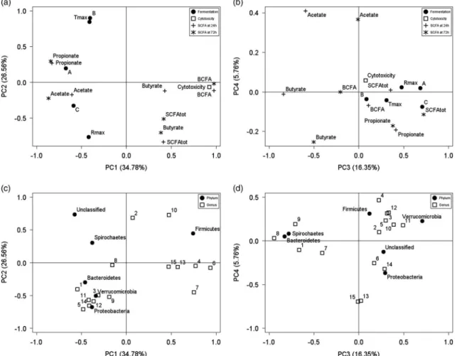
Principal component analysis of the influence of the
ingredient and the reducing agent
A PCA was performed based on the correlation matrix from
the complete data set (Figures 2 and 3). Thefirst two
prin-cipal components (PC1 to PC2), which explained 61.3% of the variability in the data set, clearly discriminated samples according to the ingredient. High values along PC1 reflected high degrees of cytotoxicity of casein, combined to high BCFA production that was counterbalanced by reduced acetate and propionate ratios compared with the other ingredients. PC2 mainly discriminated ingredients according
to the fermentation kinetics measured throughB,Tmaxand
Rmax. Similarly, PC3 was mainly influenced by the ingredient
and explained 16.3% of the observed variability. High
pro-portion ofBacteroidetesafter 24 h for soybean proteins was
translated into high negative scores along PC3. PC4, accounting for 5.8% of the variance, showed a separation according to the reducing agent for casein and potato starch.
High proportions of Fusobacterium and Escherichia, as
observed when casein was fermented with cysteine-HCl, were associated with low values along PC4. Similarly, potato starch fermented with cysteine-HCl showed a high
propor-tion of Succinivibrio, leading to a negative value along
PC4 whereas a positive value was obtained with Na2S
or the control. Discussion
Comparing fermentation kinetics, SCFA production,
cyto-toxicity of the fermentation products andfinally the
micro-biota fermenting several contrasted ingredients under three
different reducing conditions (with cysteine-HCl, with Na2S,
without any reducing agent) showed that the different reducing agents accounted for a limited part of the varia-bility. Differences in the investigated response variables that
can be obtained from suchin vitromodels were by far more
influenced by the ingredient than the reducing agent since
the three first PCs accounted for more than 75% of the
variability. This was highlighted by the low share of varia-bility (5.8%) explained by PC4 in the PCA and the fact that clusters grouped the different ingredients whatever the
reducing agent along thefirst three PCs. Looking more
spe-cifically at individual response variables, final fermentability
of the ingredients as measured through total gas production
(A) was not influenced by the reducing agent added to the
fermentation broth. Nonetheless, Morgan et al. (2004)
observed with some feed ingredients a negative impact of 0 20 40 60 80 100 Triton Caco-2 DMEM Cys Na2S Control Cys Na2S Control Cys Na2S Control Cys Na2S Control Cys Na2S Control
Blank Potato starch Casein Cellulose Soybean proteins
Cytotoxicity (%LDH release)
Figure 1 Cytotoxic effects on intestinal mucosa upon exposure of Caco-2 cells to fermentation supernatants (initial dilution) of the four tested ingredients and blank of fermentation in three incubation media: with Na2S or cysteine-HCl (Cys) or control without reducing agent. Comparison with incubation
medium alone (negative reference) or supplemented with 1% Triton X-100 (positive reference). The cytotoxicity was determined by assaying the lactate dehydrogenase (LDH) activity released in the culture medium and is given as means ± SD (n= 8). Values are representative of two independent experiments. The horizontal discontinuous line represents the threshold of toxicity.
the use of cysteine-HCl on rumen microbes activityin vitro, which were reflected in this study by changes in fermentation kinetics. The addition of cysteine-HCl to the buffer solution
reduced the maximum rate of gas production (Rmax) for the
carbohydrate ingredients. The second reducing agent (Na2S)
did not influence the fermentation parameters for any of the
tested ingredients. Casein and potato starch fermented faster than soybean proteins and in turn, soybean proteins fermented faster than cellulose. This is explained, in the absence of any enzymatic pre-digestion of the ingredients, by differences in solubility of the nutrients and water-holding capacity of the polymers.
Although the total SCFA production was not influenced by
the reducing agent for casein and potato starch, the molar ratio of propionate was higher when cysteine-HCl was used by comparison with the control devoid of reducing agent. Higher propionate was counterbalanced by a reduced ratio of acetate and is explained by the higher proportion of
Proteobacteria for casein and potato starch fermentation
with cysteine-HCl since someProteobacteriaare specifically
involved in propionate production (e.g. Succinivibrio
dextrinosolvens) (Watanabeet al., 2010).
From a phylum perspective, the microbiota, dominated by
Firmicutes (77.6 ± 11.19%) followed by Bacteroidetes
(10.6 ±5.94%), was in agreement with Guoet al. (2008) and
Kim et al. (2011), regardless of the ingredient and the
reducing agent. The domination of the bacterial communities after 24 h of fermentation by these two phyla, composed
principally of anaerobes, confirms the strict anaerobic
con-ditions in all fermentation bottles, including in the controls devoid of reducing agent. Differences in microbial com-munities and SCFA profiles between the three reducing conditions after 24 h were more important with ingredients for which the fermentation started earlier, namely casein and
potato starch. This means that although the influence of the
reducing agent is less important than that of the ingredient,
it significantly affects the fermentation pathways and
bac-teria that are growing in the broth, especially when sub-strates are being metabolized intensively by the microbial communities. So for casein and potato starch, the proportion
ofProteobacteriawas considerably higher when cysteine-HCl
was used as reducing agent. This was ascribed to a
pro-liferation of theEscherichiaandSuccinivibriogenera for the
fermentation of casein and potato starch, respectively.
Moreover among theFusobacteriaphylum, the proportion of
theFusobacteriumgenus was increased for the fermentation
of casein in the presence of cysteine-HCl in the fermentation broth. These three genera are presumed to have the capacity
to degrade cysteine-HCl. Indeed, Escherichia are able to
degrade cysteine-HCl by means of specific desulfhydrases
(Awano et al., 2005). Gomez-Alarcon et al. (1982) studied
the nutrient requirements ofS. dextrinosolvensand reported
that the addition of cysteine to the culture medium promotes its growth, leading to increased propionate production as
mentioned before. Regarding the Fusobacterium genus,
F. nucleatum, found in the oral cavity and in infected sites of
healthy and sick people, was also reported as able to
Table 3 Distribution of different phyla in the bacterial population (%) after 24 h of fermentation of the four tested ingredients and blank of fermentation by pig fecal bacteria in three incubation media: with Na 2 S or cysteine-HCl (Cys) or control (Ctrl) without reducing agent (n = 2) Casein Soybean proteins Cellulose Potato starch Blank P-values Phylum Cys Na 2 S Ctrl Cys Na 2 S Ctrl Cys N a2 S Ctrl Cys Na 2 S Ctrl Cys N a2 S C trl SEM IR I× R Bacteroidetes 5.39 d 6.03 cd 5.38 d 20.0 a 20.3 a 22.2 a 7.88 c 7.43 cd 7.36 cd 13.5 b 12.6 b 13.3 b 5.26 d 6.46 cd 6.46 cd 1.08 < 0.001 0.583 0.641 Firmicutes 87.3 a 89.0 a 91.2 a 71.8 cd 72.4 cd 70.3 d 76.6 bc 79.2 b 78.8 b 47.5 e 71.7 cd 69.7 d 87.5 a 85.5 a 86.0 a 2.04 < 0.001 < 0.001 < 0.001 Proteobacteria 2.77 c 0.330 d 0.348 d 2.15 cd 1.23 cd 0.958 cd 0.512 d 0.466 d 0.462 d 31.8 a 10.7 b 9.83 b 0.793 cd 0.674 cd 0.791 cd 1.49 < 0.001 < 0.001 < 0.001 Spirochaetes 0.220 ab 0.179 ab 0.142 b 0.638 a 0.527 ab 0.614 a 0.335 ab 0.524 ab 0.287 ab 0.151 b 0.280 ab 0.313 ab 0.153 b 0.318 ab 0.313 ab 0.04 0.041 0.801 0.945 Verrucomicrobia 0.176 bc 0.286 bc 0.219 bc 0.108 c 0.101 c 0.097 c 0.276 bc 0.284 bc 0.183 bc 0.358 ab 0.524 a 0.497 a 0.205 bc 0.366 ab 0.364 ab 0.03 < 0.001 0.173 0.703 Unclassi fied 3.23 bc 3.88 bc 2.37 c 5.15 bc 5.34 bc 5.66 bc 14.1 a 11.9 a 12.7 a 6.57 b 3.92 bc 6.29 b 5.84 bc 6.33 b 5.83 bc 0.66 < 0.001 0.63 0.725 Others 1 0.871 a 0.320 bc 0.366 b 0.203 bcd 0.140 bcd 0.117 bcd 0.222 bcd 0.189 bcd 0.212 bcd 0.0578 d 0.181 bcd 0.0701 cd 0.224 bcd 0.336 b 0.264 bcd 0.04 < 0.001 0.15 0.036 I= ingredient; R = reducing agent. a,b,c,d Values withi n a row with different supe rscripts differ signi ficantly at P < 0.05. 1Others bacteria incl ude Actino bacteri a, Fusobact eria, Lentisp haerae, Synergiste tes, Candida te_d ivision_TM7, Fibrobacteres.
degrade cysteine with specific desulfhydrases (Fukamachi
et al., 2002).
Using culture-independent molecular techniques, some studies demonstrated that dietary changes affect the complex microbiota of the gastro-intestinal tract and lead
to shifts in bacterial communities (Leser et al., 2000;
Pieper et al., 2009b). So the higher differences in phyla
according to the ingredient than the reducing agent are not really surprising. For example, potato starch led to higher
proportions ofProteobacteria as compared with the other
ingredients that could be attributed mainly toSuccinivibrio,
since its proliferation in the rumen was associated to a
high-starch diet (O’Herrin and Kenealy, 1993). Also,Bacteroidetes
were more abundant with potato starch and soybean proteins than in the blanks devoid of ingredients. This observation can be explained by the requirements in
polysaccharides of the Bacteroidetes. In the human large
intestine, the stability and coexistence of closely related members in this phylum was shown to be based on an synergetic interaction network related to the breakdown and utilization of dietary polysaccharides (Rakoff-Nahoum
et al., 2014).
Results from the Caco-2 assay indicate that the reducing agent incorporated in the fermentation broth has a negligible impact on intestinal cytotoxicity of the fermentation super-natants. Despite their colorectal origin, Caco-2 cells were
proven a valuable in vitro model of the human and pig
intestinal epithelium to assess toxicity of fermentation
pro-ducts (Arturssonet al., 2012). Indeed, differences in
ingre-dients were observed comforting the relevancy of the test. The higher cytotoxicity of casein can be ascribed to its fast fermentation and by the production of toxic metabolites by
the bacterial population such as ammonia, H2S, thiols and
amines, which are usually produced by protein fermenting
bacteria (Hugheset al., 2000).
Conclusion
It can be concluded that in vitro intestinal fermentation
models used in pig studies to investigate shifts in microbial fermentation and communities should avoid the use of
cysteine-HCl as reducing agent. The addition of Na2S
seems useless if appropriate CO2 saturation is realized
and, since it probably blunts the ability to detect some Table 4 Distribution of different genera in the bacterial population (%) after 24 h of fermentation of the four tested ingredients and blank of fermentation by pig fecal bacteria in three incubation media: with Na2S or cysteine-HCl (Cys) or control (Ctrl) without reducing agent (n= 2)
Phylum Firmicutes Genus Streptococcus spp. Mogibacterium spp. Blautia spp. Lachnospiraceae i.s. Roseburia spp. Dorea spp. Lachnospiraceae uncl. Ruminococcaceae uncl. Casein Cys 0.130b 0.281b 0.0553d 6.12a 0.000c 4.12a 22.4b 14.3c Na2S 0.0355b 2.97a 0.0484d 4.71b 0.000c 2.47b 20.6bc 15.9abc Ctrl 0.0796b 2.81a 0.135cd 4.82b 0.000c 2.53b 19.7bcd 17.6ab Soybean proteins Cys 0.0316b 0.000b 0.217c 4.81b 0.339b 0.509c 29.0a 8.56de Na2S 0.0854b 0.000b 0.105cd 4.27b 0.628a 0.211c 28.4a 10.3d Ctrl 0.0328b 0.0156b 0.132cd 4.06bc 0.605a 0.229c 30.0a 9.96d Cellulose Cys 0.121b 0.0489b 0.0827cd 1.50f 0.000c 0.438c 17.2def 15.0bc Na2S 0.124b 0.0242b 0.0644cd 2.54de 0.000c 0.213c 18.6cdef 18.3a Ctrl 0.0598b 0.000b 0.148cd 1.77ef 0.000c 0.198c 18.0cdef 16.6abc Potato starch Cys 0.154b 0.0156b 0.536b 3.29cd 0.0458c 0.346c 19.7bcde 6.09e Na2S 3.90a 0.0193b 1.16a 3.32cd 0.000c 0.340c 27.5a 9.23de Ctrl 1.03b 0.0109b 1.13a 3.39cd 0.0416c 0.0591c 28.8a 9.29de Blank Cys 0.256b 0.0127b 0.0641cd 1.48f 0.000c 0.628c 17.0def 14.6bc Na2S 0.0705b 0.0285b 0.0856cd 1.62f 0.000c 0.112c 16.6ef 16.2abc Ctrl 0.125b 0.0376b 0.101cd 1.46f 0.0125c 0.239c 16.0f 15.0bc SEM 0.192 0.186 0.0677 0.275 0.0406 0.222 0.959 0.715 P-values I <0.001 <0.001 <0.001 <0.001 <0.001 <0.001 <0.001 <0.001 R 0.024 0.004 0.002 0.191 0.108 0.016 0.084 0.008 I× R 0.002 <0.001 <0.001 0.038 0.026 0.258 0.002 0.775
i.s.= incertae sedis; uncl. = unclassified; I = ingredient; R = reducing agent.
protein fermentation metabolites, it can easily be omitted. Finally, when fermentation broths are applied to Caco-2 cells culture to simulate the effect of bacterial metabolites on the
intestinal epithelium, the impact of the ingredient is detect-able regardless of the reducing agent used, as for most of the response variables measured in the present study.
Table 5Distribution of different genera in the bacterial population (%) after 24 h of fermentation of the four tested ingredients and blank of fermentation by pig fecal bacteria in three incubation media: with Na2S or cysteine-HCl (Cys) or control (Ctrl) without reducing
agent (n= 2)
Phylum Firmicutes Bacteroidetes Fusobacteria Proteobacteria
Genus Veillonellaceae uncl. Erysipelotrichaceae i.s. Xylanibacter spp. RF16 uncl. Fusobacterium spp. Succinivibrio spp. Escherichia spp. Casein Cys 0.342cde 0.0284c 0.0284cd 0.136bcd 0.430a 0.0134c 1.96a Na2S 0.276cde 0.0129c 0.0387cd 0.291abc 0.0258b 0.0484c 0.0970d Ctrl 0.189de 0.0324c 0.0148d 0.130cd 0.0176b 0.0296c 0.0769d Soybean proteins Cys 0.144e 0.0136c 0.443b 0.126cd 0.0452b 1.16c 0.516b Na2S 0.158e 0.000c 0.498ab 0.124cd 0.000b 0.960c 0.112d Ctrl 0.183de 0.0156c 0.628a 0.0955cd 0.000b 0.644c 0.0830d Cellulose Cys 0.389cd 0.0225c 0.0905cd 0.177bcd 0.0152b 0.0529c 0.257bcd Na2S 0.273cde 0.0159c 0.111cd 0.506a 0.000b 0.0523c 0.161d Ctrl 0.327cde 0.0190c 0.136cd 0.243bc 0.000b 0.163c 0.104d Potato starch Cys 0.483c 0.106c 0.513ab 0.0313d 0.0144b 28.9a 0.177cd Na2S 1.23a 1.20a 0.188c 0.0265d 0.000b 9.08b 0.232cd Ctrl 1.01b 0.648b 0.438b 0.145bcd 0.000b 8.24b 0.138d Blank Cys 0.218de 0.0962c 0.0641cd 0.224bc 0.0193b 0.0509c 0.429bc Na2S 0.186de 0.0437c 0.0875cd 0.475a 0.000b 0.122c 0.234cd Ctrl 0.251de 0.0754c 0.113cd 0.351ab 0.000b 0.163c 0.201cd SEM 0.0586 0.0600 0.0393 0.0294 0.0212 1.38 0.0861 P-values I <0.001 <0.001 <0.001 0.001 0.002 <0.001 <0.001 R 0.071 <0.001 0.086 0.020 0.003 <0.001 <0.001 I× R 0.001 <0.001 0.045 0.232 0.003 <0.001 <0.001
uncl.= unclassified; i.s. = incertae sedis; I = ingredient; R = reducing agent.
a,b,c,d,eValues within a column with different superscripts differ significantly atP<0.05.
Figure 2 Score plot from thefirst four principal components (PC1 to PC4). Different symbols indicate the scores of the four ingredients according to the reducing agent incorporated in the fermentation broth: Na2S, cysteine-HCl (Cys) or control without reducing agent (control). Symbols are: = Casein;
Acknowledgments
The project was supported by the Fund for Scientific Research (FNRS, Brussels, Belgium – Research Credit 1.5180.12). The research was performed in the framework of the collaborative Food4Gut excellence research program of the Walloon Govern-ment. The results were partially presented at the 12thInternational Symposium on Digestive Physiology of Pigs in Keystone, CO, USA, 29 May 29–1 June 2012 and published in the Supplement 4 (Volume 90, 2012) of theJournal of Animal Science.
Supplementary material
To view supplementary material for this article, please visit https://doi.org/10.1017/S1751731117002749
References
Altschul SF, Gish W, Miller W, Myers EW and Lipman DJ 1990. Basic local alignment search tool. Journal of Molecular Biology 215, 403–410.
Andriamihaja M, Davila AM, Eklou-Lawson M, Petit N, Delpal S, Allek F, Blais A, Delteil C, Tomé D and Blachier F 2010. Colon luminal content and epithelial
cell morphology are markedly modified in rats fed with a high-protein diet. American Journal of Physiology - Gastrointestinal and Liver Physiology 299, G1030–G1037.
Artursson P, Palm K and Luthman K 2012. Caco-2 monolayers in experimental and theoretical predictions of drug transport. Advanced Drug Delivery Reviews 64, 280–289.
Attene-Ramos MS, Wagner ED, Plewa MJ and Gaskins HR 2006. Evidence that hydrogen sulfide is a genotoxic agent. Molecular Cancer Research 4, 9–14. Awano N, Wada M, Mori H, Nakamori S and Takagi H 2005. Identification and functional analysis of Escherichia coli cysteine desulfhydrases. Applied and Environmental Microbiology 71, 4149–4152.
Bindelle J, Buldgen A, Wavreille J, Agneessens R, Destain JP, Wathelet B and Leterme P 2007. The source of fermentable carbohydrates influences thein vitro protein synthesis by colonic bacteria isolated from pigs. Animal 1, 1126–1133. Bindelle J, Pieper R, Montoya CA, Van Kessel AG and Leterme P 2011. Nonstarch polysaccharide-degrading enzymes alter the microbial community and the fer-mentation patterns of barley cultivars and wheat products in anin vitromodel of the porcine gastrointestinal tract. FEMS Microbiology Ecology 76, 553–563. Cardarelli HR, Martinez RCR, Albrecht S, Schols H, BDGM Franco, SMI Saad and Smidt H 2016.In vitrofermentation of prebiotic carbohydrates by intestinal microbiota in the presence ofLactobacillus amylovorusDSM 16998. Beneficial Microbes 7, 119–133.
Christl SU, Murgatroyd PR, Gibson GR and Cummings JH 1992. Production, metabolism, and excretion of hydrogen in the large intestine. Gastroenterology 102, 1269–1277.
Figure 3 Loading plot from thefirst four principal components (PC1 to PC4) describing the relationships between fermentation parameters, short-chain fatty acids (SCFA) production after 24 and 72 h of fermentation, cytotoxicity (a and b) and microbial composition (c and d). A= maximal gas volume; B= time to reach 50% of A; C = constant; Rmax= maximum rate of gas production; Tmax= time to reach Rmax; SCFAtot= total SCFA production;
BCFA= branched-chain fatty acids; 1 = Xylanibacter spp.; 2 = RF16 unclassified; 3 = Streptococcus spp.; 4 = Mogibacterium spp.; 5 = Blautia spp.; 6= Dorea spp.; 7 = Lachnospiraceae Incertae Sedis; 8 = Roseburia spp.; 9 = Lachnospiraceae unclassified; 10 = Ruminococcaceae unclassified; 11= Veillonellaceae unclassified; 12 = Erysipelotrichaceae Incertae Sedis; 13 = Fusobacterium spp.; 14 = Succinivibrio spp.; 15 = Escherichia spp.
Cremin JD, Fitch MD and Fleming SE 2003. Glucose alleviates ammonia-induced inhibition of short-chain fatty acid metabolism in rat colonic epithelial cells. American Journal of Physiology - Gastrointestinal and Liver Physiology 285, G105–G114.
Fukamachi H, Nakano Y, Yoshimura M and Koga T 2002. Cloning and char-acterization of the l-cysteine desulfhydrase gene ofFusobacterium nucleatum. FEMS Microbiology Letters 215, 75–80.
Fukushima RS, Weimer PJ and Kunz DA 2003. Use of photocatalytic reduction to hasten preparation of culture media forsaccharolytic Clostridiumspecies. Brazilian Journal of Microbiology 34, 22–26.
Gomez-Alarcon RA, O’Dowd C, Leedle JA and Bryant MP 1982. 1,4-Naphthoquinone and other nutrient requirements of Succinivibrio dextrinosolvens. Applied and Environmental Microbiology 44, 346–350. Groot JCJ, Cone JW, Williams BA, Debersaques FMA and Lantinga EA 1996. Multiphasic analysis of gas production kinetics forin vitrofermentation of ruminant feeds. Animal Feed Science and Technology 64, 77–89.
Guo X, Xia X, Tang R, Zhou J, Zhao H and Wang K 2008. Development of a real-time PCR method forFirmicutesandBacteroidetesin faeces and its application to quantify intestinal population of obese and lean pigs. Letters in Applied Microbiology 47, 367–373.
Hughes R, Magee EA and Bingham S 2000. Protein degradation in the large intestine: relevance to colorectal cancer. Current Issues in Intestinal Micro-biology 1, 51–58.
Jha R, Woyengo TA, Li J, Bedford MR, Vasanthan T and Zijlstra RT 2015. Enzymes enhance degradation of thefiber–starch–protein matrix of distillers dried grains with solubles as revealed by a porcinein vitrofermentation model and microscopy. Journal of Animal Science 93, 1039–1051.
Kim HB, Borewicz K, White BA, Singer RS, Sreevatsan S, Tu ZJ and Isaacson RE 2011. Longitudinal investigation of the age-related bacterial diversity in the feces of commercial pigs. Veterinary Microbiology 153, 124–133.
Koecher KJ, Noack JA, Timm DA, Klosterbuer AS, Thomas W and Slavin JL 2014. Estimation and interpretation of fermentation in the gut: coupling results from a 24 h batchin vitrosystem with fecal measurements from a human intervention feeding study using fructo-oligosaccharides, inulin, gum acacia, and peafiber. Journal of Agricultural and Food Chemistry 62, 1332–1337.
Kumagai H, Sejima S, Choi Y, Tanaka H and Yamada H 1975. Crystallization and properties of cysteine desulfhydrase from Aerobacter aerogenes. FEBS Letters 52, 304–307.
Leser TD, Lindecrona RH, Jensen TK, Jensen BB and Møller K 2000. Changes in bacterial community structure in the colon of pigs fed different experimental diets and after infection with Brachyspira hyodysenteriae. Applied and Environ-mental Microbiology 66, 3290–3296.
Medani M, Collins D, Docherty NG, Baird AW, O’Connell PR and Winter DC 2011. Emerging role of hydrogen sulfide in colonic physiology and patho-physiology. Inflammatory Bowel Diseases 17, 1620–1625.
Menke KH and Steingass H 1988. Estimation of the energetic feed value obtained from chemical analysis andin vitrogas production using rumenfluid. Animal Research and Development 28, 7–55.
Morgan R, Kliem KE and Mould FL 2004. The use of a nitrogen free medium for in vitrofermentation studies. Paper presented at the British Society of Animal Science Annual Meeting, 5–7 April, York, UK, p. 232.
O’Herrin SM and Kenealy WR 1993. Glucose and carbon dioxide metabolism by Succinivibrio dextrinosolvens. Applied and Environmental Microbiology 59, 748–755.
Payne AN, Zihler A, Chassard C and Lacroix C 2012. Advances and perspectives in in vitrohuman gut fermentation modeling. Trends in Biotechnology 30, 17–25.
Pieper R, Bindelle J, Rossnagel B, Van Kessel A and Leterme P 2009a. Effect of carbohydrate composition in barley and oat cultivars on microbial ecophysiology and proliferation ofSalmonella entericain anin vitromodel of the porcine gastrointestinal tract. Applied and Environmental Microbiology 75, 7006–7016. Pieper R, Janczyk P, Urubschurov V, Korn U, Pieper B and Souffrant WB 2009b. Effect of a single oral administration of Lactobacillus plantarum DSMZ 8862/ 8866 before and at the time point of weaning on intestinal microbial com-munities in piglets. International Journal of Food Microbiology 130, 227–232. Pruesse E, Quast C, Knittel K, Fuchs BM, Ludwig W, Peplies J and Glöckner FO 2007. SILVA: a comprehensive online resource for quality checked and aligned ribosomal RNA sequence data compatible with ARB. Nucleic Acids Research 35, 7188–7196.
Rakoff-Nahoum S, Coyne MJ and Comstock LE 2014. An ecological network of polysaccharide utilization among human intestinal symbionts. Current Biology 24, 40–49.
Rodriguez C, Taminiau B, Korsak N, Avesani V, Van Broeck J, Brach P, Delmée M and Daube G 2016. Longitudinal survey of Clostridium difficile presence and gut microbiota composition in a Belgian nursing home. BMC Microbiology 16, 229. Roediger WE, Moore J and Babidge W 1997. Colonic sulfide in pathogenesis and treatment of ulcerative colitis. Digestive Diseases and Sciences 42, 1571–1579. Sappok M, Pellikaan WF, Verstegen MW and Sundrum A 2009. Assessing fibre-rich feedstuffs in pig nutrition: comparison of methods and their potential implications. Journal of the Science of Food and Agriculture 89, 2541–2550.
Schloss PD, Westcott SL, Ryabin T, Hall JR, Hartmann M, Hollister EB, Lesniewski RA, Oakley BB, Parks DH, Robinson CJ, Sahl JW, Stres B, Thallinger GG, Van Horn DJ and Weber CF 2009. Introducing mothur: open-source, platform-independent, community-supported software for describing and comparing microbial communities. Applied and Environmental Microbiology 75, 7537–7541. Su Y, Bian G, Zhu Z, Smidt H and Zhu W 2014. Early methanogenic colonisation in the faeces of Meishan and Yorkshire piglets as determined by pyrosequencing analysis. Archaea 2014, 547908.
Theodorou MK, Williams BA, Dhanoa MS, McAllan AB and France J 1994. A simple gas production method using a pressure transducer to determine the fermentation kinetics of ruminant feeds. Animal Feed Science and Technology 48, 185–197.
Tilley JMA and Terry RA 1963. A two-stage technique for thein vitrodigestion of forage crops. Grass and Forage Science 18, 104–111.
Tran THT, Boudry C, Everaert N, Théwis A, Portetelle D, Daube G, Nezer C, Taminiau B and Bindelle J 2015. Adding mucins to anin vitrobatch fermen-tation model of the large intestine induces changes in microbial population isolated from porcine feces depending on the substrate. FEMS Microbial Ecology (, https://doi.org/10.1093/femsec/fiv165, Published online by Oxford University Press 20 December 2015.
Watanabe Y, Suzuki R, Koike S, Nagashima K, Mochizuki M, Forster RJ and Kobayashi Y 2010.In vitroevaluation of cashew nut shell liquid as a methane-inhibiting and propionate-enhancing agent for ruminants. Journal of Dairy Science 93, 5258–5267.
Weiss E, Rist V, Rink F and Mosenthin R 2015. Discrepancies in porcine micro-biota composition betweenin vivomeasurements andin vitrofermentation with the modified Hohenheim Gas Test. Livestock Science 176, 141–145.