Université de Montréal
The human organic cation transporter OCT1 mediates
high affinity uptake of the anticancer drug daunorubicin
Emil Andreev
Microbiology, Infectious Diseases and Immunology, Université de Montréal Faculty of Medicine
A thesis submitted in fulfill of the requirements of the degree
Magister Scientiae (M. SC.)
Microbiology and Immunology
May,2016
Université de Montréal
Faculté des études supérieures et postdoctorales
The human organic cation transporter OCT1 mediates
high affinity uptake of the anticancer drug daunorubicin
Emil Andreev
Thesis examiners:
Dr. Elliot Drobetsky, president of the jury Dr. Dindial Ramotar, thesis supervisor Dr. Janos Filep, member of the jury
i
Résumé
Les anthracyclines tels que la doxorubicin et la daunorubicin sont une famille de médicaments anticancéreux hydrophiles qui doivent être transportés dans les cellules afin d’exercer leur action par intercalation à l’ADN dans le noyau cellulaire. Ceci mène à la perturbation du métabolisme de l’ADN et entraine la mort cellulaire. Les anthracyclines sont utilisés pour le traitement d’une variété de cancers incluant la leucémie, les lymphomes, le cancer du sein, le cancer des poumons et le cancer des ovaires. Étant donné que le transport actif des anthracyclines dans les cellules a partiellement été démontré, le transporteur spécifique impliqué dans ce processus n’est pas encore connu. En utilisant un modèle de cancer des ovaires, la lignée cellulaire TOV2223G, nous avons démontré que des substrats spécifiques au transporteur de cations organiques 1 (OCT1), notamment la ergothionéine, la thiamine et la phenformin, ont partiellement inhibé l’absorption de la daunorubicin en différence de la carnitine qui est un substrat de haute affinité des transporteurs CT2 et OCTN2. Ces résultats suggèrent que les transporteurs organiques spécifiques au transport de la carnitine ne sont pas impliqués dans le transport des anthracyclines. Ainsi, nos résultats ont démontré que l’absorption de la daunorubicin est orchestrée par le transporteur OCT1 dans les cellules TOV2223G (Km ~ 5 μM) et des concentrations micromolaires de choline ont complètement abolies l’absorption de la drogue. De plus, un ARN sh dirigé contre OCT1 a réprimé son expression protéique, ce qui a été confirmé par la technique d’immuno-buvardage en utilisant un anti-OCT1 anticorps. Les cellules déficientes en OCT1 n’ont pas été capables d’absorber la daunorubicin et ont été plus résistantes à l’action de la drogue par rapport aux cellules contrôle. La transfection des cellules HEK293T avec un plasmide construit de façon à faire exprimer OCT1 comme protéine de fusion avec la protéine fluorescente EYFP a montré que celle-ci est localisée dans la membrane plasmique. Les cellules transfectées ont été capables d’absorber cinq fois plus de daunorubicin comparé aux cellules contrôles. Cette étude est, selon nous, la première à démontrer que OCT1 est un transporteur de haute affinité des anthracyclines. Ainsi, nous avons émis l’hypothèse que des défauts de OCT1 peuvent contribuer à l’efficacité de la réponse des cellules cancéreuses à la chimiothérapie avec les anthracyclines.
ii
iii
Abstract
Anthracyclines such as doxorubicin and daunorubicin are hydrophilic anticancer agents that must be transported into cells. These drugs accumulate in the nucleus where they intercalate with DNA, thereby interfering with DNA replication in turn leading to cell death. Anthracyclines are used for treating a variety of cancers including leukemia, lymphomas, breast, lung, and ovarian. Despite evidence for active uptake of anthracyclines, the specific transporter has not been identified. Using the ovarian cancer cell line TOV2223G, we show that substrates reported for the organic cation transporter OCT1, such as ergothioneine, thiamine and phenformin, partially compete with uptake of daunorubicin, but not of L-carnitine, i.e., a high affinity substrate transported by hCT2 and OCTN2. These findings exclude the involvement of the L-carnitine organic cation family of transporters in anthracycline uptake. Moreover, we show that OCT1 actively mediates high affinity (Km ~ 5 μM) transport of daunorubicin into TOV2223G cells, whereas micromolar amounts of choline completely abolish drug uptake. shRNA-mediated downregulation of OCT1 causes defective uptake of daunorubicin, as well as significant resistance to the drug, as compared to the vector control. Transfection of HEK293T cells with a plasmid expressing OCT1 as a GFP fusion protein revealed that OCT1-EYFP was predominantly localized to the plasma membrane. These transfected cells manifested nearly 5-fold increased uptake of daunorubicin compared to the empty vector control. In summary, we show for the first time that human OCT1 is a high affinity transporter for anthracyclines. As such, we postulate that OCT1 status represents a critical determinant in the response of cancer cells to chemotherapy with anthracyclines
iv
Table of contents
Résumé ... i
Abstract ... iii
Table of contents ... iv
List of tables ... vii
List of figures ... viii
Abbreviations ... ix
Acknowledgments... xii
Chapter 1: Introduction ... 13
1. Chemotherapy and DNA-damaging agents ... 13
1.1. Anthracyclines ... 14
1.2. Anthracyclines as topoisomerase inhibitors... 15
1.3. Anthracyclines and ROS formation ... 15
1.4. Anthracyclines and cardiotoxicity ... 16
1.5. Novel generations of Anthracyclines ... 16
2. Transporters of Anthracyclines ... 18
2.1. ATP-binding cassette family (ABC) ... 18
2.1.1. The ABCB subfamily transporters... 18
2.1.2. The ABCC subfamily transporters... 19
2.1.3. The ABCG subfamily transporters ... 19
2.2. Mechanism of transport by ABC transporters ... 20
3. Organic cation transporters (OCTs and OCTNs) ... 21
3.1. OCT1... 22
3.1.1. OCT1 as metformin transporter ... 22
3.1.2. OCT1 as thiamine transporter ... 23
3.1.3. OCT1 as donepezil transporter ... 23
v
3.1.5. OCT1 as transporter for anticancer drugs ... 24
3.2. OCT2... 25
3.2.1. OCT2 as metformin transporter ... 26
3.2.2. OCT2 role in Parkinson’s disease ... 26
3.2.3. OCT2 as transporter for anticancer drugs ... 27
3.3. OCT3... 28
3.3.1. OCT3 as transporter for anticancer drugs ... 29
3.4. OCTN1 ... 29
3.4.1. OCTN1 as transporter for anticancer drugs ... 30
3.5. OCTN2 ... 31
3.5.1. OCTN2 as transporter for anticancer drugs ... 33
3.6. OCTN3 ... 33
3.7. CT2 (OCT6) ... 34
3.7.1. CT2 as anthracycline transporter ... 35
4. Organic anion transporters ... 38
5. Hypothesis and Objectives ... 41
Chapter 2. The human organic cation transporter OCT1 mediates high affinity uptake of the anticancer drug daunorubicin ... 42
ABSTRACT ... 44
INTRODUCTION ... 45
RESULTS ... 48
DISCUSSION ... 56
ACKNOWLEDGMENTS: ... 62
MATERIALS AND METHODS ... 63
REFERENCES ... 70
FIGURE LEGENDS ... 74
SUPPLEMENTAL DATA: ... viii
FIGURE LEGENDS TO SUPPLEMENTAL DATA ... ix
Chapter 3: Discussion ... xv
vi
vii
List of tables
Table 1. SLC22 organic cations/carnitine transporters Table 2. SLC22 organic anion transporters
viii
List of figures
Figure 1. Chemical structure of DOX and DNR
Figure 2. Chemical structure of various anthracyclines derivatives Figure 3 Schematic representation of the ABC transporters
Figure 4. Schematic representation of the OCTs and OCTNs transporters Figure 5. Schematic representation of the OATs and OATPs transporters
ix
Abbreviations
A
ABC- ATP-binding cassette AML- acute myeloid leukemia
B
BCRP- breast cancer resistance protein
C
CALYPSO- Caelyx in Platinum Sensitive Ovarian patients
C6–NBD–PC- Ethanamimium 2-hydroxy-C6 -N,N-dimethyl-N-[2-N-(2,1,3-benzoxadiazol-4-amine,-N-methyl,-7- nitro)-ethyl] bromide- phosphatidylcholine
CD- Carboplatin/cisplatin and paclitaxel cDNA- complementary DNA
CHP- cyclo (his-pro)
CHT1- Choline transporter 1 CT1, 2- Carnitine transporter 1, 2 CTL-1- Choline transporter-like 1 cRNA- complementary RNA
D
DAT- Dopamine transporter DOX- Doxorubicin
DNR- Daunorubicin DSB- double strand break DRG- dorsal root ganglion
x
E
EMA- European Medicines Agency
EMT- Extra neuronal monoamine transporter ETT- Ergothineine transporter
EYFP- Enhanced yellow fluorescent protein
F
FACS- Fluorescence Activated Cell Sorting FDA- Food and Drug Administration
G
GAT2- GABA transporter 2/ sodium- and chloride-dependent GABA transporter 2
H
HFN-4α- Hepatocyte nuclear factor 4 alpha
M
MDR- Multidrug resistance
MFS- Major Facilitator Superfamily 1-MPP- 1-methyl-4-phenylpyridinium MRP- Multidrug resistance protein MXR- Multixenobiotic resistance
N
NAPDH- Nicotinamide adenine dinucleotide phosphate
NBD-choline- Ethanamimium 2-hydroxy- N,N-dimethyl-N-[2-N-(2,1,3-benzoxadiazol-4-amine,-N-methyl,-7- nitro)-ethyl] bromide- choline
NCI- National Cancer Institute NKT- Novel kidney transporter
xi OAT- Organic anion transporter
OC- Organic cations
OCT- Organic cation transporter
OCTN- Novel organic cation/zwitterion transporter
P
PAH- p-aminohippuric acid PD- PEGylated DOX PFA- paraformaldehyde
R
RT-qPCR- Quantitative reverse transcription polymerase chain reaction
S
SAL- Salsolinol SLC- Solute carrier
SNc- substantia nigra pars compacta
T
TCGA- The Cancer Genome Atlas TEA- Tetraethyl ammonium
xii
Acknowledgments
First and foremost, I would like to thank my supervisor Dr. Dindial Ramotar for his mentorship and constant support throughout my master’s degree study. Dr. Ramotar has given me the opportunity to be a part of his team and to develop a passion in science and in cancer research. I also would like to thank our research assistant Tara Harihar and all lab members for their help, guidance and also for their enjoyable company. I want to thank Nicolas Brosseau for his devoted work and contribution in OCT1 research. I want to thank Dr. Euridice Carmona and Dr. Anne-Marie Mes-Masson for their expertise and contribution in completing the present thesis.
Chapter 1: Introduction
1. Chemotherapy and DNA-damaging agents
Cancer is a complex disease that is characterized by deregulated cell proliferation caused by various factors including genetic and environmental (1). Currently, cancer is responsible for more than seven million deaths worldwide according to statistics from the World Health Organization. The battle with cancer has been led for more than four decades as all began with the War on Cancer with the President Nixon’s National Cancer Act of 1971 (2). Despite the increasing success of recently introduced novel cellular therapies, such as hematopoietic stem cell transplantations, treatment of cancer is still widely based on the use of chemotherapeutic agents to eradicate cancer cells, to reduce tumor growth, and to ameliorate patient’s quality of life. During the World War I and II, when studying victims exposed to sulfur mustard gas, important discoveries were made using mice as a model. By injecting stable nitrogen mustard compounds, it was observed an important tumor regression. Moreover, leukemia patients treated with folic acid showed increased proliferation of acute lymphoblastic leukemia cells (1). This has led to the discovery of folate analogs such as aminopterin whose action led to successful, yet short, remissions. Nonetheless, the most important role of these compounds was finally established and that consisted of damaging DNA (1). However, it took another ten years before the discovery of the exact mechanisms by which these compounds caused DNA damage. Hence, it was established that nitrogen mustard act by directly alkylating DNA on purine bases. Based on this mechanism, derivatives such as cyclophosphamide, chlorambucil and melphalan were developed. In addition, platinum agents possessing DNA alkylating capabilities were also discovered and led to a new era of anticancer drug research. It all began with the accidental discovery of cisplatin by Rosenberg et al., (3) followed by the development of other platinum derivatives such as carboplatin and oxaliplatin (4). However, cisplatin remains the most used platinum drug and cisplatin therapy succeeds to treat more than 90% of all testicular cancer patients, and has a good efficacy in treating ovarian, bladder, and head and neck cancers (5). A second mechanism of DNA damage is the interference with DNA replication, and antimetabolites which are part of anticancer drugs, have the ability to mimic endogenous cellular molecules. Such antimetabolites are the pyrimidine analogs 5-fluorouracil
14
(5-FU), gemcitabine, floxuridine, and capecitabine whose action consists of incorporating purine and pyrimidine analogs into DNA during S phase which in turn prevents addition of normal nucleobases leading to abrogation of DNA replication (1). Lastly, a third mechanism of DNA damage is interference with the normal DNA function by targeting protein-DNA complexes. Topoisomerases are a class of enzymes that participate in the release of the torsional strain of the DNA double helix (1, 6). Topoisomerases act by causing transient single strand breaks (topo I) or double strand breaks (topo II) (6). Topoisomerase poisons have the ability to trap the complex formed by DNA –enzyme as intermediate which prevents religation of the break leading to multiple alterations in DNA transactions such as replication fork progression in turn causing fatal double strand breaks (DSBs) (1, 6) . Anthracyclines represent a very important class of anticancer drugs that act as topoisomerase II poison and are the most effective anticancer agents ever used in treatment of a variety of cancers (7).
1.1. Anthracyclines
The first anthracyclines, i.e., daunorubicin (DNR) and doxorubicin (DOX), were isolated in 1960 form Streptomyces peucetius. DNR (trade name Cerubidine) and DOX (trade names Adriamycin and Rubex) have similar structures characterized by aglyconic and sugar moieties. The aglyconic structure consists of a tetracycline backbone attached to the sugar moiety referred to as daunosamine by a glycosidic bond. DOX and DNR differ structurally only at C-14, terminating with alcohol and methyl groups respectively (figure 1, indicated with arrows) (6, 8)
15
1.2. Anthracyclines as topoisomerase inhibitors
Anthracyclines kill cancer cells by several mechanisms including (i) DNA intercalation and thus interference with DNA replication, (ii) generation of reactive oxygen species (ROS) which damage DNA and other cellular macromolecules, (iii) poisoning topoisomerase II (topo II), and (iv) interference with DNA unwinding and helicase activity (6). Interference with topo II activity consists of modifying DNA topology without affecting DNA structure or sequence. As mentioned above, topoisomerases act by causing transient single strand breaks (topo I) or double strand breaks (topo II) (6). Numerous studies have focused on drugs targeting topo II such as etoposide. This drug “traps” topo II at break sites, i.e., stabilizes the cleavage complex by blocking the DNA ligation step. Anthracyclines are believed to act similarly to etoposides; for example, topo II activity correlates with the anticancer efficiency of anthracyclines in a mouse model of lymphoma (9). However, DOX was shown to trigger cell death independently of topo II in a promyelocytic leukemic cell line where it rather resulted in histone eviction (9). Anthracyclines intercalate into DNA at sites adjacent to GC base pairs due hydrogen bond formation with guanine, leading to the formation of DNA adducts which are characterized as DNA fragments bound to an antineoplastic agent. Moreover, anthracycline-DNA interactions are stabilized by formation of covalent bonds mediated by cellular formaldehyde and generated by free radical reactions that result from carbon sources supplied by lipids and spermine. As a result, anthracyclines form a covalent bond on one strand of the DNA and a hydrogen bond with guanine on the opposite strand (9).
1.3. Anthracyclines and ROS formation
The entry of the anthracyclines in the cell is accompanied by the addition of one-electron to the quinone moiety present in ring C (figure 1) which results in the formation of semi-quinone. Regeneration of quinone is followed by reducing oxygen to superoxide anion and hydrogen peroxide (6). This formation is maintained by a number of NADPH-oxidases including mitochondrial NADPH, cytochrome P450 and others. Moreover, a further contribution of the semi-quinone is to oxidize the bond between ring A and the sugar moiety daunosamine which results in a deglycosylation followed by the formation of deoxyglycone. This process is
16
believed to confer more lipid solubility which allows the anthracyclines to easily incorporate into biological membranes creating reactive oxygen species (10), (6).
1.4.
Anthracyclines and cardiotoxicity
Despite the effectiveness of anthracyclines as anticncer drugs, there are important limitations, particularly cardiac toxicity in the form of myocyte damage, reduced contractility, and total cardiac failure (11). Hefti et al., (12), reported that multiple processes are involved in cardiomyocyte dysfunction, including the implication of first stage alcohol metabolites in the disruption of iron and calcium homeostasis. This alters mitochondrial calcium release which, as a consequence, impairs mitochondrial creatine kinase activity. Overall, perturbed mitochondrial function is believed to underlie severe cardiomyocyte deregulation. Cardiomyocyte failure is further promoted by release of anthracycline-induced ROS coupled with a substantial decreases in the level of detoxifying enzymes such as superoxide dismutase and deregulation of nitric oxide production (12).
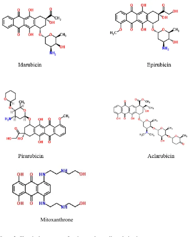
1.5. Novel generations of Anthracyclines
To counteract anthracycline toxicity, new semi-synthetic and synthetic formulations have been produced for approval by the Food and Drug Administration (FDA) and European Medicines Agency (EMA). Only few semisynthetic anthracyclines derivatives, i.e., idarubicin, epirubicin, pirarubicin, aclarubicin and mitoxanthrone, have obtained such approval (figure 2). Idarubicin, derived from DNR, appears to be more effective than DNR in several aspects including increased liposolubility and oral administration (13). Epirubicin, a close relative to DOX, has been used mostly in treatment of breast cancer with relatively good success especially when in combination with cyclophosphamide and 5-fluorouracil. However, similar to DNR and DOX, epirubicin induces significant cardiotoxicity (8). A study in rats investigated the effectiveness of pirarubicin, another close relative of DOX. The former manifested significantly lower cardiotoxicity, but cancer cell killing was also less effective compared to DOX (Adriamycin) (14). Aclarubicin and aclamycin were also shown to display only a relatively modest cardiotoxicity improvement over DNR and DOX. Mitoxanthrone which is similar to DOX has
17
been extensively tested for treatment of breast cancer, prostate cancer and leukemia because of reduced cardiotoxicity. Nonetheless, Thomas and coworkers reported that mitoxanthrone induces cardiotoxicity levels similar to DOX and DNR (6, 15)
Mitoxanthrone
18
2. Transporters of Anthracyclines
Anthracyclines are positively charged drugs that must be actively transported into and out of cells. Numerous studies have revealed two major families of anthracycline transporters which will be discussed here in some detail: (i) the ATP-binding cassette family (ABC) and (ii) the organic cation transporters (OCTs). In addition, the organic anion transporters (OATs) will also be reviewed briefly since they are part of SLC22 subfamily and have been reported to interact with typical OCT substrates.
2.1. ATP-binding cassette family (ABC)
The ABC transporter family has a role in tissue protection against toxic compounds including endogenous metabolites and exogenous xenobiotics. They have also garnered attention for their role in chemoresistance (16). This family of transporters consists of seven subfamily members designated A to G and are encoded by 48 genes (17). They display broad tissue distribution and are present in key physiological barriers such as the biliary canalicular membrane of hepatocytes, apical membrane of the proximal tubules of the kidney, brush border membrane of intestine and epithelium of the blood-brain barrier (17). Various studies have established that among all ABC transporters, only 12 members belonging to 4 subfamilies exert an important role in drug resistance (17).
2.1.1. The ABCB subfamily transporters
The ABCB subfamily of transporters, also known as multidrug resistance (MDR) proteins, includes the well characterized P-glycoprotein which transports mostly anionic compounds (16), but also various positively charged antineoplastic drugs such as anthracyclines, taxanes and vinca alkaloids. P-glycoprotein is expressed in various tissues such as liver, kidney and intestine, as well as specialized stem cells of the immune system (17). Moreover P-glycoprotein is expressed in the blood-brain barrier which prevents access of many xenobiotics to the central nervous system (18). Such xenobiotics have been reported to modulate P-glycoprotein. For example, it was reported that cationic (hydrophilic) compounds
19
can strongly compete with hydrophobic ones, thereby acting as either inhibitors or inducers of P-glycoprotein. Some drugs that specifically inhibit P-glycoprotein interact with digoxin leading to toxic accumulation of this cardiac glycoside. To prevent this accumulation, inducers of P-glycoprotein are required (18). There are two types of P-glycoprotein inhibitors: calcium channel blockers and beta blockers. Examples of the former type include nicardipine, verapamil, diltiazem, nifedipine, while the latter type includes carvedilol, bisoprolol, propranolol (18).
2.1.2. The ABCC subfamily transporters
The ABCC subfamily transporters, known as multidrug resistance-associated proteins (MRPs), differ structurally from ABCB counterparts in possessing additional domains aside from the common nucleotide binding- and transmembrane-domains. In fact, some members of the ABCC1, ABCC2, ABCC3, ABCC6 and ABCC10 subfamilies possess an N-terminal membrane domain attached to the core structure by a cytoplasmic linker, whereas ABCC4, 5, 11 and 12 lack an N-terminal domain but retain the cytoplasmic linker (17). ABCC transporters also differ in the ability to transport negatively charged compounds. This evolutionary property of the ABCC transporters confers improved protection from differently charged toxic biomolecules (17).
2.1.3. The ABCG subfamily transporters
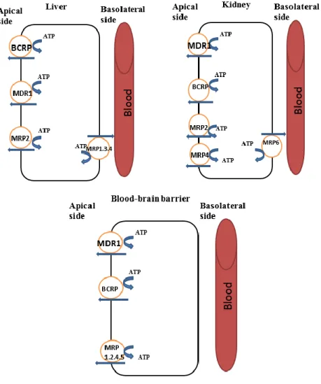
ABCG subfamily transporters contains, among others, their most important member involved in drug resistance, i.e., ABCG2 efflux transporter also known as MXR or BCRP (17). ABCG2 was first discovered based on its ability to transport mitoxanthrone and it was reported that this substrate is not transported either by P-gp or MRP1 (19). Unlike P-glycoprotein which more selectively transports negatively charged compounds, ABCG2 can transport either negatively or positively charged compounds. It was reported that unlike P-glycoprotein, ABCG2 cannot transport key substrates such as verapamil, taxols, imatinib, Vinca alkaloids and anthracyclines (17). Figure 3 presents an overview of some of the ABC transporters and their localization in various tissues.
20
Figure 3 Schematic representation of the ABC transporter localization in liver (hepatocytes), kidney (proximal tubule cells) and blood-brain barrier (brain endothelial cells). Adapted from (20).
2.2. Mechanism of transport by ABC transporters
Drug transport by ABC family members consists of two stages: ATP hydrolysis which activates transport, followed by export of the compound from the cytoplasm to the extracellular space. However, the exact mechanism by which the binding and subsequent transport occur remain unclear (16). The following mechanism has been proposed:
ATP-21
dependent drug binding (first stage) is followed by physical translocation from the cytoplasm to the extracellular space (second stage). In the first stage, ATP binding occurs in the nucleotide binding domain, consisting of three highly conserved sequences and the signature C motif. All ABC transporters display high ATPase activity in the micromolar range, but low affinity for ATP in the millimolar range (16). The second stage is highly dependent on the structure presented by the transporter. For example, P-glycoprotein (ABCB1) has 12 transmembrane domains and 2 nucleotide binding domains while ABCG2 displays only half of both. This suggests that the latter is likely to homodimerize. Because of its function in expelling toxic compounds outside the cell, the substrate binding site is located on the cytoplasmic side of the plasma membrane and consists of transmembrane helices which form a binding pocket (16). Studies have revealed that additional binding minipockets are located adjacent to the main binding pocket, together forming a funnel-like shape with the narrowest part facing the cytoplasm. Two co-substrates can easily enter the narrow opening and determine either massive and rapid efflux of the targeted compound or its inhibited transport regulated by the binding of the inhibitor substrate (16).
3. Organic cation transporters (OCTs and OCTNs)
OCTs belonging to the SLC22 family are part of the major, highly-conserved facilitator superfamily (MFS) of transporters (20). OCTs include two subclasses based on substrate specificity:1) OCT1, OCT2, and OCT3 and 2) zwitterion/cation transporters OCTN1, OCTN2, OCTN3 and CT2/OCT6 (20, 21). The OCTs subgroup (OCT1-3) exhibits close similarity in substrate specificity and transport mechanism. They all translocate various cations across the plasma membrane. The subclass of OCTNs (OCTN1, OCTN2 and CT2) are either uniporters of cations and zwitterions, and differ from OCTs in their ability to transport carnitine to the peroxisome for beta oxidation in a sodium-dependent manner (symport mechanism) (20).
22
3.1. OCT1
OCT1 (gene number SLC22A1) is located on chromosome 6. The first characterization of OCT1 was carried out by Gründemann et al., (22). Using a Xenopus oocyte-based expression system, this group successfully isolated OCT1 cDNA from a rat kidney library. The reported clone contained an open reading frame encoding a plasma membrane protein of 61.5 kDa. Overexpression of this protein in oocytes was shown to correlate with increased uptake of tetraethyl ammonium (TEA) with Km of approximately 100µM (22). Despite evidence that TEA transport by OCT1 is potential-dependent, it was shown that inwardly or outwardly proton gradients of 1 pH unit do not alter significantly uptake of this substrate (22). In mouse and rabbit, using RT-qPCR, mRNA levels were detected in kidney and to a lesser extend in liver (20). In humans, OCT1 is predominantly expressed in liver and less so in kidney. hOCT1 mRNA levels are also detected in the immune system and various organs such as small intestine, heart, skeletal muscle, brain, eye, placenta and others (20).
3.1.1. OCT1 as metformin transporter
Metformin belongs to the class of the biguanides that are used to treat type 2 diabetes; indeed, this drug efficiently reduces gastrointestinal glucose absorption and glucose production in the liver (23) (24). Metformin is initially activated through phosphorylation by AMP-activated protein kinase which in turn greatly reduces glucose production in hepatocytes thus leading to enhanced uptake of circulating glucose by the muscle and hepatocytes (24). OCT1 is a determinant of metformin uptake and it was reported that variations in response to metformin are attributable to genetic variants of OCT1 as reported by Shu et al., (24). The authors reported that several genetic variants displayed normal uptake of the model OCT1 substrate 1-methyl-4-phenylpyridinium (1-MPP), but greatly reduced metformin uptake suggesting the determinant role of OCT1 in the transport of metformin.
23
3.1.2. OCT1 as thiamine transporter
Chen et al.,(23) reported that OCT1 is a high capacity thiamine transporter. Thiamine is transported by two high affinity transporters belonging to the SLC19 family, i.e., members 2 and 3 which act in sodium-dependent manner. However, using both human and mouse hepatic cells, the authors reported that OCT1 retains the ability to take up thiamine with high affinity. This seems consistent with the fact that thiamine is directed to the liver where OCT1 is predominantly expressed, and particularly in the central vein where important metabolic reactions occur.
3.1.3. OCT1 as donepezil transporter
Kim et al., (25) reported that OCT1 has an important role in transport of donepezil, a therapeutic agent for Alzheimer’s disease which is characterized by progressive degeneration of cholinergic neurons. Donepezil is a cholinesterase inhibitor which acts similarly to tacrine by increasing levels of the neurotransmitter acetylcholine in the presynaptic cleft of the cholinergic neurons. It was shown in rat brain, that donepezil is taken up with high affinity by CHT1 and OCT1 and to a lesser extent by other organic cation/zwitterion transporters such as OCT2 and OCTN2 (25). CHT1 (CHoline Transporter 1), is the main transporter for acetylcholine in neurons and manifests the highest affinity for donepezil. However, Murakami et al., (26) showed that CHT1 is a close relative to OCT1 in terms of substrate specificity, and also reported that CHT1 in the blood-brain barrier might in fact be an isoform of the OCTs family (26)
3.1.4. OCT1 as choline transporter
Choline plays an important role in cell homeostasis by providing phospholipid compounds for the plasma membrane. In addition, it is involved in methionine metabolism as a methyl donor and in neurotransmission by providing acetylcholine (27). Moreover, deficiency in choline is associated with various neuronal and hepatic disorders and is also linked to various cancers (27). Choline is transported by three systems based on tissue localization and substrate affinity. The highest affinity choline transporter is CHT1, which delivers acetylcholine in cholinergic neurons. The role of the organic cation transporters OCT1 and OCT2 in choline
24
transport is however unclear and results vary from study to study (27). For example, overexpression of either OCT1 or OCT2 in human embryonic kidney HEK293T cells revealed increased uptake of choline by OCT1, but not by OCT2 (28). Another study showed that both OCTs were efficient in choline uptake in Xenopus oocytes (29). Various reports also claim considerable variability of the Michaelis-Menten affinity constant ranging from 100µM to 350 µM for either OCT1 or OCT2 (27). However, the evidence for OCT1 as a donepezil transporter in rat brain supports its role in choline and/or acetylcholine uptake in both neuronal and non-neuronal cells (25); nonetheless more studies are needed to determine whether OCT1 is indeed able to transport choline independently of CHT1 and/or OCT2.
3.1.5. OCT1 as transporter for anticancer drugs
Anticancer therapy outcome depends on the activity of the anticancer agent and the cellular machinery (30). For example, platinums such as picoplatin, oxaliplatin and cisplatin activities lead to differences in the formation of DNA adducts depending on the activity of the nucleotide excision repair mechanism to repair the resulting DNA damage. Thus, the cytotoxicity effect of this formation depends on the cellular expression of transporter proteins for these platinum drugs. More et al., (30) demonstrated that expression of OCT1, OCT2, but not OCT3 significantly enhances the uptake of picoplatin and the resulted formation of DNA adducts. Disposition of picoplatin (i.e. absorption, distribution, metabolism and excretion) was shown not to be dependent on OCT1 expression as demonstrated by pharmacokinetic studies (30). It also was shown that OCT1 expression greatly influences oxaliplatin uptake, but not carboplatin or cisplatin in two independent studies done by Zhang et al., (31) and Yokoo et al., (32). OCT1 expression levels were also demonstrated to correlate with imatinib action in treating patients with chronic myelogenous leukemia (CML) as shown by White et al., (33). Taxanes such as paclitaxel are important class of drugs whose action interferes with microtubule proper function leading to deregulation in mitosis (34). The role of OCT1 in the transport of paclitaxel was investigated by Gupta et al., (35) who demonstrated that OCT1 expression in model lymphoma cell lines and in primary chronic lymphocytic leukemia patients samples is a determinant of enhanced paclitaxel uptake. Moreover, the authors investigated the uptake of another anticancer agent- irinotecan, and found that OCT1 expression pattern influences the efficacy of treatment with this particular agent (35).
25
3.2. OCT2
OCT2 (gene number SLC22A2) was first cloned in 1996 by Okuda et al. (36) from rat. Functional analysis of the open reading frame revealed that rOCT2 is a membrane protein containing twelve membrane spanning helices with an extracellular loop between helices 1 and 2 and two glycosylation sites. The extracellular loop, which contains the substrate binding pocket, was reported to be highly similar to rOCT1 with 85% identity. The authors also identified four potential phosphorylation sites for protein kinase A and two phosphorylation sites for protein kinase C. By injecting OCT2 cRNA in Xenopus oocytes, uptake studies with TEA showed a marked increase in transport rate that was greatly decreased in the case of cimetidine, quinidine, and procainamide. By varying the pH, it was established that OCT2 acts independently of proton gradient and/or environmental pH (36). Further studies investigating the role of OCT2 revealed that this transporter mediates, among others, electrogenic transport of various organic cations such as N-methyl-nicotinamide and choline (37). A study from Mitohashi et al., (38) reported that human OCT2 is the main transporter in kidney and is similar to rat OCT2 which is localized on the basolateral membrane of proximal tubules. Moreover, immunohistochemistry studies on human OCT2 also showed basolateral membrane localization (38). However, there was a discrepancy over OCT2 localization from that study and a study from Gorboulev et al., (39) that showed OCT2 being localized on the apical membrane in distal tubules of human kidney. The marked difference was associated with the antibody which apparently displayed some cross-reactivity with closely related transporters in the same region (38). Several other studies investigating the role and behavior of OCT2 have also been conducted. In terms of substrate binding and stability, Barendt et al., (40) showed that among all organic cation transporters, interactions between substrates and OCT2 are greatly influenced by the charge in such way that when there were variations in the pH of the environment, an increase or a decrease of a protonated fraction of a substrate was observed. This, in consequence, affected the binding properties of substrates for OCT2. However, these changes did not affect the model substrates TEA and 1-MPP (40). Aside from its role in kidney, OCT2 was found in the brain where it participates in the transport of corticosterone (41). Although uptake of this steroid is mainly transported by extra neuronal monoamine transporter or EMT (see OCT3/EMT), the authors noted that OCT2 and OCT3 are not
26
identical and can be distinguished pharmacologically by use of O-methyllisoprenaline which appears to inhibit potently EMT, but not OCT2 (41). OCT2 can also transport several neurotransmitters, but most importantly dopamine in both kidney and brain. Evidence for OCT2 involvement in dopamine clearance came from a study reporting that proximal and distal tubules were able to express dopamine synthesizing enzyme L-aromatic amino acid decarboxylase. In support of this, cells forming the proximal tubules are known to produce dopamine which is believed to act as natriuretic hormone (41).
3.2.1. OCT2 as metformin transporter
Based on the primary localization of OCT2 in kidney, and as renal excretion is the major pathway for the elimination of xenobiotics, OCT2 plays an important role in detoxification. Among several drugs, OCT2 appears to be critical for disposition of metformin (42, 43). Numerous studies have been conducted to evaluate the relationship between OCT2 and the pharmacokinetics of metformin. Although the uptake of metformin was shown to depend on OCT1 function, Kimura et al., (44) claimed that metformin is a better substrate for OCT2 than for OCT1. Nonetheless, the role of OCT2 in metformin uptake is mostly associated with several genetic OCT2 variants such as A270S, M165I, R400C and K432Q which are nonsynonymous single nucleotide polymorphisms (SNPs) (42, 43). It was shown that metformin pharmacokinetics are deregulated in humans presenting these mutations. In addition, these mutations are ethnic-specific and the A270S variant represents the most important allele frequency for five ethnic populations including Caucasians, Afro-Americans, Asian-Americans, Mexicans and Pacific Islanders. M165I and R400C variants are present only in Afro-Americans and K432Q is present in both Afro-Americans and Mexicans (43).
3.2.2. OCT2 role in Parkinson’s disease
Parkinson’s disease is a neurological disorder which is characterized by loss of dopaminergic neurons in the substantia nigra pars compacta (SNc) region of the brain (45, 46). In healthy individuals, normal functioning of dopaminergic neurons is maintained by a normal distribution of the dopamine transporter (DAT) whose role is to remove circulating dopamine
27
from presynaptic cleft in order to terminate neurological synapse (45, 46). However, Taubert et al., (47) reported an unexpected role of OCT2 in maintaining the healthy state of these neurons. They demonstrated that OCT2, besides of its role in secretion and removal of various cations in kidney, has a particular role in human brain where it localizes in SNc and where it colocalizes with DAT and tyrosine xydroxylase. In addition, OCT2 participates in the exclusive transport of two endogenous compounds, i.e., cyclo (his-pro) (CHP), and salsolinol (SAL). The first compound has a neuroprotective role by contributing to the maintenance of presynaptic transactions and by stimulating neuronal mitochondrial respiration. Inversely, SAL appears to act as a neurocytotoxic compound by inhibiting mitochondrial respiration and therefore by promoting oxidative stress (47). Moreover, OCT2 assures a strict balance in secreting CHP and SAL in such way that the ratio between the two is maintained with increased CHP levels. It was also demonstrated that the mutant OCT2 R400C loses the ability to transport CHP while maintaining SAL levels; moreover this switch may be detrimental for proper neuronal functioning and is therefore favorable for Parkinson’s disease development (47)
3.2.3. OCT2 as transporter for anticancer drugs
As mentioned above, cisplatin is one of the most effective antineoplastic agents used for treating a variety of solid tumors. In a study from Filipski et al., (48), it was shown that mice deleted in OCT1 and OCT2 severely abrogated urinary excretion of cisplatin, although plasma levels were not affected. Moreover, mice deficient in both OCTs was efficiently protected from cisplatin induced renal tubular damage. However, patients presenting OCT2 variant rs316019 were less affected from cisplatin cytotoxicity compared to reference OCT2. Therefore, the authors concluded that OCT2 possesses a critical role in renal toxicity induced by cisplatin (48). The role of OCT2 in cisplatin handling was also reported by Tanihara et al.,(49) and the authors also demonstrated the protective role of imatinib on renal nephrotoxicity caused by cisplatin. As mentioned above, OCT2 also participates in oxaliplatin and picoplatin uptake (30-32).
28
3.3. OCT3
OCT3 (gene number SLC22A3) was independently identified in 1998 by two groups from Japan and Germany. Kekuda et al., (50) using two different expression systems, i.e., X.
laevisoocyte and mammalian expression system (HeLa cells), reported that the newly
identified protein was electrogenic cation transporter mainly expressed in fetal basal membrane of placenta and not in liver or kidney. Based on membrane localization, the authors reported that the main role of this transporter is to participate in the first line of defense against toxic xenobiotics. Data analysis also showed that interactions with OCT1 and OCT2 were negligible. OCT3 has the capacity to transport 1-MPP similarly to OCT1 and OCT2, but can also transport serotonin and desipramine (50). Transport of catecholamines occurs via two different systems: neuronal monoamine transporter (uptake1) and extra neuronal monoamine transporter (uptake2). The latter is characterized by the ability to transport catecholamines in an Na+ and Cl- independent manner as reported by Wu et al., (51). Moreover, the authors identified OCT3 as extra neuronal monoamine transporter (EMT) or uptake2 system. OCT3 can transport various compounds such as amphetamine and methamphetamine, serotonin and dopamine. Thus, a role in handling of neuroactive compounds in the brain was exclusively attributed to OCT3 (51). A functional study from Gründemann et al., (52) using amino acid sequence analysis revealed that OCT3 is similar to OCTs and organic anion transporters (OATs) with respectively 50% and 30% homology. The protein is composed of 556 amino acids and contains 12 putative transmembrane domains. Gene localization was mapped to 6q27 locus using fluorescence in situ hybridization (52). Regarding the distribution and the function of the organic cation transporters in the brain, both OCT2 and OCT3 are mainly expressed in cortex, thalamus, and hypothalamus. However, within these regions, OCT2 and OCT3 occupy different sub regions. Although both OCT2 and OCT3 are expressed in the central nervous system, OCT3 seems to be exclusively expressed in astrocytes that are localized in restricted areas (53). In the brain, OCT3 appears to perform a very important function by regulating osmotic pressure by salt ingestion (53). The exact mechanism by which OCT3 regulates the osmolarity is however unclear, but it is believed that it might participate in activation of secondary neuronal pathways responsible for insuring proper ways of neurotransmission which go through osmosensitive regions and other relay regions (53).
29
OCT3 is also believed to be strongly involved in stress, as it was labeled a corticosterone sensitive monoamine transporter. Moreover, in view of the fact that OCT3 has a high affinity for dopamine and its clearance, this transporter is strongly linked to sensitivity to psychostimulants and therefore to addiction behavior (53). These data suggest that brain transporters from the group of OCTs have a special role to play in drug transport and are targets for new drug development.
3.3.1. OCT3 as transporter for anticancer drugs
The role of OCT3 in the uptake of irinotecan, vincristine and melphalan was investigated by Shnitsar et al., (54). By stably transfecting Chinese hamster ovary (CHO) cells with human OCT3 cDNA and performing competition studies with (3) H]MPP (4-methyl-pyridinium iodide and either melphalan, irinotecan or vincristine, the authors reported a significantly inhibited transport of (3) H]MPP (4-methyl-pyridinium iodide by these three agents suggesting the important role of OCT3 in handling of these antineoplastic compounds (54).
3.4. OCTN1
In 1997, Tamai et al., (55) cloned a cDNA corresponding to the novel organic cation transporter OCTN1 (gene number SLC22A4), which was then transiently expressed in HEK293T cells. The transfected cells were evaluated for transport capacity using the model organic cation transporter substrate TEA. The results showed that transport was saturable, energy dependent, and pH-sensitive. However, based on kinetic studies, OCTN1 exhibited a higher Michaelis-Menten constant compared to both OCT1 and OCT2, i.e. 440 µM vs 20 µM -100µM. Moreover, the authors revealed that OCTN1 operates in the apical membrane of renal epithelial cells. Because of the presence of a nucleotide binding motif, OCTN1 was suggested to act as an ATP-dependent transporter and it was shown that at pH 6.0 under ATP depletion, TEA transport was greatly decreased (55). Since OCTN1 is expressed in various human cell lines, Tamai et al., (55) suggested that it may participate in the efflux of anti-cancer drugs, although functional studies needed to be done in order to identify substrate specificity. A few years later, Gründemann et al., (56) expressed OCTN1 in HEK293T cells
30
and cells were assayed for transport of various substrates including TEA, carnitine, the anti-oxidants ergothioneine, stachydrine (proline betaine) and betaine. Surprisingly, they reported that uptake of TEA and carnitine was negligible compared to that of ergothioneine, stachydrine and betaine and proposed OCTN1 be named ergothioneine transporter (ETT). However, despite their recommendation for the new appellation, both OCTN1 and ETT refer to gene number SLC22A4. Using RT-PCR, tissue distribution of OCTN1 was revealed to be highest in bone marrow and fetal liver (56). Consistent with other localization studies, OCTN1 was reported to be specifically expressed in monocytes (CD14) and absent in stem and blast cells. Strong OCTN1 expression in small intestine and kidney was linked to its function in initial absorption and reabsorption of ergothioneine (56). Moreover, Gründemann et al., (57) reported that the newly proposed OCTN1 is not expressed in human liver, and to a very low extent in heart, muscle and skin. OCTN1 appears to be expressed in various species including rat and mouse. Despite the reported lower affinity of OCTN1 for carnitine, Pochini et al., (58) reported that structural analysis of OCTN1 binding pocket can fit substrates like acetyl carnitine and acetylcholine. However, when carnitine and ergothioneine are present, there seems to be a different structural organization of these molecules that prevents overlapping with the cation site, which explains the noncompetitive interaction with TEA. Regarding the transport of acetylcholine, Pochini et al., (58) also reported that human OCTN1 is involved in the bidirectional transport of this substrate. As epithelial cells of the small intestine and immune system cells such as lymphocytes participate in extra neuronal cholinergic transport, and since OCTN1 is predominantly expressed in these regions, it was suggested that this transporter may participate in catalysis of neo synthesized acetylcholine in order to activate receptor pathways in autocrine and paracrine mode (58). In heart, OCTN1 seems to play a role in absorption and removal of cations thereby modulating myocardial biogenesis (58)
3.4.1. OCTN1 as transporter for anticancer drugs
The role of OCTN1 in anticancer drug uptake was investigated by two groups. Okabe et al., (59) performed a screen with 60 cancer cell lines from the National Cancer Institute (NCI-60) for anticancer substrates. This revealed that DOX and mitoxanthrone, a close DOX relative, are substrates for OCTN1. Jong et al., (60) investigated the role of OCTN1 and OCTN2 in oxaliplatin uptake and cytotoxicity. Using HEK293T cells overexpressing OCTN1 and
31
OCTN2 and primary dorsal root ganglion (DRG) neurons, data from this study showed an increased uptake and cytotoxicity of oxaliplatin in both model systems compared to mock. The authors also showed that uptake of ergothioneine (OCTN1) and carnitine (OCTN2) in HEK293T was decreased when competing with oxaliplatin, and as a consequence, the cytotoxicity was also reduced. In DRG neurons, the same effect was observed and the uptake was determined time, concentration, temperature and sodium dependent (60).
3.5. OCTN2
OCTN2 (gene number SLC22A5) was cloned from rat and human in 1998 by several groups including Sekine et al.,(61), Tamai et al., (62), and Wu et al., (63). OCTN2 is expressed in various tissues and organs including, among others, liver, kidney, small intestine, colon (64); skeletal muscle, lung (65); ovary, placenta and brain (66). OCTN2 is a high affinity sodium dependent carnitine transporter needed for the transport and oxidation of fatty acids in the mitochondria via a process known as β-oxidation (58), (67). Without carnitine, fatty acids cannot cross the mitochondrial inner membrane by the acyl carnitine translocase. Thus OCTN2, as a high affinity carnitine transporter, is abundantly expressed in various tissues, where it plays specific roles. For example, it was shown that in murine kidney, OCTN2 regulates glomerular reabsorption of carnitine, some of which is excreted in urine due to its inability to bind to serum protein. Immunohistochemistry studies revealed that OCTN2 is localized to the apical membrane of proximal tubular epithelial cells. Transport of carnitine by OCTN2 in kidney is sodium dependent and seems to be linked to co-expression of PDZ domain-containing proteins such as PDZK1 protein at the apical membrane, although levels of OCTN2 expression do not seem to depend on PDZK1 expression (67). PDZ domains are protein modules that regulate multiple processes including transport and signaling (68). In the intestine, OCTN2 participates in intestinal absorption of carnitine, although its role in this process is still controversial (67). In addition, although significant levels of OCTN2 are detected in small intestine and colon, there is another nutrient transporter, PEPT1, which is strongly expressed in these organs. Similarly, to kidney, OCTN2-mediated transport of carnitine in the intestine seems to be regulated by PDZK1 protein. Human and murine in vitro studies showed that uptake of carnitine is saturable between 10 µM and 20 µM (67). In heart,
32
carnitine plays an important role in preserving skeletal muscle and carnitine deficiency causes cardiomyopathy. OCTN2, which is expressed in the vascular endothelium, transports carnitine in sodium dependent manner (67). The precise localization of OCTN2 in the vascular endothelium was also suggested to indicate a role in carnitine accumulation in heart. OCTN2 is also expressed in liver where carnitine biosynthesis occurs. Tamai et al., (67) reported strong expression in liver of gamma-butyrobetaine dioxygenase which catalyzes carnitine production from gamma-butyrobetaine. In fact, OCTN2 can transport gamma-butyrobetaine in a sodium dependent manner with Km of 13 µM, although the real contribution of gamma-butyric transport comes from another transporter, i.e., GAT2 (67). Yet, based on studies using mutated OCTN2 in mice, it appears that GAT2 gamma-butyrobetaine transport prevails only when OCTN2 does not perform its function and GAT2 only transports gamma-butyrobetaine acid into the liver, where carnitine biosynthesis occurs, to supply other tissues with carnitine (67). OCTN2 is also expressed in brain capillary endothelial cells and this correlates with regulatory transport of carnitine across the blood-brain barrier. Studies have revealed that the precise localization of OCTN2 is on the brain side and not on blood side suggesting that OCTN2 might only participate in one directional transport of carnitine and acetyl carnitine from the blood to the brain (67). Moreover, OCTN2 is highly involved in carnitine and acetyl carnitine circulation in placenta. Lahjouji et al., (69) showed that OCTN2 is localized in the brush border membrane of placenta and is involved in carnitine transport in a sodium-, pH-, temperature- and osmolarity-dependent manner. They also reported that carnitine transport in placenta, mediated by OCTN2, is saturable at 11 µM which appears to be similar to the transport observed in kidney. Moreover, while in competition with TEA, valspodar and valproate, carnitine uptake is greatly inhibited suggesting that these substrates are specific for OCTN2 as well. However, in the case of verapamil, it seems that inhibition of OCTN2 is triggered by both competitive and noncompetitive modes (69). The implication of other carnitine transporters has not yet been excluded. In fact, OCTN1 is also expressed in placenta and transports carnitine in a sodium-dependent manner although the affinity of OCTN1 for carnitine has not been reported. A study from Maekawa et al., (70) has identified cartregulin which is homologous to OCTN2 in rat brain and acts by stabilizing the mRNA of OCTN2. Moreover, a novel gene encoding full length of OCTN2 was identified with the addition of 24 amino acids in the first extracellular loop. This variant, named OCTN2 N2VT, exhibited
33
several different features compared to wild type OCTN2 including reduced glycosylation which regulated retention of OCTN2 N2VT in the reticulum endoplasmic compared to normal glycosylation and subsequent expression on the cell surface. It was also found that while OCTN2 N2VT is expressed in HeLa cells, carnitine uptake is severely abrogated due to lack of expression on the cell surface (70).
3.5.1. OCTN2 as transporter for anticancer drugs
As mentioned above, OCTN2 plays a role in oxaliplatin uptake (60). Moreover, Hu et al., (71) reported a role of OCTN2 in the uptake of the topoisomerase II poison etoposide. The study was performed in cells expressing either human or mouse OCT2 challenged with both carnitine and etoposide. The results showed a marked decrease in carnitine transport suggesting the important role of OCTN2. However, the authors were able to reverse the inhibition by adding carnitine in excess.
3.6. OCTN3
OCTN3 is another member of the SLC22 family that has not received much attention. Duran et al., (72) successfully isolated mOCTN3 cDNA from small intestine. Moreover, using commercially available antibodies against OCTN2 and OCTN3, the authors also reported that OCTN3 localizes on the basolateral membrane of the enterocytes in rat and chicken small intestine compared to OCTN2 which is localized on the apical membrane. OCTN3 was also found to be responsible for carnitine uptake in the peroxisome in mammals. Using a specific antibody 629 that binds to mouse OCTN3, Lamhonwah, et al., (73) localized OCTN3 on the peroxisomes and the cisternae of the rough reticulum endoplasmic. As already pointed out, carnitine is essential for β-oxidation in mitochondria, but is also important for β-oxidation in the peroxisome. The role of carnitine in the peroxisome is to shuttle short-sided hexacosanoid acid and pristanic acid which are products of β-oxidation in the peroxisome and exports them into the mitochondria as carnitine esters (73). The evidence for the existence of carnitine acyltransferases and carnitine translocase in the peroxisome further supports the importance of carnitine transport to this organelle. mOCTN3 transports carnitine in the peroxisome with
34
affinity of 20µM (73). The authors, based on comparison with mOCTN3, have also predicted that, similarly to OCTN1 and OCTN2 which are mapped at 5q31 locus, OCTN3 may also be localized at the same region in humans and may play an important role in carnitine homeostasis. They also predicted that if OCTN3 is mutated, this should confer susceptibility to several diseases related to OCTNs such as Crohn’s disease (73).
3.7. CT2 (OCT6)
OCT6 or CT2, for carnitine transporter 2 (gene number SLC22A16), has been expressed in
Xenopus laevis followed by characterization of the corresponding protein by Enomoto et al.,
(74). Structural analysis studies performed by the authors allowed to identify a CT2 cDNA encoding 543 amino acid protein that shared up to 40% identity with the other carnitine transporter members such as OCTN1 and OCTN2 (reported to be CT1 for carnitine transporter 1). Carnitine transport was found to be saturable at 50 µM and the affinity for this substrate was evaluated to 20 µM. It was shown that uptake of carnitine is not entirely sodium dependent. Competition studies with other carnitine derivatives and betaine greatly inhibited CT2, but not choline and other selected substrates. Tissue distribution analysis revealed that CT2 is expressed in human testis, and not in any other tissues including brain, placenta, heart, ovary, small intestine, and colon (74). Nonetheless Sato et al., (75) found that CT2 was strongly expressed in normal human endometrium and that this expression was regulated by progesterone. Analysis of CT2 gene also revealed that there were no identical sequences in the 5’ regions upstream of progesterone responsive elements suggesting that, in endometrial cancer, CT2 expression should not depend on progesterone (75). In another study, Okabe et al., (76) characterized CT2 from normal vs tissues from patients with acute leukemia and reported that CT2 is predominantly expressed in bone marrow and testis and that high levels of CT2 are observed in fetal liver, but not in normal liver (76). Moreover, CT2 is expressed in a variety of leukemia and its expression is tightly related to hematopoiesis. CT2 was reported to transport typical OCTs substrates such as TEA (76). CT2 seems to have an important role in lung cancer and Kunii et al., (77) reported that its expression correlates with cisplatin uptake. The authors also reported that OCT2 is not in fact the main platinum drug uptake transporter, but is more likely to be CT2 (OCT6). Studies from our laboratory involving the role of CT2
35
have demonstrated that this protein is involved in the transport of the anti-cancer polyamine analogue Bleomycin- A5 (78). Bleomycin is currently used as antineoplastic agent for treating various cancers such as lymphomas and carcinomas. By overexpressing CT2 in various cancer cell lines, Aouida et al., (78) showed that these cells are extremely sensitive to Bleomycin- A5 compared to control cells. The results correlated with the previously shown increased uptake of polyamines mediated by Agp2 which is a high affinity carnitine transporter in
Saccharomyces cerevisiae (79).
3.7.1. CT2 as anthracycline transporter
By injecting CT2 cRNA in Xenopus oocytes and stably transfecting Jurkat cells, Okabe et al., (76) tested the behavior of this transporter towards DOX. The authors reported that transport of DOX was saturable, dose dependent and uptake was independent of sodium with apparent affinity constant of 5 µM. A slight decrease in cell viability in control cells vs ones overexpressing CT2 was observed. Nonetheless, it was claimed that CT2 is a DOX importer. In a study involving the role of CT2 in human epithelial ovarian cancer, Ota et al., (80) reported that CT2 transports DOX in clear cell adenocarcinoma cell lines. Data from this study suggested that anthracyclines might be used in combination chemotherapy with paclitaxel and other platinum drugs used in ovarian cancer. Moreover, they predicted that clear cell adenocarcinoma expressing CT2 might display a better response to combinational treatment with platinum drugs and anthracyclines compared to other ovarian cancer cell lines (80). Correlation between CT2 expression and anthracyclines action was reported by Lal et al., (81) in a study investigating the genetic variants of this transporter versus DOX and its metabolite doxorubicinol in Asian breast cancer patients. Moreover, by studying different ethnic groups, the authors revealed that CT2 presented at least four different polymorphisms. Comparison between Asian and Caucasian groups showed differences in allele and genotype frequencies and data suggested that CT2 variations present the hallmark of DOX sensitivity and even other drugs candidates. CT2 c.146G allele was reported to harbor increased exposure levels to DOX and its metabolite doxorubicinol and has, as a consequence, shown better outcome of cancer treatment (81).
36
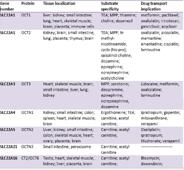
Figure 4. Schematic representation of the OCTs and OCTNs transporters localization in liver (hepatocytes), kidney (proximal tubule cells) and blood-brain barrier (brain endothelial cells). OC+- organic cations. Adapted from (20).
37
38
4. Organic anion transporters
Organic anion transporters (OATs) include more than ten transporter members (figure 4) and are part of the SLC22 subfamily of transporters (83). Localized on almost all epithelial physiological barriers, OATs are involved in the transport of numerous anionic compounds. OATs are mostly facilitator transporters, but it was shown that anionic substrates are also imported with counter transport of α-ketoglutarate. Thus, OATs that operate by such mechanism form part of a rate-limiting «tertiary» transport system as transport is driven by concentration gradient created from the sodium-decarboxylate cotransporter (NaDC3) and Na+, K+, ATPase (83). The first cloned OAT was the novel kidney transporter (NKT) which was designated OAT1 (SLC22A6). It was demonstrated that the hippuric acid derivative, the p-aminohippuric acid (PAH) is a typical OAT substrate. Moreover, OATs have the ability to transport xenobiotics such as antibiotics, antivirals, anti-inflammatory drugs, diuretics, and non-steroidals (83). A study conducted by Ahn et al., (84) demonstrated that typical OCT substrates can interact with OATs, specifically with mOAT1, mOAT3 and mOAT6. These substrates include TEA, 1-MPP, cimetidine, metformin, verapamil procainamide, clonidine and others. Moreover, some of these substrates appear to be member specific as procainamide, clonidine and metoclopramide can bind to mOAT3, but not to mOAT6. It was also demonstrated that 1-MPP can bind to mOAT1 and mOAT6 although with low affinity (Km of ~1mM and ~2mM). The authors also reported that the assayed cationic substrates have the ability to equally inhibit the cationic transport system (N1- methyl nicotinamide) and the anionic transport system (PAH) (84).
39
Table 2. SLC22 organic anion transporters. Adapted from (83)
40
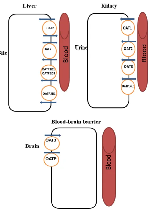
Figure 5. Schematic representation of the OATs and OATPs transporters localization in liver (hepatocytes), kidney (proximal tubule cells) and blood-brain barrier (brain endothelial cells). Adapted from (20, 83, 85).
41
5. Hypothesis and Objectives
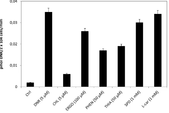
As discussed above in the Introduction and despite numerous studies aimed at investigating the role of OCTs transporters in the uptake of various anticancer agents including anthracyclines, the implication of these transporters in anthracyclines uptake has yet not been clearly demonstrated. It is noteworthy that anthracyclines are still believed to undergo cellular penetration by passive diffusion and cellular transport of the anthracycline DOX by facilitated transport exerted by CT2 was only demonstrated by Okabe et al., (76). However, as the authors only showed a slight decrease in cell viability in cells overexpressing CT2 protein compared to control, and as down regulation of CT2 was never done, the role of this transporter in anthracyclines transport remains highly inconclusive. We propose that the anthracycline DNR, a close relative to DOX, enters cells by means of facilitated transport mechanism mediated specifically by OCT1. Thus, screening of patientscurrently treated with anthracyclines, for OCT1 status might constitute a useful predictor of responsiveness towards DNR.
In view of these considerations, our general objective is to demonstrate that OCT1 is the specific transporter for DNR and we intend to accomplish this by:
Evaluating DNR accumulation in various cell lines Evaluating OCT1 expression in various cell lines
Performing competition studies with DNR and key OCTs substrates Downregulating OCT1 and subsequent evaluation of DNR transport Overexpressing OCT1 and subsequent evaluation of DNR transport
42
Chapter 2. The human organic cation transporter OCT1
mediates high affinity uptake of the anticancer drug daunorubicin
Emil Andreev1, Nicolas Brosseau1, Euridice Carmona2, Anne-Marie Mes-Masson2, and Dindial Ramotar1*
1Maisonneuve-Rosemont Hospital, Research Center, Université de Montréal, Department of Medicine, 5415 Boul. de l’Assomption, Montréal, Québec, Canada, H1T 2M4,
2Institut du cancer de Montréal/Centre de recherche du Centre hospitalier de l’Université de Montréal (CRCHUM), 900 rue Saint-Denis, Montréal, Québec, Canada, H2X 0X9 Running title: The human transporter of anthracyclines
Keywords: organic cation transporters, SLC22A1/OCT1, mammalian cells, anticancer drug
43
Background: Anthracyclines are antitumor agents used for treating various cancers.
Results: Human OCT1 displayed a high affinity for the uptake of the anthracycline
daunorubicin. Downregulation of OCT1 blocked the uptake of daunorubicin and conferred cellular resistance to the drug.
Conclusion: Human OCT1 possesses the ability to transport anthracyclines into cancer cells. Significance: Defects in OCT1 function are likely to cause drug resistance.
44
ABSTRACT
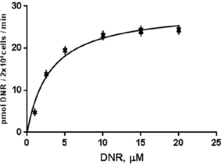
Anthracyclines such as daunorubicin are anticancer agents that are transported into cells, and exert cytotoxicity by blocking DNA metabolism. Although there is evidence for active uptake of anthracyclines into cells, the specific transporter involved in this process has not been identified. Using the high-grade serous ovarian cancer cell line TOV2223G, we show that OCT1 mediated the high affinity (Km ~ 5 μM) uptake of daunorubicin into the cells, and that micromolar amounts
of choline completely abolished the drug entry. OCT1 downregulation by shRNA impaired daunorubicin uptake into the TOV2223G cells, and these cells were significantly more resistant to the drug in comparison to the control shRNA. Transfection of HEK293T cells, which accommodated the ectopic expression of OCT1, with a plasmid expressing OCT1-EYFP showed that the transporter was predominantly localized to the plasma membrane. These transfected cells exhibited an increase in the uptake of daunorubicin in comparison to control cells transfected with an empty EYFP vector. Furthermore, a variant of OCT1, OCT1-D474C-EYFP, failed to enhance daunorubicin uptake. This is the first report demonstrating that human OCT1 is involved in the high affinity transport of anthracyclines. We postulate that OCT1 defects may contribute to the resistance of cancer cells treated with anthracyclines.