HAL Id: hal-00157543
https://hal.archives-ouvertes.fr/hal-00157543
Submitted on 26 Jun 2007
HAL is a multi-disciplinary open access
archive for the deposit and dissemination of sci-entific research documents, whether they are pub-lished or not. The documents may come from teaching and research institutions in France or abroad, or from public or private research centers.
L’archive ouverte pluridisciplinaire HAL, est destinée au dépôt et à la diffusion de documents scientifiques de niveau recherche, publiés ou non, émanant des établissements d’enseignement et de recherche français ou étrangers, des laboratoires publics ou privés.
wounding.
Patricia Guzzardi, Geneviève Genot, Elisabeth Jamet
To cite this version:
Patricia Guzzardi, Geneviève Genot, Elisabeth Jamet. The Nicotiana sylvestris extensin gene, Ext 1.2A, is expressed in the root transition zone and upon wounding.. Biochimica et Biophysica Acta - Molecular Cell Research, Elsevier, 2004, 1680 (2), pp.83-92. �10.1016/j.bbaexp.2004.08.012�. �hal-00157543�
Published in: Biochim. Biophys. Acta 1680 (2004) 83-92.
PMID: 15488988
The Nicotiana sylvestris extensin gene, Ext 1.2A, is expressed in the
root transition zone and upon wounding
Patricia Guzzardi
a, Geneviève Genot
a, Elisabeth Jamet
a, b, * aInstitut de Biologie Moléculaire des Plantes, UPR CNRS 2356 12 rue du Général Zimmer
67000 STRASBOURG, France
b
UMR CNRS-UPS 5546 24, chemin de Borderouge BP 17 AUZEVILLE
31326 CASTANET TOLOSAN, France
*
Corresponding author:Elisabeth JAMET UMR CNRS-UPS 5546 24, chemin de Borderouge BP 17 AUZEVILLE
31326 CASTANET TOLOSAN, France Tel: +33 (0)5 62 19 35 30
Fax: +33 (0)5 62 19 35 02 e-mail: jamet@scsv.ups-tlse.fr
Keywords: cell wall, extensin, HRGP, Nicotiana sylvestris, root, wounding
Abstract:
The Ext 1.2A gene of Nicotiana sylvestris L. encoding an extensin, a cell wall structural protein, was characterized. Ext 1.2A encodes a polypeptide of 311 amino acids having a highly repetitive structure and showing extensin features such as Ser-(Pro)4 repeats and a high content in Tyr and Lys. The expression profile of the gene was demonstrated using the reporter GUS (ß-glucuronidase) fused to its promoter region (-630/+124, relative to the transcription start site) and by RNA gel blots. The results show that the (-630/+124)Ext 1.2A/GUS gene fusion is expressed in the root transition zone, where cells undergo an isodiametric growth but have not reached yet the rapid elongation phase, in stem inner and outer phloems and in cortical cells at the stem/petiole junction. The Ext 1.2A gene is also induced after wounding of stems, ribs, leaves or roots. The gene fusion is expressed in stem cortical cells, in ribs and at leaf edges upon wounding. These data suggest that the (-630/+124) promoter region contains regulatory elements responsible for expression in roots and stems, as well as for response to wounding in stems and leaves.1.
Introduction
Plant cell walls are complex extracellular matrixes playing many roles during development and in response to biotic or abiotic stresses [1]. They are involved in maintenance of osmotic pressure and cell shape, thereby ensuring adhesion of cells in plant tissues [2]. They participate in cell-to-cell signaling [3] and in plant defense since they constitute physical barriers against pathogen invasion [4, 5].
Synthesis of the primary cell wall starts soon after cell plate fusion to the parental cell plasma membrane [6]. Current models describe the primary cell wall as a structure composed of two interdependent networks embedded in a pectin matrix [1, 7, 8]. The first network would be composed of cellulose microfibrils cross-linked with xyloglucans through hydrogen bonds and the second network would be constituted of structural proteins eventually cross-linked together or linked to other cell wall components via hydrogen or covalent bonds. These structural proteins are classified into three groups [9]: hydroxyproline-rich glycoproteins (HRGPs), proline-rich proteins (PRPs) and glycine-rich proteins (GRPs). During cell elongation, the cell wall is assumed to expand under the conjugated effects of 1,4-ß-D-glucanases (EGases) [10], xyloglucane endo-transglycosylases (XETs) [11] and expansins which act at the cellulose/hemicellulose interface [12]. After completion of cell expansion, structural proteins are thought to be insolubilized by peroxidases [13, 14].
The most abundant HRGPs of primary cell walls are extensins. Several extensin genes have been already characterized in monocot or dicot plants. These studies showed that all extensin genes are regulated in different ways. They are not expressed in all plant cells: they may be expressed in root or stem phloem cells, in cells under mechanical constraints, in response to wounding and in cells proliferating under hormone control [15]. However, none of these studies report on the differential expression of different members of an extensin gene family inside a plant. Moreover, description of expression patterns of extensin genes is of high interest since the roles such proteins play in cell walls is not yet really understood. The first extensin mutant affected in the RSH gene was only recently described. The RSH protein seems to be essential for correct positioning of the cell plate during embryogenesis of Arabidopsis thaliana [16].
In Nicotiana sylvestris and in N. tabacum, we already showed that there are two gene families, Ext 1.2 and Ext 1.4, encoding very different extensins [17, 18]. The Ext 1.4 gene was cloned and its pattern of expression has been analyzed in details. It was shown to be regulated by mechanical constraints occurring either during development or imposed on root or stem tissues [19, 20]. On the contrary, the structure and the fine regulation of Ext 1.2 genes have not yet been described. The present study aims (i) at the characterization of the N. sylvestris Ext 1.2A gene (ii) at the identification of cells expressing Ext 1.2A in tobacco plants and in response to wounding and at (iii) the analysis of the activity of the cloned Ext 1.2A promoter region in transgenic tobacco plants.
2. Materials and Methods
2.1. Plant material
Seeds of Nicotiana sylvestris L. were kindly donated by SEITA (Institut Expérimental du Tabac, Bergerac, France). Plants of Nicotiana tabacum L. var. Petit Havana SR1 were used for transformation, and experiments of wounding [21]. Plants were grown in vitro under a 16 h light at 24°C/8 h dark at 20°C cycle. Greenhouse culture conditions were a 12 h light/12 h dark cycle at 22±2°C.
Roots were grown in vitro in MS liquid medium (Duchefa, Haarlem, The Netherlands) diluted twice [22]. Coloration of root nuclei was performed with aceto carmine according to [23]. Wounded roots, stems, ribs and leaves were obtained by slicing organs of into 1-2 mm wide strips which were immersed for 8 h, 24 h or 48 h in MS medium diluted twice. Germination of pollen grains was done on MS medium containing 10 %sucrose and 0.5 % agar.
2.2. Nucleic acid extraction and analysis
RNA isolation was performed according to [24]. Genomic DNA purification and Northern blot analyses were performed as previously [17]. The integrity and the amount of RNAs were systematically checked using a bell pepper 25S rRNA probe kindly provided by Dr. R. Schantz (IBMP-CNRS Strasbourg). Sizes of RNAs were determined after electrophoresis through agarose-formaldehyde gels and comparison to an RNA ladder (Life Technologies, Cergy Pontoise, France). The Ext 1.2 specific probe spanned its 3’ UTR (untranslated region). The probes were labeled with [α-32P]dCTP (3000 Ci/mmol, Amersham Pharmacia Biotech UK Limited, Buckinghamshire, England) by random primer labeling [25].
2.3. Isolation and sequencing of the Ext 1.2A gene
Fragments of Ext 1.2 genes were obtained directly from N. sylvestris genomic DNA using PCR amplification. Different pairs of PCR primers located along the sequence of the 6PExt 1.2 cDNA previously characterized [17] were used. They are listed in Table 1 (oligonucleotides A to G) and their positions are indicated on Figs. 1 and 2. The 5’ as well as the 3’ distal UTR regions were amplified by inverted PCR [26]. All the PCR-amplified fragments were cloned into the pBluescript® KS+ vector (Stratagene, La Jolla, CA) according to standard procedures [27] or into the pCR®2.1-TOPO vector (Invitrogen, Carlsbad, CA). Recombinant plasmids were sequenced on both strands either by the dideoxynucleotide chain termination method using the T7 DNA polymerase [28, 29] or using an Applied Biosystems model 373A DNA sequencer (Perkin Elmer, Foster city, CA). In the latter case, a fluorescent dye-labeled dideoxy terminator kit was used (Abi Prism Dye Terminator Cycle Sequencing Ready Reaction Kit with DNA polymerase, FS Perkin Elmer). The nucleotide sequence data reported appear in the Genbank Database under the accession number AJ271871.
Table 1
PCR primers used for cloning and primer extension
Primer name Sequence (1) Restriction site Location on the gene Purpose
A 5'-CGGAATTCATGGCTAAAATAGTCTCTCTTCTC-3’ EcoR I +105 → +128 Cloning of Ext 1.2A
B 5'-CGGGATCCTCACTACTAGGAAATGCG-3’ BamH I +1029 → +1048 Cloning of Ext 1.2A, inverted PCR
C 5'-CGGGATCCAGAGAACCTTGACATAATC-3’ BamH I +1430 ← +1449 Cloning of Ext 1.2A
D 5'-CGGGATCCCCTTTAGAATGCAAAGCAAATTAC-3’ BamH I +165 → +188 Cloning of Ext 1.2A
E 5'-CGGGATCCGCATTTCCTAGTAGTGAGG-3’ BamH I +1029 ← +1048 Cloning of Ext 1.2A
F 5'-GGGGTACCGTGGAGATTTGTACACCGGA-3’ Kpn I +824 ← +846 Cloning of the Ext 1.2A coding region
G
5’-GCTCTAGAGCATTCTAAAGGAAAGCTAAGGGAC-3’
Xba I +152 ← +176 Inverted PCR, primer extension
H 5’-TCCCCCGGGAGAGAGACTATTTTAGCCATT-3’ Sma I +104 ← +124 Ext 1.2A/GUS constructs
I 5’-CCCAAGCTTCCAAGTCAGTCCAGTTTCAT-3’ Hind III -77 → -56 (-75/+124)Ext 1.2A/GUS construct
J 5’-CCCAAGCTTGAATTCCAGATTCATCTAG-3’ Hind III -630 → -611 (-630/+124)Ext 1.2A/GUS construct
2.4. Determination of the Ext 1.2A transcription initiation site
Primer extension was performed on total RNAs extracted from N. tabacum leaf strips. DNA contamination was removed with 5 U DNAse RNase free (RQ1 RNase-free DNAse, Promega, Madison, WI) in 40 mM Tris-HCl pH 7.9, 10 mM NaCl, 6 mM MgCl2,
10 mM CaCl2 buffer. Reaction was performed for 1 h at 37°C. RNAs were stored at a concentration of 0.1 µg/µL after phenol/chloroform extraction (v/v) and ethanol precipitation. Primer extension was performed in a 20 µL reaction volume with M-MLV (Moloney Murine Leukemia Virus) RT (Reverse Transcriptase) buffer (Promega), 2 mM dCTP, 2 mM dTTP, 2 mM dGTP, 4 µM dATP, 20 to 40 U RNase inhibitor (RNAguard inhibitor, porcine, Amersham Pharmacia Biotech UK Limited), 50 pmol oligonucleotide G (Table 1) and 5 µCi [α-35S]dATP (>1000 Ci/mmol, Amersham Pharmacia Biotech UK Limited). Reaction was incubated for 3 min at 65°C to remove RNA secondary structures and then for 10 min at 37°C for primer annealing. After addition of 200 U M-MLV RT (Promega, Madison, WI), the reaction was performed for 1 h at 37°C. At the same time, a plasmid containing the Ext 1.2A gene was sequenced using the oligonucleotide G (Table 1) [28, 30]. DNA fragments were analyzed on a denaturing 6% urea/polyacrylamide gel after denaturation at 72°C for 3 min.
2.5. Plant transformation and regeneration
Fragments of the Ext 1.2A promoter were amplified using either oligonucleotides H and I, or H and J (see Table 1 and Fig. 2). They were successively cloned into the Hind III and Sma I restriction sites of the pBluescript® KS+ (Stratagene), and of the pBI101 vectors (Clontech, Palo Alto, CA) [31]. The fragments resulting from PCR amplification were completely sequenced as well as the fusion between the fragments of the Ext 1.2A promoter and the coding region of the uidA gene encoding the ß-glucuronidase. The constructs were then transferred into the Agrobacterium tumefaciens strain LBA4404 by tri-parental mating, leaf discs of N. tabacum were transformed and plants were regenerated as previously described [19].
2.6. Detection of ß-glucuronidase (GUS) activity
Histochemical analyses were performed both on in vitro plantlets and on greenhouse-grown plants from primary transformants (T0). Tissues were incubated for 15 min in fixation solution (10 mM MES pH 5.6, 0.3 M mannitol, 0.3% formaldehyde) soon after sampling in order to avoid the induction of the Ext 1.2A gene by wounding. They were subsequently washed 3 times in 50 mM phosphate buffer pH 7 prior to histochemical staining. GUS activity was detected using either histochemical staining or fluorometric assay as previously described [32]. For fluorometric assays, normalization between samples was achieved by protein quantitation according to [33].
3. Results
3.1. Cloning of the N. sylvestris extensin gene Ext 1.2A
The Ext 1.2A gene was cloned using PCR (polymerase chain reaction) techniques applied on N. sylvestris genomic DNA. A series of oligonucleotides were designed from the 6Pext 1.2 cDNA sequence previously described [17]. They are listed in Table 1 and their positions are indicated in Fig. 1.
Fig. 1. Schematic drawings of the 6PExt 1.2 cDNA (A) and of the Ext 1.2A
gene (B) showing the positions and the orientations of the oligonucleotide primers used for PCR amplifications. The sequences of the oligonucleotides are given in Table 1. The schemes are not drawn to scale.
Several DNA fragments could be amplified using different combinations of primers, namely B and C, D and E or A and C (results not shown). However, it was not possible to clone full-length PCR fragments amplified with D and E, or A and C. Indeed, clones were missing the central part of the coding region, but kept a continuous open reading frame. The analysis of these clones allowed the identification of duplicated sequences located on each side of the missing fragment. As an example, two of them are boxed with thick lines in Fig. 2. Our working hypothesis was that recombination events occurred in Escherichia coli as discussed in previous publications [34, 35]. Finally, the cloning of the central part of the coding region was successful when the oligonucleotide F located in between all the identified duplicated sequences was used together with the oligonucleotide D for PCR amplification. Finally, the sequence of the coding region of Ext 1.2A was found to be identical to that of the 6Pext 1.2 cDNA with the exception of a 450 bp-insertion in the central part of the Ext 1.2A coding region. Interestingly, this sequence was flanked by repeats (TAT/CAAG/ATCTCCACCACCACCT) boxed with thin dotted lines in Fig. 2.
ATG TAG 5’UTR 3’UTR coding region PCR primer A B D C E G
A
B
intron ATG TAG H I J D Fpart of the coding region absent in the cDNA
C ATG TAG 5’UTR 3’UTR coding region PCR primer A B D C E G
A
B
intron ATG TAG H I J D Fpart of the coding region absent in the cDNA
J . . . . -630 GAATTCCAGATTCATCTAGTCCTAACGCATGTATTGGACACTCGATAAAAATATATTAAA . . . . -570 AAAACATTGTAACAAACACATCCTACTAAAGAGTCTCATATGCTCTCTACTTTTTTTTAA . . . . -510 TCTACCTGCCTTTTTTCAGCGATAAAATTTTGTTTCTCTAGAACAACTATATACTGTATT . . . . -450 GGTTTATTTATTTATTTTCTAGTGGACTAAGAATTACGGAACTAACGAAAAATCTTCCAA . . . . -390 GAGCGGAAGAGTCATGTGTCGTATATTGGTCCAAAGAACTGTTACGTGAAATTGACATTC . . . . -330 GAAATTAAATTTACATGCACGTAAAAGAGAGTTAACGAAAATGAAGTTGATAGGTTAACA . . . . -270 AGAAAATTGTAGTCATGTAATTACGAGGTCTCGTTATTAAATTCATTTGATTTTCATTAT . . . . -210 TTTGAAAGAAATAAAAGGGCAAATTAAGCATTCACACAAAACTTTCAAAGTCTTCCAAGC . . . . -150 ACACTTGTGATGACAAAAATTAATGAATGCTTTCTTTTATGTTTCCCTGTATTTTTCTGC . I . . . . . -90 CTAATAAAATATCCTCCCAAGTCAGTCCAGTTTCATAATTTTACTAGTTGATATAAGAGT . . +1 . . . -30 GCCAGTTCAAACTACTATATTTAAATAATAAACACAATTAATTATAGTTAATATCTTTGT . . . . +31 GATTAAACGATGAGTTGAAGCCTTACGTTGAGCTATAAAAGCAGGCTTTGGGGAGGATTC . . . H . . . +91 ATTGGCACACCAAAAATGGCTAAAATAGTCTCTCTTCTCGCCACTCTTGTAGTGGCTTTA 1 M A K I V S L L A T L V V A L D . . G. . . . 151 CTGTCCCTTAGCTTTCCTTTAGAATGCAAAGCAAATTACTACTATAGTTCTCCTCCTCCA 17 L S L S F P L E C K A N Y Y Y S S P P P . . . . 211 CCAACCAAGAAGTACGTGTACTCATCACCACCACCTCCAGTTTATAAGTACAAATCCCCG 37 P T K K Y V Y S S P P P P V Y K Y K S P . . Bgl II . . . . 271 CCACCACCTCTACCAATTTATAGATCTCCACCACCACCCGTTTACAAGTATAAGTCTCCA 57 P P P L P I Y R S P P P P V Y K Y K S P . . . . 331 CCACCACCTGTTTATAAGTACAAGTCTCCACCACCACCTCCACCAGTCTATAAATCCCCA 77 P P P V Y K Y K S P P P P P P V Y K S P . . . . 391 CCGCCACCTGTTTACAAGTACAAGTCCCCACCACCACCCGTTTACAAGTACAAGTCTCCA 97 P P P V Y K Y K S P P P P V Y K Y K S P . . . . 451 CCACCACCTCCACCAGTTTATAAATCCCCACCGCCACCCGTTTACAAGTATAAGTCCCCA 117 P P P P P V Y K S P P P P V Y K Y K S P . . . . 541 CCACCACCCGTTTATAAGTACAAGTCTCCACCACCACCTCCACCAGTTTATAAATCCCCA 137 P P P V Y K Y K S P P P P P P V Y K S P . . . . 571 CCACCACCCGTTTACAAGTACAAGTCCCCACCACCACCTCCACCAGTCCATAAATCCCCA 157 P P P V Y K Y K S P P P P P P V H K S P . . . . 631 CCGCCACCTATTTACAAGTACAAGTCCCCACCACCACCCGTTTACAAGTACAAGTCTCCA 177 P P P I Y K Y K S P P P P V Y K Y K S P . . . . 691 CCACCACCTCCACCAATGTACAAATCCCCACCACCACCCGTTTACAAGCATAAGTCTCCT 197 P P P P P M Y K S P P P P V Y K H K S P . . . . 751 CCACCACCTCCTCCGGTTTACAAATACAAATCACCACCACCACCCGTTTATAAATACAAA 217 P P P P P V Y K Y K S P P P P V Y K Y K . . . F. . . 811 TCACCACCACCGCCTCCTCCGGTGTACAAATCTCCACCACCACCTATTTACAAGTACAAG 237 S P P P P P P V Y K S P P P P I Y K Y K . . . . 871 TCTCCTCCACCACCTCCTCCAGTTTACAAATCCCCACCACCACCAGTTTACAAGTATAAA 257 S P P P P P P V Y K S P P P P V Y K Y K . . . . 931 TCACCGCCGCCGCCTCCACCAGTTTATAAATCTCCACCACCCCCAGTTTACAAGTCTCCA 277 S P P P P P P V Y K S P P P P V Y K S P . . . B . . . 991 CCACCCCCTTACCACTATTATTACACATCTCCACCTCCTCCTCACTACTAGGAAATGCGT 297 P P P Y H Y Y Y T S P P P P H Y * . ↓5' splice site of intron . . 1051 TTTACACTCTAGATCAAGgtaatatatctgtcatattttgcagtctttgtctatgctact . . . . 1111 aagcacattgaatagaatatttgattaatgaaaaccttcatatattttacctcatctatg 3' splice site of intron↓ . . . . 1171 gacaatttgatccatgcagGGAAAAGAGGAAGCAGAAGGAAAAGCCTCACGAGCGAATAA . . . . 1231 ATGGAGTGTTTTAAAGAAGAAGAGAAGTTAAAGCCACAAAAGGAGAATATGTCCTTTTGT . . . . 1291 TTTGTGTTTTTTTCCCATTGACTTCACGTTAGTTGTATATCTCGTGAAGATTTCGAACTA . . . . 1351 TATATGTTTTGAAAGTATTATATTTATTTACGTACCAAGCCGTACATGTTTATGTGCTTT . . . . C . . 1411 TGAGTTTTGACTTCATGTTGGATTATGTCAAGGTTCTCTGTAATAAGATCAATAAGTTCA end of cDNA 6Pext 1.2 ↓ . . . . 1471 TTGAATAATATTGTAGTTTCCGTTTCCAAGAGATACTTCTGATTTTCAATTTTTTTTTTT . . . . 1531 TTTACTGATTTTTTGTCGAGCATCATGTGCTAGACCTATTCAAAGGAGAAACATAAAGGG . . . . 1591 TCTAAAAAAGGAGAAAAAATGTTTTAAAAAAAAAACAAAAACAAAAACAAAAGGCAAAAC . . . . 1651 CTAGCTGGATTTGCATTAAGAGAAAATGACTCGATGCACGTGATAAATACTTTATTCTTT . . . . 1711 GTGTAAAAGCAGTTGTCAATTGTAGGAATATGATTCTTTTAATCCGTTTCCTATATTAGG . . . . 1771 TTGTAGTGAGTGCAGTATATCGTATATATGAATAGACTGGTGCTGCTGATCTGCAATTTA . . . . . 1831 TTCTTATGAAATTTCTTAATTAAAAGACCCTTACATTAGTAGGCATAGAGACGAATTC
Fig. 2. Nucleotide sequence of the N. sylvestris Ext 1.2A gene. The putative TATA box is underlined. The position of the transcription start is indicated by +1. The sequence of the intron is in lower-case letters. The locations of its 5’ and 3’ splice sites are indicated by arrows (after nucleotides +1068 and +1189 respectively). Putative polyadenylation signals (AATAA) are underlined and the end of the 6PExt 1.2 cDNA is indicated by an arrow. The two nucleotide sequences boxed with thick lines are examples of repetitive sequences probably involved in recombination during cloning in E. coli. The position and the orientation of the PCR primers used for cloning the central part of the coding region (D and F), for inverted PCR (G and B), for primer extension (G) and for cloning the (-75/+124) or (-630/+124)Ext 1.2A/GUS gene fusions (H and I or J and I) are indicated by arrows. The nucleotide sequence underlined with a dotted line has no counterpart in the 6Pext 1.2 cDNA. The nucleotide sequences boxed with thin dotted lines are repeats flanking the Ext 1.2A specific insertion.
The 420 bp fragment amplified with oligonucleotides B and C had a sequence identical to that of the 3’ UTR of the cDNA, but contained an intron of 121 bp in length located 27 bp downstream of the TAG stop codon. The AGGTAATATAT and ATTTGATCCATGCAG sequences were present at the 5’ and 3’ splice sites of this intron respectively. The underlined nucleotides fitted with consensus sequences for intron splice site [36]. Cloning and sequencing of the DNA fragment amplified with oligonucleotides A and C showed that the 420 bp fragment amplified with oligonucleotides B and C belonged to the proximal 3’ UTR of Ext 1.2A.
Fig. 3. Determination of the transcription initiation site of Ext 1.2A. Total
RNAs extracted from leaf strips incubated for 48 h in liquid medium as described in Material and methods were used. The oligonucleotide primer G was annealed prior to extension with M-MLV reverse transcriptase (lane PE). The same primer was used to generate the corresponding sequence from a recombinant plasmid containing the Ext 1.2A gene (lanes G, A, T, C). DNA fragments were analyzed by electrophoresis in a 6% polyacrylamide/urea gel. The thick arrow points to the major transcription start for Ext 1.2 mRNAs whereas the thin arrow indicates the transcription start which was used for sequence numbering. An arrowhead indicates the corresponding nucleotide in the sequence written on the right.
The promoter region as well as the distal 3’ UTR of Ext 1.2A were cloned by inverted PCR. Oligonucleotides G and B (Fig. 2 and Table 1) and N. sylvestris genomic DNA restricted with Dra I or EcoR I were used. The transcription initiation site Ext 1.2A was mapped by primer extension (Fig. 3). The strongest signal observed represented the major site of transcription initiation and corresponded to a leader sequence of 104 nucleotides in length. However, a weaker signal corresponding to a leader sequence of 106 nucleotides in length was used for sequence numbering. A putative TATA box (TATAA) was located 33 to 40 bp upstream of this transcription initiation site (Fig. 2).
G A T C PE T A T A T T T A A A T A A T A A A C A C A A T T A A T T A T A G
Several putative polyadenylation signals (AATAA) [37] could be located in the 3’ UTR of Ext 1.2A.
The structure of Ext 1.2A was confirmed after cloning and sequencing of PCR-amplified fragments obtained with primers J and C (Fig. 2). The calculated length of Ext 1.2A transcripts was 1.2 kb. This was in agreement with the size of mRNAs previously found in roots, in protoplasts and in leaf strips [17].
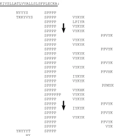
Ext 1.2A encodes a polypeptide of 311 amino acids which shows extensin features (Fig. 4). It has a putative signal peptide of 26 amino acids. The Ext 1.2A mature protein is rich in Pro (47%), Tyr (17%), Lys (14 %), Ser (10%) and Val (8%). It contains 28 (i) Ser-(Pro)4 motifs responsible for the polyproline II conformation adopted by the protein [38],
(ii) Val- or Ile-Tyr-Lys-Tyr-Lys motifs involved in isodityrosine intramolecular bonds [39] and eventually in intra or intermolecular di-isodityrosine cross-links [40] and (iii) Pro-Pro-Val-Tyr-Lys motifs which might be involved in the formation of Tyr-Lys inter-molecular cross-links [13, 14, 41]. MAKIVSLLATLVVALLSLSFPLECKA: NYYYS SPPPP TKKYVYS SPPPP VYKYK SPPPP LPIYR SPPPP VYKYK SPPPP VYKYK SPPPP PPVYK SPPPP VYKYK SPPPP VYKYK SPPPP PPVYK SPPPP VYKYK SPPPP VYKYK SPPPP PPVYK SPPPP VYKYK SPPPP PPVHK SPPPP IYKYK SPPPP VYKYK SPPPP PPMYK SPPPP VYKHK SPPPPPP VYKYK SPPPP VYKYK SPPPP PPVYK SPPPP IYKYK SPPPP PPVYK SPPPP VYKYK SPPPP PPVYK SPPPP VYK YHYYYT SPPPP HY
Fig. 4. Repetitive structure of the amino-acid sequence of the N. sylvestris
Ext 1.2A extensin. The sequence should be read from left to right and from top to bottom. Signal peptide is underlined. Repetitive units are arranged to emphasize their periodicity. The vertical arrows indicate the borders of the Ext 1.2A specific amino acid sequence .
3.2. Pattern of expression of the (-75/+124) and (-630/+124)Ext 1.2A/GUS gene fusions in tobacco plants
In order to identify cells expressing Ext 1.2A and to study the activity of the cloned promoter region, transgenic tobacco plants carrying the (-75/+124) or the (-630/+124)Ext 1.2A/GUS gene fusion were obtained. Twelve independent primary transformants were analyzed for each construct. The expression of chimeric genes was analyzed by histochemical staining after fixation of samples as described in Materials and Methods.
Fig. 5. Pattern of expression of the (-630/+124)Ext 1.2A/GUS gene fusion in transgenic
tobacco plants as revealed by in situ staining for GUS activity. (A-B) Stem internode of an adult plant: (A) Cross-section, (B) Close-up of A showing expression in inner and outer phloem. (C-D) Stem node of an adult plant: cross sections at two different levels showing expression in inner and outer phloem as well as in cortical cells at the stem/petiole junction. (E-H) Primary root grown in liquid medium in vitro: (E) Tip of the root, (F) Close-up of E showing expression in the root transition zone, (G) Cells of the root transition zone after coloration of nuclei with aceto carmine showing a metaphase indicated by an arrow, (H) Oriented line of root tip cells after coloration with aceto carmine showing the changes in shape undergone by cells in this zone (compare cells 1 and 2). (I-J) Effect of wounding on the expression of the gene fusion: (I) Cross-section of a wounded stem internode of an adult plant 48 h after wounding, (J) Leaf strip 48 h after wounding. When plant organs were sectioned prior to staining for GUS activity, samples were either fixed to avoid the effect of wounding on the expression of the gene fusion (A-D) or not fixed (I-J). (K) Germination of pollen grains: pollen grains and three steps of pollen tube growth. c = cortex, e = epidermis, ez = elongation zone, ip = inner phloem, m = meristem, op = outer phloem, p = pith, pz = piliferous zone, rc = root cap, tz = transition zone, x = xylem. A E ez pz tz I J p c e x op ip B F ez tz m rc G C D 2 1 m H K A E ez pz tz I J p c e x op ip B F ez tz m rc G C D 2 1 m H K
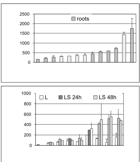
Fig. 6. Activity of the (-630/+124)Ext 1.2A/GUS gene fusion in transgenic
tobacco plants. (A) Roots of control non-transgenic plants (SR) and of 12 independent primary transformants were grown in liquid medium in vitro. Values reported are the average of 2 independent experiments for each plant. (B) Leaves were cut in thin strips and incubated in liquid medium for 24 h or 48 h. Control non-transgenic plants (SR) together with 8 independent primary transformants were analyzed. Values reported are the average of 4 independent experiments performed on each plant. In all cases, plants were ordered by increasing GUS activity from left to right and GUS activities were measured by a fluorometric assay. Numbers refer to the name of transgenic plants.
All plants carrying the (-630/+124)Ext 1.2A/GUS construct showed the same pattern of expression with different staining levels. As an example, samples from a plant showing one of the highest levels of expression are shown in Fig. 5, namely plant 48 (see Fig. 6). Expression of the (-630/+124) gene fusion was found in inner and outer phloem of stem internodes (Fig. 5A-B). In nodes, expression was observed in inner and outer phloem of both stem and petiole (Fig. 5C-D). GUS staining was systematically more intense in phloem cells at the junction of vascular tissues. Expression of the gene fusion was occasionally found in the cortex at the stem/petiole junction (Fig. 5 C-D). It was also found in mature pollen grains and during pollen germination where it follows the cytoplasm (Fig. 5K). In roots, GUS staining was observed in a region located between the root meristem where cells are actively dividing and the elongation zone (Fig. 5E-H). Staining of nuclei with aceto carmine revealed some cell divisions in this transition region as shown by the
0 500 1000 1500 2000 2500 roots 0 200 400 600 800 1000 L LS 24h LS 48h
presence of cells undergoing metaphase (Fig. 5G). These cells just started an isodiametric growth but have not yet reached the rapid elongation phase (Fig. 5H: compare sizes of cells 1 and 2). These data identified this region as the transition zone already described in roots of maize [42]. A weak tissue-specific histochemical staining was detected in 3 plants carrying the (-75/+124)Ext 1.2A/GUS gene fusion (results not shown).
The activity of the Ext 1.2A (-630/+124) promoter was also checked in roots with fluorometric assays (Fig. 6A). A high variability was observed between the different transgenic plants as previously described for other extensin genes [20, 43].
3.3. The (-630/-76) domain of the Ext 1.2A promoter contains wound-inducible regulatory elements active in stems, in ribs and in leaves
It was already shown that the Ext 1.2A gene is wound-inducible in leaf strips since 1.2 kb transcripts accumulate as early as 12 h after wounding [17]. Now the effect of wounding was analyzed in various tissues by comparing transcript accumulation in control plants and activities of the (-75/+124) and (-630/+124)Ext 1.2A/GUS gene fusions in transgenic plants.
Total RNAs were extracted from unwounded tissues and from wounded ribs, stems, leaves devoid of main ribs and roots grown in vitro in liquid medium at various times after wounding. They were analyzed on an RNA gel blot which was successively hybridized to the Ext 1.2 specific probe spanning its 3'UTR and to the 25S rRNA probe (Fig. 7). No Ext 1.2 mRNAs were detected in unwounded leaves and roots. However, 24 h and 48 h after wounding, 1.2 kb mRNAs were detected in both leaves and roots. In unwounded ribs and stems, a low level of accumulation of 0.9 kb mRNAs was found and it was not significantly modified in response to wounding. On the contrary, the 1.2 kb mRNAs level dramatically increased in wounded ribs and stems. Induction was detectable 8 h and 24 h respectively after wounding in ribs and stems respectively. It increased further after 48 h.
Histochemical GUS staining was detected at the place of the cut in all wounded stem tissues of plants carrying the (-630/+124)Ext 1.2A/GUS construct, including pith and cortex (Fig. 5: compare A and I). Induction of the gene fusion was also observed in leaf strips, especially in the rib and at the edges of the blade (Fig. 5J). These results are consistent with the accumulation of Ext 1.2A 1.2 kb mRNAs in response to wounding in stems, ribs and leaves described above. However, no additional histochemical staining was observed in wounded plants carrying the (-75/+124)Ext 1.2A/GUS construct (results not shown). In the same way, no staining was observed in wounded roots of transgenic plants carrying the (-630/+124)Ext 1.2A/GUS gene fusion (results not shown).
ribs stems UW W8 W24 W48 UW W8 W24 W48 Ext 1.2 1.2 kb 0.9 kb leaves roots UW W48 UW W8 W24 W48 1.2 kb
Fig. 7. RNA gel blot analysis of Ext 1.2 transcript levels in response to
wounding of different organs of N. tabacum plants. Ribs, stems, leaves devoid of major ribs and roots were cut in thin strips and incubated in liquid medium for 8 h (W8), 24 h (W24) or 48 h (W48). Total RNAs were extracted from control unwounded tissues (UW) and from wounded tissues (W8, W24 and W48). 10 µg of total RNAs extracted from leaves, ribs and stems and 5 µg of total RNAs extracted from roots were submitted to electrophoresis in an agarose-formaldehyde gel and subsequently transferred to a nylon membrane. Hybridizations were successively performed on the same membrane with the Ext 1.2 specific probe and a 25S rRNA probe. Sizes of Ext 1.2 transcripts are indicated in kb.
Fluorometric assays were performed on leaf strips 24 h and 48 h after wounding. In plants carrying the (-630/+124)Ext 1.2A/GUS construct, significant increase in GUS activity -at least 1.5 fold- was found 24 h after wounding in all cases. 48 h after wounding, GUS activity was found to be either stable or to increase further. However, in plants carrying the (-75/+124)Ext 1.2A/GUS gene fusion, no significant increase in GUS activity was found (results not shown).
4.
Discussion
The N. sylvestris Ext 1.2A gene was cloned by PCR. It encodes a highly repetitive protein only comprising motifs characteristic for extensins. During the cloning process, a lot of clones lacking the central part of the coding region in E. coli were obtained. These observations suggest that the 6PExt 1.2 cDNA previously isolated [17] originated from a recombination event leading to the deletion of the central part of its coding region. Such problems were already encountered during the cloning of Prunus amygdalus HRGP cDNAs [34] and of the NaPRP5 cDNA encoding a protein showing features of both extensins and arabinogalactan proteins [35].
In roots, a pattern of expression not yet described for an extensin gene was found. The (-630/+124)Ext 1.2A/GUS chimeric gene was expressed in the transition zone which is intercalated between the meristem and the rapidly elongating cells [44]. This region was also named “Postmitotic Isodiametric Growth” (PIG) [40] or “Distal Elongation Zone” (DEZ) [45]. In this zone, cells may complete mitosis before rapid elongation starts and before cell growth progressively becomes strictly isotropic in the longitudinal direction [46, 47]. Several changes were observed in these cells, among which reorientation of cortical microtubules and of cellulose microfibrils and an increase in XET activity [48, 49]. It was also shown that maintenance of elongation in the apical zone of maize primary roots
requires an increase in cell wall extension properties and in wall susceptibility to expansins [50]. The (-630/+124)Ext 1.2A/GUS chimeric gene has also been found to be expressed in germinating pollen grains that undergo tip growth. The role of the Ext 1.2 extensin in the wall of elongating cells might thus be different from that proposed in classical models. Indeed, extensins are supposed to be produced and insolubilized at the end of cell elongation by cross-links catalyzed by peroxidases [7, 13]. Instead, different levels of regulation might exist. The insolubilization of extensins might be delayed with regard to their excretion. Extensins might also be targeted to and/or insolubilized at particular places around the cells to allow polarized growth. Two recent papers support these hypotheses. A RSH-GFP fusion protein has been shown to be located at the edges of the cell plate in A. thaliana embryos [16]. The A. thaliana LRX1 chimeric protein comprising an extensin domain has been proposed to play a role in establishing and maintaining growth polarization of root hairs from the study of lrx1 knockout mutants [51].
The expression of the (-630/+124)Ext 1.2A/GUS gene fusion was also found in inner and outer phloem cells of stems and petioles. Although only 0.9 kb mRNAs could be observed in stems after Northern blot analysis, more sensitive RT-PCR experiments performed with PCR primers specific for the Ext 1.2A gene revealed a faint level of expression of this gene in stems (results not shown). This type of expression was also found for the Ext 1.4 tobacco extensin gene [19]. The expression of the (-630/+124)Ext 1.2A/GUS gene fusion was also occasionally found in cortical cells at the stem/petiole junction. Expression of other extensin genes was already described in such cells and was related to the existence of tensile stress exerted by the weight of the leaf [19, 43, 52]. However, expression of the (-630/+124)Ext 1.2A/GUS gene fusion was never found in roots at the emergence or at the branching of lateral roots. Such cells are also under mechanical constraints and they may express extensin genes such as the bean HRGP4.1 gene [43], the soybean SbHRGP3 gene [53], the tobacco Ext 1.4 gene [19] or the tobacco HRGPnt3 gene encoding a protein with a C-terminal extensin domain [54]. So, the Ext 1.2 protein seems to play a role in the reinforcement of the cell wall in response to mechanical constraints in some but not all plant tissues.
The Ext 1.2A gene is differentially induced upon wounding of stems, ribs, leaves devoid of ribs or roots. This regulation is thus superimposed on a tissue-specific expression and is driven by the (-630/+124)bp domain of its promoter except in roots. It means that additional regulatory elements involved in the root wound response are probably located upstream -630. Positive or negative regulations of extensin genes upon wounding were already reported for several extensin genes. The level of accumulation of tomato class I and class II extensin mRNAs increases in wounded leaves whereas it decreases in wounded roots [55]. The B. napus extA gene is induced in wounded leaves, stems and petioles whereas it is repressed in wounded roots [56]. It is also the only other gene known to have a wound responsive region called WAR (Wound Activating Region) located far upstream in its promoter between -3500 and -940 [57]. Histochemical analyses of transgenic plants carrying the (-630/+124)Ext 1.2A/GUS construct showed that, in response to wounding, the gene fusion is expressed in all stem tissues whereas in leaves, it is only expressed in ribs and in cells located at the edges of blade. This spatial pattern of wound activation is clearly different from what was already observed for the B. napus extA, the bean HRGP4.1 and the soybean SbHRGP3 extensin genes. Indeed, in wounded stems, the extA promoter/GUS and the HRGP4.1 promoter/GUS gene fusions were found to be expressed in inner and outer phloems and occasionally in the pith [43, 57].
Combining results obtained from histochemical and fluorometric analyses, it could be shown that the (-630/+124)bp domain of the Ext 1.2A promoter allows expression in stems, in roots as well as in response to wounding of stems and leaves. A few motifs showing homologies to elements previously described in promoters of two genes encoding cell wall structural proteins could be noticed. The CAAACTACTATATTTAAAT motif at position -23 shows homologies to the RSE (Root Specific Element) (9/18 mismatches) of the bean GRP1.8 promoter [58] and to the RSR (Root Specific Region) (5/17 mismatches) of the B. napus extA promoter [57]. However, although these two genes are also expressed in roots, it is not in the root transition zone like Ext 1.2A. The TACGTG motif at position -348, which is very close to a Gbox (CACGTG), is also found in the QAR (Quantitative Activator Region) of the extA promoter [57]. The GTGG motif at position -428 was found in a negatively acting element of the GRP1.8 promoter (Torres-Schumann et al. 1996) and in the NRR (Negative Regulatory Region) of the extA promoter [57]. It is proposed to be part of a bZip nuclear protein binding site [59]. But, these three genes are regulated in very different ways and only a detailed functional study of the Ext 1.2A promoter will allow to identify cis-regulatory elements active in vivo.
In tobacco plants, the two extensins, Ext 1.2A and Ext 1.4, appear to play different roles during growth and development and in response to stresses. Ext 1.2A is mainly expressed in the root transition zone where it may play a role in polarization of elongation (this work) whereas Ext 1.4 is expressed in cells submitted to mechanical constraints [19, 20]. Ext 1.2A is tightly regulated in response to wounding (this work) whereas Ext 1.4 is specifically induced in response to imposed mechanical constraints [20]. Both Ext 1.2A and Ext 1.4 probably contribute to reinforcement of cell wall structures in all these situations, but each of them in a specific way due to their different primary structures.
Ackowledgements
We are indebted to Dr. B. Kloareg, Dr. N. Chaubet-Gigot and Prof. M.-T. Esquerré-Tugayé for fruitful discussions. We acknowledge Dr. R. Bronner for help in histochemical analyses, Alexandre Mader and Sylvia Notter for skilful technical assistance. We wish to thank P. Keltz and R. Wagner for taking care of transgenic plants and P. Hammann for the sequencing work. This work was supported by CNRS and by a grant from "Ministère de l’Éducation Nationale, de l’Enseignement Supérieur, de la Recherche et de la Technologie" to P. G.
References
[1] K. Roberts, How the cell wall acquired a cellular context, Plant Physiol. 125 (2001) 127-130.
[2] H. Shi, Y. Kim, Y. Guo, B. Stevenson, J.K. Zhu, The Arabidopsis SOS5 locus encodes a putative cell surface adhesion protein and is required for normal cell expansion, Plant Cell 15 (2003) 19-32.
[3] R. Pennell, Cell walls: structures and signals, Cur. Opin. Plant Biol. 1 (1998) 504-510.
[4] D.J. Bradley, P. Kjellbom, C.J. Lamb, Elicitor- and wound-induced oxidative cross-linking of a proline-rich plant cell wall protein: a novel, rapid defense response, Cell 70 (1992) 21-30.
[5] H. Thordal-Christensen, Z. Zhang, Y. Wei, D.B. Collinge, Subcellular localization of H2O2 in plants. H2O2 accumulation in papillae and hypersensitive response during the
barley-powdery mildew interaction, Plant J. 11 (1997) 1187-1194.
[6] A.L. Samuels, T.H. Giddings Jr, L.A. Staehelin, Cytokinesis in tobacco BY-2 and root tip cells: a new model of cell plate formation in higher plants, J. Cell Biol. 130 (1995) 1345-1357.
[7] N.C. Carpita, D.M. Gibeaut, Structural models of primary cell walls in flowering plants: consistency of molecular structure with the physical properties of the walls during growth, Plant J. 3 (1993) 1-30.
[8] D.J. Cosgrove, Expansive growth of plant cell walls, Plant Physiol. Biochem. 38 (2000) 109-124.
[9] G.I. Cassab, Plant cell wall proteins, Annu. Rev. Plant Physiol. Plant Mol. Biol. 49 (1998) 281-309.
[10] F. Nicol, H. Höfte, Plant cell expansion: scaling the wall, Cur. Opin. Plant Biol. 1 (1998) 12-17.
[11] R. Yokoyama, K. Nishitani, Functional diversity of xyloglucan-related proteins and its implications in the cell wall dynamics in plants, Plant Biol. 2 (2000) 598-604.
[12] D.J. Cosgrove, Enzymes and other agents that enhance cell wall extensibility, Annu. Rev. Plant Physiol. Plant Mol. Biol. 50 (1999) 391-417.
[13] S. Schnabelrauch, M. Kieliszewski, B.L. Upham, H. Alizedeh, D.T.A. Lamport, Isolation of pI 4.6 extensin peroxidase from tomato cell suspension cultures and identification of Val-Tyr-Lys as putative intermolecular cross-link site, Plant J. 9 (1996) 477-489.
[14] P.A.P. Jackson, C.I.R. Galinha, C.S. Pereira, A. Fortunato, N.C. Soares, S.B.Q. Amâncio, C.P. Pinto Ricardo, Rapid deposition of extensin during the elicitation of grapevine callus cultures is specifically catalyzed by a 40-kilodalton peroxidase, Plant Physiol. 127 (2001) 1065-1076.
[15] E. Jamet, P. Guzzardi, I. Salvà, What do transgenic plants tell us about regulation and function of cell wall structural proteins like extensins? Russ. J. Plant Physiol. Engl. Tr. 47 (2000) 318-326.
[16] Q. Hall, M.C. Cannon, The cell wall hydroxyproline-rich glycoprotein RSH is essential for normal embryo development in Arabidopsis, Plant Cell 14 (2002) 1161-1172. [17] Y. Parmentier, A. Durr, J. Marbach, C. Hirsinger, M.C. Criqui, J. Fleck, E. Jamet, A novel wound-inducible extensin gene is expressed early in newly isolated protoplasts of Nicotiana sylvestris, Plant Mol. Biol. 29 (1995) 279-292.
[18] C. Hirsinger., Y. Parmentier, A. Durr, J. Fleck, E. Jamet, Characterization of a tobacco extensin gene and regulation of its gene family in healthy plants and under various stress conditions, Plant Mol. Biol. 33 (1997) 279-289.
[19] C. Hirsinger, I. Salvà, J. Marbach, A. Durr, J. Fleck, E. Jamet, The tobacco extensin gene Ext 1.4 is expressed in cells submitted to mechanical constraints and in cells proliferating under hormone control, J. Exp. Bot. 50 (1999) 343-355.
[20] I. Salvà, E. Jamet, Expression of the tobacco extensin gene upon mechanical constraints and localization of regulatory regions, Plant Biol. 3 (2001) 1-10.
[21] P. Maliga, A. Sz.-Breznovits, L. Marton, Streptomycin-resistant plants from callus culture of haploid tobacco, Nature New Biol. 244 (1973) 29-30.
[22] T. Murashige, F. Skoog, A revised medium for rapid growth and bioassays with tobacco tissue cultures, Physiol. Plant. 15 (1962) 473-497.
[23] R. Martoja, M. Martoja-Pierson, Initiation aux Techniques de l'Histologie Animale, Masson, Paris, 1967.
[24] G.J. Goodall, K. Wiebauer, W. Filipowicz, Analysis of pre-mRNA processing in transfected protoplasts, Meth. Enzymol. 88 (1990) 148-161.
[25] A.P. Feinberg, B. Vogelstein, A technique for radiolabelling DNA restriction endonuclease fragments to high specific activity, Anal. Biochem. 132 (1983) 6-13.
[26] T. Triglia, M.G. Peterson, D.J. Kemp, A procedure for in vitro amplification of DNA segments that lie outside the boundaries of known sequences, Nucleic Acids Res. 16 (1988) 8186.
[27] J. Sambrook, E.F. Fritsch, T. Maniatis, Molecular Cloning: A Laboratory Manual, 2nd edition, Cold Spring Harbor Laboratory Press, New-York, 1989.
[28] F. Sanger, S. Nicklen, A.R. Coulson, DNA sequencing with chain-terminating inhibitors, Proc. Natl. Acad. Sci. USA 74 (1977) 5463-5467.
[29] S. Tabor, C.C. Richardson, DNA sequence analysis with a modified bacteriophage T7 DNA polymerase, Proc. Natl. Acad. Sci. USA 84 (1987) 4767-4771.
[30] S. Tabor, C.C. Richardson, Effect of manganese ions on the incorporation of dideoxynucleotides by bacteriophage T7 DNA polymerase and Escherichia coli DNA polymerase I, Proc. Natl. Acad. Sci. USA 86 (1989) 4076-4080.
[31] R.A. Jefferson, T.A. Kavanagh, M.W. Bevan, GUS fusions: ß-glucuronidase as a sensitive and versatile gene fusion marker in higher plants, EMBO J. 6 (1987) 3901-3907. [32] R.A. Jefferson, Assaying chimeric genes in plants: the GUS gene fusion system, Plant Mol. Biol. Rep. 5 (1987) 387-405.
[33] M.M. Bradford, A rapid and sensitive method for the quantitation of microgram quantities of protein utilizing the principle of protein-dye binding, Anal. Biochem. 72 (1976) 248-254.
[34] J. Garcia-Mas, R. Messeguer, P. Arùs, P. Puigdomènech, The extensin from Prunus amygdalus, Plant Physiol. 100 (1992) 1603-1604.
[35] C.J. Schultz, K. Hauser, J.L. Lind, A.H. Atkinson, Z.Y. Pu, M.A. Anderson, A.E. Clarke, Molecular characterisation of a cDNA sequence encoding the backbone of a style-specific 120 kDa glycoprotein which has features of both extensins and arabinogalactan proteins, Plant Mol. Biol. 35 (1997) 833-845.
[36] B.A. Hanley, M.A. Schuler, Plant intron sequences: evidence for distinct groups of introns, Nucleic Acids Res. 16 (1988) 7159-7176.
[37] C.P. Joshi, Putative polyadenylation signals in nuclear genes of higher plants: a compilation and analysis, Nucleic. Acids Res. 15 (1987) 9627-9640.
[38] G.J. Van Holst, J.E. Varner, Reinforced polyproline II conformation in a hydroxyproline-rich cell wall glycoprotein from carrot root, Plant Physiol. 74 (1984) 247-251.
[39] L. Epstein, D.T.A. Lamport, An intramolecular linkage involving isodityrosine in extensin, Phytochem. 23 (1984) 1241-1246.
[40] J.D. Brady, I.H. Sadler, S.C. Fry, Di-isodityrosine, a novel tetrameric derivative of tyrosine in plant cell wall proteins: a new potential cross-link, Biochem. J. 315 (1996) 323-327.
[41] M.J. Kieliszewski, D.T.A. Lamport, Extensin: repetitive motifs, functional sites, post-translational codes, and phylogeny, Plant J. 5 (1994) 157-172.
[42] F. Baluska, S. Kubica, M. Hauskrecht, Postmitotic "isodiametric" cell growth in the maize root apex, Planta 181 (1990) 269-274.
[43] K.L. Wycoff, P.A. Powell, R.A. Gonzales, D.R. Corbin, C. Lamb, R.A. Dixon, Stress activation of a bean hydroxyproline-rich glycoprotein is superimposed on a pattern of tissue-specific developmental expression, Plant Physiol. 109 (1995) 41-52.
[44] F. Baluska, D. Volkmann, P.W. Barlow, Specialized zones of development in roots: view from cellular level, Plant Physiol. 112 (1996) 3-4.
[45] H. Ishikawa, M.L. Evans, Specialized zones of development in roots, Plant Physiol. 109 (1995) 725-727.
[46] F. Baluska, P.W. Barlow, S. Kubica, Importance of the post-mitotic isodiametric growth (PIG) region for growth and development of roots, Plant Soil 167 (1994) 31-41. [47] V.B. Ivanov, Relationship between cell proliferation and transition to elongation in plant roots, Int. J. Dev. Biol. 41 (1997) 907-915.
[48] F. Baluska, J.S. Parker, P.W. Barlow, Specific patterns of cortical and endoplasmic microtubules associated with cell growth and tissue differentiation in roots of maize (Zea mays L.), J. Cell Sci. 103 (1992) 191-200.
[49] J. Pritchard, P.R. Hetherington, S.C. Fry, A.D. Tomos, Xyloglucan endotransglycosylase activity, microfibril orientation and the profiles of cell wall properties along growing regions of maize roots, J. Exp. Bot. 44 (1993) 1281-1289.
[50] Y. Wu, R.E. Sharp, D.M. Durachko, D.J. Cosgrove, Growth maintenance of the maize primary root at low water potentials involves increase in cell-wall extension properties, expansin activity, and wall susceptibility to expansins, Plant Physiol. 111 (1996) 765-772.
[51] N. Baumberger, C. Ringli, B. Keller, The chimeric leucine-rich repeat/extensin cell wall protein LRX1 is required for root hair morphogenesis in Arabidopsis thaliana, Genes Dev. 15 (2001) 1128-1139.
[52] A.H. Shirsat, A. Bell, J. Spence, J.N. Harris, The Brassica napus extA extensin gene is expressed in regions of the plant subject to tensile stresses, Planta 199 (1996) 618-624. [53] J.H. Ahn, Y. Choi, Y.M. Kwoon, S.G. Kim, Y.D. Choi, J.S. Lee, A novel extensin gene encoding a hydroxyproline-rich glycoprotein requires sucrose for its wound-inducible expression in transgenic plants, Plant Cell 8 (1996) 1477-1490.
[54] B. Keller, C.J. Lamb, Specific expression of a novel cell wall hydroxyproline-rich glycoprotein gene in lateral root initiation, Genes Dev. 3 (1989) 1639-1646.
[55] A.M. Showalter, A.D. Butt, S. Kim, Molecular details of tomato extensin and glycine-rich protein gene expression, Plant Mol. Biol. 19 (1992) 205-215.
[56] A.H. Shirsat, D. Wieczorek, P. Kozbial, A gene for Brassica napus extensin is differentially expressed on wounding, Plant Mol. Biol. 30 (1996) 1291-1300.
[57] K.A. Elliott, A.H. Shirsat, Promoter regions of the extA extensin gene from Brassica napus control activation in response to wounding and tensile stress, Plant Mol. Biol. 37 (1998) 675-687.
[58] B. Keller, C. Baumgartner, ) Vascular-specific expression of the bean GRP 1.8 gene is negatively regulated, Plant Cell 3 (1991) 1051-1061.
[59] S. Torres-Schumann, C. Ringli, D. Heierli, N. Amrhein, B. Keller, In vitro binding of the tomato bZIP transcriptional activator VSF-1 to a regulatory element that controls xylem-specific gene expression, Plant J. 9 (1996) 283-296.