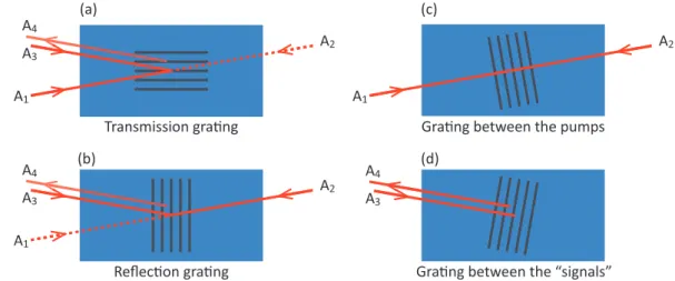
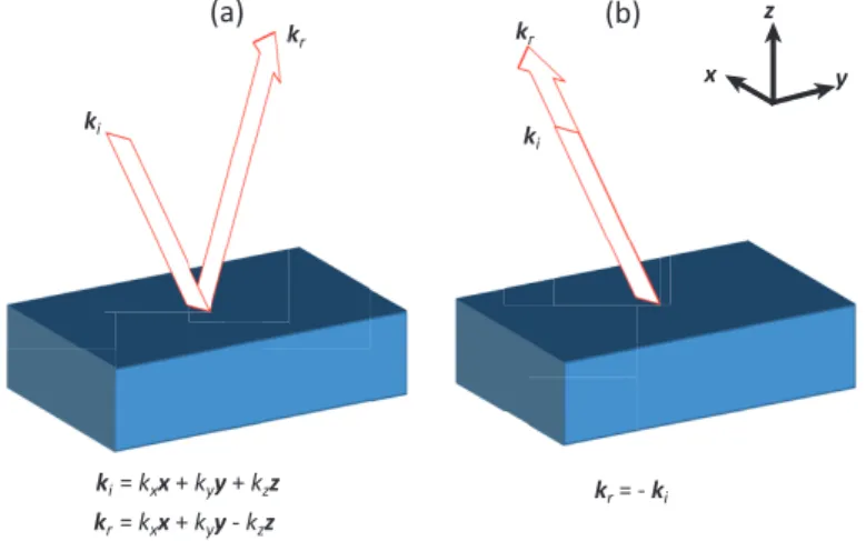
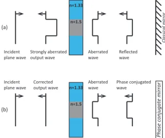
Acousto-optic and photoacoustic imaging of scattering media using wavefront adaptive holography techniques in NdYO4
Texte intégral
Figure


Documents relatifs
Somatomedin C and other hormones, as well as blood metabolites, were measured during the dry period and during lactation in dairy cows, given different amounts of energy and protein,
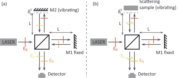
The light probe– phonon scattering process can be monitored, thanks to an interferometric effect involving the femtosecond light probe pulse reflection at the free surface (beam 1
When the movie depicted the participant's self right hand (Self Hand condition) he/she felt the illusory sensation that his/her right hand was actually moving, whereas when the
Using these reconstructed activity of each of the 68 brain regions, Phase Locking Values (PLV) were computed between each pair of channels (Hassan and Wendling 2015). Computing
The proof of the Main Theorem is then contained in Sections 4 & 5 : at first we show the result under the stronger assumption that F is µ−uniformly convex everywhere; then we
The integrated model used in the analysis included 0.1% modal damping, a 100 Hz bandwidth optical (fringe tracking) control loop, and reaction wheel disturbance
[r]
Although aspirin and ticagrelor used separately slightly decreased platelet aggregation compared to untreated and infected PRP ( Figures 1B and 2C ), the highest decrease was
