Université de Montréal
Identifying the Causal SNP(s) Determining Dalcetrapib
Responses
par Magdalena Burchert
Département de Biologie moléculaire Faculté de médecine
Mémoire présenté
en vue de l’obtention du grade de Maîtrise en Biologie Moléculaire
Option Générale
Février 2019
i
RÉSUMÉ
Introduction: Le dalcétrapib est un inhibiteur de la protéine de transfert des esters de cholestérol (CETP) qui augmente le niveau du cholestérol-HDL. Des études d’association pangénomiques ont révélé une association entre les polymorphismes du gène adénylate cyclase de type 9 (ADCY9) et les réponses au dalcétrapib. Le but de cette étude était d’identifier le polymorphisme nucléotidique (SNP) causal, ce qui pourrait mener à comprendre le mécanisme moléculaire modifiant les effets du dalcétrapib sur les bénéfices cardiovasculaires.
Méthodes: Des essais d’EMSA (electrophoretic mobility shift assay) ont été réalisés afin d’analyser les effets modificateurs de douze SNPs candidats sur la liaison de protéines nucléaires, provenant de cellules monocytaires THP-1. Ensuite, des essais de transfections avec un gène rapporteur ont été utilisées pour évaluer l’effet transcriptionnel de ces SNPs. La liaison des protéines au SNP rs12920508 a par la suite été étudiée par des chromatographies d’affinité d’ADN suivies par des spectrométries de masse et par MC-EMSA (multiplexed competitor EMSA).
Résultats: Sept sur douze SNPs ont démontré une liaison spécifique à un allèle qui n’a pas été influencée par l’exposition des cellules au dalcétrapib. Le résultat des transfections de vecteurs rapporteurs dans les cellules THP-1 a montré que les constructions plasmidiques portant les variants rs1967309 et rs12920508 augmentaient l’activité transcriptionnelle. Onze protéines ont été identifiées comme des candidats potentiels pouvant se lier à la région du SNP rs12920508. De plus, la région contenant les deux variants rs1967309 et rs12920509 a présenté une activité transcriptionnelle accrue et significativement plus élevée pour l’haplotype délétère.
Conclusion: Le polymorphisme rs1967309 semble causer la majorité des effets fonctionnels dans la lignée cellulaire THP-1. Cependant, une interaction avec le SNP rs12920508 ou la présence de la région de ce SNP pourrait être nécessaire pour l’activité optimale de rs1967309. Des travaux supplémentaires sont nécessaires pour élucider le lien entre le SNP potentiellement causal et les réponses cardiovasculaires induites par le dalcétrapib.
ii
ABSTRACT
Introduction: Dalcetrapib is a cholesteryl ester transfer protein (CETP) inhibitor that increases the circulating level of HDL-cholesterol. Genome-wide association studies have revealed an association between polymorphisms found in the adenylate cyclase type 9 (ADCY9) gene and responses to dalcetrapib, including its cardiovascular benefits. The purpose of this study was to identify the causal single nucleotide polymorphisms (SNP) which could lead to understand the molecular mechanisms altering dalcetrapib effects on cardiovascular outcomes.
Methods: Electrophoretic mobility shift assays (EMSA) were performed to analyze the allele-specific effects of the best causal SNP candidates on binding with nuclear proteins obtained from a THP-1 monocytic cell line. Afterwards, a dual luciferase reporter assay was used to assess the effect of selected genetic variants on gene transcription. Protein binding to SNP rs12920508 was investigated by DNA-affinity chromatography followed by mass spectrometry and multiplexed competitor EMSA.
Results: Seven out of 12 SNPs demonstrated allele-specific protein binding, which was not influenced by dalcetrapib exposure of the cells. Results from dual luciferase reporter assay showed that plasmid constructs bearing variants rs12920508 and rs1967309 increased transcriptional activity when transfected into THP-1 undifferentiated monocytic cells. Eleven proteins were identified as potential candidates binding to region of SNP rs12920508. Additionally, region containing both SNPs rs1967309 and rs12920508 displayed increased transcriptional activity with significantly higher activity for deleterious haplotype.
Conclusion: Polymorphism rs1967309 seems to be causing most functional effects in the THP-1 monocytic cell line. However, an interaction with rs12920508 or presence of the DNA region of this SNP may be necessary for optimal activity of rs1967309. Further work is required to elucidate the link between potentially causal SNPs and cardiovascular responses induced by dalcetrapib.
iii TABLE OF CONTENTS Résumé ... i Abstract ... ii List of tables ... v List of figures ... vi
List of abbreviations ... vii
Acknowledgements ... xi
1 Introduction ... 1
1.1 Atherosclerosis... 1
1.1.1 Risk factors for atherosclerosis ... 1
1.1.2 Pathogenesis of atherosclerosis... 2
1.2 Existing treatments ... 5
1.3 HDL and their atheroprotective properties ... 7
1.3.1 The HDL hypothesis and epidemiological studies ... 8
1.3.2 HDL composition and structure ... 9
1.3.3 HDL metabolism ... 11
1.3.4 Atheroprotective properties of HDL ... 12
1.4 CETP inhibitors ... 14
1.4.1 Mechanism of action of CETP inhibitors ... 14
1.4.2 Different CETP inhibitors ... 14
1.4.3 Evacetrapib and anacetrapib clinical trials ... 15
1.4.4 Dalcetrapib clinical trials ... 15
1.5 Genome-wide association study ... 16
1.5.1 GWAS Dalcetrapib ... 17
1.5.2 Association of dalcetrapib treatment and C-reactive protein levels... 20
iv
1.5.4 Linkage disequilibrium block located at ADCY9 gene ... 21
1.6 Adenylyl cyclases ... 22
1.6.1 Adenylyl cyclase 9 ... 22
1.6.2 Expression of ADCY9 ... 22
1.6.3 Regulation of ADCY9 activity ... 22
1.6.4 ADCY9 in cardiovascular disease ... 24
1.6.5 ADCY9 inactivation protects from atherosclerosis only in the absence of CETP... 25
1.7 From association to function ... 27
1.7.1 Prioritization of functional SNP... 27
1.7.2 How intronic SNP(s) can influence gene expression? ... 28
1.7.3 Bioinformatic analysis to prioritize putative causal SNP(s) ... 36
1.7.4 Experimental approaches to identify causal SNP(s) ... 37
1.8 Justification of the cell type choice for identification of causal SNP associated with cardiovascular responses of patients treated with dalcetrapib ... 42
2 Hypothesis and objectives ... 44
3 Article ... 46
4 Supplementary methods ... 87
5 Complementary results ... 88
6 Discussion ... 93
7 Conclusion and perspectives ... 101
v
LIST OF TABLES
Table I : Genetic variants identified by genome-wide association study in dal-OUTCOMES
(P value < 1 × 10⁻⁶) and dal-PLAQUE-2 (P value < 0.05) ... 19
Table II : Polymorphisms selected for functional analysis... 70
Table III : Sequences of 5’-biotinylated oligonucleotides used in EMSA. ... 71
Table IV : List of primers used for PCR amplification of cassettes ... 72
Table V : Location and size of cassettes used for luciferase reporter assay ... 73
Table VI : Transcription factors covered in seven competitor probe cocktails used in multiplexed competitor EMSA ... 74
Table VII : Antibodies used in chromatin immunoprecipitation assays. ... 75
Table VIII : Primers used for amplification of DNA regions containing SNP rs12920508, enriched by chromatin immunoprecipitation ... 76
Table IX : Summary of EMSA results for 12 tested SNPs ... 77
Table X : Eleven proteins identified by mass spectrometry binding significantly more to the C allele of SNP rs12920508 ... 78
vi
LIST OF FIGURES
Figure 1 : Monocytes in atherogenesis ... 4
Figure 2: Principal stages of HDL metabolism ... 12
Figure 3 : Cumulative incidence of cardiovascular events from dal-OUTCOMES study for the dalcetrapib and the placebo arm separately ... 18
Figure 4 : Graphical representation of adenylyl cyclase 9 gene ... 21
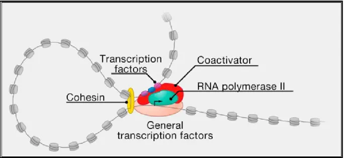
Figure 5 : Assembly of preinitiation complex ... 29
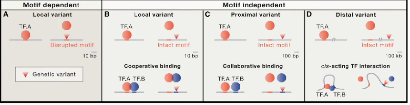
Figure 6 : Different mechanisms of TF-DNA binding variation by genetic variants ... 31
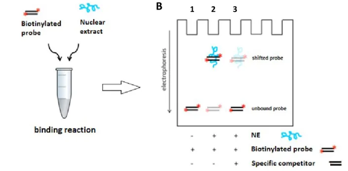
Figure 7 : Schematic representation of electrophoretic mobility shift (EMSA) assay ... 40
Figure 8 : SNPs selected for functional analysis with supplementary ENCODE and FANTOM5 data. ... 79
Figure 9 : SNP rs1967309 shows allele-specific protein binding. ... 80
Figure 10 : Exposure of cells to dalcetrapib did not influence protein binding... 81
Figure 11 : Luciferase reporter assays for SNPs rs1967309 and rs12920508 after transfection of THP-1 cell lines. ... 82
Figure 12 : TARDBP binds to the region of SNP rs12920508 in vivo. ... 84
Figure 13 : Multiplexed competitor EMSA with cocktails against the probe bearing C allele of variant rs12920508 assayed using nuclear extract derived from THP-1 monocytes. ... 85
Figure 14 : Luciferase reporter assay for DNA region containing SNPs rs1967309 and rs12920508 after transfection of THP-1 cell lines. ... 86
Figure 15 : Chromatin immunoprecipitation results analyzed by droplet digital PCR. ... 90
Figure 16 : EMSA with ISRE and GAS competitor probes. ... 91
Figure 17 : Dual luciferase reporter assay with IFN-γ, INFα-2a and INF-β treatment in THP-1 monocytic cell line. ... 92
vii
LIST OF ABBREVIATIONS
AAV8 : adeno-associated virus serotype 8 ABCA-1 : ATP-binding cassette transporter 1 ABCG-1 : ATP-binding cassette sub-family G 1 AC : adenylyl cyclase
ACS : acute coronary syndrome ADCY9 : adenylate cyclase type 9 ApoA-I : Apolipoprotein AI ApoA-II : Apolipoprotein AII ApoB: Apolipoprotein B Ago2 : Argonaute 2
ARNT : aryl hydrocarbon receptor nuclear translocator ATP : -adrenergic receptor
β2AR : β2-adrenergic receptor BMI : body mass index
cAMP : cyclic adenosine monophosphate CCR2 : C-C chemokine receptor type 2 CCL2 : C-C motif chemokine ligand 2 cDNA : complementary DNA
cd5/p35 : cyclin-dependent protein kinase 5/p35 complex CE : cholesteryl esters
CETP : cholesteryl ester transfer protein ChIP : chromatin immunoprecipitation CK1 : casein kinase 1
CoQ10 : ubiquinone
CRISPR : clustered regularly interspaced short palindromic repeats CVD : cardiovascular diseases
DGAT2 : hepatocyte microsomal diacylglycerol DHSs : DNase I hypersensitivity site
viii EMSA : electrophoretic mobility shift assay eNOS : endothelial nitric oxide synthase eQTL : expression quantitative trait loci GSPx : glutathione selenoperoxidase GTP : guanosine-5’-triphosphate
GWAS : genome-wide association study HDL : high-density lipoproteins
HL : hepatic lipase
HMG-CoA : 3-hydroxy-3-methyl-glutaryl-coenzyme A HOMER1 : homer scaffolding protein 1
hs-CRP : high-sensitivity C reactive protein Hsp20 : heat shock protein 20
ICAM : Intercellular adhesion molecule I IGF-1 : insulin-like growth factor 1 IL-1β : interleukin 1 β
IL-6 : interleukin 6
IMT : intima-media thickness
LCAT : lecithin-cholesterol acyltransferase LD : linkage disequilibrium
LDL : low-density lipoproteins
LDLR : low-density lipoprotein receptor lincRNA : long intergenic non-coding RNA lncRNA : long non-coding RNA
MCP-1 : monocyte chemotactic protein-1 M-CSF : macrophage colony stimulating factor miRNA : micro RNA
mRNA : messenger ribonucleic acid mTOR2 : rapamycin complex 2 Myo II : myosin II
ix ncRNA : non-coding RNA
NO : nitric oxide
PAF : platelet-activating factor
PCSK9 : proprotein convertase subtilisin/kexin type-9 PDGF : platelet-derived growth factor
PGI2 : prostacyclin
PIC : transcription preinitiation complex PKA : protein kinase A
PKC : protein kinase C
PLTP : phosholipid transfer protein Pol II : RNA polymerase II
PON-1 : paraoxonase 1
PPARα : peroxisome proliferator-activated receptor-α pre-mRNA : precursor mRNA
pri-mRNA : primary RNA PSL : piglet splay leg
P-TEFb : positive transcription elongation factor b qPCR : quantitative polymerase chain reaction RBP : RNA-binding proteins
RCT : reverse cholesterol transport RISC : RNA-induced silencing complex ROS : reactive oxygen species
SEC : super elongation complex SMC : smooth muscle cells
SNP : single nucleotide polymorphism SR-B1 : scavenger receptor class type 1 TAE : Tris-acetate buffer
TBE : Tris-borate buffer TBP : TATA-binding protein
x TF : transcription factor
TG : triglyceride TM : transmembrane
TNFα : tumor necrosis factor α TSS : transcription start site UTR : untranslated region
VCAM-1 : vascular cell adhesion protein 1 VLDL : very-low-density lipoprotein
xi
ACKNOWLEDGEMENTS
I would like to express my sincere appreciation towards all the great people I encountered during my master studies. First of all, I would like to thank my director Dr Jean-Claude Tardif and my co-director Dr Eric Rhéaume for giving me the opportunity to work on such an interesting and revolutionary project. Their extensive knowledge and valuable advices guided me throughout my master studies. I would also like to thank members of the Dr Tardif’s laboratory, in particular Dr Rocio Sanchez and Gabriel Théberge-Julien for teaching me new laboratory techniques. Moreover, I have to thank former master students Julien Renaud and Matthieu Blanchard for helpful tips about various laboratory techniques and administrative part of master program.
Outside of our laboratory, I would like to thank Isabel Gamache for all the lunches eaten together and long discussions about ADCY9 and favorite SNPs.
Finally, I would like to thank my parents and my fiancé for their support. Their continuously encouraged me during my studies and never stopped believing in me.
1
1 INTRODUCTION
1.1 Atherosclerosis
Cardiovascular disease is the leading cause of human death in North America and other developed countries. In most cases, the major cause of the cardiovascular event is atherosclerosis. Atherosclerosis is a chronic inflammatory disease characterized by the accumulation of lipids, inflammatory cells and fibrous elements in medium-sized and large arteries. In the heart, atherosclerosis can result in myocardial infarction and heart failure caused by coronary artery stenosis. In the brain, rupture of atherosclerotic plaques or stenosis may induce a stroke [1-3]. 1.1.1 Risk factors for atherosclerosis
Numerous risk factors for atherosclerosis were identified over the past years. Some of them, called modifiable risk factors, can be controlled in order to delay or prevent atherosclerosis progression. To the modifiable factors we can include: elevated levels of cholesterol and low-density lipoprotein (LDL), reduced levels of high-low-density lipoprotein (HDL), cigarette smoking (>10/day), hypertension, diabetes, severe obesity (>30% overweight) and inactive lifestyle [4]. The other risk factors such as sex, age, ethnicity and family history cannot be regulated and are therefore called non-modifiable risk factors.
The majority of enumerated risk factors are not independent and contribute to global risk profile. For example, diabetic patients often exert elevated levels of low-density lipoprotein-cholesterol (LDL-C) and hypertension that might contribute to atherogenesis. Also, active lifestyle is known to have protective effect on atherosclerosis, likely due to reduced blood pressure and body weight, increase in high-density lipoprotein-cholesterol (HDL-C) levels and decrease in LDL-C. Therefore, understanding the global risk profile of a patient could provide improved predictive power for atherosclerosis [5-7].
In addition, total risk profile of atherosclerosis is complicated even more by presence of genetic factors. One of the first genetic factors affecting cardiovascular diseases was discovered in a patient with hypocholesterolemia and his mother. Both patients possessed a 5-kb deletion in low-density lipoprotein receptor (LDLR) gene that result in reduced production of LDLR on the cell surface. Decreased levels of LDLR subsequently impair receptor–mediated hepatic uptake of LDL and lead to elevated levels of circulating cholesterol [8]. Another family-based study
2
identified mutations in two additional genes, ApoB and PCSK9, which cause hypercholesterolemia and elevated levels of LDL [9, 10].
1.1.2 Pathogenesis of atherosclerosis
Atherosclerosis is a chronic inflammatory condition, which remains unnoticed during its first phases of development. Formation of lesions begins with endothelial dysfunction, endothelial cell activation, accumulation of atherogenic LDL, inflammation and may lead to an acute clinical event induced by plaque rupture and thrombosis.
Endothelial dysfunction
The basic structure of blood vessel is divided into three morphologically distinct layers. The outermost layer called adventita is built from connective tissues with scattered fibroblasts and smooth muscle cells (SMCs). The middle layer, media, consists essentilally of SMCs. The innermost layer, which is in direct contact with the blood flow is called the intima. On the luminal side, the intima consists of a monolayer of endothelial cells, which play an important role in development of atherosclerosis [2]. The endothelium regulates vascular homeostasis, serving as a selectively permeable barrier between blood and tissues. The endothelium also produces a vast range of factors, which regulate processes affecting creation of atherosclerotic lesions, such as vascular tone, adhesion of cells, thromboresistance, proliferation of SMCs, and inflammation of vessel wall. One of the most important factors released by endothelium is a potent vasodilatator called nitric oxide (NO). Nitric oxide opposes the effect of endothelium-derived vasoconstrictors and inhibits oxidation of low-density lipoproteins. Defect in its production or activity leads to endothelial dysfunction [11, 12]. Oxidative stress, which may originate from smoking, hypertension, hyperlipidemic states and diabetes, can also interfere with the production and activity of nitric oxide and as a result provoke an inflammatory response, vascular remodeling and higher permeability of endothelium [3].
Monocyte adhesion and accumulation of LDL
Beside causing increased permeability of endothelium, endothelial dysfunction is also implicated in the inflammatory response, known as endothelial cell activation. Factors inducing endothelial cell activation are: certain bacteria and viruses, pro-inflammatory cytokines such as interleukin 1 and tumour necrosis factor, physical and oxidative stress, oxidised low density lipoproteins.
3
During endothelial cell activation, the endothelium express cell surface molecules, such as vascular cell adhesion protein 1 (VCAM-1), intercellular adhesion molecule 1 (ICAM-1), and endothelial leukocyte adhesion molecule (E-selectin). Expression of these surface molecules facilitates the recruitment and attachment of circulationg monocytes and T-lymphocytes to the vessel wall [13, 14] .
Increased permeability of arterial wall also favors accumulation of atherogenic lipoproteins, mainly LDL, which extravasate through the leaky and defective endothelium into the subendothelial space. Retained lipoprotein particles undergo oxidative modifications and become cytotoxic, proinflammatory and proatherogenic. The molecules responsible for such modifications are reactive oxygen species (ROS) as well as myeloperoxidases and lipoxygenases released by inflammatory cells and responsible for the production of HOCl and oxygenated lipids, respectively [15, 16].
Plaque progression
Oxidized lipids trigger secretion of chemokines (chemotactic cytokines) by overlying endothelial cells. Next, chemokines direct transmigration of adherent monocytes across endothelium into the intima. Monocyte chemotactic protein-1 (MCP-1) is the most important atherogenic chemoatractant. Its receptor on monocytes C-C chemokine receptor type 2 (CCR2) may be upregulated significantly during plaque development. Monocytes located within subendothelial space, differentiate into macrophages. The macrophages express scavenger receptors that bind oxidized lipoproteins. The expression of scavenger receptors is influenced by other important cytokines secreted by endothelial cells, namely macrophage colony stimulating factors (M-CSF) [2]. These receptors are not down-regulated by cholesterol accumulation, thus the macrophages internalize continously modified LDL, what leads to foam cell formation [1]. Foam cells trapped within arterial intima ultimately die and contribute to the formation of destabilizing lipid-rich core in the plaque. Transformation of the macrophages to foam cells may be inhibited by removal of excess cholesterol by HDL in a process called reverse cholesterol transport (RCT) [2].
Macrophages and T cells located within lesions produce growth factors, such as insulin-like growth factor 1 (IGF-1) and platelet-derived growth factor (PDGF), which induce smooth muscle cells (SMC) migration to arterial intima [17]. Subsequently, SMCs proliferate and produce collagen-rich matrix which cause plaque increase in size. Advanced lesions can grow sufficiently
4
large to block completely blood flow [2]. Nevertheless, smooth muscle cells confer stability to plaques, protecting them from more important consequences such as plaque rupture that can lead to thrombosis [15].
Additionally, cholesterol crystals can form in foam cells and cause release of interleukin 1 β (IL-1β), which further provokes SMC to produce interleukin 6 (IL-6). Both inteleukins are pro-inflammatory, and induce synthesis of C-reactive protein (CRP) in the liver. Increased production of CRP results in its secretion into the circulation and subsequent rise in CRP serum concentration. Since the concentration of CRP in the serum is known to correlate with the occurence of cardiovascular disease, CRP levels are considered as a risk marker [18].
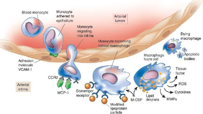
Figure 1 : Monocytes in atherogenesis. Monocytes are recruited to defective endothelium by
adhesion molecules. Adherent monocytes migrate into the intima and differentiate into macrophages. Macrophages internalize oxidized low-density lipoproteins and become foam cells. Trapped foam cells eventually die and contribute to atherosclerotic plaque formation. Figure presented with permission (Peter Libby, 2002) [19].
5
1.2 Existing treatments
Prevention is an important part of atherosclerosis treatment. Change of lifestyle including modification of diet, stimulation of physical activity and suspension of smoking may greatly reduce progression of the disease. However, if the modification of lifestyle is not sufficient, introduction of medical therapy is neccesary.
The most popular therapies reducing cardiovascular events focus on lowering the levels of atherogenic LDL cholesterol. Although LDL-C-lowering medications significantly improve cardiovascular health of the patients, remaining risk needs to be targeted by modulation of other factors, such as triglycerides (TGs) and HDL-C. In addition, there is more and more evidence that therapy outcomes vary depending on the patient genotype. Therefore, the future of cardiovascular treatment should focus on personalized medicine to deliver right treatment to the patients, taking into account their genetic profiles [20].
LDL-C-lowering treatments
Until now 3-hydroxy-3-methyl-glutaryl-coenzyme A (HMG-CoA) reductase inhibitors (statins) are the most effective and the most often prescribed medicines for atherosclerosis. Statins competitively inhibit HMG-CoA reductase and cause an elevated expression of LDL receptors on the hepatocytes surface. Upregulated expression of LDL receptors then leads to decreased plasma levels of LDL-C and other apo B-containing lipoproteins. Apart from their lipid lowering action, statins have been reported to exert anti-inflammatory activity by lowering serum levels of C-reactive protein [21]. Statins are generally safe and well-tolerated in patients. At the maximum dosage, the most effective statins reduce LDL-C levels by 55% to 60% [22]. Clinical trials with patients with and without coronary heart disease, reported that statins decrease the relative risk of major coronary events by ~30%. Also, patients with higher baseline risk, benefit more from statin treatment [23].
Although statins greatly reduce cardiovascular risk, patients often stop statin therapy due to adverse effects. Two causally related adverse effects of statins are myopathy and impairement of insulin resistance. Myopathy is the most common adverse effect of statins, and is caused by depletion of muscle ubiquinone (CoQ10), what results in impairment of mitochondrial function. In relation to insulin resistance, statins increase the risk of incident diabetes by ≈ 9% to 28% [24].
6
Ezetimibe is another medicine that reduces levels of LDL-C by inhibiting cholesterol absorption.
No significant adverse effects were observed for ezetimibe thus it is ofen used as second-line therapy in patients with contraindications to statins [20]. Also, therapy combining ezetimibe with statins was found to lower the risk of cardiovascular events in high-risk patients in comparison to statin therapy alone [25].
Bile acid sequestrants can produce a reduction in LDL-C of 18-25%, when used at the highest
dose. But their use is limited due to gastrointestinal secondary effects and interactions with other often prescribed drugs [20].
Recently discovered proprotein convertase subtilisin/kexin type-9 (PCSK-9) inhibitors are promising medicines, which significantly decrease LDL-C levels. PCSK9 was found to regulate LDL receptor degradation. Its inhibition prevents degradation of LDL receptors, improves absorption of LDL-C particles by liver and therefore decreases LDL-C plasma concentration. The limiting factors for PCSK-9 inhibitors are their current relatively high costs and lack of solid outcomes in large randomised controlled trials [26].
TG-lowering treatments
Beside LDL-lowering ability, statins also reduce plasma levels of TG-rich particles throughout inhibition of HMG-CoA reductase [21].
Another type of drugs which reduce concentrations of triglycerides are the agonists of peroxisome proliferator-activated receptor-α (PPARα), called fibrates. PPARα is a nuclear hormone receptor, which binds to DNA and modulates the transcription of genes involved in lipid and glucose metabolism. Fibrates decrease the plasma levels of TG by stimulating the catabolism of TG-rich lipoproteins [27].
HDL-C-increasing treatments
Treatment with fibrates is also associated with a moderate increase in HDL-C concentrations. Raise of HDL-C levels by fibrates results from the increased synthesis of the major HDL apolipoproteins, apoA-I and apoA-II , mediated by PPARα. Most studies conducted with fibrates reported coronary risk reduction after fibrate treatment [28]. However, the response of hypercholesterolemic patients to fibrates was found to vary. For example, patients with higher body mass index (BMI>29) and with higher probability of having the metabolic syndrome
7
benefited more from fibrate treatment. Also, fibrate treatment was associated with significantly less ischemic heart disease in patients with diabetes [29, 30]. The Helsinki Heart Study analysed the baseline levels of HDL-C in patients treated with fibrate (gemfibrozil) and found that patients with lower levels of baseline HDL-C showed greatest benefit from treatment [31]. However, there is controversy as to wheter the beneficial effects of fibrates are related to the increase of HDL-C or to an absolute reduction in LDL-C [32]. Surprisingly, in some patients with dyslipidemia, fibrate treatment resulted in increase of LDL-C levels [33]. Therefore, treatment with fibrates should be prescribed with consideration of patient profile.
Niacin increases the levels of HDL-C throught non-competitive inhibition of enzyme involved in
triglyceride synthesis, called hepatocyte microsomal diacylglycerol (DGAT2). Additionally, niacin raise HDL-C by selective inhibition of apoA-I uptake. In two recent clinical trials (AIM-HIGH and HPS2-THRIVE) niacin failed to significantly reduce cardiovascular events. In addition, niacin treatment is limited by adverse side-effects [34].
Another promising group of medications increasing levels of HDL-C are the cholesteryl ester transfer protein (CETP) inhibitors. CETP inhibitors increase HDL-C levels by inhibition of cholesteryl esters transfer from HDL to apoB-containing proteins, and are described in detail in chapter 1.3.
1.3 HDL and their atheroprotective properties
Although statins constitute an essential part of the standard of care forin cardiovascular disease (CVD) secondary prevention, there remains a significant CV risk in these patients. Therefore, development of new therapies with different targets is necessary [35]. Low plasma concentrations of high-density lipoprotein cholesterol are commonly associated with higher risk for cardiovascular disease in patients. Thus, raising the levels of HDL-C became a target of many novel therapies. Disappointingly, the results from the majority of clinical trials demonstrate so far that despite significant rises of HDL-C levels in patients, HDL-raising drugs show no significant improvement in cardiovascular events. Such results emphasize the complexity of high-density lipoprotein molecule structure, metabolism and function. It is important for further studies in HDL field to consider different subclasses of HDL particles and their various involvement in atheroprotective mechanisms, such as reverse-cholesterol transport [34].
8
1.3.1 The HDL hypothesis and epidemiological studies
Early studies in 1951, reported by Barr et al. showed that patients with coronary artery disease have reduced plasma levels of high-density lipoprotein cholesterol [36]. A subsequent Framingham study of Gordon et al. in 1977, demonstrated that low level of HDL-C is a risk factor for coronary heart disease [37]. These discoveries initiated multiple HDL studies, which reported numerous beneficial effects of increased levels of HDL-C and led to formulation of the HDL hypothesis. This hypothesis states that the intervention to raise plasma levels of HDL-C protects against atherosclerosis. Following this belief, strategies to raise plasma HDL levels were developped but they did not bring the expected results so far. Therefore, the HDL hypothesis started to be questioned.
In the last few decades many epidemiological studies supported the HDL hypothesis [38]. A meta-analysis of four large studies, including in total 15252 individuals, showed that each 1 𝑚𝑔 𝑑𝐿⁄ raise of HDL-C levels was associated with a 2-3% decrease in risk of cardiovascular disease [39]. However, the fact that many factors affect both CVD risk and HDL-C levels brings uncertainty to the causality of HDL-C alone. For example, it was shown that women have on average higher levels of HDL-C than men [40]. Smokers show 14% lower levels of HDL-C than nonsmokers [41]. Also, it was reported that regular aerobic exercise raise HDL-C levels by around 2.5 𝑚𝑔 𝑑𝑙⁄ [42]. Moreover, the abdominal obesity is associated with lower HDL-C levels, whereas weight loss is associated with raise of HDL-C levels [43, 44]. Finally, systemic inflammation that is a known risk factor for CVD was shown to be associated with low levels of HDL-C [45].
Studies investigating human genetics of HDL led to better understanding of HDL metabolism, but also suggested that HDL-C may not be significantly associated with cardiovascular risk. Three mendelian disorders causing the loss of the capacity to produce mature HDL, were identified up to date [46]. These include the apoA-1 structural mutations, ABCA1 deficiency (Tangier disease), and familial LCAT deficiency. Although, studying these disorders helped identification of genes and proteins playing important role in regulation of HDL-C levels, they did not demonstrate convincing association with increased risk of CVD.
The other evidence causing doubt in HDL hypothesis are the clinical trials with HDL-raising drugs, niacin and CETP inhibitors [47]. Niacin is a medication used to raise HDL-C levels and
9
lower LDL-C and TGs. A trial called Coronary Drug Project reported that niacin reduced cardiovascular events in hypercholesterolemic men [48]. Two more recent trials AIM-HIGH and HPS2-THRIVE, which studied treatment of patients with cardiovascular heart disease who had well-controlled LDL-C levels, failed to demonstrate significant reduction of cardiovascular events. However, there is uncertainty associated with interpretations of these two trials. The number of individuals taking part in AIM-HIGH study was relatively low, and there was a modest increase in HDL-C levels in the treated group compared to placebo [49]. The HPS2-THRIVE trial was larger but included a additional drug called laropiprent, which reduces the flushing associated with nacin [50]. Although HPS2-THRIVE study failed to demonstrate significant association of niacin therapy with lower cardiovascular risk, a tendency was observed with significant benefit in a group of patients with higher baseline LDL-C levels.
Another HDL-raising group of drugs, which failed to confirm HDL hypothesis is called CETP inhibitors. Torcetrapib was the first CETP inhibitor to enter in a phase III clinical trial called ILLUMINATE [51]. In the ILLUMINATE study, torcetrapib raised HDL-C levels by 72.1 %, but this trial was prematurely terminated due to elevated rate of cardiovascular events and mortality in the torcetrapib group. Such unexpected results were further explained by the fact that torcetrapib has off-target effect on blood pressure and aldosterone that may explain the increased cardiovascular risk [52]. What is more, group of researchers investigating effects of torcetrapib in animal species, claimed that the blood pressure response caused by torcetrapib is independent on CETP inhibition [53]. They showed that torcetrapib increases blood pressure to an equivalent extent in normal and CETP transgenic mice. Also, the same group demonstrated that treatment of CETP transgenic mice with another CETP inhibitor, anacetrapib results in an equal rise of HDL-C like with torcetrapib treatment, but has no influence on blood pressure. Explanations of failure of niacin and torcetrapib trials and the fact that HDL has been demonstrated with many anti-atherogenic properties (described in section “Atheroprotective properties of HDL”) bring hope for HDL-raising therapies.
1.3.2 HDL composition and structure
Plasma HDL represents an heterogenous group of small discoidal and spherical particles. The HDL particles distinguish themselves from other lipoprotein classes by they small particle size (7-12 nm diameter) and high protein content (30-70% by weight). The HDL proteins may be divided into four groups: apolipoproteins, enzymes, lipid transfer proteins and minor proteins that
10
make up <5% of total HDL proteins. Apolipoproteins and enzymes play an important role in HDL metabolism and function. Additionally, minor proteins, such as acute phase response proteins and proteins involved in regulation and protection against infectious disease gained increased attention in the past few years. The major HDL structural apolipoprotein is apolipoprotein (apo) A-I. The ApoA-I accounts for around 70% of total HDL proteins, and is significantly involved in HDL biogenesis, and function. Apolipoprotein (apo) A-II is the second main HDL apolipoprotein, and accounts for around 20% of total HDL proteins. Less aboundant HDL apolipoproteins include among others: apoA-IV, apoA-V, apoC-I and apoE. Some of the enzymes carried by HDL particles are involved in important mechanisms. For example, lecithin/cholesterol acyltransferase (LCAT) plays and important role in lipid metabolism. Other enzymes, paraoxygenase 1 (PON1), platelet-activating factor-acetyl hydrolase (PAF-AM) and glutathione selenoperoxidase (GSPx) have antioxidative activities. The HDL lipidome comprises 40-60% of phospholipids, 30-40% of cholesteryl esters, 5-12% of triglycerides and 5-10% of free cholesterol. Additionally, more than 200 individual lipid species were identified in HDL particles due to advances in lipidomic analyses. HDL was also found to carry multiple copies of microRNAs that may be delivered to cells and tissues [54].
The subclasses of plasma HDL particles differ significantly in their physicochemical properties, metabolism and biological function. The heterogeneity in HDL particles size and structure is caused by different conformations of apoA-I, induced by different quantity of attached lipids [55]. The smallest class of HDL particles is called pre-β-1 HDL. This subclass is represented by lipid-poor (lipid content ≤ 30%), ≤ 8 𝑛𝑚 diameter discoid particles. Pre-β-1 HDL particles mainly contain apoA-I together with small amounts of phospholipids and free cholesterol. Larger class of HDL particles, called spherical HDL are > 8 𝑛𝑚 in size and contain hydrophobic core of cholesteryl esters and triglycerides. Two subclasses of spherical HDL can be recognised, HDL2 and HDL3. The HDL2 are larger, lipid-rich particles, containing: 9% of TGs, 18% of CE, 6% of free cholesterol and 25% of PLs, of total HDL mass. The HDL3 particles are smaller and denser than HDL2, and contain lipid-poor HDL subfraction, including: 7% of TGs, 14% of CEs, 3% of free cholesterol and 23% of PLs of total HDL mass [54].
In-depth understanding of different properties of HDL subclasses is crucial for comprehension of HDL protective properties. A recent meta-analysis of two large studies (Framingham Offspring
11
Study and Jackson Heart Study) showed that HDL2 and HDL3 subclasses were differently associated with cardiovascular events. Only HDL3 was found to significantly reduce cardiovascular risk [56].
1.3.3 HDL metabolism
The main function of HDL proteins is to transport cholesterol and other lipids between circulating cells, lipoproteins, tissues and organs. During this process, HDL particles undergo dynamic remodeling (Figure 2) [54]. At first small precursors of mature HDL, lipid-free apoAI and lipid-poor pre-β-1 HDL particles are synthesized by liver and intestine. Next, these particles collect increasing quantities of free cholesterol from peripheral tissues, via ATP binding cassette transporter 1 (ABCA-1) mediated efflux. Afterwards, lecithin/cholesterol acyltransferase (LCAT) transfers fatty acid residues from lecithin to the hydrozyl group of cholesterol, what results in formation of cholesteryl esters. Cholesteryl esters have hydrophobic properties, therefore they migrate into the hydrophobic core of HDL particles, converting discoidal HDL into lighter and larger spherical HDL3. Further, acquired cholesterol can be converted to cholesterol esters by LCAT enzyme and create less dense spherical HDL2 particles. Phospholipids for the LCAT reaction are transfered from VLDL into HDL by phospholipid transfer protein (PLTP) [57]. Spherical HDL particles can promote cholesterol efflux from peripheral cells, such as macrophage foam cells, by aqueous diffusion, ATP-binding cassette sub-family G member 1 (ABCG-1) or scavenger receptor class type 1 (SR-B1) [58]. HDL2 then transfer cholesterol esters to the liver through SR-B1 receptor. After HDL2 particles return CE to liver they become HDL3 particles again. The other pathway which involves HDL2 and HDL3 is the CETP-mediated transfer of cholesteryl esters from spherical HDL to apoB-containing lipoproteins, such as LDL and VLDL. Throughout heteroexchange of cholesteryl esters and triglycerides between HDL and apoB-containing lipoproteins, TG-rich particles are created. These TG-rich HDLs can be hydrolysed by hepatic lipase (HL) to small TG-rich particles. Next, upon action of both CETP and HL, the HDL size is reduced and lipid-poor HDL particles are generated, which then can interact with ABCA1 in next lipidation cycle [59]. Therefore, the HDL lipids are catabolised in the liver upon selective uptake via scavenger receptor class type 1 (SR-B1), or upon CETP-mediated transfer to apoB-containing particles.
12
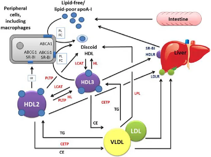
Figure 2: Principal stages of HDL metabolism. HDL discoidal particles, synthesised by the liver
acquire cholesterol from peripheral cells by ABCA1-mediated cholesterol efflux. Free cholesterol in discoidal HDL is converted into cholesterol ester by LCAT. Cholesterol esters migrate into hydrophobic core of HDL and generate spherical HDL. Mature HDL can further acquire free cholesterol from peripheral tissues by aqueous diffusion or cholesterol efflux mediated by either SR-B1 receptor or ABCG1. Finally, HDL cholesterol esters and free cholesterol are transferred to the liver, via SR-B1 receptor for elimination. Figure presented with permission (Fabian H. Rached, 2015) [54].
1.3.4 Atheroprotective properties of HDL
The main objective of HDL research nowadays concentrates on understanding the mechanisms by which HDL protects against atherosclerosis. The most important antiatherogenic function of HDL is reverse-cholesterol transport (RCT), a process crucial for HDL particle maturation. In RCT process, HDL particles collect free cholesterol from lipid-rich macrophage foam cells and as a result facilitate plaque regression. Acquired cholesterol is further excreted from organism by the liver and biliary system [58]. Epidemiological studies showed strong inverse association between cholesterol efflux capacity and coronary and peripheral atherosclerosis [34].
13
Reverse cholesterol transport starts with the cholesterol efflux from macrophages to HDL particles. Four cellular efflux pathways were identified, which involve key proteins, such as SR-B1, ABCG1 and ABCA1. Different subclasses of HDL particles were shown to interact differently with these proteins, possibly influencing the cholesterol efflux capacity. The first pathway that mediates the bidirectional flux of cholesterol between the cell plasma membrane and HDL is called aqueous diffusion. The direction of mass transport of cholesterol is determined by the cholesterol concentration gradient in the donor and acceptor molecules [60]. Since, cholesterol efflux in this pathway is not significantly affected by the size of HDL particle, the efflux by aqueous diffusion is equally effective for all HDL subclasses [61]. In the second pathway, transfer of cholesterol to HDL particles is facilitated by B1 [62]. HDL binds to SR-B1 and forms complex, which possess hydrophobic channel via which cholesterol molecules may diffuse. Larger HDL particles are more effective in mediating cholesterol efflux by this pathway, because they bind better to SR-B1 receptor [63, 64]. In the third pathway, ABCG1 increases the pool of free cholesterol in plasma membrane and reorganises it so it desorbs better into the extracellular medium. Larger HDL2 and smaller HDL3 are equally effective acceptors in this pathway. Discoidal HDL, created by the apoAI/ABCA1 reaction are also an effective acceptors of cholesterol effluxed by ABCG1 [65, 66]. Last pathway for cellular cholesterol efflux is mediated by ABCA1 receptor. In this case pre-β-1 HDL particles are the main acceptors. Subsequently, cholesterol collected by HDL particles is transferred through the plasma compartment by diverse HDL remodeling steps described in the previous section, “HDL metabolism”, and is delivered to the liver. The cholesterol uptake by the liver is mediated by SR-B1 receptor expressed at hepatocytes’ surface. HDL particles bind to the receptor and free cholesterol and cholesterol esters are diffused into the cell plasma membrane. Large HDL particles (10 nm diameter) bind better to SR-B1 receptor, thus deliver more cholesterol ester than small HDL particles (8 nm diameter) [57].
Other antiatherogenic properties of HDL include its antioxidant, inflammatory and anti-thrombotic functions. HDL exert antioxidant activity thanks mainly to its associated enzyme called paraoxonase-1 (PON1). PON1 reduces formation of lipid peroxide known to oxidize LDL. Oxidized LDL accumulates on endothelium and disrupts its structural integrity and function. Thus, PON1 activity decrease the risk of cardiovascular event [34, 67]. Anti-inflammatory properties of HDL concern the ability of HDL particles to inhibit expression of adhesion
14
molecules in endothelial cells, such as VCAM-1, ICAM-1 and E-selectin. Down-regulation of these molecules causes reduced recruitment of monocytes to the arterial wall [34, 68]. Few HDL functions are responsible for their anti-thrombotic properties. HDL upregulate the expression of NO synthase (eNOS), which reduces vasorelaxation. HDL also activate prostacyclin (PGI2). PGI2 is known to inhibit platelet activation and proliferation of smooth muscle cells [34].
1.4 CETP inhibitors
CETP inhibitors belong to the group of drugs developed to increase HDL-C levels in the blood. The interest in CETP inhibition as a therapeutic approach began with observation that patients with mutations in the CETP gene possess high levels of HDL-C and often a substantial reduction in LDL-cholesterol and apoB levels. According to many epidemiological studies, elevated concentration of HDL-C and decrease in LDL-C concentration reduce the cardiovascular risk. Nevertheless, the exact association between inhibition of CETP function in patients and cardiovascular outcomes remains complex and not fully understood [69].
1.4.1 Mechanism of action of CETP inhibitors
CETP is a plasma protein, mainly secreted by the liver. It binds to HDL in circulation and takes part in transfer of cholesteryl esters (CE) from HDL to apoB-containing lipoproteins LDL and VLDL (heterotypic transfer). In exchange for cholesteryl esters, apoB-containing lipoproteins transfer triglycerides (TGs) to HDL particles. Inhibition of CE transfer results in increased concentration of HDL-C and lower levels of LDL-C, indicating that CETP has proatherogenic properties. Contrary, CETP also facilitates the transfer of CE among HDL subtypes (homotypic transfer) and the conversion of apoA-I-containing α-HDL (HDL2 and HDL3) to small lipid-poor pre-β-HDL particles (remodeling of HDL). Homotypic transfer with remodeling of HDL particles play an important role in RCT thus is antiatherogenic. Generated pre-β-HDL particles are involved in ABCA1-dependent cholesterol efflux [70, 71].
1.4.2 Different CETP inhibitors
There are two types of drugs targeting cholesteryl ester transfer protein, potent CETP inhibitors (torcetrapib, anacetrapib, evacetrapib) and CETP modulators (dalcetrapib). They belong to two different chemical classes, thus their mechanism of action and inhibition of CETP activity presumably differs. Potent CETP inhibitors are 3,5-bis-trifluoromethyl-benzene derivatives and they increase plasma HDL-C levels in humans up to 130%. Their high-affinity binding to CETP
15
results in formation of CETP-lipoprotein complex. Created complex is inactive and cannot facilitate the transfer of lipids between different lipoproteins, thus blocks both heterotypic and homotypic transfer [71]. CETP modulator dalcetrapib is a benzenethiol derivative and it raises HDL-C levels in humans up to 36%. Dalcetrapib was shown to form disulfide bond with Cys13 of CETP and to impair its conformational change required for its proper function. Such a modulation of CETP activity limits heterotypic CE transfer from HDL to LDL but does not influence CETP activity among HDL subtypes and sustained formation of pre-β-HDL particles [71]. These findings suggest that in comparison to potent CETP inhibitors, dalcetrapib maintains or even potentially enhances the reverse cholesterol efflux.
1.4.3 Evacetrapib and anacetrapib clinical trials
The efficacy of the CETP inhibitor evacetrapib was assessed in placebo-controlled, phase III clinical trial, called ACCELERATE. Study enrolled 12092 patients with high cardiovascular risk. The end point was the occurrence of cardiovascular event, such as death from cardiovascular causes, myocardial infraction, stroke, coronary revascularization or hospitalization from unstable angina. The results showed improvement of patients lipid profile after 3 months of treatment, with 37% absolute decrease in the mean concentration of LDL-C and 131.6% absolute increase in the mean levels of HDL-C. Despite this improved lipid profile in treated patients, evacetrapib did not reduce the number of cardiovascular events among patients with high-risk vascular disease [72, 73]. More favorable results were obtained from phase III clinical trial REVEAL, which aimed to evaluate safety and efficacy of another CETP-inhibitor, anacetrapib. Study enrolled 30449 adults with atherosclerotic vascular disease and demonstrated that treatment with anacetrapib for around 4 years reduced the occurrence of major coronary events by 9% [74]. The lack of observed benefits in clinical trial testing efficacy of evacetrapib may result from insufficient number of study group and too short length of treatment.
1.4.4 Dalcetrapib clinical trials
Dalcetrapib entered phase III clinical trials. A first study named dal-ACUTE was a double-blind, placebo-controlled study which aimed to evaluate efficacy and safety of dalcetrapib in 300 patients hospitalized for an acute coronary syndrome. The results showed 33.7% increase of HDL-C after 4 weeks of treatment and 11.8% raise of apolipoprotein A1. Despite one-third increase in HDL-C the total cholesterol efflux raised only by 9,5%, suggesting that increase of HDL-C levels may not correlate with improved HDL function [75].
16
Next, a significantly bigger phase III clinical trial called dal-OUTCOMES intended to test dalcetrapib influence on cardiovascular risk in patients with a recent acute coronary syndrome (ACS). The 15,871 clinically stable patients after a recent ACS were randomly assigned to 600 mg daily treatment with dalcetrapib or placebo on top of basic treatment. The primary endpoint of the study was the time to occurrence of first cardiovascular event, such as coronary heart disease death, nonfatal myocardial infraction, unstable angina, cardiac arrest with resustitation, or ischemic stroke. The results of the study demonstrated that dalcetrapib increased HDL-C levels by around 30% and had minimal effect on LDL-C concentration. Study was terminated sooner than expected due to futility. Dalcetrapib did not influence the risk of cardiovascular events comparing to placebo without significant effect on any component of the primary end point. Patients treated with dalcetrapib had higher levels of inflammatory marker, called C-reactive protein, than placebo group [76, 77].
The dal-PLAQUE-2 is a phase III substudy designed to evaluate the effect of dalcetrapib on progresion of atherosclerosis in 988 patients with evidence of coronary artery disease. Patients were randomized to receive treatment with 600 mg of dalcetrapib or placebo. Planned time of trial was 24 months, however study was terminated earlier, after 12 months, due to discontinuation of dal-OUTCOMES clinical trial [77]. Dalcetrapib and its increase of HDL-C in patients from dal-PLAQUE-2 study had no effect on carotid intima media thickness (IMT) [78]. Generally all clinical trials confirmed safety of dalcetrapib and lack of off-target effects caused by the other CETP-inhibitor torcetrapib.
1.5 Genome-wide association study
In the past few years big projects such as sequencing of whole human DNA (Human Genome Project) and development of a haplotype map of human genome (HapMap) contributed to significant progress in understanding of genetic causes of human disease. Collected data on human genome and common patterns of human genetic variation enabled identification of numerous loci associated with diseases and drug response. Genome wide association study (GWAS) allows such a discoveries by using genotyping platforms with a large number of markers spread across the human genome, called single nucleotide polymorphisms (SNP). This hypothesis free approach tests if there is a significant association between SNP(s) and phenotype of interest, frequently disease. Due to high number of performed tests, analysis often result in a
17
high false positive rate, therefore the genomewide statistical significance is usually set at P values of 5 × 10⁻⁸ or lower. To be able to reach statistical significance very large sample sizes are
needed. Results obtained by GWAS are usually repeated in an independent sample set focusing only on identified SNPs or loci, to validate genuineness of association. Single nucleotide polymorphisms that lay in close proximity tend to be in linkage disequilibrium (LD), what means that their specific alleles are correlated in population. Frequently, GWAS genotyping arrays contain only few variants per LD what limits power of detection. Thus, other SNPs from the identified loci may be additionally imputed in order to discover other potential associations [79]. 1.5.1 GWAS Dalcetrapib
After obtaining results showing the lack of benefit from dalcetrapib dal-OUTCOMES trial, the group of researchers under lead of Dr Jean-Claude Tardif and Dr Marie-Pierre Dubé decided to conduct pharmacogenomics analysis on dal-OUTCOMES study using genome-wide association study (GWAS) approach [77]. The goal of this GWAS analysis was to determine if there is an association between genetic variants with cardiovascular events in patients treated with dalcetrapib.
The DNA samples from 5749 white participants of dal-OUTCOMES study that gave informed consent for genomic research were tested in GWAS using Illumina array with 2567845 genetic variants. The genome-wide significant threshold was set at P value < 5 × 10⁻⁸ to detect genetic
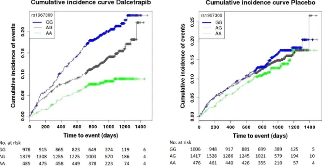
variant(s) associated with cardiovascular events in dalcetrapib treatment arm. Next, the significant variants were tested in the placebo arm to verify if detected association was only observed with dalcetrapib treatment. Tested phenotype included all cardiovascular events from dal-OUTCOME primary outcome with the addition of unanticipated coronary revascularization. A single region located in adenylate cyclase 9 gene was found to be associated with cardiovascular events in the dalcetrapib arm (Figure 3). In the identified region, genetic variant rs1967309 passed the significance threshold with P value = 2.41 × 10⁻⁸. In the same region, 7
other single nucleotide polymorphisms were identified with P value < 1 × 10⁻⁶ (Table 1). In
patients with AA genotype (minor allele) for SNP rs1967309, 39% reduction in the cardiovascular risk was observed when treated with dalcetrapib compared to placebo. Contrary, for patients with GG genotype (major allele), 27% increase in cardiovascular events was detected
18
in the dalcetrapib arm versus placebo. For heterozygote patients AG and placebo arm alone, there was no significant effect observed [77].
Figure 3 : Cumulative incidence of cardiovascular events from dal-OUTCOMES study for the
dalcetrapib and the placebo arm separately. Data stratified by the three genotypes of variant rs1967309 located within ADCY9 gene. Figure from Tardif JC et al., 2015 [77].
To validate the results obtained, 27 SNPs located in the ADCY9 gene were selected to genotype the samples of participants of dal-PLAQUE-2 study, which gave informed consent for genomic research (n=386). Tested endpoint for dal-PLAQUE-2 studies was the mean intima media thickness of common carotid arteries measured after 6 and 12 months of dalcetrapib treatment. Due to high correlation of tested genetic variants there was no need for adjustment of significance threshold for multiple testing, thus associations were considered as significant reaching P value < 0.05.
Ten single nucleotide polymorphisms displayed association with IMT measures and reached significance threshold (Table 1). Interestingly, significant association was observed for SNP rs2238448 (P value = 0.009), which is in high linkage disequilibrium with rs1967309 (𝑅2 =
0.80) and was also found to be associated with cardiovascular events in dal-OUTCOMES (P value = 8.88 × 10⁻⁸). In patients with TT genotype (minor allele) at variant rs2238448
19
and in participants with CC genotype (common allele) 0.009 ± 0.038. Again, there was no observable effect in dalcetrapib arm alone. SNP rs1967309, which provided significant association with cardiovascular outcomes in dal-OUTCOMES studies, did not reach significance threshold for association with change in IMT (P=0.114). However, observed changes in intima media thickness measure were consistent with findings in dal-OUTCOMES [77].
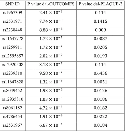
Table I : Genetic variants identified by genome-wide association study in dal-OUTCOMES (P
value < 1 × 10⁻⁶) and dal-PLAQUE-2 (P value < 0.05).
SNP ID P value dal-OUTCOMES P value dal-PLAQUE-2
rs1967309 2.41 × 10⁻⁸ 0.114 rs2531971 7.74 × 10⁻⁸ 0.1415 rs2238448 8.88 × 10⁻⁸ 0.009 rs11647778 1.72 × 10⁻⁷ 0.0087 rs1259911 1.72 × 10⁻⁷ 0.0205 rs12595857 2.02 × 10⁻⁷ 0.0193 rs12920508 3.18 × 10⁻⁷ 0.114 rs2239310 9.58 × 10⁻⁷ 0.6456 rs11647828 1.32 × 10⁻⁶ 0.0051 rs8049452 1.93 × 10⁻⁶ 0.0126 rs12935810 1.03 × 10⁻⁵ 0.0186 rs8061182 4.72 × 10⁻⁵ 0.0182 rs4786454 1.91 × 10⁻⁴ 0.0222 rs2531967 6.67 × 10⁻⁴ 0.0184
In patients treated with dalcetrapib, genotypes of the SNP rs1967309 were also found to be associated with change in total cholesterol after 1 month. The homozygote carriers of common allele (GG) had a smaller total cholesterol increase compared with AG and AA carriers [10.0 ± 23.3 𝑚𝑔 𝑑𝐿⁄ (0.2586 + 0.6025 𝑚𝑚𝑜𝑙 𝐿)⁄ , 12.9 ± 30.3 𝑚𝑔/𝑑𝐿 (0.3336 + 0.7836 𝑚𝑚𝑜𝑙 𝐿)⁄ and 13.8 ± 23.3 𝑚𝑔/𝑑𝐿 (0.3569 + 0.6025 𝑚𝑚𝑜𝑙 𝐿)⁄ , respectively]. For the changes in LDL-C after 1 month, similar genotype-dependent pattern was observed. Additionally, rs1967309 also
20
influenced the changes in body mass index, weight, plasma LDL/HDL cholesterol ratio and triglycerides. Surprisingly, the changes were in opposite direction than could have been anticipated [77].
The association of genetic variant rs1967309, reported to interact with dalcetrapib, was examined in DNA samples obtained from patients participating in ACCELERATE trial. Analysis revealed similar pattern of genetic association as observed with dalcetrapib, but did not reach statistical significance for evacetrapib-treated arm [80]. Meta-analysis of dal-OUTCOMES and ACCELERATE studies aimed to further investigate the evidence that ADCY9 genotype affects the treatment response to CETP-inhibitors on incidence of cardiovascular events. Analysis demonstrated the evidence of heterogeneity across genetic groups (P=0.004) with regard to relative risk, thus lending support to a potential interaction between CETP inhibition and ADCY9 genotype for cardiovascular events [81].
1.5.2 Association of dalcetrapib treatment and C-reactive protein levels
HDL are commonly considered to have atheroprotective properties, partly due to their anti-inflammatory functions. One of the standard markers of inflammation is high-sensitivity C-reactive protein (hs-CRP). Studies measuring hs-CRP levels in patients from dal-OUTCOMES trial showed unexpected results. After 3 months of treatment, patients taking dalcetrapib had +13.7 𝑚𝑔 𝑑𝐿 (+0.3543 𝑚𝑚𝑜𝑙 𝐿)⁄ ⁄ increase of HDL-C from baseline and +0.21 𝑚𝑔 𝑑𝐿 (+0.005431 𝑚𝑚𝑜𝑙 𝐿)⁄ ⁄ raise in hs-CRP. Patients from placebo group had slight increase in mean change in HDL-C +1.7 𝑚𝑔 𝑑𝐿 (+0.04396 𝑚𝑚𝑜𝑙 𝐿)⁄ ⁄ and reduction in hs-CRP −0.49 𝑚𝑔 𝑑𝐿 (−0.012671 𝑚𝑚𝑜𝑙 𝐿)⁄ ⁄ [82]. Considering different genotype of patients for variant rs1967309 an association between genotype groups and treatment was observed. Placebo-adjusted geometric mean percent changes in hs-CRP from baseline (12 months) to the end of trial were 18.1% for patients with common genotype GG, 18.7% for participants with AG and -1.0% for patients with minor genotype AA. The increase in hs-CRP levels after treatment with dalcetrapib, which increases HDL-C thought to possess anti-inflammatory properties is difficult to understand but has also been observed for other CETP inhibitors such as anacetrapib and evacetrapib [83]. The fact that only patients with protective genotype AA showed no increase in C-reactive protein levels supports the results obtained from GWAS and suggests that
21
inflammatory responses may be differently regulated depending on the genotype of the patient [84].
1.5.3 Association of dalcetrapib treatment and cholesterol efflux
Cholesterol efflux is an important step of RCT. In this process HDL particles remove excess cholesterol from cells of peripheral tissues and deliver it to the liver for its excretion [85]. The mean change in cholesterol efflux capacity of serum HDL in patients from dal-PLAQUE-2 trial as measured by the standard J774 macrophage assay was similar between study arms. However, the increase in cholesterol efflux capacity differed between participant groups treated with dalcetrapib. Patients with GG genotype had 7.8 ± 18.0% increase in cholesterol efflux capacity, participants with AG 12.9 ± 16.9% and patients with AA 22.3 ± 22.3%. The highest increase in cholesterol efflux capacity for patients with protective genotype is another evidence supporting the genotype-dependent effects of dalcetrapib [84].
1.5.4 Linkage disequilibrium block located at ADCY9 gene
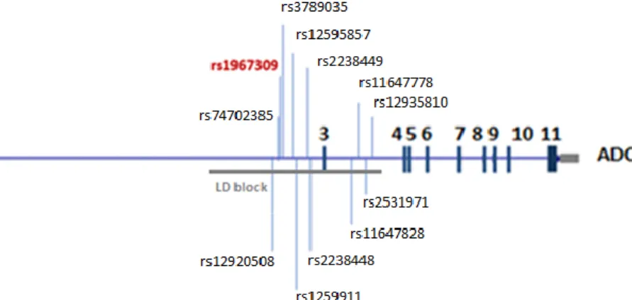
Single nucleotide polymorphisms associated with responses of patients treated with dalcetrapib are located within the ADCY9 gene on chromosome 16. Majority of identified SNPs are nonrandomly associated and create a linkage disequilibrium block (Figure 4). This LD block occupies a 27-kb region overlapping part of intron 2 and 3 of the ADCY9 gene.
Figure 4 : Graphical representation of adenylyl cyclase 9 gene. Linkage disequilibrium (LD)
block lies within second and third intron of ADCY9 gene. Polymorphism shown in bold red (rs1967309) was found to be significantly associated with cardiovascular response of patients treated with dalcetrapib. The other SNPs presented in black are polymorphisms selected for functional analysis, majority of them is in high linkage disequilibrium with rs1967309.
22
1.6 Adenylyl cyclases
The adenylyl cyclases (AC) are membrane-associated enzymes that catalyse the conversion of adenosine triphosphate (ATP) to 3’,5’-cyclic AMP (cAMP). They play an essential role in signal transduction following stimulation of G-protein coupled receptors, as they are the sole source of cAMP. Cyclic AMP is a second messanger involved in many biological processes, such as hormone responses. Nine membrane-bound adenylyl cyclase isoforms have been identified with a tenth distinct isoform that lacks membrane spans. The primary structure of the first nine isoforms consist of twelve transmembrane (TM) regions featuring cytosolic N and C termini. The first six TM domains (TM1-TM6) are followed by a cytosolic region with catalytic domain (C1a) and C1b domain. The second TM region (TM7-TM12) finishes with a second catalytic domain C2a and C-terminal C2b [86-88]. The tenth, soluble isoform has different regulatory and catalytic properties, and resembles more cyanobacteria enzymes [89].
1.6.1 Adenylyl cyclase 9
In comparison to the previous eight adenylyl cyclases, the ninth isoform (ADCY9) is the least characterized and the most divergent one. Human ADCY9 gene is located at chromosome 16 and its cDNA contains a 4059 bp open reading frame, which encodes a 1353 amino acid protein. Human ADCY9 cDNA is 85% identical to the mouse Adcy9 cDNA. On the protein level, ADCY9 is 90% identical to the mouse protein and all the structural motifs predicted in mouse Adcy9, such as 12 transmembrane domains, Asn-linked glycosylation sites and cAMP-dependent protein kinase phosphorylation sites, are conserved in the human form [90].
1.6.2 Expression of ADCY9
Each isoform of adenylyl cyclases has unique tissue distribution. ADCY9 expresses high levels of mRNA and protein in a wide variety of tissues. The mRNA levels of ADCY9 are present in most human tissues and the highest levels occur in brain, skeletal muscle, heart and pancreas [90-92]. According to the EMBL-EBI expression atlas, ADCY9 is also expressed in lower level in CD14-positive, CD16-negative classical monocytes.
1.6.3 Regulation of ADCY9 activity
Like all adenylyl cyclases, ADCY9 main function is catalysis of second messenger cyclic AMP formation. Despite the same basic function, each of the isoforms possess unique regulatory properties. Depending on these properties and expression levels of the isoforms in a tissue or cell
23
type and cell compartments, extracellular signals received by G-protein coupled receptors can be differently integrated [91]. All the isoforms are known to be upregulated by G-protein (𝐺𝛼𝑠),
which is involved in well known signaling pathway mediated by one of the G-protein coupled receptors, called β-adrenergic receptor (β2AR). The agonists of β2AR induce its interaction with α subunit of heterotrimeric G-protein (𝐺𝛼𝑠). Next, 𝐺𝛼𝑠 binds GTP and in turn dissociates from
G-protein γ subunit. The GTP-bound 𝐺𝛼𝑠 then binds and activates adenylyl cyclase, which produces
cAMP. Increased levels of cAMP may lead to activation of protein kinase A (PKA) and other important signalling cascades. G proteins were found to bind directly to the catalytic core of AC, which includes two homologous cytoplasmic domains (C1a and C2a). However, each of the AC isoforms consists of additional structural elements that are crucial for the correct assembly of G-proteins and proper enzymatic function of ACs. A team of researchers, which determined the 3D structure of the bovine ADCY9 in complex with 𝐺𝛼𝑠, reported that the C2b region plays an important role in the G protein-bound state of ADCY9. The occlusion of ADCY9 reaction center by the C2b region reduces the affinity for the substrate (ATP) and as a result decreases production of cAMP. The 3D structure of bovine ADCY9 also revealed that transmembrane helices TM6 and TM12 are laterally exposed to the lipid bilayer and they extend into the cytosol becoming helices h1.1 and h2.1. Therefore, it is likely that direct interactions of TM6 and TM12 with lipids and other molecules could directly influence the catalytic domain by altering orientation of helices h1.1 and h2.1 [88]. Additionally, Pálvölgyi et al. reported that the short motif in the C2b isoform-specific domain of ADCY9 is responsible for supression of ADCY9 activation by 𝐺𝛼𝑠 protein. The authors also suggested that the autoinhibitory effect of C2b domain
on ADCY9 activation in the heart may be released by proteolytic cleavage of this domain [93]. The other agents known to regulate the activity of all the isoforms of AC are 𝐶𝑎2+ and 𝑀𝑔2+.
The rest of regulatory agents are isoform-specific. Since ADCY9 is the least characterized isoform, its regulation is poorly characterized. Unlike other adenylyl cyclase isoforms, the activity of ADCY9 is unaffected by forskolin and calcineurin inhibitors. Also, ADCY9 is stimulated by 𝐺𝛼𝑠-coupled receptors activation but is not weaken by 𝐺𝑖-coupled receptors [90]. ADCY9 was reported to be phosphorylated by four different types of protein kinases, protein kinase C (PKC), protein kinase A (PKA), casein kinase 1 (CK1) and cyclin-dependent protein kinase 5/p35 complex (cd5/p35). Additionally, ADCY9 contains two N-glycosylation sites