Ultrasonographic assessment of uterine involution and ovarian activity in West African Sahelian goats.
6
0
0
Texte intégral
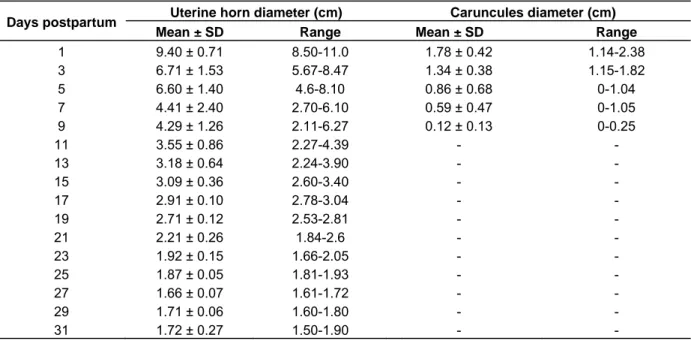
(2) 72. J. Vet. Med. Anim. Health. Greyling, 2000). Different research reports have shown different intervals to complete uterine involution. While Degefa et al. (2006) demonstrated complete macroscopic uterine involution by day 19 PP, Greyling and van Niekerk (1991) reported day 28 PP as the day of complete uterine involution. Moreover, histochemical study of caprine endometrium indicated complete regression of endometrium and re-epithelialization by day 16 PP (Sanchez et al., 2002). It is difficult to judge the time of uterine involution in the goat, because the uterus cannot be examined by rectal or abdominal palpation. In most studies, uterine involution was investigated after slaughter (Van Wyk et al., 1972; Rubianes and Ungerfeld, 1993) or by laparotomy (Rubianes et al., 1996) or using radio-opaque markers (Tian and Noakes, 1991). Ultrasonography provided a non-invasive technique to image directly in the reproductive tract. In goat, ultrasonography is routinely used for pregnancy diagnosis (Hesselink and Taverne, 1994; Buckrell, 1988). Recently, it was shown that transrectal ultrasonography is a useful and reliable method to observe the uterine involution and follicular dynamics in goats (Kandiel et al., 2012; Badawi et al., 2014). The Sahelian goat is one of the most common breed in West Africa, which is used for meat production. Its rapid reproductive rate is its most important advantage. It shows estrous activity throughout the year (Traoré et al., 2006). However, there is still little information concerning the course of postpartum period in goat of this breed. Moreover, the ultrasonic characterization of reproductive function in goat is very scanty in the literature. The objectives of this study were to characterize uterine involution and early ovarian activity in West African Sahelian goats. MATERIALS AND METHODS This study was undertaken at the station of Ouagadougou University from November 2011 to May 2013. A total of 21 nulliparous West African Sahelian goats were used in this study. Their ages and weights at the beginning of the experiment ranged from 12 to 15 months and 25 to 35 kg, respectively. They were clinically free from any infectious, parasitic or genital diseases. During the study period, all goats were kept under uniform standard management practice and housed in the Agriculture Farm of Ouagadougou University. They were kept in a shed attached with an open yard. Each animal was given daily 1 kg concentrated ration, in addition to a green fodder. The goats were synchronized using a 12-days chronogest® CR treatment with intravaginal sponges containing 20 mg of flurogestone acetate (FGA, Intervet International B.V., Intervet Ireland Ltd, Iternet productions S.A., Rue de Lyons, France) (Baril et al., 1993). They were mated with a fertile buck and examined for pregnancy one month later using a linear array ultrasound scanner. All does were confirmed to be pregnant and gave birth in October after normal gestation period and normal parturition. They were allowed to nurse their kids. The ultrasonographic inspection was performed transrectally by a single operator employing the Chison Ultrasonic Scanner (Chison Medical Imagin Co. Ltd, 8300) equipped with a 5-MHz transducer.. The doe was lightly restrained by one person against railing in standing position. One of the hind legs was folded up at the time of scanning for proper placement of the probe. An ultrasound coupling gel was applied each time to the probe to develop good contact and to remove air between probe and animal skin. Thereafter, the rectum was evacuated from feces and air with the aid of the lubricated fingers of the operator. The ultrasound probe fixed to an extension rod was inserted into the rectum. For scanning of the uterus and ovaries, the probe was moved approximately 60° to each side around its longitudinal axis. Uterine horns were scanned once every other day starting from day one PP until there was no further reduction in the uterine diameter for two successive weeks. Ovarian structures were scanned daily for the first PP week (Hayder and Ali, 2008). Parameters for the determination of the endpoint of uterine involution were the transversal diameter of uterine horns of ≤2 cm and the lack of contents in the uterine cavity (Hauser and Bosted, 2002). All follicles with a diameter greater than 2 mm were sketched and the video image recorded in external disk to allow individual structure to be monitored. Parameters for the present investigation were the transversal cross-sectional diameter and number and size of follicles of the right and left ovaries. The interval from parturition to caruncules disappearance and their mean size were recorded by determining the diameters of three to five caruncles in each uterine horn. Data were presented as mean ± standard deviation and were analyzed using repeated-measures of ANOVA. Differences were considered to be statistically significant at P ≤ 0.05. RESULTS AND DISCUSSION This is the first study to provide baseline information on the use of real-time ultrasonography for the assessment of PP uterine involution and ovarian activity in West African Sahelian goats. All included does showed normal gestation period ranging from 145 to 157 days. In all cases, the delivery was uneventful, and placenta was expelled within 10 h after kidding. The kids started suckling within 1 h after kidding. At the term of the gestation period, a total number of 26 kids were born with about 38.46% of twinning kids (5 animals delivered twin’s kids). Average birth weights of male (n = 17) and female kids (n = 9) were 2.26 ± 0.7 and 2.32 ± 0.7 kg, respectively. Non-significance difference was found between male and female birth weights. During transrectal scanning, the does showed no signs of distress apart from short avoidance behavior when the scanner passes through the anal sphincter. The average PP uterine and caruncules diameters, as estimated ultrasonographicaly, at the different postpartum periods are shown in Table 1. The uterine diameter could not be estimated by day 0 in most of does, as it was too large to fit effectively on the screen. The readings were taken from day 1 onwards (Figure 1A). The does showed a physiological regression of the uterus and caruncules with transversal diameters of 9.40 ± 0.71 and 1.78 ± 0.42 cm on day 1 PP decreasing to 1.72 ± 0.27 and 0.12 ± 0.13 cm on days 31 and 9 PP, respectively (Figure 1; A, B, C and D). The mean time required for uterine complete involution was 22.44 ± 1.54 days (range 18 - 25 days) and was characterized by a small cross-sectional diameter (<2 cm).
(3) Zongo et al. Table 1. Uterine and caruncules mean (± SD) and range diameters (cm) of Sahelian goats on different days postpartum.. Uterine horn diameter (cm) Mean ± SD Range 9.40 ± 0.71 8.50-11.0 6.71 ± 1.53 5.67-8.47 6.60 ± 1.40 4.6-8.10 4.41 ± 2.40 2.70-6.10 4.29 ± 1.26 2.11-6.27 3.55 ± 0.86 2.27-4.39 3.18 ± 0.64 2.24-3.90 3.09 ± 0.36 2.60-3.40 2.91 ± 0.10 2.78-3.04 2.71 ± 0.12 2.53-2.81 2.21 ± 0.26 1.84-2.6 1.92 ± 0.15 1.66-2.05 1.87 ± 0.05 1.81-1.93 1.66 ± 0.07 1.61-1.72 1.71 ± 0.06 1.60-1.80 1.72 ± 0.27 1.50-1.90. Days postpartum 1 3 5 7 9 11 13 15 17 19 21 23 25 27 29 31. Caruncules diameter (cm) Mean ± SD Range 1.78 ± 0.42 1.14-2.38 1.34 ± 0.38 1.15-1.82 0.86 ± 0.68 0-1.04 0.59 ± 0.47 0-1.05 0.12 ± 0.13 0-0.25 -. Table 2. Early postpartum ovarian structure in West African Sahelian goat: Characteristics of follicular development during the first week postpartum.. Days postpartum 1 2 3 4 5 6 7. Mean number of follicles Right ovary Left ovary 2 3 3 1 3 2 3 3 2 1 1 1 2 1. Mean diameter (mm) ± SD Right ovary Left ovary 5.77 ± 1.51 7.05 ± 2.62 6.97 ± 1.7 5.88 ±1.04 7.56 ±1.97 6.69 ±1.85 4.95 ± 1.45 7.38 ±1.54 7.68 ± 0.59 8.25 ± 2.05 5.51 ± 0.93 5.44 ±1.13 8.57 ± 1.79 8.56 ± 0.66. UD. UD. UD. CD. A (day 1 PP). Foll. Foll. B (day 6 PP). C (day 12 PP). D (day 18 PP). Figure 1. Ultrasonic images of uterine horns at different days postpartum. UD: Uterine diameter, CD: caruncule diameter, Foll.: follicle.. 73.
(4) 74. J. Vet. Med. Anim. Health. Figure 2. Profiles of uterine involution in does bearing single. (▬, R2 = 0.933) and twins (…..., R2 = 0.931) fetuses.. of uterine horns and absence of lochia in uterus (Figure1D). These findings were in close agreement with previous reports on goats (Takayama et al., 2010; Ababneh and Degefa, 2005; Baru et al., 1983; Sanchez et al., 2002). In Jordan local balady goats, microscopic involution and complete regeneration of the caruncular epithelium was also evident by the same period (Degefa et al., 2006). However, the present findings were earlier compared to the reports of 28 days in Boer goats (Greyling, 2000; Greyling and van Niekerk, 1991). In comparison with sheep, most studies reported slower involution and found that the uterine regression in ewes finished approximately after 30 days PP (Hayder and Ali, 2008; Rubianes and Ungerfeld, 1993), in contrast to other authors which observed the end of the uterine regression in sheep by day 17 PP (Hauser and Bostedt, 2002). The variability regarding the time required for a complete uterine involution may result from differences in breed, reproductive status and methods. During the experiment, one goat (F96) lost her kid on day 1 PP and ended uterus involution 10 days later. She was mated on day 18 PP and was diagnosed pregnant one month later. Another goat (F85) showed an abnormally higher uterine diameter on day 13 PP onward up to days 20 PP than the previous diameter with accumulated fluid within. the lumen. These two goats were excluded from the general profile. The 19 other goats exhibited a very similar profile of uterine involution. 9/19 and 10/19 females completed uterine involution by the third (ranging from days 18 to 21) and fourth (ranging from days 22 to 25) weeks postpartum, respectively. The calculated rates of involution during the first, second and third PP weeks were 0.71, 0.17 and 0.14 cm/day, respectively. Furthermore, regression analysis showed a high positive correlation between diameters of the uteri in both goats and days PP (r = 0.98). The course of uterine regression was established on these data and was described by the logarithmic equation: y = -2.33ln(x) + 9.43, (R2 = 0.98, P <0.05) where y = uterine cross sectional diameters (cm), x = postpartum period in days. In ewes, Hauser and Bostedt (2002) described the course of uterine regression by exponential function as: Y= E + A*e-b*t. The data revealed significantly faster reduction between the first and second weeks (p < 0.01) than between the second and third weeks (P < 0.01). This was evidenced by 67.43% of the total reduction in diameter recorded on day 7 postpartum. After day 21 PP, the reduction in size was not significant. At this point of time,.
(5) Zongo et al. 97.16% of the hypertrophy as a result of pregnancy was reduced. The faster reduction of uterine diameter during the early postpartum has been reviewed previously and seem to be a response of myometrial contractility that plays a major role in clearing lochial debris from the uterus after parturition (Ababneh and Degefa, 2005; Hauser and Bostedt, 2002). The statistical analysis showed significant (P = 0.02) higher values of the uterine diameter during the first week PP in females bearing twins fetuses than the females bearing single fetus. However, no significant difference was observed in uterine diameter during the second, third and fourth weeks between females bearing twins and females bearing single fetuses. The females bearing single fetus (n = 14) showed a physiological regression of the uterus with a transversal diameter of 9.16 ± 0.48 cm on day 1 postpartum, decreasing to 2.16 ± 0.31 cm on day 21, while the does bearing twins fetuses showed a transversal diameter of 9.76 ± 0.79 cm on day 1 postpartum, decreased to 2.25 ± 0.08 cm on day 21 postpartum, and complete involution was delayed as the diameter of the uterine horn was reduced to 1.97 ± 0.06 cm on day 24 postpartum. The typical ultrasonographic patterns of uterine regression in does bearing single and twin fetuses were established as shown in Figure 2. The uterine wall covered with caruncules and uterine lumen was readily identified by different ultrasonographic echotextures (Figure 1A). In does bearing single fetus, the caruncules were completely degenerated on an average by days 5 postpartum, whereas in the twins, they were delayed until day 9 postpartum. At those points of time, their echotextures appeared similar to the endometrium and ultrasonography differentiation and measurements were impossible to be carried out. The regression of the caruncules in the first week PP was in good agreement with different studies (Hauser and Bosted, 2002; Rubianes and Ungerfeld, 1993; van Wyk et al., 1972). However, in Balady goat, complete regression of the caruncules was not completed until day 19 PP (Degefa et al., 2006). In addition, lochia was cleared as early as day 7 PP in primiparous goats. Ovarian follicular dynamic of the experimental does during the early postpartum period (days 1 to 7 PP) are summarized in Table 2. In all does (n = 19), ovarian anatomical events during the early post-partum period (days 1 to 7) were similar. At least one follicle was examined on each ovary examined within 7 days after kidding (Figure 1C). The mean size of follicles ranged from 4.95 ± 1.45 to 8.56 ± 0.66 mm on both ovaries. These findings were fairly in close corroboration with the result reported in Serrana goat (Simoes et al., 2006). In Jennies, Dadarwal et al. (2004) found three to seven follicles of size 10 to 15 mm diameter on one or both ovaries examined within 8 to 24 h after foaling. Those follicles reached to >25 mm in diameter on days 5 to 12. 75. PP. In non-nursing Shiba goats, the first postpartum ovulation was observed between days 7 and 13 following parturition (Takayama et al., 2010). The early return to active follicular development in West African Sahelian goats. demonstrated the ability of the ovary to resume activity early after kidding. In Shiba Goats, three to six follicles, whose diameters ranged from 1 to 7 mm, were observed throughout the cycle; a few follicles grew to more than 5 mm in diameter and most of them atrophied during the luteal phase (Orita et al., 2000). No significant differences (P ≥ 0.05) were detected between number and size of follicles between does bearing single and twin fetuses. It could be concluded that, complete uterine involution in West African Sahelian goats occurred during the 3rd and fourth weeks post kidding, while the ovarian follicular dynamics started on the first week PP. The early return to active follicular development suggests that ovarian responsiveness may not be the major reason for the variable duration of the post-partum periods commonly observed in tropical goats. However, the period of ovarian observations may extend until standing estrus period to permit definitive conclusions about quality of follicular dynamic and ovulation in this breed. These findings should assist practitioners and Sahelians goat breeders to decide when goats should be bred following parturition. In a future study, factors responsible for the shorter involution period in West Africa Sahelian goats should be investigated.. ACKNOWLEDGEMENTS This work was supported by the West Africa Agricultural Production/ECOWARD/CORAF/FCN/Burkina Faso, International Atomic Energy Agency (IAEA)/Country Program for Support Development of Small Ruminants Production in Burkina Faso and International Foundation for Sciences (IFS). The authors would like to thank Pr. Beckers JF., Pr. Drissa SANOU, Drs. Maria Teresa Rubio Pellicer, Jean Luc TOUZE, Noélita Melo De Sousa and Philippe Horlait for their assistance in ultrasonic training. Special thanks to Prs. Mario PODESTA and Mohammad Shamsudin for their advices and their hope to the research in Africa. Thank to Dr. KIMSE Moussa for his assistance in data collection.. Conflict of Interest Authors have no conflict of interest. REFERENCES Ababneh MM, Degefa T (2005). Ultrasonic assessment of puerperal uterine involution in Balady goats. J. Vet. Med. A Physiol. Pathol. Clin. Med. (52):244-248. Badawi ME, Makawi SEA, Abdelghafar RM, Ibrahim MT (2014)..
(6) 76. J. Vet. Med. Anim. Health. Assessment of postpartum uterine involution and progesterone profile in Nubian goats (Capra hircus). J. Adv. Vet. Anim. Res. 1(2):36-41. Baril G, Leboeuf B, Saumande J (1993). Synchronization of estrus in goats: the relationship between time of occurrence of estrus and fertility following Artificial Insemination. Theriogenology 40:621-628. Baru P, Khar SK, Gupta RC, Luthra RA (1983). Uterine involution in goats. Vet. Med. Small Anim. Clin. 11:1773-1776. Buckrell BC (1988). Applications of ultrasonography in reproduction in sheep and goats. Theriogenology 29:71-84. Dadarwal D, Tandonb SN, Purohitc GN, Pareekc PK (2004). Ultrasonographic evaluation of uterine involution and postpartum follicular dynamics in French Jennies (Equus asinus). Theriogenology 62:257-264. Degefa T, Ababneh MM, Moustafa MF (2006). Uterine involution in the postpartum Balady goat. Vet. Arhiv. 76 (2):119-133. Delgadillo JA, Flores JA, Villarreal O, Flores MJ, Hoyos G, Chemineau P, Malpaux B (1998). Length of postpartum anestrus in goats in subtropical Mexico: Effect of season of parturition and duration of nursing. Theriogenology 49:1209-1218. Elsheikh AS, Omer NNE, Alqurashi AM (2013). Management of Postpartum Interval of Nubian goats with PGF2α and GnRH. J. Am. Sci. 9(3):181-184. Greyling JPC (2000). Reproduction traits in the Boar goat doe. Small Rum. Res. 36:171-177. Greyling JPC, van Niekerk CH (1991). Macroscopic uterine involution in the postpartum Boer goat. Small Rum. Res. 4:277-283. Hauser B, Bostedt H (2002). Ultrasonographic observations of the uterine regression in the ewe under different obstetrical conditions. J. Vet. Med. A (49):511-516. Hayder M, Ali A (2008). Factors affecting the postpartum uterine involution and luteal function of sheep in the subtropics. Small Rum. Res. 79:174-178. Hesselink JW, Taverne MAM (1994). Ultrasonography of the uterus of the goat. Vet. Q. 16(1):41-45. Kandiel Mohamed MM, Watanabé G, Abou-El-Roos ME, Abdel-Ghaffar AE, Sosa GA, EL-Azab AESI, Nagaoka K, Li JY, Manabé N, Kazuyoshi T (2012). Follicular Turnover and Hormonal Association in Postpartum Goats During Early and Late Lactation. J. Reprod. Dev. 58 (1):61-68. Orita J, Tanaka T, Kamomae H, Kaneda Y (2000). Ultrasonographic observation of follicular and luteal dynamics during the estrous cycle in Shiba Goats. J. Reprod. Dev. 46:31:37. Rubianes E, Ungerfeld R, Vinoles C, Carbajal B, de Castro T, Ibarra D (1996). Uterine involution time and ovarian activity in weaned and suckling ewes. Can. J. Anim. Sci. 76:153-155.. Rubianes E, Ungerfeld R (1993). Uterine involution and ovarian changes during the early postpartum of autumn-lambing Coniedale ewes. Theriogenology 40:365-372. Sanchez MA, Garcia P, Menendez S, Sanchez B, Gonzalez M, Flores JM (2002). Fibroblastic growth factor receptor (FGF-R) expression during uterine involution in goat. Anim. Reprod. Sci. 69:25-35. Simoes J, Almeida JC, Valentim R, Baril G, Azevedo J, Fontes P, Mascarenhas R (2006). Follicular dynamics in Serrana goats. Anim. Reprod. Sci. 95:16-26. Takayama H, Tanaka T, Kamomae H (2010). Postpartum ovarian activity and uterine involution in non-seasonal Shiba goats, with or without nursing. Small Rum. Res. 88:62-66. Tian W, Noakes DE (1991). A radiographic method for measuring the effect of exogenous hormone therapy on uterine involution in ewes. Vet. Rec. 129:463-646. Traoré A, Tamboura HH, Kaboré A, Yaméogo N, Bayala B, Zaré I (2006). Caractérisation morphologique des petits ruminants (ovins et caprins) de race locale “Mossi” au Burkina Faso. AGRI 39:39-50. Ungerfeld R, Sanchez-Davila F (2012). Oestrus synchronization in postpartum autumn-lambing ewes: effect of postpartum time, parity, and early weaning. Spanish J. Agric. Res. 10(1):62-68. Van Wyk LC, van Niekerk CH, Belonje PC (1972). Involution of the postpartum uterus of the sheep. J. South Afr. Vet. Assoc. 43:19-26..
(7)
Figure

Documents relatifs
We now describe MV polytopes for the only other rank two affine root system. One could perhaps rework the proof as in the sl b 2 case and directly prove that the MV polytopes
Comparison with monthly total energy density calculated with temperature only sup- ports that observed small-scale temperature and wind pertur- bations are mostly associated
Time series of annual rainfall (mm) in the AMMA-CATCH Niger (ACN) site versus the corresponding number of Mesoscale rain events as determined from the ground network (left) and
In this study, the genetic diversity of four indigenous Algerian goat breeds (i.e., Arabia, Makatia, M’Zabite and Kabyle, with n = 12 for each breed) has been investigated for the
With univariate analysis, there was no signi fi cant age- related difference in the PDGF-BB concentration between the two groups (p = 0.25), whereas with the Mann-Whitney test, the
2.2.2 Carbon flux and soil moisture measurements We collected net ecosystem C exchange (NEE) measure- ments from 12 flux tower sites, hereinafter called the core sites, in the
In an effort to codify and grant a specific status to marabouts in France, the newly elected Socialist government in 1981 focused on their religious role, which it
difficult to obtain*. Using some consumption expenditure patterns, a uniform elasticity measure for both beer and other alcoholic beverages is estimated to be 1.3. For soft drinks
