Comparaison de la survie fonctionnelle des disques
intervertébraux chez le mini-porc ayant subi une chirurgie de
scoliose avec système d’ostéosynthèse fixe versus
ostéosynthèse mobile.
par
Marco Bérard
Université de Montréal
Sciences Biomédicales
Mémoire présenté â la Faculté des études supérieures
en vue de l’obtention du grade de
Maîtrise en Sciences Biomédicales
Août2005
/‘y
W
H
053
2çOt
L’auteur et les coauteurs le cas échéant conservent la propriété du droit
d’auteur et des droits moraux qui protègent ce document. Ni la thèse ou le mémoire, ni des extraits substantiels de ce document, ne doivent être imprimés ou autrement reproduits sans l’autorisation de l’auteur.
Afin de se conformer à la Loi canadienne sur la protection des renseignements personnels, quelques formulaires secondaires, coordonnées
ou signatures intégrées au texte ont pu être enlevés de ce document. Bien
que cela ait pu affecter la pagination, il n’y a aucun contenu manquant.
NOTICE
The author of this thesis or dissertation has granted a nonexciusive license
allowing Université de Montréal to reproduce and publish the document, in part or in whole, and in any format, solely for noncommercial educational and
research purposes.
The author and co-authors if applicable retain copyright ownership and moral
rights in this document. Neither the whole thesis or dissertation, nor substantial extracts from it, may be printed or otherwise reproduced without the author’s permission.
In compliance with the Canadian Privacy Act some supporting forms, contact
information or signatures may have been removed from the document. While this may affect the document page count, it does flot represent any Ioss cf
Université de Montréal
Faculté des études supérieures
Ce Mémoire intitulé:
Comparaison de la survie fonctionnelle des disques
intervertébraux chez le mini-porc ayant subi une chirurgie de
scoliose avec système d’ostéosynthèse fixe versus
ostéosynthèse mobile.
présenté par:
Marco Bérard
a été évalué par un jury composé des personnes suivantes:
Dr Nicholas Newman
président rapporteur
Dr Charles-Hilaire Rivard
directeur de recherche
Dr Maurice Duhaime
membre du jury
Titre: Comparaison de la survie fonctionnelle des disques intervertébraux chez le mini-porc ayant subi une chirurgie de scoliose avec système d’ostéosynthèse fixe versus
ostéosynthèse mobile.
Matériel et Méthode: Une scoliose thoraco-lombaire est induite chez 12 mini-porcs et maintenue avec un système d’ostéosynthèse vertébrale: 6 avec un système fixe non-mobile et 6 avec le système mobile d’ostéosynthèse (Orthobiom). Aucune greffe osseuse n’est effectuée. Trois (3) mini-porcs, chez qui aucune scoliose n’est induite, servent de groupe contrôle pour l’analyse des disques intervertébraux. Après un suivi post-opératoire de 12 à 18 mois, les disques sont retirés après euthanasie des sujets et des analyses immunohistochimiques sont réalisées pour démontrer l’apparition de Collagènes Type X, signifiant la dégénérescence dans les disques intervertébraux. La quantification des proportions du Collagène Type X dans les différents groupes est effectuée via logiciel informatique. Une étude radiologique des facettes et une évaluation subjective de la mobilité des segments instrumentés sont réalisées
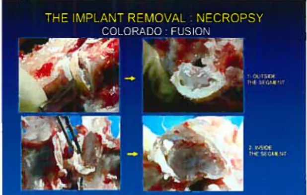
Résultats: Les 15 mini-porcs ont été euthanasiés et les disques prélevés. Chez les sujets avec ostéosynthèse fixe, une fusion vertébrale est observée à tous les niveaux inclus dans la fixation. Aucun nucleus pulposus n’est observé et seulement quelques fibres de fibrosus sont isolées. Aucun mouvement facettaire n’est noté. Chez les sujets avec l’ostéosynthèse mobile, nous constatons une préservation du nucleus pulposus et de I’ annulus fibrosus aux niveaux inclus dans la fixation. Un mouvement facettaire est observé. L’immunohistochimie et la quantification informatique (du Collagène Type X) démontre une apparition du Collagène type X chez les les mini-porcs instrumentés avec le système fixe.
DiscussionlConclusion: Macroscopiquement, nous notons une différence importante quant à l’aspect radiologique et quant à la mobilité des segments instrumentés: la préservation est meilleure avec le système Orthobiom. L’ immunohistochimie démontre que la quantité de Collagène type X est statistiquement supérieure dans le groupe fixe non-mobile comparé au groupe Orthobiom, signifiant une préservation de la fonction dans ce dernier groupe.
Mots clés français: Scoliose Idiopathique, Immunohistochimie, Collagène type X, Orthobiom, Ostéosynthèse vertébrale
groups. The concentration of Type X Collagen, a sign of disc degeneration, was compared among the three groups. A radiologic assessment of the intervertebral facets and a subjective evaluation of the spine mobility were performed.
Results: Macroscopically, in the non-mobile group, fusion of ail levels fixed by the instrumentation was observed: no nucleus pulposi were seen at any of the levels. Only a few fibers of the annulus fibrosus were found. In the Orihobiom minipigs, the pulposi were preserved. The immunohistochemistry, done by computerized analysis, showed significant amounts of Type X Collagen in the non-mobile fused discs of the Colorado mini-pigs. The Orihobiom mini-pigs showed smaller proportions of Type X Collagen in the discs, similar to control pigs. The mobility and the facet preservation were significantly higher in the mobile Orthobiom group.
Discussion: With the immunochemical analysis, it was shown that the morphology of the discs is better preserved with a system that allows movement compared with a system that is rigid and does not allow any movement. The quantity of Type X Collagen was significantly Iess in the mobile Orthobiom group, indicating a beffer survival of the intervertebral discs. The mobility and the preservation of the facets as seen with the CT-scan analysis point toward the same affirmation. Conclusion: a mobile osteosynthesis system permits better function and preservation of the intervertebral discs. Fewer long-term disc complications may be expected in a patient treated with this instrumentation as compared with conventional fusion-osteosynthesis system.
English key words: Idiopatic Scoliosis, Type X Collagen, Immunohistochemistry, Vertebral Osteosynthesis, Orthobiom
C
Dédicace 13-14
Remerciements 15-16
Avant-Propos 17-18
Corps de l’ouvrage 19-59
Les Sources Documentaires 60-64
Liste des Tableaux
Fixed System 1- 6 month = O 2- l2months=1 3- l8months=5 4- 2 years = O Total = 9 Orthobiom 1- 6 months = I 2- 12 months = 3 3- l8months=3 4- 2 years = 2 Total = 3 Control 1- l2months=2 2- 18 months =1
Liste des Figures
5
Figure 3: disques intervertébraux avec ostéosynthèse fixe I
intervertebral discs with non-mobile osteosynthesis
Figure 4: disques intervertébraux avec ostéosynthèse mobile I
intervertebral discs with mobile osteosynthesis
7
Figure 7: Clemex Technologies
I Fond
—Background (rougelred)
Contrôle - 0.72774 0.00040
Orthobiom 0.72774 - 0.00044
Colorado 0.00040 0.00044
-Hôpital Ste-Justine
Figure 10 : ANOVA Collagen X
Collagen X
(%)
P Value
Contrôle Orthobiom ColoradoContrôle - 0.43063 0.00074
Orthobiom 0.43063 - 0.00094
Colorado 0.00074 0.00094
9
Fîgure 11: Ct-Scan Facettes Articulaires sujet Contrôle
I
Articular Facet Control subject
Figure 12
: Ct-Scan Facettes Articulaires sujet ostéosynthèse mobile I Articular Facet mobile osteosynthesis subjectDédicace
un
professeur,qui par une ééterminante rencontre en un jour &automne mit neuf cent quatre-vingt-quatorze, guicfa ma carrière. (Depuis ce jour,
it
cfevint mon mentor et mon inspiration et ce, pour heaucoup ptusque ta recftercfie: en chaque patient que je
sozne,
ity a unpeu ée
tui.(Dr Rjvard
cfu ptus profoncfée mon
coeur,
je vous remercie et vous serai toujours rec[evant.Remerciements
_,j
Souad
Sans qui, je n ‘auraisjamais atteint ce 6ut.
Par ta
simpte
écoute, ton support et tes consei(sjudciezv.,tu as su
me
faire progresser etce, non
seufementdans
ta recfiercfie,mais
sans
mon cfzeminementen
tant qu ‘ortîiopédiste.gvlerci mitte
fois
pour tout ce que tu asfait pour moi!Avant-propos
déformation
du
rachis
rencontrée
dans
la
Scoliose
Idiopathique.
La fonction de cette instrumentation est de corriger une
déformation vertébrale et maintenir cette correction tout en
préservant l’intégrité et la fonction ultérieure des disques
intervertébraux.
Ce faisant, les patients ayant subi une chirurgie du rachis
avec une telle instrumentation, seraient moins susceptibles
aux
complications
futures
associées
avec
la
fusion
évaluation macroscopique de la mobilité 30 imagerie des facettes articulaires intervertébrales 30
Résultats 31-33
immunohistochimie 32-33
mobilité segmentaire 33
imagerie des facettes articulaires intervertébrales 33
Discussion 34-36
Conclusion 37-39
Introduction
Les disques intervertébraux, en tant que composantes primordiales de la colonne vertébrale chez l’humain, sont des structures complexes permettant une répartition des forces biomécaniques et une flexibilité au rachis, permettant à la colonne vertébrale flexion et rotation.1 Cependant, pour soutenir ce rôle primordial de fonction biomécanique, une organisation structurée de tous les composantes majeures des disques intervertébraux est requise. Sous des conditions normales, les constituants majeurs des disques sont les protéoglycans, l’eau et le collagène. Il est bien établi que dans le disque intervertébral normal, sept types de collagène sont présents et leurs proportions varient selon les structures discales. 1,2 Ghosh et al. ont démontré que, en conditions normales avec une morphologie intacte, l’anneau fibreux est constitué majoritairement de collagène type I et le noyau pulpeux de collagène type Il.
23
Avec les changements dégénératifs, Nerlich et al. ont démontré que dans le noyau pulpeux, il existe une quantité détectable par immunohistochimie de collagène type X qui est non détectable dans les disques fétaux ou d’adolescents I jeunes adultes2. Ce type de collagène est donc hautement suggestif de changements dégénératifs du rachis. Il est démontré que la quantité de collagène type Il est également significativement réduite lors de tels changements. Le collagène type IV démontre une hausse précoce et une baisse lors des stades avancés de dégénérescence discale. De tels changements dégénératifs sont rencontrés dans plusieurs pathologies chez l’humain et plus particulièrement chez des sujets atteints de Scoliose Idiopathique (SI) ayant requis une fusion vertébrale.
La SI, condition caractérisée par une déformation géométrique du rachis, est diagnostiquée selon une évaluation clinique et des analyses radiologiques des plans frontaux et sagittaux du rachis. C’est une des conditions ? orthopédiques rachidiennes? les plus prévalentes chez l’adolescent. La prévalence estimée de cette condition varie de 2% à 14% de la population nord américaine dépendant des critères diagnostics utilisés 5-11 Les filles sont plus atteintes que les garçons
5,6,8,10
et cette pathologie progresse dans la période de croissance accélérée. Le pronostic demeure toujours difficile à établir 1215
Le traitement envisagé dépend de l’amplitude de la scoliose, évaluée selon la méthode de Cobb 16 Selon l’évolution
60°, tenant compte de l’âge, de la maturité du patient, de la localisation de la scoliose et de l’expérience clinique de l’orthopédiste, un traitement chirurgical est planifié. Jusqu’à 0.6% de la population ayant une SI vont nécessiter une chirurgie2° dont le but ultime est de contrer les effets nocifs sur les organes internes d’une déformation progressive du rachis 21-23 Cependant, même si 23 à 54% des adolescents avec SI présentent des douleurs au dos 24-25 la douleur ne semble pas corréler avec l’amplitude de la déformation et n’est donc pas un élément décisif dans la décision d’opter pour le traitement chirurgical, contrairement à la population scoliotique adulte 26
Depuis le début des années 1900, la fusion segmentaire du rachis a été réalisée pour limiter la progression de la SI. Harrington 27 avec l’utilisation de barres métalliques, a été le premier à introduire le concept de la correction de la déformation spinale sans fusion osseuse inter segmentaire. Cette approche révolutionnaire n’a eu qu’un succès très limité. Dans les faits, l’implant a à soutenir la correction de la déformation du rachis tout en contrant la mobilité des vertèbres et les
25
larges forces créés par la marche du patient. La fusion a été donc réintroduite pour stabiliser et maintenir la correction de la du rachis instrumenté. L’implant n’a donc plus à soutenir des forces de contrainte pour une période prolongée car la fusion accomplie déjà seule cette tâche approximativement 6 mois après la chirurgie. De ces idées et concepts innovateurs ont émergé plusieurs nouvelles techniques chirurgicales, mais tous se basaient sur le principe de l’instrumentation vertébrale avec qu’implant fixe associée à la fusion osseuse définitive du rachis.
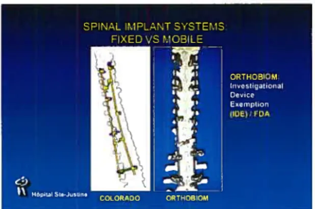
L’instrumentation vertébrale conventionnelle est une procédure irréversible qui peut engendrer des impacts négatifs. L’instrumentation—fusion résulte en une perte de la mobilité proportionnelle à l’étendue du segment fusionné. Cette mobilité réduite induit d’énormes stress sur les zones mobiles adjacentes aux segments fusionnés. Les structures caudales à la fusion subissent d’énormes forces biomécaniques. Des dommages permanents et de la douleur peuvent subvenir durant la vie adulte 28-37 De ces faits, l’idée de créer un système d’ostéosynthèse vertébral mobile qui exclurait la fusion osseuse émergea de nouveau.
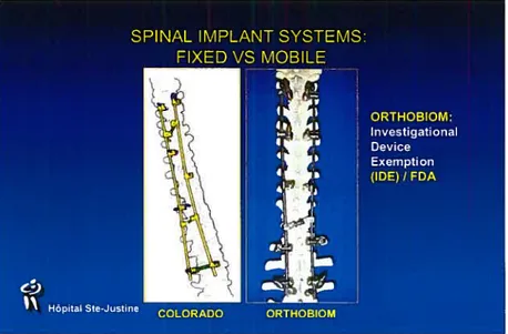
Le but ultime recherché est de créer un système d’ostéosynthèse vertébral mobile, Orthobiom (figure 1, rubrique liste des figures), qui sera utilisé pour correction du rachis atteint de SI, avec angle de Cobb inférieur à 5Q0,
L’objectif propre de l’étude ci-présentée est la comparaison de la survie fonctionnelle des disques intervertébraux chez le mini-porc ayant subi une chirurgie de scoliose avec système d’ostéosynthèse fixe versus ostéosynthèse mobile. Cette comparaison se base sur l’analyse quantitative immunohistochimique du collagène de type X. Les autres objectifs primordiaux de cette étude sont d’évaluer qualitativement la mobilité du segment vertébral instrumenté et de réaliser une évaluation radiologique des facettes vertébrales. Le tout se réalise via groupes comparatifs de mini-porcs.
C
28
Matériel et méthode
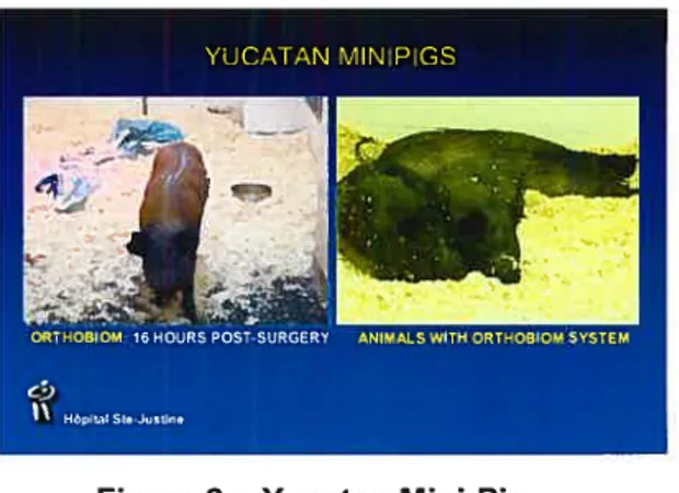
Population à l’étude. 18 mini-porcs (figure 2, rubrique liste des figures) sont utilisés: 6 ont subi une chirurgie avec système d’ostéosynthèse non-mobile fixe (TRSH (1) et Colorado (5)
)
et 9 avec le système d’ostéosynthèse mobile Orthobiom.Une scoliose thoraco-lombaire de 200 est induite chez 18 mini-porcs. De plus, trois servent de groupe contrôle. L’âge au moment de le chirurgie varie de 5.5 mois à 7 mois.
À
l’euthanasie, les sujets sont âgés entre 6 mois et deux ans. L’âge des sujets contrôle à l’euthanasie est de 12 mois pour deux sujets et de 18 mois pour un sujet. Les spécifications de la population à l’étude sont données au Tableau I (Tableau 1, rubrique liste des tableaux).Manipulation Tissulaire. L’euthanasie des mini-porcs est réalisée par induction par Somnotol (65mglkg) immédiatement suivie de l’injection d’Euthanyl (240mg/ml, 1 ml/2.5kg). Les segments fusionnés sont excisés par une approche postérieure. Les niveaux excisés incluent deux segments vertébraux non fusionnés de part et d’autre de l’instrumentation. Si l’instrumentation incluse L4 ou L5, le sacrum est excisé.
Les disques intervertébraux sont excisés avec un scalpel (lame #23) et avec l’aide d’un ostéotome si l’excision par scalpel s’avère impossible secondaire à la fusion intervertébrale (figure 3-4, rubrique liste des figures). Toutes les étapes sont documentées avec caméra digitale.
Immunihistochïmie. Les sections appropriées de disque intervertébral sont pré-traitées enzymatiquement pour en augmenter l’immunoréactivité avec H202 et trypsine 0.lgr / 50cc de solution tampon dicialane à 35° Celcius pour 20 minutes. Après cette étape, les échantillons sont traités avec PBS et la réaction neutralisante est réalisée pendant 10 minutes. Les échantillons sont ensuite traités pendant 24 heures à 4° Celcius avec l’anti-corps approprié selon les concentrations suivantes négative, 1/20, 1/50, 1/100. Ensuite, un autre traitement avec PBS, Dab et Mayer termine la préparation des échantillons.
L’anti-corps utilisé est un anticorps de collagène X53 monoclonal anti humain issu d’un hybridôme de souris contenant 10% de sérum fétal de bouvillon. Les anticorps sont une gracieuseté du Centre de Médecine Moléculaire, Université Nikolaus-Fiebiger, en Allemagne.
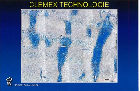
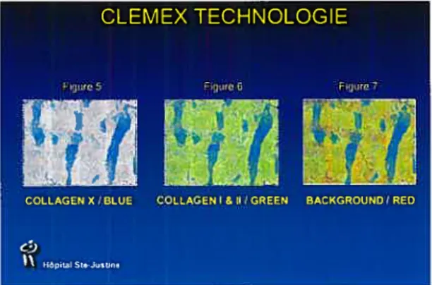
La technologie utilisée pour quantifier le taux de collagène X dans les échantillons est issue de Clemex Technologies Processing.
Trente-30
deux échantillons des mini-porcs contrôle et 16 échantillons des deux groupes instrumentés sont analysés par Clemex Technologie Processing.
Évaluatïon macroscopique de la mobilité des segments
vertébraux fusionnés. Avant l’excision des disques intervertébraux, et après le retrait de l’instrumentation vertébrale, une évaluation de la mobilité intervertébrale est réalisée. Des mouvements de rotation et de flexion sont induits. Une caméra digitale est utilisée pour enregistrer et ensuite évaluer la mobilité des segments. Une évaluation qualitative de la mobilité entre les systèmes d’ostéosynthèse fixe et le système d’ostéosynthèse mobile, Orthobiom, est réalisée. Le tout, comparé au groupe contrôle.
Imagerie des facettes articulaires intervertébrales. Un Ct-Scan des facettes du groupe contrôle et des groupes instrumentées est réalisé. Le but est d’analyser l’aspect macroscopique des facettes articulaires intervertébrales. L’imagerie est réalisée avec coupes de lmm à L4-L5 et L5-S1. L’imagerie est réalisée dans le plan axial.
(background). Le nombre de pixels est ensuite converti en pourcentage de collagène X, collagène normal et bruit de fond pour le champs analysé (figures5-7, rubrique liste des figures).
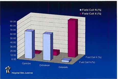
Pour le groupe contrôle, le pourcentage moyen de collagène normal est de 64.84% par champs analysé. Le pourcentage moyen pour le collagène type X est de 1.49%. Le bruit de fond correspond à 21.10% du champs étudié (figure 8, rubrique liste des figures).
Pour les échantillons Orthobiom, le collagène normal est représenté à 62.96%, le collagène X à 7.84% et le bruit de fond à 14.81%. Pour les échantillons du groupe fixe non-mobile, le collagène normal es indécelable, le collagène X est représenté à 92.84% et le bruit de fond à 6.42% (figure 8, rubrique liste des figures).
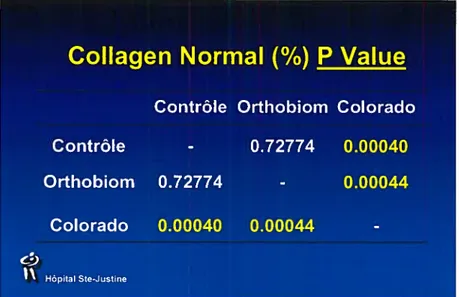
Une analyse statistique de variance (ANOVA) est réalisée pour comparer les différents groupes. Entre le groupe contrôle et le groupe Orthobiom, l’analyse démontre une valeur P de o.72774 pour le collagène normal et de 0.43063 pour le collagène X. Entre le groupe
33 contrôle et le groupe fixe (Colorado), l’on obtient une valeur P de 0.00040 pour le collagène normal et de 0.00074 pour le collagène X. L’analyse entre le groupe Orthobiom et Colorado révèle une valeur P de 0.00044 pour le collagène normal et de 0.00094 pour le collagène X (figure 9-10, lrubrique liste des figures).
Mobilité segmentaire. L’évaluation de la mobilité segmentaire révèle que pour le système fixe non-mobile d’ostéosynthèse vertébrale, il n’y a aucun mouvement décelé entre chaque niveau instrumenté. L’analyse qualitative de la mobilité du groupe contrôle et du groupe Orthobiom révèle que la mobilité entre facettes instrumentées est maintenue et comparable entre ces deux derniers groupes.
Imagerie des facettes articulaires intervertébrales. L’imagerie par CT-Scan des facettes lombo-sacrées révèle que pour le groupe contrôle et le groupe Orthobiom, un espace radio transparent entre deux facettes adjacentes est encore visible (figure 11-12, rubrique liste des figures). Cet espace radio transparent défini le cartilage articulaire encore présent composant la surface articulaire facettaire. Pour le groupe fixe non-mobile, aucune évidence d’espace radio transparent n’est notable, Il y a même disparition des délimitations facettaires (figure 13, rubrique liste des figures).
prématurée des segments instrumentés et ce, même si aucune greffe osseuse n’a été réalisée lors de l’instrumentation.
En effet, l’étude immunohistochimique révèle la présence statistiquement significative de collagène type X chez le groupe non-mobile lorsque comparé aux groupe Contrôle et au groupe Orthobiom (figure 9-10, rubrique liste des figures). La comparaison entre le groupe fixe non-mobile et le groupe Orthobiom révéle une différence encore une fois statistiquement significative quant au collagène normal et collagène X (figure 9-10, rubrique liste des figures)
L’évaluation qualitative de la mobilité segmentaire intervertébrale et l’évaluation par CT-scan des facettes lombo-sacrées permettent de réaliser que le groupe Orihobiom a maintenu une morphologie facettaire plus près du groupe contrôle lorsque comparé au groupe fixe non-mobile. Et comme ces deux évaluations sont deux des buts premiers de cette étude, il est intéressant de voir l’ensemble de ces données s’orienter vers le même constat soit que la détérioration des disques intervertébraux est statistiquement plus importante chez le groupe fixe non-mobile. Le fait que le système
36
Orthobiom maintient un mouvement entre les segments vertébraux fusionnés nous mène à penser que l’intégrité des disques est
préservée.
Un fait a été soulevé sur la méthodologie de cette étude en ce qui concerne l’utilisation d’un quadrupède comme animal modèle et l’utilisation des données de telles études pour le développement d’une instrumentation vertébrale qui sera utilisée chez l’humain bipède. Un doute peut être soulevé quant aux charges auxquelles les disques intervertébraux sont soumis: ces charges sont-elles équivalentes à celles de l’humain. Smit et al.38 ont décrit une étude validant l’utilisation de quadrupède pour de telles recherches sur le rachis. Les considérations biomécaniques étaient le principal aspect étudié. Ils conclurent que le quadrupède est un modèle valide en ce sens que le quadrupède subit des forces intervertébrales axiales compressives de loin supérieures à celles rencontrées chez l’humain. Selon leur étude, les résultats de telles recherches sont donc significatives pour le développement d’instrumentation vertébrale destinée à l’utilisation chez l’humain.
l’hypothèse que la survie fonctionnelle des disques intervertébraux est meilleure avec l’utilisation du système vertébral mobile Orthobiom.
L’étude réalisée a mis au point une nouvelle technique pour analyser la survie des disques intervertébraux basée sur l’apparition de Collagène type X et sa quantification par le logiciel informatique de Clemex Technologies Processing. Cette technique n’avait jamais été utilisée auparavant.
En analysant dans leur ensemble les résultats de l’immunihistochimie, l’analyse de la mobilité segmentaire et l’imagerie des facettes, toutes les données s’orientent vers le même constat: le système d’ostéosynthèse vertébral mobile Orthobiom permet une survie des disques intervertébraux. Donc, le développement de ce système d’ostéosynthèse vertébral mobile doit continuer et mener à une instrumentation qui sera utilisée pour le traitement de la SI chez l’humain.
39
Ce système d’ostéosynthèse vertébral mobile, Orthobiom, préviendra la fusion des segments intervertébraux instrumentés tout en conservant la correction de la déformation du rachis scoliotique.
Ultimement, le système Orthobiom préviendra les complications rencontrées chez la population adulte ayant subi dans leur adolescence une chirurgie avec fusion vertébrale pour la correction de la Scoliose Idiopathique.
4f
Analysis of Mini-pig Intervertebral Disc Survival
Comparison of surgical specimens between a non-mobile osteosynthesis system and a mobile osteosynthesis system
Marco Berard, M.D., D.M.V., Souad Rhalmi, MSc , Christine
Coillard, M.D., and Charles-Hilaire Rivard, M.D.
Ste-Justine Hospital Research Center, Montreal, Canada.
Department of Surgery, University of Montreal Medical School, Montreal, Canada.
The human intervertebral discs are highly specialized structures that permit load transmission and flexibility throughout the vertebral spine, allowing the vertebral spine to bend and rotate.1 The proper organization of ail the components of the intervertebral discs s required to sustain this primary role of mechanical function. Under normal conditions, the major constituents of the discs are large aggregating proteoglycans, water, and collagen. It is weIl established that in the normal intervertebral discs, seven type of collagen are present and their proportion varies among structures.26 Ghosh et al. demonstrated that there is an inverse gradient of collagen Types I and II from the outer Annulus Fibrosus to the inner Nucleus Pulposus.2 Thus, under normal conditions, the Annulus Fibrosus contains more Type I collagen that Type Il , whereas the Nucleus Pulposus is
decrease in areas with severe degenerative changes and the collagen Type IV was expressed in younger individuals with early signs of degeneration.2 In the Annulus Fibrosus, Nerlich et al. demonstrated increased amounts of collagen Type X in the degenerated discs and changes in the collagen Type IV similar to those seen in the Nucleus Pulposus.2 Such degenerative changes were found to be present in clinical conditions that require spinal fusion as treatment. One of these is Idiopathic Scoliosis.
Idiopathic Scoliosis, a condition characterized by a geometrical deformation of the spine, is diagnosed by a clinical examination and the analysis of the frontal and sagittal radiographs of the patient’s trunk. It is one of the most common orthopaedic conditions affecting adolescents The estimated prevalence of this condition ranges from 2% to 14% of the population in North America depending on the criteria used 5-11 Girls are more affected by IS than boys 5,6,8,10 the deformity is progressive during growth spurts and the prognosis is difficult to establish 12-15
The treatment varies with the magnitude of the spinal curvatures as quantified with the technique proposed by Cobb 16
43 When the spinal deformity progresses in the immature 17 skeleton to a Cobb angle between 15 and 40 degrees, brace treatment is the option normally accepted. Six to ten percent of the patients showing progressive lS require brace treatment 5,6,18,19 Some patients will, however, show severe deterioration, even during the brace treatment. When the Cobb angle reaches 45 to 60 degrees, depending on the age and maturity of the patient, on the location of the curie and on the physician’s clinical experience, a more definitive treatment is required, usually surgery. Up to 0.6% of the lS patient population will require surgery2° mainly to avoid negative effects of the growing spinal deformation on internai organs 21-23 However, even if 23 to 54% of the adolescent IS patients have back pain 24-25 this pain does not seem to be correiated with the magnitude of the deformation. Pain does not represent a major consideration in the decision for surgery in the adolescent as opposed to the adult scoliosis population 26
Since the beginning of the 1900’s, fusion of the spine segments has been used to limit the progression of the scoliosis curies. Harrington27 was the first to introduce the concept of spinal deformation correction without fusion, using metai rods. This revolutionary approach had, unfortunately, Iittle success. In fact, the fixed implant had to maintain the spinal correction in the face of large forces created by the movement of the patient. Fusion was subsequently re-introduced to stabilize the instrumented spine after correction with the metallic implant. The rod no longer needed to
changes the nature of the system with some negative implications. The instrumentation-fusion resuits in a loss of mobility proportional to the length of the fused segment of the spine. This mobility limitation will induce enormous stresses above and beiow the fusion. Structures at the caudal end of the rigid spine segment will have to sustain the iargest forces. Damages to these structures and pain could develop during adulthood 28-37 Thus, the idea to create a mobile osteosynthesis system that will avoid fusion re-emerged.
The ultimate goal is to creates a mobile osteosynthesis system, Orthobiom (figure 1) for use in IS with less than 50 degrees Cobb angle. The system will be kept in place until maturity and wili serve as an internai tutor that wilI permit the intervertebral discs to continue their growth despite the deformity correction. The anticipated outcome is iess degenerative change in the intervertebral discs.
45
And as a part of this global project, the objective of this study is to compare the mini-pig intervetebral disc survival between a group who received a non-mobile osteosynthesis vertebral system and a group who received a mobile osteosynthesis vertebral system. Immunohistologic quantitative analysis of collagen type X is used as a measure of discs degeneration. Others measures used are qualitative mobility of the instrumented segments and a radiologic assessment of the facets.
Materials and methods
Study population. In this study, 18 mini-pigs (figure 2) were enrolled: 6 of them had surgery with a non-mobile ostheosynthesis model (TSRH for 1 and Colorado System for 5 of them) and 9 received the mobile osteosynthesis system.
We induced a thoracolumbar scoliosis of 20 degrees in ail of them. The
Iast 3 mini-pigs are the control group. The age at the surgery was
between 5.5 and 7 months and the follow-up period was from 6 months
to 2 years. The age of the controls at the euthanasia was 18months (1
mini-pig) and 12 months (2 mini-pig). The specifications of the study
population are given in Table I.
Experimental Design: Surgery on 6-month old minipigs
Spinal Implant Minipigs Follow-up Euthanasia
Total =6
Fixed System 5- 6 month =O
6- l2months=1 7- 18 months = 5 8- 2years=O 9-Total 9 Orthobiom 5- 6 months = 1 6- l2months3 7- l8months3 8- 2years=2 Total = 3 Control 3- 12 months = 2 4- l8months=1
Figure 2: Yucatan Mini-Pig
47 Tissue processing. The euthanasia of each mini-pig was performed by induction with somnotol (65m glkg) followed by the injection of Euthanyl (240mg/ml, Jml/2.5kg), immediately. The fused segments were removed by a posterior approach to the spine, including two levels proximal and two levels distal to the instrumentation. If the instrumentation included L4 or L5, the pelvis was removed.
The intervertebral discs were removed by scalpel excision with #23 blade and with the use of an osteotome if removal by scalpel was impossible due to intervertebral disc fusion (figure 3-4).
Fogure 3: intervertebral discs with non-mobile osteosynthesis
Immunohistochemïstry. The appropriate tissue sections were enzymatically pretreated to enhance immunoreactivity using H202 and trypsine 0.1 gram per fifty cc tampon dicialane at thirty-five degrees Celsius for twenty minutes. After this, the samples were treated with PBS and the blocking reaction was performed for ten minutes. The next step was to process the samples with the proper antibody in the negative solution in the 1/20,1/50,1/1 00 dilution for twenty-four hours at four degrees Celsius. After this ail the samples were treated again with PBS and finally processed with Dab and Mayer.
The antibody used is a monoclonal anti-human collagen antibody X53. This is a mouse hybridoma supernatant containing 10% fetal calf serum. For the immunohistology the dilution went up to 1% dilution. The antibodies came from Nikolaus-Fiebiger Molecular Medicine Center of the Friedrich-Alexander-University, Germany.
Thirty-two samples of control mini-pigs and sixteen samples of each instrumented group were processed with the Clemex Technologies technique for measurement of the percentage of Collagen X.
49 Assessment of macroscopic mobility of the spine. To remove the instrumentation, we used the appropriate screwdrivers and osteotomes. We used a digital camera to film the mobility of the spines and performed subjective assessment of the mobility of the fixed and mobile Orthobiom systems and compared with the control group.
Imaging of the articular intervertebral facets. The control and instrumented spine facets were imaged with axial CT-scan at 1 mm intervals to permit a subjective comparison of the intervertebral articular facets.
Resu Its
lmmunohistochemistry. The Clemex Technologies’ assessment is based on the attribution of color pixels to the collagen X, to the normal collagen and to the background of the samples. The number of pixels was then transformed into a percentage content of collagen X, of normal collagen and of background (figure 5-7).
CLEMEX TECHNOLOGIE
Figure5 Figure 6 Figure 7
COLCAGEN XIBLUE COLLAGEN I & II !GREEN BACKGROUNDI RED
For the Orihobiom samples the normal collagen represent 62.96% of the field, the collagen X represented 7.84% and the background 14.81% (figure 8). For the non-mobile systems the collagen X represented 92.84% of the field and the background up to 6.42% of the field (figure 8).
The anova statistic analysis for the comparison between the control system versus the Orthobiom revealed a P value of 0.43.The P value for the comparison between the control and Colorado system revealed a P value of 0.00074. The comparison between the Orthobiom and Colorado system revealed a P value of 0.00094 (table2) (figure 9-10).
51
Collagen Normal
f%)
P ValueContrôle Orthobiom Colorado
Contrôle - 0.72774 0.00040
Orthobiom 0.72774 - 0.00044
Colorado 0.00040 0.00044
-3’ HâpiIM
Figure 9: ANOVA Normal Collagen
Collagen X
f%)
P ValueContrôle Orthobiom Colorado
Contrôle - 0.43063 0.00074
Orthobiom 0.43063 - 0.00094
Colorado 0.00074 0.00094
U6plII St-Jtho
Figure 10 : ANOVA Collagen X
Macroscopic mobility. The subjective macroscopic evaluation
revealed that for the non-mobile vertebral osteosynthesis system, there was no mobility of the instrumented spine segments. The subjective assessment of the control group and Orthobiom group revealed that the mobility of the facets were maintained and were subjectively si mi la r.
Imaging of the lumbo-sacral facets. The CT-scan imaging of the
lumbar-sacral facets revealed that for the control group and the Orihobiom group (figure 11-12) a radio-lucent line was visible between the facets of two adjacent levels. This lucency represents the articular cartilage of the joint facet.
Figure 12 : Ct-Scan Articular Facet mobile osteosynthesis subject
For the non-mobile group, there is no evidence of any radio-lucency at any of the instrumented lumbar-sacral segments (figure 13).
Figure 11: Ct-Scan Articular Facet Control subject
53 Discussion.
The results show that the Orthobiom mobile osteosynthesis system preserves the integrity of the intervertebral discs and the non-mobile osteosynthesis instrumentation leads to deterioration of the intervertebral discs and to a fusion of the instrumented segments even if no bone graffing is done.
The immunohistochemistry shows much more of collagen X in the non-mobile groups then in the non-mobile osteosynthesis group (Orthobiom).
The macroscopic evaluation of the mobility and the CT scan imaging of the intervertebral lumbar-sacral facet bring more evidence to support the use of the Orthobiom mobile osteosyhthesis system.
A question may be raised regarding the relevance of a quadruped model for the development of spine instrumentation for usein the upright human. Smith and al38 described a study to validate the use of quadruped model for the study of a spine via the biomechanical properties. They concluded that the quadruped can be a valuable animal model for spine research from the fact that the quadruped has to sustain even higher spinal axial compression and torsion stresses than the human. The forces sustained by the quadruped spine are similar to those sustained by the human spine and the quadruped model is relevant for studies of human spinal instrumentation.
Conclusion.
The fact that the macroscopic mobility and the analysis of the CT scan imaging of the facets point in the same direction as the immunohistochemistry allows us to say that the mobile osteosynthesis system leads to better survival of the mini-pig intervertebral discs. The development of a mobile osteosynthesis system may lead to analogous instrumentation that can be used in humans for treatment of Idiopathic Scoliosis.
The future human Orthobiomtm instrumentation will prevent the fusion of the instrumented spine level while maintaining correction of the deformity. Ultimately, we hope the Orthobiom system will lead to fewer complications in the adult population after surgical correction of adolescent Idiopathic Scoliosis.
55 Bibliog raphy:
1. Ghosh, P. The biology of the intervertebral disc. CRC Press: Florida, 1998
2. Nerlich, A. Immunohistologic markers for age-related changes of human lumbar intervertebral discs. Spine 1997; 22 (24): 2781-2795 3. Caillet R. Scoliosis: diagnosis and management. FA Davis: Philidelphia, 1975.
4. Kelsey 1. Epidemiology of musculoskeletal disorders. New-York: Oxford University Press, 1982.
5. Rogala EJ, Drummond DS, Gurr 1. Scoliosis: incidence and natural history. A prospective epidemiological study. JBJS-A 1978; 60 (2): 173-6.
6. Stirling AJ, Howel D, Miliner P A, Sadiq S, Sharpies D, Dickson RA. Late-onset idiopathic scoliosis in chiidren six to fourteen years old. A cross-sectional prevalence study. JBJS-A 1996; 78(9): 1330-6. 7. Brooks HL, Azen SP, Gerberg E, Brooks R, Chan L. Scoliosis: A prospective epidemiological study. JBJS-A 1975; 57(7): 968-72.
8. Morais T, Bern ier M, Turcotte F. Age- and sex-specific prevalence of scoliosis and the value of school screening programs. Am J Public Health 1985; 75 (12): 1377-80.
9. Kane Wl. Scoliosis prevalence: a cali for a statement of terms. Clinical Orthopaedics 1977; 126: 43-6.
10. Robitaille Y, Villavicencio-Pereda C, Gurr 1. Adolescent idiopathic scoliosis: epidemiology and treatment outcome in a large cohort of chiidren six years after
screening. Int J Epidemioll984; 13(3): 31 9-23.
11. Payne WK 3rd, Ogilvie JW, Resnick MD, Kane RL, Transfeldt EE, Blum RW. Does scoliosis have a psychological impact and does gendermake a difference? Spine 1997;22 (12): 1380-4.
12. Skaggs DL, Basseif GS. Adolescent idiopathic scoliosis: an update. Am Fam Physician 1996; 53 (7): 2327-35.
untreated idiopathic scoliosis during growth. JBJS-A 1984; 66 (7): 1061-71.
16. Cobb JR. Outiine for the study of scoliosis. In Instructional Course Lectures, The American Academy of Orthopaedic Surgeons, Ann Arbor, JW Edwards; vol. 5, 1948.
17. Labelle H, Dansereau J. Orthotic treatment of pediatric spinal disorders and diseases. In Spine: State of the art reviews,
6Philadelphia, Hanley & Belfus, mc; vol. 4(1), 1990.
18. Emans JB. Scoliosis: diagnosis and current treatment. Women Health 1984; 9: 81-1 02.
19. Farady JA. Current principles in the nonoperative management of structural adolescent idiopathic scoliosis. Physical Therapy 1983; 63 (4): 512-23.
20. Goldberg CJ, Dowiing FE, Fogarty EE, Moore DP. School scoliosis screening and the United States Preventive Services Task Force. An examination of Iong-term resuits. Spine 1995; 20 (12): 1368-74. 21. Ascani E, Bartolozzi P, Logroscino CA, Marchetti PG; Ponte A, Savini R et al.
Natural history of untreated idiopathic scoliosis after skeletal maturity. Spine 1986; 11(8): 784-9.
22. Weinstein SL, Zavala DC, Ponseti IV. Idiopathic scoliosis: long term follow-up and prognosis in untreated patients. JBJS-A 1981; 63 (5): 702-12.
23. Pehrsson K, Larsson S, Oden A, Nachemson A. Long-term follow up of patients with untreated scoliosis. A study of mortality, causes of death, and symptoms. Spine 1992; 17(9): 1091-6.
57 24. Ramirez N, Johnston CE, Browne RH. The prevalence of back pain in chiidren who have idiopathic scoliosis. JBJS-A 1997; 79 (3): 364-368.
25. Joncas J, Labelle H, Poitras B, Duhaime M, Rivard CH, Le Blanc R. Douleur dorsolombaire et scoliose idiopathique de l’adolescence. Ann Chir 1996; 50 (8): 637-640.
26. Poitras B, Mayo N, Goldberg M et al. The Sainte-Justine
adolescent idiopathic scoliosis cohort study. Part III: back pain. Spine 1994; 19: 1573-1 781.
27. Harrington PR. Treatment of scoliosis. Correction and internai fixation by spine instrumentation. JBJS-A 1962; 44: 591-611.
28. Sawatzky BJ, Tredwell SJ, Jang SB, Black AH. Effects of three dimensional assessment on surgical correction and on hook strategies in multi-hook instrumentation for adolescent idiopathic scoliosis. Spine 1998; 23(2): 201-205.
29. Ecker ML, Betz RR, Trent P5, Mahboubi S, Mesgarzadeh M, Bonakdapour A et al. Computer tomography evaluation of Cotrel Dubousset instrumentation in idiopathic scoliosis. Spine 1988; 13 (10): 1141 -1144.
30. Puno RM, Johnson JR, Ostermann P A, HoIt RT. Analysis of the primary and compensatory curvatures following Zielke instrumentation for idiopathic scoliosis.
Spine 1989;-14 (7): 738-743.
31. Wojcik AS, Webb JK, Burwell RG. Harrington-Luque and Cotrel Dubousset instrumentation for idiopathic thoracic scoliosis. A
postoperative comparison using segmental radiologic analysis. Spine 1990; 15(5): 424-431.
32. Winter RB, Lonstein JE, VanderBrink K, Anderson MB. Harrington rod with sub laminar wires in the treatment of adolescent idiopathic thoracic scoliosis: A study of sagittal plane correction. Proceedings of the 21 sI Annual Meeting of the Scoliosis Research Society, combined with British Scoliosis Society, Hamilton, Bermuda,September 21-25, 1986.
33. Dickson RA, Archer lA. Surgical treatment of late-onset idiopathic thoracic scoliosis: The Leeds procedure. J.B.J.S.-B 1987; 69: 709-714.
scoliosis: A new method of analysis of the deformity. Spine 1987; 12 (2): 228-232.
37. Cheung KM, Luk KD. Prediction of correction of scoliosis with use ofthefulcrum bending radiograph. JBJS-A 1997; 79(8): 1144-1150.
61
1. Ghosh, P. The biology of the intervertebral disc. CRC Press: Florida, 1998
2. Nerlich, A. Immunohistologic markers for age-related changes of human lumbar intervertebral discs. Spine 1997; 22 (24): 2781-2795 3. Caillet R. Scoliosis: diagnosis and management. FA Davis: Philidelphia, 1975.
4. Kelsey 1. Epidemiology of musculoskeletal disorders. New-York: Oxford University Press, 1982.
5. Rogala EJ, Drummond DS, Gurr 1. Scoliosis: incidence and natural history. A prospective epidemiological study. JBJS-A 1978; 60 (2): 173-6.
6. Stirling AJ, Howel D, Miliner P A, Sadiq S, Sharpies D, Dickson RA. Late-onset idiopathic scoliosis in children six to fourteen years olU. A cross-sectional prevalence study. JBJS-A 1996; 78 (9): 1330-6. 7. Brooks HL, Azen SP, Gerberg E, Brooks R, Chan L. Scoliosis: A prospective epidemiological study. JBJS-A 1975; 57 (7): 968-72.
8. Morais T, Bernier M, Turcotte F. Age- and sex-specific prevalence of scoliosis and the value of school screening programs. Am J Public Health 1985; 75 (12): 1377-80.
9. Kane Wl. Scoliosis prevalence: a cail for a statement of terms. Clinical Orthopaedics 1977; 126: 43-6.
10. Robitaille Y, Villavicencio-Pereda C, Gurr 1. Adolescent idiopathic scoliosis: epidemiology and treatment outcome in a large cohort of chiidren six years after
screening. Int J Epidemioll984; 13 (3): 31 9-23.
11. Payne WK 3rd, Ogilvie JW, Resnick MD, Kane RL, Transfeldt EE, Blum RW. Does scoliosis have a psychological impact and does gendermake a difference? Spine 1997;22 (12): 1380-4.
et al. Assessment of curve progression in idiopathic-scoliosis. European Spine J 1998; 7(4): 270-7.
15. Lonstein JE, Carlson JM. The prediction of curve progression in untreated idiopathic scoliosis during growth. JBJS-A 1984; 66 (7): 1061-71.
16. Cobb JR. Outline for the study of scoliosis. In Instructional Course Lectures, The American Academy of Orthopaedic Surgeons, Ann Arbor, JW Edwards; vol. 5, 1948.
17. Labelle H, Dansereau J. Orthotic treatment of pediatric spinal disorders and diseases. In Spine: State ofthe
art reviews,
Philadelphia, Hanley & Belfus, Inc; vol. 4 (1), 1990.
18. Emans JB. Scoliosis: diagnosis and current treatment. Women
Health 1984; 9: 81-1 02.
19. Farady JA. Current principles in the nonoperative management of
structural adolescent idiopathic scoliosis. Physical Therapy 1983; 63
(4): 512-23.
20. Goldberg CJ, Dowiing FE, Fogarty EE, Moore DP. School scoliosis
screening and the United States Preventive Services Task Force. An
examination of Iong-term results. Spine 1995; 20(12): 1368-74.
21. Ascani E, Bartolozzi P, Logroscino CA, Marchetti PG; Ponte A,
Savini R et al.
Natu rai history of untreated idiopathic scoliosis after skeletal maturity.
Spine 1986; 11(8): 784-9.
63 22. Weinstein SL, Zavala DC, Ponseti IV. Idiopathic scoliosis: long term follow-up and prognosis in untreated patients. JBJS-A 1981; 63 (5): 702-12.
23. Pehrsson K, Larsson S, Oden A, Nachemson A. Long-term follow up of patients with untreated scoliosis. A study of mortality, causes of death, and symptoms. Spine 1992; 17(9): 1091-6.
24. Ramirez N, Johnston CE, Browne RH. The prevalence of back pain in chiidren who have idiopathic scoliosis. JBJS-A 1997; 79 (3): 364-368.
25. Joncas J, Labelle H, Poitras B, Duhaime M, Rivard CH, Le Blanc R. Douleur dorsolombaire et scoliose idiopathique de l’adolescence. Ann Chir 1996; 50 (8): 637-640.
26. Poitras B, Mayo N, Goldberg M et al. The Sainte-Justine
adolescent idiopathic scoliosis cohort study. Part III: back pain. Spine 1994; 19: 1573-1 781.
27. Harrington PR. Treatment of scoliosis. Correction and internaI fixation by spine instrumentation. JBJS-A 1962; 44: 591-611.
28. Sawatzky BJ, Tredwell SJ, Jang SB, Black AH. Effects of three dimensional assessment on surgical correction and on hook strategies in multi-hook instrumentation for adolescent idiopathic scoliosis. Spine 1998; 23 (2): 201-205.
29. Ecker ML, Betz RR, Trent P5, Mahboubi S, Mesgarzadeh M, Bonakdapour A et al. Computer tomography evaluation of Cotrel Dubousset instrumentation in idiopathic scoliosis. Spine 1988; 13 (10): 1141 -1144.
30. Puno RM, Johnson JR, Ostermann P A, HoIt RT. Analysis ofthe primary and compensatory curvatures following Zielke instrumentation for idiopathic scoliosis.
the 21 sI Annual Meeting of the Scoliosis Research Society, combined with British Scoliosis Society, Hamilton, Bermuda,September 21-25, 1986.
33. Dickson RA, Archer lA. Surgical treatment of late-onset idiopathic thoracic scoliosis: The Leeds procedure. J.B.J.S.-B 1987; 69: 709-714. 34. Willers D, Hediund R, Aaro S, Nonnelli H, Westman L. Long-tenn resuits of Harrington instrumentation in idiopathic scoliosis. Spine 1993; 18(6): 71 3-71 7.
35. Stokes lA, Roncheffi PI, Aronsson DD. Changes in shape ofthe adolescent idiopathic scoliosis curie after surgical correction. Spine 1994; 19 (9): 1032-1 037.
36. Shuffeiberger HL, Wesley FK. Composite measurement of scoliosis: A new method of analysis of the deformity. Spine 1987; 12 (2): 228-232.
37. Cheung KM, Luk KD. Prediction of correction of scoliosis with use ofthefulcrum bending radiograph. JB]S-A 1997; 79(8): 1144-1150. 38. Smit, T. The use of a quadruped as an in vivo model for the study ofthe spine-biomechanical considerations. EurSpine J 2002; 11:137-144.
67
Master in Biomedical Sciences, University of Montreal, 2000-2002
Comparison between a Fixed versus a
Mobile vertebral osteosynthesis system
on the Physiology of the minipig
Intervertebral Discs
BERARD M, MD, DVM, RHALMI S, MSc, DEMONTIGNY A, KENISBERG, R, MSc, PhD, COILLARD, C, MD, RIVARD C-H, MD.
Sainte-Justïne Hospital, MONTREAL, Canada.
Q)
Hôpital Ste-Justine
Introduction
• Idiopathic Scoliosis=3D deformation
• Prevalence: 2% to 14% I Girls
• continuum: Evolution / Treatment
• Surgery: Stop the deformation
• Harrington: Concept of Correction without Fusion...Failed
Emergence of the Spine Fusion
• Fusion: Growth limitation
Restricted Mobility
Distal Discs: Higher Loading / Stress Disc Degeneration
L!)
-Appearance of Carboxymethyl-lysine -Appearance of COLLAGEN X
Hôpital Ste-Justine
HYPOTH ESIS
«
THE INTERVERTEBRAL DISCS
MORPHOLOGY AND PHYSIOLOGY
WILL SE PRESERVED WITH A
VERTEBRAL OSTEOSYNTHESIS
SYSTEM PERMITTING A
MICRO-MOVEMENT»
69
GOALS
(1) Create a vertebral osteosynthesis system -Mobile -Idiopathic Scoliosis -Cobb 35*-50*! 20% reductible -Cobb 51*-60*! 30% reductible -Cobb »61* I 40% reductible -T1-L5
-Removed at the end of growth
-Preserve the Intervertebral disc Physiology (2) Comparison of the Minipigs intervertebral discs
morphology between a mobile an a fixed system
71
SPINAL IMPLANT MINIPIGS FOLLOW-UP EUTHANASIA
1 6 Months = O 2- 12 Months = I TOTAL=6 COLORADO 3- 18 Months = 5 4- 2Years =0 6 Months =I 1 6 Months = I ORTHOBIOM 12 Months =2 2- 12 Months = 3 18 Months 2 3- 18 Months 4 2 Years =0 4- 2 Years =1 CONTROL i. 12 Months 2 12 Months =2 2- 18 Months 1 18 Months =I
ORTHOBIOM REMOVAL After 12 Months = 2 AFTER I YEAR
AND A MINIPIG SURVIVAL
79
Collagen X
(%)
P Value
Contrôle Orthobiom Colorado
Contrôle
-0.43063
0.00074
Orthobïom
0.43063
-0.00094
Colorado
0M0074
0.00094
-Hôpital Ste-Justine
Collagen Normal
(%)
P Value
Contrôle Orthobiom Colorado
Contrôle
-0.72774
0.00040
Orthobiom
0.72774
-0.00044
Colorado
0.00040
0.00044
83
CONCLUSION
AFTER 18 MONTHS, GOOD INTERVERTEBRAL
DISC PHYSIOLOGY IS PRESENT WITH
THE MOBILE SYSTEM.
•.
Neyer been done
...Moreaccurate
.Applications: menisci ; cartilage...
4)
1’
Hôpital Ste-JustineGrateful Thanks...
Audite (Demontiqny
pda 7(enis6erg
(Canaéa.
•SwitzertEand:. •.Çermany!!!)SouaéRjia(mi lU
4,
85
Grateful Thanks...
cDr Cfiartes-W’i(aire
Rjvaré
I!!
• more tfianjust a researcli cDirector!
L!)