Université de Montréal
Transcriptional activation by Spi ami post
transcriptional repression by muscle-specific microRNA
miR-133 of expression of human ERGJ and KCNQ1 genes
and potential implication in arrhythmogenesïs
par
Xiaobin Luo
Programme des Sciences biomédicales Département de Médecine
Faculté de Médecine
Thèse présentée à la Faculté des études supérieures en vue de l’obtention du grade de Maîtrise
en Sciences biomédicales Avril 2007 © Xiaobin Luo, 2007 /Grad2 corr àcomp2rL \ 200? WT 4 dc
o
de Montréal
Direction des bib1othèques
AVIS
L’auteur a autorisé l’Université de Montréal à reproduire et diffuser, en totalité ou en partie, par quelque moyen que ce soit et sur quelque support que ce soit, et exclusivement à des fins non lucratives d’enseignement et de recherche, des copies de ce mémoire ou de cette thèse.
L’auteur et les coauteurs le cas échéant conservent la propriété du droit d’auteur et des droits moraux qui protègent ce document. Ni la thèse ou le mémoire, ni des extraits substantiels de ce document, ne doivent être imprimés ou autrement reproduits sans l’autorisation de l’auteur.
Afin de se conformer à la Loi canadienne sur la protection des
renseignements personnels, quelques formulaires secondaires, coordonnées ou signatures intégrées au texte ont pu être enlevés de ce document. Bien que cela ait pu affecter ta pagination, il n’y a aucun contenu manquant.
NOTICE
The author cf this thesis or dissertation has granted a nonexclusive license allowing Université de Montréal to reproduce and publish the document, in part or in whole, and in any format, solely for noncommercial educational and research purposes.
The author and co-authors if applicable retain copyright ownership and moral rights in this document. Neither the whole thesis or dissertation, nor substantial extracts from it, may be printed or otherwise reproduced without the author’s permission.
In compliance with the Canadian Privacy Act some supporting forms, contact
information or signatures may have been removed ftom the document. While this may affect the document page count, it does flot represent any loss of content from the document
Faculté des études supérieures
Cette thèse intitulée
Transcriptional activation by Spi and post-transcriptional repression by muscle-specific microRNA miR-133 of expression of human ERG] and KCNQ] genes and potential
implication in arrhythmogenesis
présentée par
Xiaobin Luo
a été évaluée par un jury composé des personnes suivantes:
Dr. Bruce G. Allen
Président-rapporteurDr. Zhiguo Wang
Directeur de rechercheDr. Alvin Slirier
Membre du juryperméables aux ions K. Ces protéines canal participent activement au processus de repolarisation des myocytes cardiaques conmie dans le phénomène de «réserve de repolarisation» et ainsi constituent des acteurs déterminants dans l’apparition des arythmies. L’expression de ces gènes présente une disparité régionale dépendant à la fois de facteurs de différenciation mais aussi du profil physiopathologique du muscle cardiaque. Dans cette étude, nous avons identifié l’ensemble des régions promotrices et les sites d’initiation de la transcription de tous les isoformes de HERGJ et KCNQ]. Nous avons aussi caractérisé un transactivateur commun, la protéine SPi qui permet de contrôler leur transcription. Pour la première fois nous apportons des évidences expérimentales montrant que les gènes codant pour l’expression des canaux ioniques constituent des cibles de régulation pour les microARNs (miARNs): miR-133 réprimait l’expression de HERGJ et KCNQ] et miR-1 réprimait l’expression de KCNEJ. Les ARNm codant pour l’expression de l’ensemble des isoformes HERG1 et KCNQ1 étaient distribuées de façon asymétrique au sein du muscle cardiaque avec une prédominance dans les cavités de la partie droite du coeur en comparaison avec la partie gauche. Nous avons observé une hétérogénéité spatiale de la distribution de KCNQJ et KCNEJ suivant trois axes (interventriculaire, transmural et apical-basal) avec une disparité notable entre les niveau d’expression des ARNm et des protéines correspondantes. Cette disparité était associée à une hétérogénéité dans la distribution spatiale de SPi et miR-1/miR-133. Nos données ont ensuite permis de montrer clairement que des interactions entre SPi et miR-i/miR-i33 déterminent l’expression des gènes HERGJ et KCNQJ, par la même, seul le profil d’expression régional de SP-i et miR 1/miR-133 pouvait expliquer la différence régionale reconnue des courants ‘Kr et ‘Ks,
démasquant une implication potentielle dans la genèse des arythmies. Ainsi notre étude a permis de caractériser un nouvel aspect de la fonction cellulaire des miARNs visant à réguler l’électrophysiologie cardiaque. Offrant une explication des différences observées entre les quantités d’ARNm et les niveaux d’expression des protéines issus d’un même gène.
Mots-clés: HERG1, KCNQ1, Canaux potassiques, Spi, microARNs, miR-i, miR-133, Arythmies, Différences régionales, Repolarisation.
ABSTRACT
HERGJ and KCNQJ genes encode two major repolarizïng K channel a subunits that critically determine the repolarization rate and repolarization reserve in cardiac celis thereby the likelihood of anhythmias. Expressions of these genes are regionally heterogeneous and change dynamically depending on differentiation status and pathologicai states of the heart. Here we identified the core promoter regions and transcription start sites for ail HERG1 and KCNQJ isoforms, and revealed Spi as a common transactivator for their transcriptions. We also demonstrated ffiat the mRNA levels of ail HERG1 and KCNQ] isoforms are asymmetrically distributed within the heart, being predominant in the right relative to the lefi chamber. Notably, we for the first time experimentally validated ion charmel genes as targets for microRNA regulation by showing the ability of miR-133 to repress HERGJ and KCNQJ and of miR-1 to repress KCNE1. We further demonstrated the spatial heterogeneity of KCNQJ and KCNEJ distributions along three axes (interventricular, transmural and apical-basal) and disparity between mRNA and protein expression of ifiese genes, and the spatial heterogeneity of Spi and miR-1/miR-133 distributions as well. Our data strongly indicate that the interplay between
Spi
and miR4/miR-133 determines the expression levels of HERGJ and KCNQJ genes and the unique regional expression profiles of Spi and miR-1/miR-133 may be one of the mechanisms for the weli recognized regional differences of I and I1c, which might have a potential implication in arrhythmogenesis. Moreover, our study unraveled a novel aspect of the cellular function of miRNAs in regulating cardiac electrophysiology and offered an explanation for disparities between mRNA and protein expressions of genes.Keywords : HERG1, KCNQ1, Potassium channels, Spi, microRNA, 1, miR-133, Arrhythmia, Regional difference, Repolarization.
TABLE
0f CONTENT
RÉSUMÉ
ABSTRACT ii
TABLE 0F CONTENT iii
LIST 0F TABLES ix
LIST 0F FIGURES x
LIST 0F ABBREVIATIONS xii
ACKNOWLEDGEMENTS xiv
CONTRIBUTION 0F AUTHORS xvi
DEDICATION xviii
PART I. INTRODUCTION AND REVIEW 0F THE LITERATURE 1
1. Delayed rectifier potassium currents I and I in the heart 1
1.1 Overview of cardiac action poteutial 1
1.2 Biophysical properties of rapîd and slow delayed rectifier potassium
channels 3
1.2.1 Overview ofI and I 3
1.2.2 Electrophysiological and pharmacological properties ofI and I.4
1.2.2.1 and I in general 4
1.2.2.2 Electrophysiological and pharmacological properties ofI)c 5 1.2.2.3 Electrophysïological and pharmacological properties of Iic 5 1.2.3 Contributions ofI. and Iic to repolarization 6
2. Molecular composition of I and I 7
2.1 Overview of molecular composition of I. and I 7
2.2 Molecular composition of I 7
2.3 Molecular composition ofI 8
2.4 Isoforms ofHERG1 and KCNQJ 8
2.4.1 Isoforms ofHERGJ $
2.4.2 Isoforms ofKCNQ1 9
3. Regional heterogeneities of I. and Ic 9
3.2 Apex-base differences ofIy and Ic .10
3.3 Inter-chamber differences of Ic1 and Ic 10
3.4 Molecular bases underlying the regional heterogeneities of lIc. and iKs 11
3.5 Implications of regional heterogeneities of Iic and ‘Ksin
arrhythmogenesis 12
4. Alterations of delayed rectifier potassium channel (Age and diseases) 12
4.1 Age-dependent changes in I and I 12
4.2 I. and I under pathological conditions 13
4.2.1 Congenital and acquired long QT syndromes 13
4.2.2 Atrial fibrillation (AI) 14
4.2.3 Hypertrophy 14
4.2.4 Ischemia 15
4.3.5 Heart failure 15
4.3.6 Dïabetes 16
4.3.7 Molecular mechanism underlying pathological changes of I and 16 5. Molecular regulation ofHERG and KCNQ1 expression 17 5.1 Transcriptional regulation of ion channel-encoding genes 17
5.1.1 Importance and feasibility 17
5.1.2 Current progress in study ofhuman ion channel promoters 18 5.1.3 The role of stimulating protein 1 (Spi) in transcriptional regulation
19 5.2 Post-transcriptional regulation of ion channel genes 19
6 References 21
HYPOTHESIS 39
OBJECTIVES 40
PART II.ORIGINAL CONTRIBUTIONS 41
CHAPTER 1.Genomic structure, transcriptional control and tissue distribution
1.1 Abstract .43
1.2 Introduction 44
1.3 Resuits 45
1.3.1 Identification of the transcription start sites of the HERG1 and KCNQJ
genes 45
1.3.2 Genomic arrangement of the HERGJ and KCNQ1 genes and their
promoter regions 46
1.3.3 Structural analysis of the 5’-flanking regions of the HERG1 and KCNQJ
genes 47
1.3.4 Characterization of the promoter regions of the HERG1 and KCNQJ
genes 4$
1.3.5 Multiple Spi cis-elements and CpG islands in HERGJ and KCNQ1a core
promoter regions 49
1.3.6 Expression and tissue distribution ofHERGJ and KCNQ1 genes 50
1.4 Discussion 50
1.4.1 Transcriptional control ofHERGJ and KCNQJ genes 51 1.4.2 Expression profiles ofHERGJ and KCNQ1 genes 52
1.5 Materials and methods 54
1.5.1 Rapid amplification of cDNA ends (5’RACE) 54
1.5.2 RNase protection assay (RPA) 54
1.5.3 PCR amplification of putative promoter regions and construction of
promoter-luciferase fusion plasmids 55
1.5.4 Cdl culture 55
1.5.5 Transfection and luciferase assay 55
1.5.6 Real-time RT-PCR 56
1.5.7 Data analysis 56
1.6 Acknowledgements 57
1.7 References 57
CHAPTER 2. Transcriptional Activation by Stimulating Protein 1 and Post Transcriptional Repression by Muscle-Specific MicroRNAs of I-Encoding Genes and Potential Implications in Regional Heterogeneity of Their
Expressions 77
2.1 Abstract 79
2.2 Introduction 79
2.3 Materiais and Methods $1 2.3.1 Rapid amplification of cDNA ends (5’RACE) 81
2.3.2 Construction of promoter-luciferase fusion plasmids 82 2.3.3 Synthesis of mIRNAs and anti-miRNA antisense inhibitors 82
2.3.4 Mutagenesis $2
2.3.5 Construction of chimeric miRNA-target site—luciferase reporter vectors 82
2.3.6 Ccii culture 83
2.3.7 Transfection and lucïferase assay 83
2.3.8 Real-time RT-PCR 84
2.3.9 Western blot 85
2.3.10 Drug treatment 85
2.3.11 Data analysis $6
2.4 Results 86
2.4.1 Transcription start sites ofKCNQJ $6 2.4.2 Spi as a transcription activator ofKCNQJ and KCNE1a 87 2.4.3 MiR-133 as a post-transcriptional repressor ofKCNQ1 89 2.4.4 MiR-1 as a post-transcriptional repressor ofKCNEJ 91
2.4.5 SpI and rniR-lImiR-133 and their rotes in regional heterogeneity of KCNQJ and KCNEJ expressions 91
2.5 Discussion 92
2.6 Acknowledgments 96
2.7 References 96
Chapter 3. MicroRNA miR-133 Represses HERG
iC
Channel Expression 1123.1 Abstract 114
3.2 Introduction 114
3.3 Experimental Procedures 116 3.3.1 Preparation of Rabbit Model of Diabetes Mellitus (DM) 116 3.3.2 Isolation of Rabbit Ventricular Myocytes and Ceil Culture 116 3.3.3 Whole-Cell Patch-Clamp Recording 116 3.3.4 Synthesis of mIRNAs and Anti-miRNA Antisense Inhibitors and Their
Mutant Constructs 116
3.3.5 Construction of Chimeric miRNA-Target Site—Luciferase Reporter
Vectors 116
3.3.6 SmaH Interference RNA (sIRNA) Treatment 117
3.3.7 Cel] Culture 117
3.3.8 Transfection and Luciferase Assay 117 3.3.9 Quantification of mRNA and miRNA Levels 117
3.3.10 Western Blot 118
3.3.11 Data Analysis 118
3.4 Results 118
3.4.1 Overexpression of miR-1 and miR-133 and Downregulation of ERG Protein Level in Diabetic Hearts 118
3.4.2 Post-Transcriptional Repression ofHERG Expression by miR-133 119 3.4.3 Potential Role of Serum Responsive Factor (SRF) in miR-133
Overexpression 120
3.5 Discussion 121
3.6 References 123
3.7 Ackuowiedgements 124
3.8 Figures and Figure Legends 125 3.9 Supplementary Materials 131
1. Major findings .140
2. Summary and Conclusion .141
2.1 Identification ofthe core promoter regions ofHERGJ and KCNQJ
isoforms 141
2.2 Spi as the essential transactivators for HER Gi,KCNQ1 and KCNE1 genes 142 2.3 HERG1, KCNQi and KCNEi are targets for post-transcriptional
repression by the muscle-specific microRNAs miR-1 and miR-133 143 2.4 The spatial distribution pafterns of Spi and miR-1/miR-133 and the
potential roles in regional heterogeneity of I and 144
3. Potential implications 146
4. Possible limitations of the present study 147
5. Future Directions 14$
6. References 149
APPENDIX xxi
Appendix 1. Additïona] Publications xxi
LIST 0F TABLES
Table 1. Number of consensus binding sites for cardiac-specific or -related transcription factors within a 3-kb frame of the 5’-flanldng regions of HERGJ and
KCNQ] genes 75
Table 2. Number of consensus binding sites for stimulating protein 1 (Spi) and CCAAT boxes within the core promoter regions and CpG islands within a 3-kb frame upstream the translational start sites (TSSs) ofHERGJ and KCNQ]genes 76
LI$T 0F FIGURES
Part I INTRODUCTION
Figure 1 .Cardiac action potential and ion channels 2 figure 2. Action potential waveforms are variable in different regions of the heart. .
Part II ORIGINAL CONTRIBUTIONS Chapter 1
Figure 1. Identification oftranscripfion start sites ofHERGJ and KCNQ1 genes 65 Figure 2. Genomic structure ofHERG gene subfamily 69 Figure 3. Genomic structure ofKCNQJ gene subfamily 70 figure 4. Analysis of the HERGJa and HERG1b promoter activities in various ceil
unes 71
Figure 5. Analysis ofthe KCNQJa (A) and KCNQJb (B) promoter activities in H9c2 rat ventricularce!! une and HEK293 human kidney embryonic ceil une 72 Figure 6. CpG islands ofHERGJ and KCNQJ genes predicted using the CpG Island
Searcher 73
Figure 7. Distribution of HERG1 and KCNQ1 transcripts in human tissues and
regional differences of expression in the heart 74
Chapter 2
figure 1. The 5 ‘-flanking regions containing the core promoter sequence and
transcription start sites of the KCNQ1 105
Figure 2. Ma!ysis ofthe KCNQJ promoter activities in various ceil unes 106 Figure 3. Role of stimulating protein 1 (Spi) as a common driving factor for KCNQJ
and KCNE]a transcriptions 107
Figure 4. Post-transcriptional repression of KCNQ1 by the muscle-specific
microRNA miR-133 108
Figure 5. Post-transcriptional repression of KCNF1 by the muscie-specific
microRNA miR-1 109
Figure 6. Regiona! differences of expressions of KCNQJ and KCNE] at protein and
Figure 7. Regional differences of expressions of Spi and miR-1 and miR-133 geies in
human hearts 111
Chapter 3
Figure 1. Upregulation of miR-1 and miR-133 and downregulation of ERG in rabbit hearts of diabetes model and in human hearts from subjects with diabetes mellitus...128 Figure 2. Post-transcriptional repression ofHERG by miR-133 129 Figure 3. Role of serum response factor (SRF) in enhancing expressions of miR 1/miR -133 in the heart of diabetic rabbits 130 Supplementary Figure 1. Sequences of the muscle-specific miRNAs and their putative target sites in human ether-a-go-go-related gene (HERG) and rabbitether-a
go-go-related gene (rbERG) 137 Supplementary Figure 2. Comparison of miR-1 and miR-133 expression levels in various ceil unes indicated 138 Supplementary Figure 3. Whole-cell patch-clamp recordings of the rapid delayed rectifier K current in lefi ventricular myocytes isolated from diabetic and healthy
LIST 0F ABBREVIATIONS
AF: Atrial fibrillationAP: Action potential
APD: Action potential duration AVB: Atrioventricular block AVN: Atrioventricular node
CACNA1C: L-type-calcium channel alpha 1C subunit EAD: Early afierdepolarizaiton
EAG: ether-a-go-gorelated gene Epi: Epicardial
Endo: Endocardial
HEK293: Human embryonic kidney 293 celi une HERG: Human ether- a-go-go related gene
HERG1a: Human ether- a-go-go related gene isoform 1 HERG1b: Human ether- a-go-go related gene isoform 2
ICa,L L-type calcium current/channel
IK: Delayed rectifier potassium currentJchannel
IKur: Ultrarapidly-activated delayed rectifier potassium current/channel ‘Na: Voltage-gated sodium channel/charinel
Ito: Transientoutward potassium current/channel
I: Rapidly-activated delayed rectifier potassium current/channel I: Slowly-activated delayed rectifier potassium channellchannel
IK1: Inward recitifer potassium current/channel
frx5: Iroquois homeobox transcription factor K: Potassium
KCNQ1 (KvLQT1): Slow delayed rectifier K channel alpha subunit member 1
KCNQ1a: Slow delayed rectifier K channel alpha subunit member 1 isoform 1 KCNQYb: Slow delayed rectifier K channel alpha subunit member 1 isoform 2 Kv: Voltage-gated potassium
LA: Lefl atrium LV: Lefi ventricle
LPC: Lysophosphatidylcholine
minK (KCNE1): Minimal potassium channel MiRP1 (or KCNE2): MinK-related peptide 1 M-cell: Midmyocardial celis
Mid: Midmyocardial miRNA(s): microRNA(s) miR-1: microRNA-1 miR-133: microRNA-133
Mef2: Myocyte enhancer factor 2
Nkx2-5: NK2 class ofhomeobox transcription factor NFAT: Nuclear factor of activated T-cells
NF-KB: Nuclear factor-kappa B RA: Rightatrium
RV: Right ventricle
RACE: Rapid amplification ofcDNA ends P-/GW-bodies: Processing bodies
SAN: Sinoatrial node
siRNAs: short interfering RNAs Spi: Stimulating protein 1
SCN5A: Sodium channel alpha subunit SRF: Serum response factor
Stat3: Signal transducer and activator of transcription-3 SV4O promoter: Simian virus 40 (SV4O) promoter TK promoter: Thymidine kinase (1K) promoter TdP: torsades de pointes
Tbx: T-Box transcription factor TSS: Transcription start site 3’UTR: 3’-untranslated region 5‘UTR: 5 ‘-untranslated region
Speciai recognition is due as weli to the administrative office at the Centre de Recherche de 1’ICM, to the staff in the Faculte de medicine and the Faculte de medicine and the Faculte de etudies superieurs of the Universite de Montreal, for their help in my graduate studies and preparing for this thesis. I would like to extend my gratitude to Dr. Bruce G. Allen, Dr. Alvin Shrier for their kindness in spending time to review my thesis and providing insightfiil suggestions and constructive comments in my thesis.
I want to deeply thank my wonderful parents (Zhihong Wang and Shiguang Luo), my brother-in-law Chengwei and my sister Xiaolin for their endless care and love; to my giriffiend Ling who has been supporting me facing any obstacle and sharing our happiness; and to ail others of my families. This work would not be done without their support.
CONTRIBUTION 0F AUTIIORS
The following is a statement regarding the contribution of co-authors and myseif to ffie four articles which have been published or submitted for publication, and included in this thesis.
1. Luo X, Xiao J, Lin H, Shan H, Yang B, Wang Z. (2007) Genomic structure, transcriptional control and tissue distribution ofhuman ERG] and KCNQ] genes. fEBS Lett. (Submitted in August 2007).
Dr. Zhiguo Wang and I proposed the original research plan. I designed and performed the experiments, analyzed the data, and wrote the manuscript. Dr. Jiening Xiao and Dr. Huixian Lin were involved in the experiments related to cloning promoters of HERGJa and HERG]b. Dr. Hongli Shan and Dr. Baofeng Yang participated in experiments of Real-time RT PCR. Dr. Zhiguo Wang had the original idea and fully supervised the work and finalized the manuscript for publication.
2. Luo X, Xiao J, Lin H, Lu Y, Li B, Yang B, Wang Z. (2007) Transcriptional activation by stimulating protein 1 and post-transcriptional repression by muscle specific microRNAs of Ii-encoding genes and potential implications in regional heterogeneity of their expressions. J (‘et! Physiot. 2007 Aug;2 12(2): 358-67.
Dr. Zhiguo Wang and I proposed the original research plan. I designed and performed the experiments, analyzed the data, and wrote the manuscript. Dr. Jiening Xiao and Dr. Huixian Lin were involved in experiments of Western blot. Dr. Yanjie Lu, Dr. Baoxin Li and Dr. Baofeng Yang contnbuted to experiments ofReal-tirne RT PCR. Dr. Zhiguo Wang fully supervised the work and finalized the manuscript for publication.
3. Xiao J, Luo X, Lin H, Zhang Y, Lu Y, Wang N, Zhang Y, Yang B, Wang Z.
MicroRNA miR-133 Represses HERG K Channel Expression Confributing to QT Prolongation in Diabetic Hearts. JBiot Chem. 2007 Apr 27;2$2(17):12363-7.
In tins work, I designed and performed ail experiments conceming the effect of miR-133 on I{ERG in ceil une, and analyzed the data for tins part. Dr. Jiening Xiao performed the exp eriments and analyzed the data about the effect of miR- 133 in diabetic rabbit and human myocytes. Dr. Huixian Lin and Ning Wang were involved in experiments of Western blot. Dr. Yanjie Lu, Ying Zhang and Dr. Baofeng Yang participated into experiments of HERG current recording under application of miR-133 and its antisense. Dr. Yiqiang Zhang performed the Patch-clamp recording of HERG current in diabetic rabbit myocytes. Dr. Zhiguo Wang fuiiy supervised the work and finalized the manuscript for publication.
DEDICATION
This thesis is dedicated to:
My mother myfather my sister my brother-in-Ïaw, and my girsfriend for their love, patience, understanding, encouragements, and supports. Wereitnotfor their sacrfice, this thesis may have neyer been compteted.
PART I. INTRODUCTION AND REVIEW 0F THE.
LITERATURE
1. Delayed rectifier potassium currents I and I in the heart
1.1 Overview of cardiac action potentïal
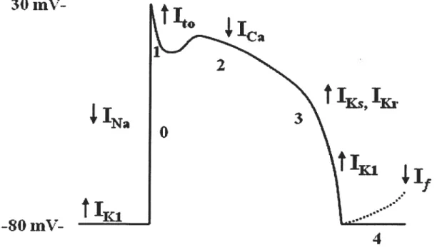
The normal electrophysiological behavior of the heart is determined by the orderly propagation of electrical impulses resulting in rapid depolarization and slow repolarizafion, thereby generating action potentials in individual myocytes1. The cardiac action potential (AP) reflects a balance between inward (depolarizing) and outward (repolarizing) currents. It consists of five phases. Phase O represents depolarization of the myocytes, in which voltage-gated sodium channels (‘Na) are
rapidly activated to depolarize the celi membrane. Phase 1 of the cardiac AP occurs right after the peak of depolarization, which underlies an early rapid phase of the repolarization in ventricular and atrial celis. This rapid repolarization is due to the inactivation of ‘Na and activation of transient outward potassium current (Ito).
following phase 1 is the long lasting plateau phase 2 repolarization, reflecting the balance between slowly decreasing inward depolarizing calcium (iCa,L) currents
through L-type calcium channels and gradually increasing outward repolarizing potassium currents mainly through rapid delayed rectifier potassium curren (Ic). The net amount of ions fluxing across the ceil membrane during the plateau phase is small, resulting in high impedance. Therefore a relatively small change in the ion current can have a significant impact on the membrane potential thereby action potential duration (APD). Phase 3 repolarization is primarily due to activation ofIKr and slow
delayed rectifier potassium channels (Ic), along with inactivation of‘Ca,L. Phase 4 is
the final stage of the cardiac action potential, during which the ceil membrane retum to its resting potential, which is accomplished by the outward potassium current (IKI)
through inward rectifier channels. Figure I shows the relationships among different cardiac ion channels and an AP.
30
;nV
-$0 rnV
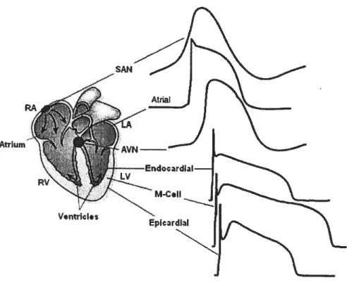
The configuration and duration of AP differ in specific regions (e.g., atrium
versus ventricle; apex versus basal, etc.) as well as in different layers within those regions (e.g., epicardium, midmyocardium and endocardium) (as shown in Figure 2). These physiologic heterogeneities of cardiac AP are likely due to differential expression of ion channels. For example, smaller I accounts partly for longer APD in midmyocardium. Other factors, such as genetic defects (mutations in ion channel genes), sympathetic regulation, modulation by dmgs and alterations in response to a variety of diseases (e.g., ischemia, myocardial infarction, hypertrophy, diabetes, etc.), can exaggerate these heterogeneities generating a substrate for arrhythmogenesis.
HCa
2
0
jIN
t
‘
‘Ks, ‘Ki
3
‘Ki4
Figure 1. Cardiac action potential and ion channels. Typical ventricular action
potential (AP) with inward currents (downward arrow) and outward currents (upward arrow). Numbers indicate various phases of an AP (Modified from Schram G, et al.2)
Atrium
1.2 Biophysical properties of rapid and slow delayed rectifier potassium channels
1.2.1 Overview0f ‘K. and I
In cardiac myocytes, voltage-gated potassium (Kv) channels are the primary determinants of action potential repolarization. In general, based on differences in time- and voltage-dependent properties and pharmacological sensitivities, there are two types of cardiac Kv have been distinguished6, transient outward potassium currents (Ito) and delayed rectifier potassium cunents (IK). Tto rapidly activates and
inactivates when ce!! membrane depolarizes to positive potentia! around -30 mV, which underlies the early phase (phase 1) of the action potential in atrial and
V.ntricI.
Figure 2. Action potential waveforms are variable in different regions of the heart. Schematic representation of the heart; AP waveforms recorded in different regions of the heart are illustrated.
ventricular cells7. ‘K activate more slowly compared to ‘to following initial
depolarization. Three distinct components, ‘Kur, I and I, comprise the delayed
rectifier potassium current in the heart8”. ‘Kur activates extremely fast (named
ultrarapid), almost instantaneously when compared to the other two components of‘K•
I activates fast and exhibits a strong inward rectification, and Iic activates very slowly. While I and I are found in both of atial and ventricle myocytes in different species (except aduit rodent), ‘Kur exists only in atrial myocytes in most species
(including human)’1-16, but flot in human ventricular myocytes and Purkinje fibers, suggesting that it is a suitable target for specific treatment of atrial arrhythmias. Since my study is focusing on the regulation of pore forming Œ-subumts of and ‘Ks
channels, for the rest of this chapter, I will mainly discuss these two major components.
1.2.2 Electrophysiological and pharmacological properties ofIKr and I7(s
1.2.2.1 ‘Kr and I in general
I and Ii are distinguished by their kinetics of activation, deactivation and inactivation, and by their highly variable sensitivity to dmg blockers. They were first distinguishably recorded in guinea pig atrial and ventricular myocytes, basing on the distinct differences in time- and voltage-dependent properties’7’9. They have also been found co-expressed in human atrial and ventricularmyocytes’5 6;2021; as well as in canine42225 and rabbit26’27 ventricles and in canine Purldnje fibers25. In some species, for example, feline283’ and fetal mice32 and rat33, composes the major repolarizing current,but it is rapidly supplanted postnatally by a very large It. In contrast, thecurrentsofIlCj and I are undetectable in small aduit rodent ventricles35,
I channels activate during the upstroke (Phase 1) and plateau (Phase 2) phases of an AP. Activation of Ifc starts with precipitous voltage dependence and its haif-maximal activation appears approximately at the potential of -25 mV. The amplitude of Ig increases as the membrane potential arises to O mV, and then decreases upon further depolarization, resulting in a unique ‘bell’ shape of the current-voltage relationship. Iy activates rapidly and inactivates faster at more
positive potential, and thus limits the time that the channels stay in the open state, showing strong inward rectification36. On repolarization the rate of recovery from inactivation through the open state is much more rapid then deactivation, which resuits in a large outward current in the voltage negative to O mV and provides a basis for promoting phase 3 repolarization.
I/HERG was originally classified to be a component of‘K due to its specific
sensitivity to methanesulfonanilide class III antiarrhythmic agent E-403 i37, dofetilide and d-sotalol38. Ijj./IIERG is also blocked by class E antiarrhythmic agents such as propafenone39, quinidine40, flecainide4’ and mexiletine42, as well as some other non cardiovascular drugs (e.g. cisapride43, terfenadine”, astemizole45). Blockage of Ijf[TERG channels by pharmacological agents is pro-arrhythmic, which will potentially produce marked QT prolongation and distinctive ventricular tachycardia, torsades de pointes, or acquired long QT syndrome (LQTS).
1.2.2.3 Electrophysiological and pharmacological properties ofIj
Compared to Ic activates much more slowly when cell membrane depolarizes to above -30 mV. Its haif-maximal activation is around +20 mV’537468. I activates much slowly, and an extreme]y long depolarization period is required for it to reach the steady-state level. I mainly serves as a “repolarization reserve” in cardiac repolarization in both human atria and ventricles4950, preventing excessive APD prolongation and development of arrhythmogenic early afierdepolarizations49,
particularly when Iic is substantial depressed by drugs or in the presence of cardiac disease.
I is resistant to blockade by methanesulfonanilide antiarrhythmic agents37, but selectively blocked by chromanols (293B, HMR-1556)51’52. Iic specific blockers have been developed but flot commercialized because ofthe potential.risk of torsades de pointes. It has been shown that there is a tendency of developing a homogeneous APD prolongation in ventricular tissue by blockingiy3.
1.2.3 Contributions of I and I to repolarization
The relative contributions of Ig and Ijc to repolarization have been broadly studied in different species for the last two decades. In guinea pig ventricular myocytes, similar amplitudes of these two currents were recorded when the membrane potential was set to the potential corresponding to plateau phase of repolarization, indicating their equal contributions to the repolarization37. Blockade of either I or I caused a similar moderate prolongation of repolarization, whereas concomitant blockade of both Ic- and I lcd to a much greater prolongation54. In rabbit and dog ventricular myocytes, the relatively amplitude of Iic under the similar condition (membrane potential depolarized to 3O or 4O mV) is larger than Ic2555,
which implicates that the overall contribution of Ic3 to ventricular repolarization is greater than that of‘Ks• Therefore, in dog and rabbit (perhaps in human), I may be
more important than I in determining ventricular repolarization. However, interestingly, when repolarization is delayed pharmacologically or pathologicaÏly, the prolonged APD will favor activation of I to restrict the excessive APD prolongation, reducing the risk of early afierdepolarization (EAD).
2. Molecular composition of 1c and I
2.1 Overview of molecular composition of Ic and I
The molecular identity of Igj and Ic remained unknown until revealed by molecular genetic analysis of an inherited disease, long QT syndromes. HERG encodes subunits to form channel complex constituting lic1. KCNQ1 and KCNEÏ encode subunits that co-assemble to form channels that mediate Mutations in any of these genes can cause LQTS, a disorder of cardiac repolarization that predisposes to lethal ventricular arrhythmias.
2.2 Molecular composition of Ii
HERG (Human ether-a-go-go) is a member of the ether-a-go-go (EAG) channel familyoriginally cloned from a hippocampal cDNA library and was found to be highly expressed in human heart tissue56. Several independent heterologous expression studies confirmed that HERG encodes ffie u-subunit underlying 11Cr5758. HERG has the same channel structure as the other voltage-gated ion channels’960. Four repeats of six transmembrane domains (Si -S6) constitute each HERG channel with a reentrant ‘pore-loop’ between $5-$6 to form the channel pore605963. The biophysical properties of expressed HERG are nearly identical to those of native Iic
in cardiac myocytes5737. Some studies also proposed that additional channels subunits, minK (KCNE1) or Mink-related peptide 1 (MiRP1 or KCNE2) may associate with the pore-forming a subunit (HERG) to form the native I6466. Although this notion is further potentiated by the finding of mutations in KCNE2 gene are associated with congenital and acquired LQTS6567, but it stiil remains somewhat uncertain and are challenged by other studies. For examples, Weerapura et al. reported that the biophysical and pharmacological properties of HERG channel without the co expression of MiRP1 were quite similar with Ic1 recorded in guinea pig68. And meanwhile, some other studies have also failed to confirrn the biophysical interactions between HERG and MiRP169. Mutations in pore-forming a-subunit
(HERG) or in putative regulatory subunit Mink-related peptide 1 (MiRP-1) of I chaimels are associated with congenital (LQT2 and LQT6) and acquired long QT syndrome, which can-ies increased risk of life-threatening cardiac arrhythmias57’65’70’.
Taken together, I is composed primarily of the pore-forming a-subumt HERG which might be associated with a function-altering 13-subunit MiRP1 or minK, but the t3-subunits underlying Ifcj, and thefr interactions with HERG are stili under controversy, and remain to be further investigated.
2.3 Molecular composition of I
The pore-forming a-subunit KCNQ1 (formerly called KvLQTI) together with -subunit mink (KCNE1) forms the slow delayed rectifier potassium channel (Ig) when co-expressed in heterogeneous systems, whule expressing any ofthem alone can not produce I1c-like current47’7174. KCNQ1 was originally identified with the purpose of searching for a gene associated wiffi LQTS by the positional cloning approach72and is found to express strongly in the pancreas and the heart72. KCNQ1 has 676 amino acids which consists of six transmembrane domains and forms typical pore ioop structure71 75,76 MinK is a single transmembrane protein containing 129 amino acids, and functions as the regulatory accessoiy to increase the macroscopic current amplitude by siowing the charinel activation kinetics and shifiing the activation at more positive potential6673’77. Loss-of-function mutations in either KCNQ1 or minK cause fonns of LQTS (LQTS1 and LQTS5, respectively)47627’;72;78;79
2.4 Isoforms ofHERG1 and KCNQ1
2.4.1 Isoforms ofHERGJ
In mammalian heart, N-terminal spiice variants of ERG have been found in both human and mouse80’81. The long isoform la (HERG1a) fias 396 amino acids in the N-terminus, whereas the short isoform lb (HERG15) has only 36 amino acids in
the N-tenninus8081. The lacking part of isoform lb is important for ifie slow deactivation process of isoform 1a8084. A recent study proposed that cardiac I
channels are minimally composed of HERG1a and HERG1b Œ-subunits that co assemble in the membrane83. Jnterestingly, HERG1b subunits co-expressed in heterologous systems preferentially form heteromultimers with HERG1 a and modify the deactivation gating properties previousiy attributed to the HERG1a N-terminus84. A smdy carried out by Nerbonne et al using specific antibodies against the N- and C tennini of HERG in human, rat and mouse, showed that HERG1 a is expressed in aduit hearts of ail three species, but there is no detectable expression of HERG1b85. However, HERG1b was shown to contribute to Ig channel function in the neonatal hearts86. These findings indicate that there is a developmental change in ERG isoform expression.
2.4.2 Isoforms ofKCNQ1
Isoform 1 (long isoform) and isoform 2 (short isoform) are the two major splice variants in the heart when detected at the mRNA level8789. Two isoforms are different in their N-termini, with deletion of nearly the whole N terminus of isoform
28790. When expressed in a heterologous context, the isoform 2 protein functions as a
dominant negative isoform87. Notably, a detectable amount of protein originating from isoform 2 transcript has neyer been reported in cardiac tissue91. For convenience, here we designate the two major isoforms KCNQIa (long isoform or isoform 1) and KCNQ1b (short isoform or isoform 2).
3. Regional heterogeneities of Ic. and I
There exist marked differences in the densities of Ic1 and Ic in different myocardial ceil types, which contribute to the regional properties of AP waveforms in the heart. These intrinsic regional heterogeneities of Ig and ‘Ks help to maintain the
normal heterogeneous property of cardiac AP through out the heart, and thereby assure the proper propagation of electrical signal. The regional differences are
reflected in sinoatrial node (SAN), atrioventricular node (AVN), apex versus base, lefi atrium (LA) versus right atrium (RA), lefi ventricle (LV) versus right ventricle(RV)) or layers (epicardial (Epi), midmyocardial (Mid) and endocardial (Endo)
)
ofthe atria and ventricles.3.1 Transmural differences of I- and I
Two independent studies have shown that the density of Ic- in ceils isolated from guinea pig lefi ventricular free walls was higher in epicardial than in midmyocardial or in endocardial myocytes, whereas both Ic and Ig densities at the base of the heart were smaller in sub-endocardial myocytes than those in mid myocardial and sub-epicardiaÏ myocytes9293. In dog ventricles, I density was found significantly higher in epicardial and endocardial celis than in midmyocardial (M) ceils, whereas, Ici density was comparable among ffiose tbree layers22. The lower density of Ii in the M celis is supposed to contribute to the longer APD in midmyocardial region22. In rabbit ventricular celis, I density was shown to be significantly greater in epicardial myocytes than in endocardial myocytes, whereas the densities of Iic in these two layers were found to be similar94.
3.2 Apex-base dïfferences of‘Kr and I
A substantial electrophysiological difference between I and Ic was also recognized along the apico-basal axis of ventricle in some animal species, for example, in rabbit ventricular myocytes, I density was higher in base than in apex, whereas the density of I was fourid to be lower in the base that in the apex55.
3.3 Inter-chamber differences ofIc and Ii
I is larger in lefi atrial free-wall than in right atrium, which accounts for the
shorter APDs in this region in guinea pig95. Two recent studies have investigated the interventricular differences of Ig and I expression in canine hearts: the density of
I was found to be comparable between lefi and right ventricles, whereas I density was almost two-fold higher in right ventricle than in lefi ventricle2396.
3.4 Molecular bases underlying the regional heterogeneities of I and I (Discrepancy between mRNA and protein)
The molecular bases for the regional differences in I and Ii have been evaluated based on differential expressions of the channel subunits at both mRNA and protein levels. For example, ERG protein levels in dogs are greater in the LA than in the RA, consistent with a larger Ii. in LA95. Human minK mRNA levels are flot significantly different among epicardial, midmyocardial, and endocardial tissues88. However, KCNQ1b (isoform 2, dominant negative spiice variant of KCNQ1) expresses more abundantly in the midmyocardium, potentially accounting for lower Ic in M cells88. k ferret, a larger I with more abundant KCNQJ transcripts are observed in RA than in LA, and ERG mRNA and protein expressions are more abundant in the apex than in the base, winch is in accordance with I in these
regions2829.
Notably, in addition to the regional difference of I and I subunits’ expressions, there is also a consistent discrepancy between the protein and mRNA expressions ofthese genes. A typical example is the-well recognized heterogeneity of Ii. According to the previous studies, KCNQ1 and KCNE1 distribute with significant inteiventricular gradients (RV>LV) at both mRNA and protin levels, winch is in agreement with I interventricular difference97. The protein levels of both KCNQ1 and KCNE1 are higher in apical than in basal area, despite that their mRNA levels are flot significantly different98. KCNQ1 protein level is greater in Epi than in Mid99, whereas that of KCNEI is the opposite, and there is flot fransmural difference in mRNA levels ofKCNQ1 and KCNEI88.
Tremendous work has been done in the past decade regarding the ionic basis of electrical heterogeneity in different regions of the heart. However, the molecular bases responsible for these regional heterogeneities are stiil poorly understood: how
ion channel genes tum on or off and what are the determining factors that control their differential expressions in the heart.
3.5 Implications of regïonal heterogeneities of 1c. and I in arrhythmogenesis
Under normal physiological conditions, the regional difference is genetically programmed with a certain pattem with APD gradient from long to short following the sequence of activation of myocardial mass during an excitation, which constitutes an intrinsic protection mechanism against arrhythmias which could be induced due to radial and retrograde excitation propagation. Under pathological situations, the spatial heterogeneity is abnormally increased and the intrinsic paffem of spatial heterogeneity may also be broken. These changes render the heart a loss of the intrinsic antiarrhythmic mechanism and a vulnerability to arrhythmogenesis. For example, enlarged interventricular differences have been shown to cause acquired
LQTS’°°’101. The spatial heterogeneity of cardiac repolarization is largely due to
diversity and varying densities of repolarizing K currents. IICj and 1ic are the two
most important repolarizing currents responsible for ventricular repolarization. It is therefore likely that regional heterogeneities of Ii and Ii play important roles in determining the spatial dispersion ofelectncal activities22.
4. Alterations of delayed rectifier potassium channel (Age and diseases)
4.1 Age-dependent changes in I and I
Expression of both HERG and KCNQJ genes is dynamic depending on differentiation status and ceil cycle of the ceils, contributing importantly to the developmental evolution of myocardial AP morphology. For example, I is the sole component of delayed rectifier K current in fetal day 18 mouse ventricles, yet both
I and ‘Ks can be observed on postnatal day 1. By day 3, Tics is the dominant component. With flirther development into adulthood, neither 1 nor IK can be
weeks, at which time I is the major repolarizing current95. With progression toward adulthood, I appears and increases in density while 1 density diminishes. It appears that expressions ofHERG and KCNQJ genes are tightly controlled by certain factors according to a defined genetic program related to morphogenesis during development. Therefore, understanding expression regulation of these genes will provide a Setter insight on this issue.
4.2 I- and I under pathological conditions
The current densities of I and I in cardiomyocytes are modulated under a variety ofpathological conditions in different species.
4.2.1 Congenïtal and acquired long QT syndromes
The long QT syndrome is a heart disease in which there is an abnormally long delay between the depolarization and repolarization of the ventricles of the heart. It is associated with syncope and sudden cardiac death due to ventricular anhythmias°3. It could 5e either congenital or acquired, depending on whether it is induced by mutations in genes or by medications103°4. Two clinical phenotypes of congenital LQTS have been recognized: The Romano-Ward syndrome (autosomal dominant)lO5lO6 and the Jerveil-Lange-Nielsen syndrome (autosomal recessive)107. Romano-Ward syndrome is most common forms of congenital LQTS and is associated with mutations in Icj, I, and‘Na channels genes5772108. On the other hand, Jerveli-Lange-Nielsen syndrome is relatively rare and is normally associated with deaffiess solely due to defects in The LQTS-associated mutations in the K channels decrease outward current through Ij or Ic by loss-of-function or dominant negative mechanisms57’72’°8. Acquired LQTS or drug-induced LQTS is more common than its congenital counterpart, mainly due to the sensitivity of I (HERG) to agents with class III antiarrhythmic action109, antifimgal or antihistamine agenes°’1 and macrolide antibiotics112;113
Recent studies have demonstrated that a number of non-genetic and genetic risk factors67’79”4 could increase susceptibility to
acquired form of LQTS. Non-genetic factors include female gender, hypokalçmia, and otherheart diseases”5”6.
To date, more than eight LQTS genes have been identified, which when genetically defected can lead to different forms of LQTS. Among these LQTS genes, KCNQ] and HERG are responsible for a majority (-85-90%) of cases of inherited LQTS, LQT1 and LQT2. Moreover, HERG protein is also a pharmacological target
fora majority of acquired LQTS as a resuit ofdrug blockade.
4.2.2 Atrfal fibrillation (AF)
11e current densities of I and I, do flot change in animal models with atrial fibrillation (atrial tachypacing-induced)”7 Thus far, voltage-clamp data regarding I, and in human atria have not been available24. However, several studies have reported alterations in Ij and Ic subunits in AF patients, including decreased mRNA expression of ERG and KCNQJ along with increased expression of minK”8”9 and decreased ERG and minK protein expression’20.
4.2.3 Uypertrophy
In hypertrophic rat hearts, both Ic and Ic were significantly decreased, resulting in significant prolongation of APD90 (90% repolarization)’21. In chronic complete atrioventricular block (AVB)-induced rabbit hypertrophy model, a prominent QT prolongation and high incidence of spontaneous TdP and sudden cardiac death were observed, and both Ic and I, were significantly smaller in AVB myocytes than in control’22. Xu et al. reported a significant reduction of I, density in both Epi- and Endo- left ventricular myocytes with no significant changes in
1ic-density, in rabbit lefi ventricular hypertrophy model94. Moreover, in AVB-induced hypertrophic dog hearts, Volders et al. found that I, and I, had a similar voltage dependence of activation and time course of deactivation in chronic AVB and control, whereas I density was similar in LV myocytes but smaller in RV rnyocytes of chronic AVB versus control, and I, densities in both LV and RV ceils were
significantly lower in chronic AVB than control96. However, due to the variations in species as well as the methods in creating hypertrophy animal models, some studies showed no significant changes in IECj and Ii under hypertrophic condition, for example, in a guinea pig model with aortic banding’23.
4.2.4 Ischemia
In a dog model of myocardial infarction, ventricular myocytes in the border zone 5 days afier the coronary occlusion showed significantly less densities of both
Ii- and when compared with the non-infarcted region, and a significant decrease
in mRNA of dERG and dminK were also observed in the infarcted hearts with no change of KCNQ1 mRNA’24. During the very early phase of acute ischemia and infarction, I is increased’25”26, which might be due to the direct interaction between lysophosphatidylcholine (LPC) and HERG’27’28.
4.3.5 Heart failure
A recent study showed that sustained tachycardia and bradycardia downregulate ‘Ks subunits (at both mRNA and protein levels), but bradycardia also
suppresses ERG/I, causing prominent repolarization delays and spontaneous TdP. These resuits point to a crucial role for delayed-rectifier subunit remodeling in TdP susceptibility associated with rate-related cardiac remodeling. In a rabbit model of tachypacing-induced heart failure, both Ic- and I were significantly down-regulated in ventricular myocytes, accompanying a significant prolongation ofAPD’29, whereas in tachypacing-induced heart failure dogs, only reduction of Ic density was observed in atrial myocytes130’31. Although the resuits of different studies ofien vary, the most consistent electrophysiological changes in the ventricles are APD prolongation, especially at slow heart rates, with a reduction in I,, I and I, and ‘K1
C
4.3.6 Diabetes
Abnormal QT prolongation in diabetic patients lias become a non-negligible clinical problem and has attracted increasing attention from basic scientists, because it increases the risk of lethal ventricular arrhythmias. In type-1 diabetic dog, the QTc interval and the ventricular action potential duration were moderately prolonged, accompanied by significant reduction in the density of Ii. No differences were observed in the density of Ij. Western blot analysis revealed a reduced expression of minK in diabetic dogs, while other charmel proteins were unchanged (HERG, MiRP1) or increased (KvLQT 1)132
However, another recent study, using rabbit mode! of type-1 diabetes, reported that a significant downregulation of ‘1Cr current density in
diabetic heart and rERG is severely depressed in its expression at the protein level but not at the mRNA level’33.
4.3.7 Motecular mechanism underlying pathological changes of Ic and
(Discrepancy between mRNA and Protein)
The reduction of I andlor I under the pathological conditions described above may provide the substrates for arrhythmias in the diseased hearts through regional inhomogeneous prolongation ofAPD.
It is important to note that a phenomenon of disparate changes of IICj and ‘Ks subunits expressions at protein and mRNA levels have been observed in faiing hearts, ischemic myocardium and diabetic hearts. For example, several studies found that I current density was significant!y diminished in myocytes from failing hearts and diabetic hearts that are electrophysio!ogically characterized by repo!arization s!owing and QT prolongation, despite that the mRNA !evel ofHERG was barely a!tered under these conditions’2931”3334. Another example is shown in infarcted heart, where the current density ofI was reduced, with no detectab!e change in KCNQJ transcripts’24. The molecu!ar mechanism underlying this disparate expression pattem of mRNA and protein is largely unknown and needs to be further investigated.
5. Molecular regulation of HERG and KCNQ1 expression
Many factors, such as neural hormone, metabolic stress and medications, were showu to regulate the expressions of HERG and KCNQ1 via the complex signaling pathways. However, littie is known concerning their gene regulations, especially on transcriptional and post-transcriptional levels.
5.1 Iranscriptional regulation of ion channet-encodïng genes
5.1.1 Importance and feasibility
Regulation of transcription is a complex set of events controlled by DNA sequences positioned in proximity to the genes (promoters) and by elements acting at a distance (enhancers). Promoters and enhancers that activate polymerase II transcribed mRNA genes are formed by a combinatorial puzzle of short sequences recognized by sequence-specific regulators. Generally, It is well accepted that an ideal model systems for the study of physiologically controlled transcriptional regulation will be monomeric proteins such as metabolic enzymes’35. Ion channels, which are typically both heteromeric and multimeric membrane proteins, seem, at least at the first glance, to be very unlikely a suitable candidate for transcriptional regulation of their expression’90•
However, large amount of recent studies showed that, ion channel expression, either during the course of organogenesis or pathogenesis of the heart, seems to be mediated primarily at the level of transcription of their encoding a- or 3-subunits’37’52. Moreover, heart development is welI govemed by a core set of evolutionarily conserved transcription factors GAlA fami1ies’539, Mef2’60, Tbx 5161
Nkx2-5’57 and Hand2’57 which controls cardiac ccli fates, the expression of genes encoding the contractile proteins, and the morphogenesis of cardiac structure16265. These transcription factors also regulate each other’s expression, forming a genetic network to program cardiac organogenesis157. Many other transcription factors such as NFAT’66, NF-ic&67 and
C
Stat3’68 have also been shown to actively participate in developing many cardiacdiseases including hypertrophy, congenital heart failure, ischemia and etc.’66-174
R is
obvious that there is a missing link between the rapidly increasing knowledge of transcription factor function and the developmental and pathological changes of ion channel genes, prompting our further studies on detail analysis of the transcriptional control of ion channel genes’75’77.
Specificity and precision are two most important advantages of transcriptional regulation of gene expression, and are especially important for good control of ion channel genes expressiofl”2 In mammalian heart, various flmctionally distinct ion channels act in concert to maintain normal function of the heart. The functions of these channels are either non-overlapping or only partially overlapping1”36, and, more importantly, different ion channels have their own distinct encoding genes. Expression of each ion channel gene can in principle be regulated independently at the transcriptional level by recruiting of multiple different transcription factorst5778 On the other hand, transcriptional regulation can produce precisely graded levels of gene expression’79. Notably, the precise regulation of channel expression is very important for some channels, such as the channels contributing to the plateau phase of the action potential, where small changes in current level can result in large changes in the membrane potential22” so$3
5.1.2 Current progress in study of buman ion channel promoters
To date, there have been only a few studies regarding the detail analysis of ion channel promoters in human, including SCN5A (encoding a-subunit ofhuman sodium channel)’84, CACNA]C (also named Cavl.2, encoding a-subunit of human L-type Ca2 channel)’85 and KCNE1 (encoding [3-subunit of human slow delayed rectifier potassium channel)’86”87. A number of K channels have been investigated on their genomic structures for transcriptional regulation, with their promoter regions identified and characterized. However, a majority of these studies were conducted with rat and mouse genes and the findings may flot be applicable to human genes, considering large interspecies variations in the 5’-flanking regions of genes. Research on promoter elements of human K channel pore-forming a-subunits has
been sparse despite a recent report describing the transcriptional control of several human KCNE genes (KCNE1 -5) encoding a family of single-transmembrane-domain K channel -subunits that modulate the properties of several K channel a subunits’
5.1.3 The role of stïmnlating protein 1 (SpI) in transcriptional regulation
In mammalian ceils, Spi transcription factor is an extremely versatile protein which functions as transactivator to enhance gene transcription by direct binding to target DNA through its zinc finger protein motifs’88”89. It was originally identifled as the transcription factor which binds to multiple GC-boxes in the simian virus 40 (SV4O) promoter’90”9’ and the thymidine kinase (TK) promoter’92 to activate transcription. Spi is also known to activate very large number of genes, such as housekeeping, tissue-specific and celi cycle-regulated genes, and is required to prevent methylation of CpG islands’93”94. Notably, one of the most important features for Spi to exert its transactivating action is to bind to promoters which contain GC rich elements such as GC-box (GGGGCGGGG) or GT/CACCC-box (GGTGTGGGG)’88’89”9°’9’
5.2 Post-transcriptional regulation of ion channel genes
In addition to transcriptional regulation, several other regulatory mechanisms are also involved in determining the ultimate level of protein expression, such as post-transcriptional regulation, translational regulation and post-translational modification (or protein maturation). Since my study is focusing on the gene regulation of the ion channel genes, I will mainly discuss the post-transcriptional regulation.
It has recently become increasingly apparent that small regulatory RNAs, including the short interfering RNAs (siRNAs) and microRNAs (miRNAs), are also important gene regulatory factors. MiRNAs are 22 nucleotide (nt)-long RNA molecules which bind to partially complementary sequences within the
3’-untranslated region (3 ‘-UTR) of target mRNAs and suppress their translation with or without mRNA cleavage, resulting in gene silencing’95197. Although the exact silencing mechanism is flot well known, more and more recent evidence indicates that miRNAs might repress gene expression by sequestering targeted mRNAs into processing bodies (P-/GW-bodies)’98’99, where the miRNAs-bound mRNAs are unavailable for protein synthesis but are subject to de-capping and degradation by resident nucleases’98”99.
MiRNAs were originally identified in nematodes200, and soon thereafier ffiey were also confirmed to exist endogenously in higher eukaryotes, including plants and mammals’95’97’201. Most of the miRNAs are evolutionarily conserve&95197. Since the first cloning of miRNA, une-4 miRNA, in 1993 by Lee et ai.200, miRNAs have attracted more and more researchers’ interest in understanding the molecular details underlying miRNA-guided gene regulation, because of their robustness in nature, and important roles in global developmentai regulation as well as in cell differentiation and proiiferation195’197.
More ffian 300 miRNAs have been identified to date, among winch, microRNA-1 (miR-1) and microRNA-133 (miR-133) are known to be specificaÏly and strongly expressed in adult cardiac and skeletai muscle and play an important role in regulating deveiopment of the heart202’203. Whether these miRNAs are involved in regulation of ion channel gene expression remained largely unknown.
6 Referen ces
1. Roden DM, Baiser JR, George AL, Jr., Anderson ME. Cardiac ion channels.
Annu RevPhysiol. 2002;64:431-475.
2. Schram G, Poun-ier M, Melnyk P, Naffel S. Differential distribution of cardiac ion channel expression as a basis for regional speciaiization in electrical fimction. Circ Res. 2002;90:939-950.
3. Antzeievitch C, Fish J. Electrical heterogeneity within ffie venfficular wall.
Basic Res cardioL 2001;96:517-527.
4. Liu DW, Gintant GA, Antzelevitch C. Tonic bases for electrophysiological
distinctions among epicardiai, midmyocardial, and endocardial myocytes from the free wail ofthe canine lefi ventricle. Circ Res. 1993;72:671-687.
5. Clark RB, Bouchard RA, Salinas-Stefanon E, Sanchez-Chapula J, Giles WR.
Heterogeneity of action potential waveforms and potassium currents in rat ventricle. Cardiovasc Res. 1993 ;27:1795-1799.
6. Barry DM, Nerbonne 1M. Myocardial potassium channels: electrophysiological and molecular diversity. Annu Rev Physiol. 1996;58:363-394.
7. Noble D, Tsien RW. Outward membrane currents activated in the plateau
range ofpotentials in cardiac Purkinje fibres. JPhysiot. 1969;200:205-231.
8. fedida D, Eldstrom J, Hesketh JC, Lamorgese M, Castel L, Steele DF, Van
Wagoner DR. Kvl .5 is an important component of repolarizing K current in canine atrial myocytes. Circ Res. 2003;93:744-751.
9. Deal KK, England SK, Tamkun MM. Molecular physiology of cardiac
potassium channels. PhysioÏ Rev. I 996;76:49-67.
10. Wang Z, Fermini B, Nattel S. Effects of fiecainide, quinidine, and
4-aminopyridine on transient outward and ultrarapid delayed rectifier currents in human atrial myocytes. JPharmacol Exp ]7ier. 1995;272:184-196.
11. Yue L, Feng J, Li GR, Nattei S. Characterization of an ultrarapid delayed
rectifier potassium channel involved in canine atrial repolarization. J Physiol. 1996;496 (Pt 3):647-662.
12. Boyle WA, Muralidharan S, Maher GM, Nerbonne 1M. Vascular actions of
‘caged’ phenylephrine analogs depend on the structure and site of attachment
13. Boyle WA, Nerbonne JM. A nove! type of depolarization-activated K
‘ç
current in isolated aduit rat atnal myocytes. Am JPhysïol.1991;260:H1236-H1247.
14. Wang Z, Fermini B, Naftel S. Delayed rectifier outward current and repolarization in human atrial myocytes. Circ Res. 1993;73:276-285.
15. Wang Z, Fermini B, Natte! S. Rapid and slow components of delayed rectifier current in human atrial myocytes. Cardiovasc Res.
1994;28:1540-1546.
16. Wang Z, Fermini B, Nattel S. Sustained depolarization-induced outward cunent in human atrial myocytes. Evidence for a novel delayed rectifier K current similar to Kvl .5 cloned channel currents. Circ Res. 1993 ;73:1061
-1076.
17. Hone M, Hayashi S, Kawai C. Two types of delayed rectifying K channels in atrial celis ofguinea pig heart. Jpn JPhysiol. 1990;40:479-490.
18. Sanguinetti MC, Jurkiewicz NK. Delayed rectifier outward K current is composed of two currents in guinea pig atrial ceils. Am J Physiol. 1991 ;260:H393-H399.
19. Sanguinetti MC, Jurkiewicz NK. Role of extemal Ca2 and K in gating of cardiac delayed rectifier K currents. Pflugers Arch. 1992;420:1$0-186. 20. Li GR, Feng J, Yue L, Carrier M, Natte! S. Evidence for two components of
delayed rectifier K current in human ventricu!ar myocytes. Circ Res. 1 996;78:689-696.
21. Varro A, Nanasi PP, Lathrop DA. Potassium currents in iso!ated human atrial ami ventricu!ar cardiocytes. Acta Physiol $cand. 1993;149:133-142. 22. Liu DW, Antzelevitch C. Characteristics of the delayed rectifier cunent tIKr
and I) in canine ventricular epicardial, midmyocardial, and endocardial myocytes. A weaker I contributes to the longer action potentia! of the M ccl!. CircRes. 1995;76:351-365.
23. Volders PG, Sipido KR, Carmeliet E, Spatjens RL, Wellens HJ, Vos MA. Repolarizing K currents ‘TOi and Ij are larger in right than lefi canine
ventricular midmyocardium. Circulation. 1 999;99 :206-210.
24. Yue L, Feng J, Li GR, Nattel S. Transient outward and de!ayed rectifier currents in canine atrium: properties and ro!e of isolation methods. Am J Physiol. 1996;270:H2157-H2168.
25. Varro A, Balati B, Iost N, Takacs J, Virag L, Lathrop DA, Csaba L, Talosi L, Papp JG. The role of the delayed rectifier component Ig. in dog
ventricular muscle and Purkinje fibre repolarization. JFhysiol. 2000;523 Pt 1:67-$1.
26. Veldkamp MW, van Ginneken AC, Bouman LN. Single delayed rectifier channels in the membrane of rabbit ventricular myocytes. Circ Res. 1993;72:865-878.
27. Salata JJ, Iurkiewicz NK, Jow B, Folander K, Guinosso PI, Jr., Raynor B, Swanson R, Ferrnini B. ‘K of rabbit ventricle is composed of two currents: evidence forIi.Am JPhysiot. 1996;271 :H2477-H2489.
28. Brahmajothi MV, Morales MI, Reimer KA, Strauss HC. Regional localization of ERG, the channel protein responsible for the rapid component of the delayed rectifier, K current in the ferret heart. Circ Res. 1997;81 :128-135.
29. Brahmajothi MV, Morales MI, Rasmusson RL, Campbell DL, Strauss HC. Heterogeneity in K channel transcnpt expression detected in isolated ferret cardiac myocytes. Facing Clin Electrophysiot. l997;20:388-396.
30. Foilmer CH, Colatsky TJ. Block of delayed rectifier potassium cunent, ‘K, by flecainide and E-4031 in cat ventricular myocytes. Circulation.
1990;82:289-293.
31. Veldkamp MW. Is the slowly activating component of the delayed rectifier current, I, absent from undiseased human ventricular myocardium? Cardiovasc Res. 1998;40:433-435.
32. Wang L, Feng ZP, Kondo CS, Sheldon RS, Duff HI. Developmental changes in the delayed rectifier K channels in mouse heart. Czrc Res.
1996;79:79-85.
33. Dukes ID, Morad M. Tedisamil inactivates transient outward K current in rat ventricular myocytes. Am J Physiol. 1 9$9;257 :H 1746-Hi 749.
34. Tseng-Crank JC, Tseng GN, Schwartz A, Tanouye MA. Molecular cloning and functional expression of a potassium channel cDNA isolated from a rat cardiac library. FEBS Lett. 1 990;268 :63-68.
35. Xu H, Guo W, Nerbonne 1M. Four kinetically distinct depolarization activated K currents in aduit mouse ventricular myocytes. J Gen Physiol. i999;1 13:661-678.
36. Tseng GN. I(): the bERG channel. JMo1 Ceil Cardiot. 2001;33:835-849. 37. Sanguinetti MC, Jurkiewicz NK. Two components of cardiac delayed
rectifier K current. Differential sensitivity to block by class III antianhythmic agents. JGen Physiol. 1990;96:195-215.
38. Carmeliet E. Voltage- and time-dependent block of the delayed K current in cardiac myocytes by dofetilide. J PharrnacoÏ Exp Ther. 1992;262:809-817.
39. Delpon E, Valenzuela C, Perez O, Casis O, Tamargo J. Propafenone preferentially blocks the rapidly activating component of delayed rectifier K current in guinea pig ventricular myocytes. Voltage-independent and time-dependent block of the slowly activating component. Circ Res.
1 995;76:223-235.
40. Carmeliet E. Use-dependent block and use-dependent unblock of the delayed rectifier K current by almokalant in rabbit ventricular myocytes. Circ Res. 1993;73:857-868.
41. Wang DW, Kiyosue T, Sato T, Arita M. Comparison ofthe effects of class I anti-arrhythmic drugs, cibenzoline, mexiletine and flecainide, on the delayed rectifier K current of guinea-pig ventricular myocytes. J Mol Celi Cardiol. 1996;28:893-903.
42. Mitcheson JS, Hancox JC. Modulation by mexiletine of action potentials, L-type Ca current and delayed rectifier K current recorded from isolated rabbit atrioventricular nodal myocytes. Pflugers Arch. 1 997;434:855-858.
43. Rampe D, Roy ML, Dennis A, Brown AM. A mechanism for the proarrhythmic effects of cisapride (Propulsid): high afflnity blockade of the human cardiac potassium channel HERG. FEBSLett. 1997;417:28-32. 44. Salata JJ, Jurkiewicz NK, Wallace AA, Stupienski RF, III, Guinosso PI, Jr.,
Lynch JJ, Ir. Cardiac electrophysiological actions of the histamine Hi receptor antagonists astemizole and terfenadine compared with chlorpheniramine and pyrilamine. Circ Res. 1995;76:1 10-119.
45. Suessbrich H, Waldegger S, Lang f, Busch AE. Blockade of HERG channels expressed in Xenopus oocytes by the histamine receptor antagonists terfenadine and astemizole. FEBS Lett. 1 996;3 85:77-80.
46. Zhou Z, Gong
Q,
Epstein ML, January CT. HERG channel dysfunction in human long QT syndrome. Intracellular transport and ffinctional defects. J Biol Chem. 1998;273:21061-21066.47. Sanguinetti MC, Zou A. Molecular physiology of cardiac delayed rectifier K chanriels. Heart Vessels. 1997;Suppl 12:170-172.
48. Kurokawa J, Abriel H, Kass RS. Molecular basis of the delayed rectifier currentI(kS)inheart. JM0Ï Ceit Cardiol. 2001;33:873-882.
49. Silva J, Rudy Y. Subunit interaction determines Iic. participation in cardiac
50. Jost N, Virag L, Bitay M, Takacs J, Lengyel C, Biliczki P, Nagy Z, Bogats G, Lathrop DA, Papp 1G, Varro A. Restricting excessive cardiac action potential and QT prolongation: a vital role for I in human ventricular muscle. Circulation. 2005;1 12:1392-1399.
51. Busch AE, $uessbrich H, Waldegger S, Sailer E, Greger R, Lang H, Lang F, Gibson U, Maylie JG. Inhibition of Iic in guinea pig cardiac myocytes and guinea pig IsK channels by the chromanol 293B. Fflugers Arch. 1996;432: 1094-1096.
52. Gogelein H, Bruggemann A, Gerlach U, Brendel J, Busch AE. Inhibition of
I channels by HMR 1556. Naunyn Schmiedebergs Arch Pharmacol.
2000;362:480-488.
53. Shimizu W, Antzelevitch C. Cellular basis for the ECG features of the LQT1 form of the long-QT syndrome: effects of beta-adrenergic agonists and antagonists and sodium channel blockers on transmural dispersion of repolarization and torsade de pointes. Circulation. 1998;98:2314-2322. 54. Geelen P, Drolet B, Lessard E, Gilbert P, O’Hara GE, Turgeon J.
Concomitant Block of the Rapid (I(Kr)) and Slow (I(Ks)) Components of the Delayed Rectifier Potassium Current is Associated With Additional Dmg Effects on Lengthening of Cardiac Repolarization. J Cardiovasc Pharmacot Ther. 1999;4:143-150.
55. Cheng J, Kamiya K, Liu W, Tsuji Y, Toyama J, Kodama I. Heterogeneous distribution of the two components of delayed rectifier K current: a potential mechanism of the proarrhythmic effects of methanesulfonanilideclass III agents. Cardiovasc Res. 1999;43:135-147. 56. Warmke 1W, Ganetzky B. A family of potassium channel genes related to
eag in Drosophila and mammals. Froc Natt Acad Sci US A. 1994;91 :3438-3442.
57. Sanguinetti MC, Jiang C, Curran ME, Keating MI. A mecharnstic link between an inherited and an acquired cardiac arrhythmia: HERG encodes the Ipotassium channel. Ceil. 1995;81:299-307.
58. Trudeau MC, Warmke 1W, Ganetzky B, Robertson GA. HERG, a human inward rectifier in the voltage-gated potassium channel family. Science. 1995;269:92-95.
59. Roden DM, George AL, Jr. Structure and function of cardiac sodium and potassium channels. Am JPhysiol. 1997;273:H51 1-H525.
+ 2+ +
60. Guy HR, Dure!! SR. Structural models of Na , Ca , and K channels. Soc