Accepted Manuscript
Title: Bioaccumulation and subcellular partitioning of Cr(III) and Cr(VI) in the freshwater green alga Chlamydomonas reinhardtii
Author: Imad Aharchaou Maikel Rosabal Fengjie Liu Eric Battaglia Davide A.L Vignati Claude Fortin
PII: S0166-445X(16)30322-8
DOI: http://dx.doi.org/doi:10.1016/j.aquatox.2016.11.004
Reference: AQTOX 4527
To appear in: Aquatic Toxicology Received date: 20-6-2016
Revised date: 31-10-2016 Accepted date: 4-11-2016
Please cite this article as: Aharchaou, Imad, Rosabal, Maikel, Liu, Fengjie, Battaglia, Eric, Vignati, Davide A.L, Fortin, Claude, Bioaccumulation and subcellular partitioning of Cr(III) and Cr(VI) in the freshwater green alga Chlamydomonas reinhardtii.Aquatic Toxicology http://dx.doi.org/10.1016/j.aquatox.2016.11.004
This is a PDF file of an unedited manuscript that has been accepted for publication. As a service to our customers we are providing this early version of the manuscript. The manuscript will undergo copyediting, typesetting, and review of the resulting proof before it is published in its final form. Please note that during the production process errors may be discovered which could affect the content, and all legal disclaimers that apply to the journal pertain.
1
Bioaccumulation and subcellular partitioning of Cr(III) and Cr(VI) in the freshwater green alga Chlamydomonas reinhardtii
Imad Aharchaoua, Maikel Rosabalb1, Fengjie Liub, Eric Battagliaa, Davide A.L Vignatia
and Claude Fortinb,*
a Laboratoire Interdisciplinaire des Environnements Continentaux, UMR 7360, Université
de Lorraine and CNRS, 8 rue du Général Delestraint, 57070 Metz, France
b Institut National de la Recherche Scientifique, Centre Eau Terre Environnement
(INRS-ETE), 490 rue de la Couronne, Québec (Québec), Canada G1K 9A9 Tel/Fax: + 1 418 654 3770 / + 1 418 654 2600
*Corresponding author: claude.fortin@ete.inrs.ca
1 Present address: Université du Québec à Montréal, Département des sciences
biologiques, Case postale 8888, Succursale Centre-ville, Montréal (Québec), Canada H3C 3P8
2
Highlights
C. reinhardtii accumulated similar levels of Cr(III) and Cr(VI) The subcellular partitioning of Cr(III) and Cr(VI) was similar
Cr(III) and Cr(VI) associated mainly with organelles and heat-stable proteins Metallomic analysis showed two main Cr-binding biomolecules after 72 h of
exposure
Abstract
Chromium occurs in aquatic environments under two main redox forms, namely Cr(III) and Cr(VI), with different geochemical and biochemical properties. Cr(VI) readily crosses biological membranes of living organisms and once inside the cells it undergoes a rapid reduction to Cr(III). The route of entry for the latter form is, however, poorly
known. Using the radioactive tracer 51Cr we compared the accumulation (absorption and
adsorption) of the two Cr forms by the green unicellular alga Chlamydomonas reinhardii after 1 h and 72 h of exposure to 100 nM of either Cr(III) or Cr(VI) at pH 7. Both Cr forms had similar accumulation, with a major part in the extracellular (adsorbed) fraction after 1 h and a major part of total accumulated Cr in the intracellular (absorbed) fraction after 72 h. We also investigated the intracellular partitioning of Cr using an operational fractionation scheme and found that both Cr forms had similar distributions among
fractions: Cr was mostly associated with organelles (23 ± 12% after 1 h and 37 ± 7% after 72 h ) and cytosolic heat-stable proteins and peptides (39 ± 18% after 1 h and 35 ± 3% after 72 h) fractions. Further investigations using a metallomic approach (SEC-ICP-MS)
3
were performed with the heat-stable proteins and peptides fraction to compare the distribution of the two Cr forms among various biomolecules of this fraction. One Cr-binding biomolecule (~ 28 kDa) appeared after 1 h of exposure for both Cr species. After 72 h another biomolecule of lower molecular weight (~ 0.7 kDa) was involved in binding Cr and higher signal intensities were observed for Cr(VI) than for Cr(III). We show, for the first time, that both Cr(III) and Cr(VI) have similar fate within algal cells, supporting the tenet that a unique redox form occurs within cells.
Keywords: Subcellular partitioning, unicellular alga, trivalent chromium, hexavalent
chromium, SEC-ICP-MS.
1 Introduction
Chromium is widely used in various industrial processes such as production of refractory materials, metallurgy and chemical manufacturing. Large quantities of chromium compounds are released into the terrestrial and aquatic (both freshwater and marine) environments where they can have significant adverse ecological effects (Cervantes et al., 2001). In surface waters, this transition element occurs mainly in two redox forms, Cr(VI) and Cr(III), with different mobilities, bioavailabilities and toxicities. Trivalent Cr is relatively immobile in the environment, poorly soluble and considered of limited ecotoxicological concern compared to the highly mobile, soluble and bioavailable Cr(VI) (Dhal et al., 2013; Kotaś and Stasicka, 2000; Markiewicz et al., 2015).
4
Hexavalent chromium is highly toxic because of its elevated oxidation potential and ability to quickly penetrate biological membranes (Ramírez-Díaz et al., 2008). This latter ability originates from the structural similarity between chromate and sulfate or phosphate anions, which are transported into cells via oxyanion transporters in various microorganisms (as reviewed in Viti et al., 2014). In contrast, the routes of entry of Cr(III) into the cells are much less well-established. Uptake via Fe(III) transporter has been hypothesized in prokaryotes, while Cr(III) ions could alter the fluidity and
permeability of the outer region of the cell membrane in eukaryotes (Parker et al., 2011; Viti et al., 2014; Volland et al., 2012). Inside living cells, Cr(VI) is rapidly reduced to Cr(III) which is the major redox form found in the intracellular environment (Cervantes et al., 2001; Viti et al., 2014). Reduction of Cr(VI) to Cr(III) inside the cell occurs via different nonspecific reductants such as glutathione (GSH), glutathione reductase (GR), cysteine, carbohydrates, reduced Nicotinamide Adenine Dinucleotide (NADH) and Nicotinamide Adenine Dinucleotide Phosphate (NADPH), nucleotides and ascorbic acid (Asatiani et al., 2004). Inside the cell, toxicity and tolerance mechanisms towards trace elements are linked to their subcellular partitioning (Campbell and Hare, 2009; Crémazy et al., 2013); i.e., the distribution among the different intracellular fractions (organelles, granule-like, heat-stable proteins and peptides (HSP), heat-denaturatable proteins (HDP) and cellular debris). The metal accumulation in the HDP and organelle fractions are generally considered as biologically active pools (metal-sensitive compartments), while those measured in the HSP and granule-like fractions are associated with metal
detoxification processes (Crémazy et al., 2013; Lavoie et al., 2009b; Wallace et al., 2003). In fact, microalgae are able to control the biologically available intracellular
5
concentration of metals by controlling their rates of uptake and loss, or by making them unavailable by complexation with binding proteins and accumulation into metal-rich granules (Lavoie et al., 2009b). These mechanisms preclude the binding of metals to physiologically important, metal-sensitive sites (Rosabal et al., 2012; Wallace et al., 2003).
With this in mind, we first compared the intracellular (absorbed) vs. extracellular (adsorbed) accumulation of Cr by the freshwater green alga Chlamydomonas reinhardtii exposed to radiolabeled Cr(III) and Cr(VI) over short (1 h) and long (72 h) time periods and determined the subcellular partitioning of Cr among operationally defined
intracellular fractions. These experiments were designed to determine if exposure to the two different redox forms of Cr eventually leads to similar subcellular distribution inside the cells. Furthermore, we applied a metallomic approach to characterize the molecular weight of the biomolecules involved in the binding of Cr in the HSP fraction of C.
reinhardtii cells exposed to either trivalent or hexavalent chromium.
2 Materials and methods
2.1 Maintenance of algal culture
Cultures of the freshwater green alga Chlamydomonas reinhardtii (CPCC11 wild strain), originally obtained from the Canadian Phycological Culture Center (University of Waterloo, Canada), were inoculated and grown in a sterile modified high salt medium (MHSM-1, table S1) at pH 7 (Crémazy et al., 2013; Lavoie et al., 2009a). Algae were kept in an environmental growth chamber (Conviron, CMP3023, Controlled
6
light (100 µmol·m-2.s-1) and rotary agitation (100 rpm). Twice a week, aliquots of 2 mL of algal suspension in the exponential growth phase were transferred to 100 mL of freshly prepared and sterile MHSM-1 medium for culture maintenance. All culture glassware was soaked for at least 24 h in 10% (v/v) HNO3, thoroughly rinsed five times with
deionized water and three times with ultrapure water and dried under a laminar flow hood before use. Media were autoclaved at 121 °C for 15 min before use.
2.2 Exposure of algae to Cr(III) and Cr(VI)
Exposure experiments to Cr(III) or Cr(VI) for comparing Cr accumulation and intracellular distribution were performed at pH 7 in sterile algal culture medium MHSM-1. The pH did not significantly vary and remained around 7.00 ± 0.15 pH values over both 1-h and 72-h exposures. Trivalent and hexavalent chromium were spiked as radiolabeled 51CrCl3(lot number 14M16j4, radionuclidic purity >99.9%, initial specific
activity of 649.54 mCi·mg-1 (24.03 GBq·mg-1), PerkinElmer, USA)or Na
251CrO4 (lot
number CR120314, radionuclidic purity >99.9%, initial specific activity of 244.33 mCi·mg-1 (9.04 GBq·mg-1), PerkinElmer, USA), respectively.In both cases, cold
(non-radioactive) Cr was added to the radiolabeled Cr (initial activity of 20,000 disintegrations per minute·mL-1 – DPM·mL-1) solution to reach a total exposure concentration of 100 nM. This relatively low concentration was used to limit the possible formation of Cr(III)-precipitated species (i.e., Cr hydroxides). The losses in Cr concentrations in the test medium were less than 5% over 48 h, and 12% over 72 h with or without filtration (0.2 µm, polycarbonate, Millipore). After spiking, the pH was re-adjusted to 7.00 ± 0.05 using
7
a 1 M solution of NaOH (99.9%, SigmaAldrich, Canada). All exposure solutions were prepared one day before their use.
Chlamydomonas reinhardtii cells in mid-exponential growth phase were collected
on a 2 µm filter membrane using a vacuum pressure of 10 cm Hg or less, and rinsed three times with MHSM-R (Table S1). The rinsing step was employed to remove any algal exudates present in the original algal culture. Cells were then resuspended into the
desired exposure solution previously spiked with either Cr(III) or Cr(VI) to give an initial cell density of 100,000 cells mL-1. Algal suspensions were incubated in the
environmental growth chamber for 1 or 72 h under the same growth conditions described above.
Cellular densities, volumes and surface areas were determined with an electronic particle counter (MultisizerTM 3 Coulter Counter® with a 70 µm aperture; Beckman,
USA) after appropriate dilution in Isoton III electrolyte (Beckman, USA). At the end of the exposure duration, the cells were collected by centrifugation and soaked for 10 minutes in 10 mL of E (Extracting solution; Table S1), corresponding to MHSM-R (rinse solution) supplemented with 0.1 mM ethylenedaminetetraacetic acid (EDTA, Sigma, Sigma Grade 98.5 – 101.5%, Germany). This step was performed to remove the extracellular metal adsorbed on the cell surface (Crémazy et al., 2013). The algal cells were then centrifuged and resuspended twice in 10 mL of fresh MHSM-R solution with pH adjusted to 8 to remove any residual MHSM-E solution. The intracellular fraction corresponds to the metal inside the cells after washing with MHSM-E and MHSM-R. The extracellular fraction corresponds to the quantity of metal in the extracting and rinse solutions. Hence, the sum of the two fractions corresponds to the total accumulated metal.
8
After the third centrifugation, algae were transferred to 2 mL of MHSM-R solution. Aliquots of this suspension were sub-sampled for the measurements of cell density and total intracellular chromium content. Three replicates and one negative control (no Cr added) were used for each exposure. All 51Cr measurements were
performed on a gamma counter (peak at 320 keV; counting window 250-380 keV; counting time of 300 sec; counting efficiency of 7%; Wallac Wizard2, PerkinElmer, USA).
2.3 Chromium subcellular partitioning procedure
The study of subcellular partitioning of Cr in algae after exposure to Cr(III) or Cr(VI) for 1 and 72 h was performed using a protocol involving an initial cell
homogenization step, followed by a differential centrifugation approach designed to separate different fractions (Fig. SI.1). The protocol previously optimized for C.
reinhardtii by Lavoie et al. (2009a) was used and homogenization efficiencies were
determined with a particle counter. The following five subcellular fractions were isolated: organelles (ORG: mitochondria, Golgi apparatus, endoplasmic reticulum), granules-like (GR: NaOH-resistant materials), debris (DE: membranes, cell walls), heat-stable proteins and peptides (HSP: glutathione, phytochelatins and other unidentified thermostable peptides) and heat-denatured proteins (HDP: enzymes) (see Fig SI.1).
The algal cells (1.5 mL) were first disrupted by ultrasonication (Branson 250 sonication probe, USA) for 4 min at a pulse frequency = 0.2 s/s and a temperature of 4 °C. Aliquots of 500 µL were collected from the algal homogenate for mass-balance quality-control measurements, while the remaining algal homogenate was kept on ice to
9
prevent protein denaturation. The broken cells were centrifuged at 1500 × g for 15 min at 4 °C to separate the organelles + cytosol fraction (S1 : supernatant 1) and the debris + granule-like fraction (P1 : pellet 1) (Fig SI.1). After collecting supernatant 1, pellet 1 was resuspended in 500 µL of ultrapure water and heated for 2 min at 100 °C in an oil bath (Cole-Parmer, Canada). Sodium hydroxide solution (500 µL, 1 M NaOH; 99.9%,
SigmaAldrich, Canada) was then added and the mixture heated again at 65 °C for 60 min. The suspension was centrifuged at 10,000 × g for 14 min at 20 °C to separate the NaOH-resistant fraction (also referred to as granule-like (GR); P2) from the supernatant
comprising the debris fraction (S2). The organelle + cytosol fraction, corresponding to supernatant 1 (S1) obtained from the first centrifugation, was split into the organelle (P3) and cytosol fraction (S3) by ultracentrifugation at 100,000 × g for 60 min at 4 °C. The cytosol fraction was further separated into HSP (S4) and HDP (P4) fractions by heating for 10 min at 80 °C in an oil bath, cooling on ice for 60 min at 4 °C and then centrifuging at 50,000 × g for 15 min at 4 °C.
The 50,000 × g and 100,000 × g centrifugations were carried out using a WX ULTRA 100 centrifuge (Sorval, Ultra Thermo Scientific, Canada) equipped with a F50L-24 × 1.5 rotor (FisherScientific, Canada), while an IEC Micromac centrifuge (Thermo IEC, USA) was used in all the other cases. All 51Cr measurements were performed by gamma counting as previously mentioned and in order to minimize sample geometry effects, the subcellular fractions have been put into the same water volume (1.5 mL).
10
Chromatographic separation of the HSP fractions collected from algae exposed to either Cr(III) or Cr(VI) for 1 and 72 h was conducted with a Thermo Spectra HPLC system equipped with an autosampler containing a 100 µL injection loop and a multiple wavelength detector. The sample flow-path of the chromatographic equipment contained metal-free components (mainly PEEK; polyether ether ketone) to preserve the integrity of Cr-biomolecule complexes and minimize unwanted surface interactions. A Thermo Elemental X series (Winsford, England) ICP-MS fitted with a nebulizer and platinum cones was used for 52Cr detection. To minimize isobaric interferences from 40Ar12C+, a collision cell (with He as collision gas) was used which decreased background signal to about 600 counts per second (cps).
Separation of the 100-µL HSP fractions were performed using a Superdex peptide (SECpep) 10/30 size exclusion column ensuring a linear mass separation range between 0.1
– 7 kDa (GE Healthcare, Uppsala, Sweden). The Superdex peptide column was calibrated with carbonic anhydrase (10 kDa), Cd-metallothionein2 (Cd-MT2) (6.8 kDa), vitamin B12 (1.3 kDa) and cysteine (0.12 kDa). Fractionation was carried out using 100 mM ammonium acetate (pH = 7.4) as the mobile phase at a flow rate of 0.7 mL·min-1. Before each analysis,
the column was flushed with 100 µL of the mobile phase spiked with 10 mM EDTA. The chromatographic effluent was directly introduced via PEEK tubing into the HPLC UV-visible detector for evaluation of protein absorbance at 570 nm, corresponding to Cr(III) d–d transitions (Döker et al., 2010; Peterson et al., 2008), and subsequently delivered to the ICP-MS for 52Cr analysis. Molecular masses of Cr-binding biomolecules from the HSP
11
for the metal-ligand complexes that eluted outside the linear mass separation range reported for the SECpep column.
2.5 Calculation and statistical analysis
Mass balances for the intracellular fractionation procedure were calculated based on the ratio between the sum of the Cr burdens in the five fractions (DE, GR, ORG, HSP and HDP) divided by the total intracellular Cr burden. For each exposure, and for all measurements, chromium burden was normalized on a per cell basis (in amol/cell). In the 72 h Cr exposure experiment, the recovery mean for all the subcellular
measurements accounted for 105 ± 3% of the total internalized Cr(III) and 111 ± 15% in the case of Cr(VI). For the 1 h Cr exposure experiment, the percentage recoveries were 172 ± 99% for Cr(III) and 159 ± 91% for Cr(VI). These high mean values and standard deviations are likely due to the low metal accumulation over 1 h. The Cr activities measured by gamma counter in the different subcellular components after a 1 h exposure ranged between 1 and 50 counts per minute (cpm) (compared with values of about 2,000 cpm for the 72 h experiment). In the case of Cr measurements, the background noise of the instrument was around 1 cpm which could translate in large standard deviations associated with some measurements. All numerical data are expressed as arithmetic means ± standard deviations (SD); n =3. Differences in Cr concentrations among the cellular compartments (total accumulated, intracellular and extracellular) and among the various subcellular fractions were assessed by one-way ANOVA using the
non-parametric Kruskal-Wallis test on ranks. For percentages values (peak area %), the data were arcsine transformed before applying parametric statistical tests, one-way ANOVA
12
followed by the Tukey Kramer HSD test. P-levels of 0.05 were used as the threshold for statistical significance. Statistical analyses were performed using JMP Pro 11 (SAS Institute Inc., USA) and graphs were prepared with SigmaPlot 10.0 (Systat Software, Inc., USA)
3 Results
3.1 Cr(III) and Cr(VI) accumulation in C. reinhardtii
Chlamydomonas reinhardtii accumulated detectable quantities of Cr after 1 and
72 h of exposure to 100 nM of each Cr form and no growth inhibition was observed over 72 h. After 1 h of exposure, chromium content in the extracellular (adsorbed)
compartment was significantly higher than in the intracellular (absorbed) one for both Cr(III) (4.59 ± 0.59 vs. 0.97 ± 0.26 amol·cell-1) and Cr (VI) (7.28 ± 2.45 vs. 1.76 ± 0.54
amol·cell-1; Fig. 1A, B). In the case of Cr(III), the opposite situation was observed after 72 h of exposure with extracellular vs. intracellular contents being 2.17 ± 0.29 vs. 5.06 ± 0.97 amol·cell-1 (Fig. 1A). However, no significant differences were observed between the extracellular and the intracellular concentration in the case of Cr(VI) ( 3.76 ± 1.72 vs. 5.60 ± 0.85 amol·cell-1; Fig.1B).
The total Cr(III) accumulation significantly increased over time, with values of 5.56 ± 0.49 amol·cell-1 after 1 h and 7.23 ± 1.10 amol·cell-1 after 72 h (Fig.1A). However,
no differences in total accumulated Cr between the two different exposure periods were observed in the case of Cr(VI) (9.04 ± 2.29 vs. 9.36 ± 2.56 amol·cell-1, Fig.1B). Finally,
13
but did not show significant differences between the two Cr forms after 72 h of exposure (Fig SI.2).
3.2 Subcellular partitioning of Cr(III) and Cr(VI)
In the case of Cr(III), no statistically significant differences were observed among the subcellular fractions after a 1 h exposure (Fig. 2). However, the Cr concentration in the HSP (0.54 ± 0.34 amol·cell-1) and ORG (0.35 ± 0.16 amol·cell-1) fractions together
represented 59% of the total bioaccumulated Cr. Similarly, the fractions HSP (2.10 ± 0.49 amol·cell-1) and ORG (2.21 ± 0.89 amol·cell-1) represented 64% the total bioaccumulated
Cr after 72 hours. Also, at 72 h, Cr accumulation in the GR fraction was significantly higher (p < 0.05) than in DE and HDP (Fig. 2). Overall Cr concentrations increased significantly in ORG, GR and HSP fractions between 1 and 72 h of exposure (Fig.2).
In cells exposed to Cr(VI), chromium accumulation in the DE and HDP fractions was significantly lower than in the remaining fractions. No significant differences were observed between these two fractions (DE and HDP) after either 1 h or 72 h of exposure. As in the case of Cr(III) experiments, Cr content in the ORG, GR and HSP fractions increased significantly between 1 h (85% of the total bioaccumulated Cr) and 72 h (94% of the total bioaccumulated Cr) of exposure (Fig. 2).
For Cr accumulation in cells exposed to different redox forms of the element, significant differences were observed only for the HSP fraction after 1 h (0.54 ± 0.35 for Cr(III) vs. 1.47 ± 0.78 amol·cell-1 for Cr(VI)) and for the GR fraction after 72 h with 0.89
± 0.26 amol·cell-1 vs. 1.83 ± 0.51 for Cr(III) and Cr(VI), respectively (Fig SI.3). The Cr content in the other fractions was similar after 72 h or did not show statistically
14
significant differences after 1 h, albeit mean values were higher in cells exposed to Cr(VI) than to Cr(III) (Fig. SI.3).
For the algal cells exposed to Cr(VI) during either 1 h or 72 h, Cr distribution and content did not show statistically significant differences between metal-sensitive
compartments (ORG and HDP) and metal-detoxified compartments (GR and HSP) (Fig. SI.4). The Cr content in the metal-sensitive compartments of the cells exposed to Cr(VI) after 1 h (1.09 ± 0.87 amol·cell-1) and 72 h (2.11 ± 0.24 amol·cell-1) were not
significantly different. Similarly, Cr content in the metal-detoxified compartments after an exposure to Cr(VI) for 1 h (2.39 ± 1.07 amol·cell-1) or 72 h (3.71 ± 0.93 amol·cell-1)
were not significantly different (Fig SI.4). However, in the Cr(III) exposures, a
significant increase of Cr content was observed between 1 h and 72 h of exposure in both metal-sensitive and metal-detoxified compartments.
Moreover, both subcellular compartments (metal-sensitive and metal-detoxified) of the cells exposed to Cr(VI) for 1 h contained relatively similar chromium content as those exposed to Cr(III) for the same duration (Fig. SI.5). After a 72 h-exposure, the cells exposed to Cr(III) contained similar Cr content in both sensitive and
metal-detoxified compartments as the same compartments of the cells exposed to Cr(VI). However, metal-detoxified compartments of the cells exposed to Cr(VI) contained
significantly more Cr than the metal-sensitive compartments of the same cells (Fig. SI.5). Furthermore, for both Cr(III) and Cr(VI) and for both exposure durations, the Cr content in the metal-sensitive compartments was mainly (> 80%) present in the ORG fraction and Cr content was very low and sometimes not statistically different from zero in the HDP fraction (Fig. 2).
15
3.3 SEC-ICP-MS analysis
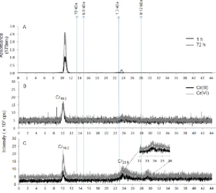
The HSP fractions of C. reinhardtii algae exposed for 1 or 72 h were analyzed by SECpep-ICP-MS to perform a screening of the Cr-binding biomolecules (Fig. 3). The
HPLC-UV detection data at 570 nm for Cr(III) showed two peaks (Fig. 3A), which overlap with the metal elution peaks (Fig. 3B, C). After 72 h of exposure, two Cr peaks were observed at 10.2 min and 23.9 min, while for the cells exposed for 1 h, only one peak was observed at a retention time of 10.2 min (Fig. 3). The area of the Cr10.2min peak
ranged around 40% of the total Cr signal for both Cr(III)- and Cr(VI)-exposed cells (data not shown). However, for both Cr(III) and Cr(VI) cells, the Cr23.9 min-peak area
percentages ranged around 60% of total Cr but no significant difference was observed between the two chromium forms in terms of percentage area of the total signal. Following exposures to both Cr forms for 72 h, SECpep-ICP-MS analysis indicated
association of Cr with a high MW biomolecule (~ 28 kDa), corresponding to the peaks denoted as Cr10.2 (retention time: 10.2 min), but also with a lower MW bioligand (0.7
kDa) denoted as Cr23.9 (Fig. 3C). For both Cr forms and especially for Cr(VI), the higher
molecular weight biomolecule (Cr10.2 , peak mean area: 5.9 × 10⁴ cps2, n = 3) contained
more chromium than the molecular component eluted at 23.9 min (peak mean area: 5.2 × 10⁴ cps2, n = 3; Fig. 3C).
4 Discussion
16
Typically, metal adsorption reactions occur over short time scales (e.g. minutes (Fortin et al., 2004; Hassler et al., 2004; Schenck et al., 1988)) and our extracellular data indeed show little changes in extracellular Cr concentrations between 1 h and 72 h exposures for the different redox Cr forms. On the other hand, accumulation inside the cell increases over time until a steady-state between accumulation, excretion and biodilution through cell division is achieved (e.g. for Cd this occurred after about 24 h; Paquet et al., 2015).
Studies comparing Cr(III) and Cr(VI) uptake are very scarce, especially in algae perhaps due to a general consensus considering Cr(VI) as the most toxic Cr form in the environment, making it the most studied form. However, this consensus was recently questioned by several studies considering Cr(III) speciation during ecotoxicity tests (Bencheikh-Latmani et al., 2007; Kováčik et al., 2015; Thompson et al., 2002; Vignati et al., 2010). Our results contribute to revisiting this tenet as the internalized Cr contents for both Cr forms after exposure to an environmentally relevant concentration of 100 nM were not significantly different when the same incubation time was considered (Fig. SI.2). It is however important to note that our exposure medium, while major cations (Ca and Mg) are representative of soft freshwaters, contains relatively high levels of NH4+
(about 1 mM) and PO43- (about 0.1 mM) which are required for cell growth. Both
compounds have been reported to exert a protective effect against Cr(VI) toxicity to
Chlorella vulgaris at levels of 3–10 mM for NH4+ and 0.1–1 mM for PO43- (Liu et al.,
2015; Qian et al., 2013). While we have been unable to find similar studies for Cr(III), we acknowledge that extrapolation of our results to other exposure media should be done with care and consider differences and similarities in their composition.
17
The similar accumulation of Cr(III) and Cr(VI) by C. reinhardtii also bears relevance for a correct interpretation of the ecotoxicity of Cr2O3 nanoparticles which are
increasingly used in leather tanning, pigment manufacture and cosmetics. Release of both Cr(III) and Cr(VI) from these nanoparticles has been reported in studies with C.
reinhardtii (Costa et al., 2016), D. magna and A. fischeri (Puerari et al., 2016) and human
cell cultures (Horie et al., 2013). Considering our results, studies on the Cr nanoparticles should systematically examine the bioavailability and toxicity of both forms of released Cr. In the next section we examine the fate of Cr once inside the cells, whether they were exposed to Cr(III) or Cr(VI).
4.2 Cr(III) and Cr(VI) subcellular partitioning
Metal distribution in algal subcellular fractions can provide insights in terms of toxic action or detoxification mechanism, as reported in previous studies (Crémazy et al., 2013; Lavoie et al., 2009b). Fractions are operationally defined and need to be interpreted with caution (Giguère et al., 2006). This being said, Lavoie et al. (2009a) showed that over 80% of the citrate synthase activity and over 90% of the cytochrome c oxidase activity, both mitochondrial enzymes, were found in the organelle fraction, indicating a good integrity of organelles.
After 1 h exposure, no significant differences were observed in subcellular partitioning of Cr(III), although, based on arithmetic mean values, Cr(III) content decreased in the order: HSP > ORG > GR > DE > HDP. On the other hand, Cr(VI) partitioning after 1 h exposure showed a significantly higher Cr burden in the HSP, GR
18
and ORG fractions compared to DE and HDP fractions. Based on arithmetic means values, Cr(VI) burden decreased in the order: HSP > ORG > GR > DE > HDP. However, after 72 h exposure more pronounced differences were noted, with Cr(III) partitioning into subcellular fractions decreasing in the order: ORG ~ HSP > GR > DE ~ HDP and Cr(VI) partitioning decreasing in the order: ORG ~ HSP ~ GR > DE > HDP. These results showed that the majority of Cr accumulated in C. reinhardtii was associated with organelles, HSP and granules fractions without significant differences between Cr(III) and Cr(VI) after 72 h exposure (Fig. 2).
Crémazy et al. (2013) have studied the subcellular distribution of two trivalent metals (Al and Sc) in C. reinhardtii after 72 h exposures. Both Al and Sc were mostly found in the organelle fraction followed by debris and granules. However, in contrast with our findings for Cr(III) and Cr(VI), very small proportions of these metals were found in the HSP fraction, suggesting different intracellular pathways for these trivalent elements compared to Cr.
Many reviews have reported that Cr(VI) is rapidly reduced to Cr(III) once inside cells (Cervantes et al., 2001; Viti et al., 2014 and the references therein) and that Cr(VI) can be reduced to Cr(III) at the surface of Chlorella miniata (Han et al., 2007). This could explain the relative similarity in the subcellular distribution of Cr(III) and Cr(VI). In biological systems, Cr(III) and Cr(VI) are expected to bind to several chemical groups. Han et al. (2007) showed that Cr(VI) ions interact with the positively charged groups (i.e., amine group) at the algal surface, and suggested that Cr(III) interacts with the oxygen-containing groups, such as carboxylate group. Cr(III) is known for its ability to bind to carboxyl groups (Cervantes et al., 2001; Gardea-Torresdey et al., 2002; Nieboer
19
and Richardson, 1980; Sobol and Schiestl, 2012) and it behaves as many trivalent
elements readily forming complexes with a variety of ligands having hydroxyl functional groups (Richard and Bourg, 1991). Moreover, Cr interaction with phosphate groups was also previously observed (e.g. the phosphate groups of di- and triphosphate nucleotides (Arakawa et al., 2000; Wolf et al., 1989)). Subcellular distribution of metals may be greatly influenced by ligand preferences, but also by competition for cellular ligands with other cations present in the subcellular fractions (Crémazy et al., 2013).
The chromium content in the ORG fraction increased between 1 and 72 h, while it did not vary or decreased over time in the HDP fraction, which may be linked to the dynamic character of this latter fraction. An important proportion of Cr(III) (around 24% after 1 h and around 40% after 72 h) and Cr(VI) (around 22% after 1 h and around 34% after 72 h) was found in the organelle fraction. This might be the result of the strong binding of chromium (considering that only Cr(III) is expected to be stable in the intracellular environment) with nucleotides and nucleic acids, e.g. adenosine di- and tri-phosphates (ADP, ATP). ADP and ATP are mainly present in mitochondria, which produce energy as ATP and are contained in the organelle fractions. Wolf et al. (1989) have examined the interaction of Cr(III) and Cr(VI) with the phosphate groups of di- and triphosphate nucleotides. They found that the formation of Cr-nucleotide complexes, could only be detected with Cr(III) and that Cr(III), generated from intracellular reduction of Cr(VI), interacted also with the phosphate groups of nucleotides. Janson and Cleland (1974) have reported that trivalent chromium ions compete with Mg ions for binding to ADP and ATP, making it unavailable for metabolism. The chromium nucleotide CrATP was found to bind to the glycerokinase tighter than the magnesium nucleotide by factors
20
up to 30 (Janson and Cleland, 1974). Similar results were observed in the case of yeast phosphoglycerate kinase, the acetate kinase of Escherichia coli, and yeast hexokinase (Janson and Cleland, 1974). Similarly, Viola et al. (1980) observed a strong inhibition of the yeast enzyme hexokinase in the presence of other trivalent metals such as Al. This inhibition of enzymes was mainly due to metal binding to ATP or ADP, which are substrates for those enzymes and could explain the important presence of Cr in the organelle fraction in our study. Other previously reported aspects of chromium interaction with organelles are the reduction in chloroplast integrity in Cr(VI)-treated
Euglena (between 1.5 and 9 µg·ml-1 of K
2Cr2O7), accompanied by an abnormal increase
of the number of plastids, enlarged mitochondria and nuclei (Fasulo et al., 1983).
Important proportions of Cr(III) (around 36% of the total internalized Cr after 1 h and around 39% after 72 h) and Cr(VI) (around 42% after 1 h and around 30% after 72 h) were also found in the cytosolic HSP fraction which includes phytochelatins, GSH and other unidentified peptides. These cysteine-rich thiolated molecules play an important role in metal sequestration and detoxification, especially for “soft” and borderline metals such as Cd and Pb (Lavoie et al., 2009b; Scheidegger et al., 2011). As expected from its hard character, the literature suggests that PC synthesis is not induced by chromium in algae and higher plants (Gorbi et al., 2006; Toppi et al., 2003 and references cited therein). A previous study in Euglena gracilis has shown that Cr(VI) induced an increased cysteine and glutathione content, but not in phytochelatins (García-García et al., 2009). Schiavon et al. (2008) have found that, once internalized, Cr(VI) induces transcription of genes associated with sulfur assimilation, and cysteine and glutathione synthesis, suggesting that these thiol compounds play a role in Cr tolerance in Brassica
21
juncea. In fact, cysteine and GSH are molecules able to reduce Cr(VI) and chelate Cr(III)
(Valko et al., 2005).
The partitioning of Cr(III) and Cr(VI) in the granule-like fraction could be associated with sequestration into polyphosphate bodies. In fact, this mode of metal detoxification has been observed and reported in different organisms, including algae. Nishikawa et al. (2003) have observed that cadmium treatment in Chlamydomonas
acidophila caused ultrastructural changes, polyphosphate degradation and increased
vacuolar short-chain and orthophosphates accompanied by the presence of starch grains and vacuoles. Moreover, previous studies have reported the existence of X-ray dense granules containing linear chains of polyphosphates in the cytoplasm and in vacuoles of several algal species exposed to various metals like Cd, Hg and Zn (Jensen et al., 1982; Komine et al., 2000; Visviki and Rachlin, 1992). The relatively important chromium content in the granule-like fraction of the cells in our exposure conditions (16% of the total internalized Cr(III) in the cells after 1 h, around 18% after 72 h; around 20% of the total internalized Cr(VI) after 1 h and about 30% after 72 h) could be due to its affinity to phosphate contained in these granules. Such structures seem to play an important role in chromium detoxification in C. reinhardtii.
The small proportions of Cr(III) and Cr(VI) observed in the debris fraction (around 16 % of the total internalized Cr(III) after 1 h and only about 2% after 72 h; around 9% of the total internalized Cr(VI) after 1 h and around 5% after 72 h) could be the result of a strongly effective EDTA wash and rinse procedure and suggest a low accumulation in the nucleus.
22
Finally, only very small Cr(III) and Cr(VI) proportions (not exceeding 9% after 1 h for both Cr forms, and not exceeding 1 %, after 72 h for either Cr(III) or Cr(VI)) were found in the HDP fraction, considered as metal-sensitive fraction. This fraction probably has a very dynamic nature and could act as a conveyor belt of Cr to the other fractions via the various enzymes that could be involved in the metal-handling mechanisms of the other fractions such as organelles.
The relative similarity of Cr(III) and Cr(VI) distribution in the different
subcellular fractions after both exposure durations (1 and 72 h) agrees with the expected rapid reduction of Cr(VI) to Cr(III) inside the cells (Viti et al., 2014), leading to a similar intracellular behavior despite some significant differences between the proportions of the two Cr forms in some fractions. Moreover, even if the 1 h-experiment resulted in poor metal recoveries due to the low activities after only 1 h, it is nevertheless noteworthy that the distribution remains relatively similar for Cr(III) and Cr(VI). Note also that in some fractions, especially DE and HDP fractions, Cr content was not significantly different from zero.
Grouping the five different fractions into the previously defined subcellular
compartments (metal-sensitive and metal-detoxified compartments) reveals no significant differences in Cr partitioning between the two subcellular compartments in C. reinhardtii exposed to 100 nM Cr(III) for 1 h . However, Cr concentration increased significantly over time in the two subcellular compartments of the cells exposed to Cr(III) while in those exposed to Cr(VI) it did not (Fig. SI.4), suggesting higher initial uptake fluxes for Cr(VI) compared to Cr(III).
23
The similar accumulation and subcellular partitioning observed for the two Cr redox forms does not always agree with the corresponding physiological effects that may be observed in algae exposed to Cr. Szivak et al. (2009) showed that a moderate increase in intracellular ROS in C. reinhardtii was induced by Cr(VI), but not by Cr(III). In the case of Cr(VI), effects on the photosynthetic activity (Didur et al., 2013), chloroplast morphology, pigment composition and photoaccumulation (Rodriguez et al., 2007) were also reported in C. reinhardtii, but no comparisons with Cr(III) were performed. Studies with other species reached different conclusions depending on the actual physiological endpoints taken into consideration (Kováčik et al., 2015; Jasso-Chávez et al., 2010; Lira-Silva et al., 2011; Volland et al., 2012). Comparisons between the effects of Cr(VI) and Cr(III) should become more systematic to get a better understanding of their relative effects on algal physiology.
4.3 SEC-ICP-MS analysis
SEC-ICP-MS analyses allowed for the determination of the distribution of Cr bound to biomolecules differing in molecular weight in the HSP fraction of C.
reinhardtii. Such screening provides insight on the nature and identity of the observed
chromium-biomolecule complexes. Amino acids, low molecular weight peptides such as glutathione, phytochelatins (PC; e.g., PC2-PC4) and other unidentified peptides (Lavoie et
al., 2009b) are cytosolic ligands expected to be found in the HSP fraction. Such thermostable ligands are involved in the detoxification of soft metals in microalgae (Crémazy et al., 2013; Lavoie et al., 2009b) but the ligands involved in the detoxification of hard metals such as chromium are not as well known. Chromium showed two peaks
24
denoted as Cr10.2min and Cr23.9min in the HSP fraction of algae exposed for 72 h to either
Cr(III) or Cr(VI) and only one peak denoted as Cr10.2min in the HSP fraction of the algae
exposed for 1 h to either Cr(III) or Cr(VI). The fact that the peaks observed in the case of Cr(III) and Cr(VI) are similar in terms of the retention times in the chromatograms lead us to suggest that only Cr(III) is detectable in the cells following rapid reduction of Cr(VI) to Cr(III) (Cervantes et al., 2001; Viti et al., 2014; and references cited therein).
As anticipated, chromium did not elute with cysteine as well as with the other compounds used as calibration standards, and neither with glutathione (0.3 kDa) or phytochelatins PC2-4 (0.5 kDa, 0.8 kDa, 1.0 kDa). Since Cr is classified as a hard metal
(da Silva and Williams, 2001), it is more likely to interact with oxygen-containing ligands than with thiolated ligands.
The Cr23.9min peak observed at 0.7 kDa could correspond to Cr(III)-Nicotinamide
Adenine Dinucleotide (Cr(III)-NAD+). Such a complex has already been reported as an end-product of Cr(VI) reduction in Escherichia coli (Puzon et al., 2002) and has the same molecular weight. Similar peaks with retention times around 23 min and molecular weight ranging between 0.12 kDa and 1.3 kDa were observed in the case of Hg-GSH and Zn-GSH in metal-exposed microalgae (Gómez-Jacinto et al., 2015), but Cr-GSH complex molecular weight (0.3 kDa) does not correspond to the observed peak even if GSH is a molecule able to reduce Cr(VI) and chelate Cr(III) (Valko et al., 2005). The Cr10.2min peak
could correspond to Cr-carbonic anhydrase 3 (Cr-Cah3; 28 kDa) which was previously observed in cells of C. reinhardtii (Mitra et al., 2005) and matches in molecular weight to the observed peak. The appearance of the second peak Cr23.9min after 72 h, could be linked
25
exposed to either Cr(III) or Cr(VI) for 72 h compared to those exposed for 1 h. Even if no significant difference was observed in terms of Cr content in the HSP fractions between the cells exposed for 72 h to either Cr(III) or Cr(VI) (Fig. SI 3), the peak Cr10.2min is
higher for Cr(VI)- than Cr(III)-treated cells. These unidentified ligands may play an important role in the internal handling and/or reduction of Cr(VI) and their identification requires further investigations.
5 Conclusion
Our result showed rapid adsorption (1 hour exposure) onto cells followed by steady-state intracellular Cr content in algal cells exposed to an environmentally relevant concentration of Cr(III) and Cr(VI) over 72 h. Furthermore, Cr(III) and Cr(VI) had similar subcellular partitioning, suggesting that they have similar intracellular fate. To the best of our knowledge, this is the first time that the partitioning of Cr(III) and Cr(VI) to subcellular components in an algal cells has been assessed and a metallomic approach applied to algal HSP fractions. The metallomic analyses showed that Cr eluted at identical times regardless of if cells are exposed to Cr(III) or Cr(VI), indicating intracellular binding of Cr by the same ligands. For a 72 h exposure, the peak eluting after 10.2 min was greater for the cells incubated in the presence of Cr(VI) than for Cr(III). These ligands are tentatively identified as Cr-carbonic anhydrase (Cr10.2min peak)
and Cr-NAD+ (Cr23.9min peak), but their exact identification needs further investigation.
All the observed similarities can be explained by the rapid reduction that Cr(VI)
undergoes once internalized into cells and may indicate a common toxicity and resistance mechanisms for Cr(III) and Cr(VI) in C. reinhardtii algal cells.
26
6 Acknowledgements
We gratefully acknowledge the technical assistance provided by K. Racine, L. Rancourt and S. Prémont. Financial support was provided by the French National Research Agency through the national program “Investissements d’avenir” (ANR-10-LABX-21-01 / LABEX RESSOURCES21), the Ministry of National Education, Higher Education and Research (France) and the Natural Sciences and Engineering Research Council of Canada. C. Fortin is supported by the Canada Research Chair Program.
References
Arakawa, H., Ahmad, R., Naoui, M., Tajmir-Riahi, H.A., 2000. A comparative study of calf thymus DNA binding to Cr(III) and Cr(VI) ions. Evidence for the guanine N-7-chromium-phosphate chelate formation. J. Biol. Chem. 275, 10150–10153. doi:10.1074/jbc.275.14.10150
Asatiani, N.V., Abuladze, M.K., Kartvelishvili, T.M., Bakradze, N.G., Sapojnikova, N.A., Tsibakhashvili, N.Y., Tabatadze, L.V., Lejava, L.V., Asanishvili, L.L., Holman, H.-Y., 2004. Effect of chromium(VI) action on Arthrobacter oxydans. Curr. Microbiol. 49, 321–326. doi:10.1007/s00284-004-4351-2
Bencheikh-Latmani, R., Obraztsova, A., Mackey, M.R., Ellisman, M.H., Tebo, B.M., 2007. Toxicity of Cr(III) to Shewanella sp. strain MR-4 during Cr(VI) reduction. Environ. Sci. Technol. 41, 214–220. doi:10.1021/es0622655
Campbell P.G.C., Hare, L.. 2009. Metal detoxification in freshwater animals. Roles of metallothioneins, in: Metallothioneins and Related Chelators: Metal Ions in Life Sciences. Vol 5. The Royal Society of Chemistry, pp 239–277.
Cervantes, C., Campos-García, J., Devars, S., Gutiérrez-Corona, F., Loza-Tavera, H., Torres-Guzmán, J.C., Moreno-Sánchez, R., 2001. Interactions of chromium with microorganisms and plants. FEMS Microbiol. Rev. 25, 335–347.
27
Crémazy, A., Levy, J.L., Campbell, P.G.C., Fortin, C., 2013. Uptake and subcellular partitioning of trivalent metals in a green alga: comparison between Al and Sc. Biometals 26, 989–1001. doi:10.1007/s10534-013-9675-6
da Costa, C.H., Perreault, F., Oukarroum, A., Melegari, S.P., Popovic, R., Matias, W.G., 2016. Effect of chromium oxide (III) nanoparticles on the production of reactive oxygen species and photosystem II activity in the green alga Chlamydomonas
reinhardtii. Sci. Total Environ. 565, 951–960.
doi:10.1016/j.scitotenv.2016.01.028
da Silva, J.J.R.F., Williams, R.J.P., 2001. The Biological Chemistry of the Elements: The Inorganic Chemistry of Life, second ed. Oxford University Press, New York. Dhal, B., Thatoi, H.N., Das, N.N., Pandey, B.D., 2013. Chemical and microbial
remediation of hexavalent chromium from contaminated soil and
mining/metallurgical solid waste: A review. J. Hazard. Mater. 250–251, 272–291. doi:10.1016/j.jhazmat.2013.01.048
Didur, O., Dewez, D., Popovic, R., 2013. Alteration of chromium effect on photosystem II activity in Chlamydomonas reinhardtii cultures under different synchronized state of the cell cycle. Environ. Sci. Pollut. Res. Int. 20, 1870–1875.
doi:10.1007/s11356-012-1389-8
Döker, S., Mounicou, S., Doğan, M., Lobinski, R., 2010. Probing the metal-homeostatis effects of the administration of chromium(VI) to mice by ICP MS and size-exclusion chromatography-ICP MS. Metallomics 2, 549–555.
doi:10.1039/C004508J
Fasulo, M.P., Bassi, M., Donini, A., 1983. Cytotoxic effects of hexavalent chromium in
Euglena gracilis. II. Physiological and ultrastructural studies. Protoplasma 114,
35–43. doi:10.1007/BF01279866
Fortin, C., Dutel, L., Garnier-Laplace, J., 2004. Uranium complexation and uptake by a green alga in relation to chemical speciation: The importance of the free uranyl ion. Environ. Toxicol. Chem. 23, 974–981. doi:10.1897/03-90
García-García, J.D., Rodríguez-Zavala, J.S., Jasso-Chávez, R., Mendoza-Cozatl, D., Moreno-Sánchez, R., 2009. Chromium uptake, retention and reduction in photosynthetic Euglena gracilis. Arch. Microbiol. 191, 431–440.
doi:10.1007/s00203-009-0469-8
Gardea-Torresdey, J.L., Dokken, K., Tiemann, K.J., Parsons, J.G., Ramos, J., Pingitore, N.E., Gamez, G., 2002. Infrared and X-ray absorption spectroscopic studies on the mechanism of chromium(III) binding to alfalfa biomass. Microchem. J. 71, 157–166. doi:10.1016/S0026-265X(02)00007-3
Giguère, A., Campbell, P.G.C., Hare, L., Couture, P., 2006. Sub-cellular partitioning of cadmium, copper, nickel and zinc in indigenous yellow perch (Perca flavescens) sampled along a polymetallic gradient. Aquat. Toxicol. 77, 178–189.
doi:10.1016/j.aquatox.2005.12.001
Gómez-Jacinto, V., García-Barrera, T., Gómez-Ariza, J.L., Garbayo-Nores, I., Vílchez-Lobato, C., 2015. Elucidation of the defence mechanism in microalgae Chlorella
sorokiniana under mercury exposure. Identification of Hg–phytochelatins. Chem.
Biol. Interact. 238, 82–90. doi:10.1016/j.cbi.2015.06.013
Gorbi, G., Torricelli, E., Pawlik-Skowrońska, B., di Toppi, L.S., Zanni, C., Corradi, M.G., 2006. Differential responses to Cr(VI)-induced oxidative stress between
Cr-28
tolerant and wild-type strains of Scenedesmus acutus (Chlorophyceae). Aquat. Toxicol. 79, 132–139. doi:10.1016/j.aquatox.2006.06.002
Han, X., Wong, Y.S., Wong, M.H., Tam, N.F.Y., 2007. Biosorption and bioreduction of Cr(VI) by a microalgal isolate, Chlorella miniata. J. Hazard. Mater. 146, 65–72. doi:10.1016/j.jhazmat.2006.11.053
Hassler, C.S., Slaveykova, V.I., Wilkinson, K.J., 2004. Discriminating between intra- and extracellular metals using chemical extractions. Limnol. Oceanogr. Methods 2, 237–247. doi:10.4319/lom.2004.2.237
Horie, M., Nishio, K., Endoh, S., Kato, H., Fujita, K., Miyauchi, A., Nakamura, A., Kinugasa, S., Yamamoto, K., Niki, E., Yoshida, Y., Iwahashi, H., 2013. Chromium(III) oxide nanoparticles induced remarkable oxidative stress and apoptosis on culture cells. Environ. Toxicol. 28, 61–75. doi:10.1002/tox.20695 Janson, C.A., Cleland, W.W., 1974. The specificity of chromium nucleotides as inhibitors
of selected kinases. J. Biol. Chem. 249, 2572–2574.
Jasso-Chávez, R., Pacheco-Rosales, A., Lira-Silva, E., Gallardo-Perez, J.C., Garcia, N., Moreno-Sanchez, R., 2010. Toxic effects of Cr(VI) and Cr(III) on energy metabolism of heterotrophic Euglena gracilis. Aquat. Toxicol. 100:329–338. doi:10.1016/j.aquatox.2010.08.006
Jensen, T.E., Baxter, M., Rachlin, J.W., Jani, V., 1982. Uptake of heavy metals by
Plectonema boryanum (cyanophyceae) into cellular components, especially
polyphosphate bodies: An X-ray energy dispersive study. Environ. Pollut. Ser.A. 27, 119–127. doi:10.1016/0143-1471(82)90104-0
Kotaś, J., Stasicka, Z., 2000. Chromium occurrence in the environment and methods of its speciation. Environ. Pollut. 107, 263–283. doi:10.1016/S0269-7491(99)00168-2
Komine, Y., Eggink, L.L., Park, H., Hoober, J.K., 2000. Vacuolar granules in
Chlamydomonas reinhardtii: polyphosphate and a 70-kDa polypeptide as major
components. Planta 210, 897–905. doi:10.1007/s004250050695
Kováčik, J., Babula, P., Hedbavny, J., Kryštofová, O., Provaznik, I., 2015. Physiology and methodology of chromium toxicity using alga Scenedesmus quadricauda as model object. Chemosphere 120, 23–30. doi:10.1016/j.chemosphere.2014.05.074 Lavoie, M., Bernier, J., Fortin, C., Campbell, P.G.C., 2009a. Cell homogenization and
subcellular fractionation in two phytoplanktonic algae: Implications for the assessment of metal subcellular distributions. Limnol. Oceanogr. Methods. 7, 277–286. doi:10.4319/lom.2009.7.277
Lavoie, M., Le Faucheur, S., Fortin, C., Campbell, P.G.C., 2009b. Cadmium
detoxification strategies in two phytoplankton species: Metal binding by newly synthesized thiolated peptides and metal sequestration in granules. Aquat. Toxicol. 92, 65–75. doi:10.1016/j.aquatox.2008.12.007
Lira-Silva, E., Ramirez-Lima, I.S., Olin-Sandoval, V., Garcia, J.D., Garcia-Contreras, R., Moreno-Sanchez, R., Jasso-Chavez, R., 2011. Removal,
accumulation and resistance to chromium in heterotrophic Euglena gracilis. J. Hazard. Mater. 193:216–224. doi: 10.1016/j.jhazmat.2011.07.056
Liu, J., Sun, Z., Lavoie, M., Fan, X., Bai, X., Qian, H., 2015. Ammonium reduces chromium toxicity in the freshwater alga Chlorella vulgaris. Appl. Microbiol. Biotechnol. 99, 3249–3258.doi: 10.1007/s00253-014-6218-1
29
Markiewicz, B., Komorowicz, I., Sajnóg, A., Belter, M., Barałkiewicz, D., 2015. Chromium and its speciation in water samples by HPLC/ICP-MS – technique establishing metrological traceability: A review since 2000. Talanta 132, 814– 828. doi:10.1016/j.talanta.2014.10.002
Mitra, M., Mason, C.B., Xiao, Y., Ynalvez, R.A., Lato, S.M., Moroney, J.V., 2005. The carbonic anhydrase gene families of Chlamydomonas reinhardtii. Can. J. Bot. 83, 780–795. doi:10.1139/b05-065
Nieboer, E., Richardson, D.H.S., 1980. The replacement of the nondescript term “heavy metals” by a biologically and chemically significant classification of metal ions. Environ. Pollut. Ser. B. 1, 3–26. doi:10.1016/0143-148X(80)90017-8
Nishikawa, K., Yamakoshi, Y., Uemura, I., Tominaga, N., 2003. Ultrastructural changes in Chlamydomonas acidophila (Chlorophyta) induced by heavy metals and polyphosphate metabolism. FEMS Microbiol. Ecol. 44, 253–259.
doi:10.1016/S0168-6496(03)00049-7
Paquet, N., Lavoie, M., Maloney, F., Duval, J.F.L., Campbell, P.G.C., Fortin, C., 2015. Cadmium accumulation and toxicity in the unicellular alga Pseudokirchneriella
subcapitata: Influence of metal-binding exudates and exposure time. Environ.
Toxicol. Chem. 34, 1524–1532. doi:10.1002/etc.2927
Parker, D.L., Borer, P., Bernier-Latmani, R., 2011. The response of Shewanella
oneidensis MR-1 to Cr(III) toxicity differs from that to Cr(VI). Front. Microbiol.
2. doi:10.3389/fmicb.2011.00223
Peterson, R.L., Banker, K.J., Garcia, T.Y., Works, C.F., 2008. Isolation of a novel chromium(III) binding protein from bovine liver tissue after chromium(VI) exposure. J. Inorg. Biochem. 102, 833–841. doi:10.1016/j.jinorgbio.2007.12.003 Puerari, R.C., da Costa, C.H., Vicentini, D.S., Fuzinatto, C.F., Melegari, S.P., Schmidt,
E.C., Bouzon, Z.L., Matias, W.G., 2016. Synthesis, characterization and toxicological evaluation of Cr2O3 nanoparticles using Daphnia magna and
Aliivibrio fischeri. Ecotoxicol. Environ. Saf. 128, 36–43.
doi:10.1016/j.ecoenv.2016.02.011
Puzon, G.J., Petersen, J.N., Roberts, A.G., Kramer, D.M., Xun, L., 2002. A bacterial flavin reductase system reduces chromate to a soluble chromium(III)–NAD+ complex. Biochem. Biophys. Res. Comm. 294, 76–81. doi:10.1016/S0006-291X(02)00438-2
Qian, H.F., Sun, Z.Q., Sun, L.W., Jiang, Y.F., Wei, Y., Xie, J., Fu, Z.W., 2013. Phosphorus availability changes chromium toxicity in the freshwater alga
Chlorella vulgaris. Chemosphere 93, 885–891.
doi:10.1016/j.chemosphere.2013.05.035
Ramírez-Díaz, M.I., Díaz-Pérez, C., Vargas, E., Riveros-Rosas, H., Campos-García, J., Cervantes, C., 2008. Mechanisms of bacterial resistance to chromium compounds. Biometals 21, 321–332. doi:10.1007/s10534-007-9121-8
Richard, F.C., Bourg, A.C.M., 1991. Aqueous geochemistry of chromium: A review. Water Res. 25, 807–816. doi:10.1016/0043-1354(91)90160-R
Rodríguez, M.C., Barsanti, L., Passarelli, V., Evangelista, V., Conforti, V., Gualtieri, P., Effects of chromium on photosynthetic and photoreceptive apparatus of the alga
Chlamydomonas reinhardtii. Environ. Res. 105, 234–239.
30
Rosabal, M., Hare, L., Campbell, P.G.C., 2012. Subcellular metal partitioning in larvae of the insect Chaoborus collected along an environmental metal exposure gradient (Cd, Cu, Ni and Zn). Aquat. Toxicol. 120–121, 67–78.
doi:10.1016/j.aquatox.2012.05.001
Scheidegger, C., Behra, R., Sigg, L., 2011. Phytochelatin formation kinetics and toxic effects in the freshwater alga Chlamydomonas reinhardtii upon short- and long-term exposure to lead(II). Aquat. Toxicol. 101, 423–429.
doi:10.1016/j.aquatox.2010.11.016
Schenck, R.C., Tessier, A., Campbell, P.G.C., 1988. The effect of pH on iron and manganese uptake by a green alga. Limnol. Oceanogr. 33, 538–550. doi:10.4319/lo.1988.33.4.0538
Schiavon, M., Pilon-Smits, E.A.H., Wirtz, M., Hell, R., Malagoli, M., 2008. Interactions between chromium and sulfur metabolism in Brassica juncea. J. Environ. Qual. 37, 1536–1545. doi:10.2134/jeq2007.0032
Sobol, Z., Schiestl, R.H., 2012. Intracellular and extracellular factors influencing Cr(VI) and Cr(III) genotoxicity. Environ. Mol. Mutagen. 53, 94–100.
doi:10.1002/em.20679
Szivak, I., Behra, R., Sigg, L., 2009. Metal-induced reactive oxygen species production in
Chlamydomonas reinhardtii (Chlorophyceae). J. Phycol. 45, 427–435.
doi:10.1111/j.1529-8817.2009.00663.x
Thompson, S.L., Manning, F.C.R., McColl, S.M., 2002. Comparison of the toxicity of chromium III and chromium VI to cyanobacteria. Bull. Environ. Contam. Toxicol. 69, 286–293. doi:10.1007/s00128-002-0059-9
Toppi, L.S.D., Gremigni, P., Pawlik-Skowrońska, B., Prasad, M.N.V., Cobbett, C.S., 2003. Response to heavy metals in plants: a molecular approach, in: di Toppi, L.S., Pawlik-Skowrońska, B. (Eds.), Abiotic Stresses in Plants. Springer Netherlands, pp. 133–156.
Valko, M., Morris, H., Cronin, M.T.D., 2005. Metals, toxicity and oxidative stress. Curr. Med. Chem. 12, 1161–1208. doi:10.2174/0929867053764635
Vignati, D.A.L., Dominik, J., Beye, M.L., Pettine, M., Ferrari, B.J.D., 2010.
Chromium(VI) is more toxic than chromium(III) to freshwater algae: A paradigm to revise? Ecotoxicol. Environ. Safe. 73, 743–749.
doi:10.1016/j.ecoenv.2010.01.011
Viola, R.E., Morrison, J.F., Cleland, W.W., 1980. Interaction of metal(III)-adenosine 5’-triphosphate complexes with yeast hexokinase. Biochemistry-US 19, 3131–3137. doi:10.1021/bi00555a003
Visviki, I., Rachlin, J.W., 1992. Ultrastructural changes in Dunaliella minuta following acute and chronic exposure to copper and cadmium. Arch. Environ. Contam. Toxicol. 23, 420–425. doi:10.1007/BF00203803
Viti, C., Marchi, E., Decorosi, F., Giovannetti, L., 2014. Molecular mechanisms of Cr(VI) resistance in bacteria and fungi. FEMS Microbiol. Rev. 38, 633–659. doi:10.1111/1574-6976.12051
Volland, S., Lütz, C., Michalke, B., Lütz-Meindl, U., 2012. Intracellular chromium localization and cell physiological response in the unicellular alga Micrasterias. Aquat. Toxicol. 109, 59–69. doi:10.1016/j.aquatox.2011.11.013
31
Wallace, W.G., Lee, B., Luoma, S.N., 2003. Subcellular compartmentalization of Cd and Zn in two bivalves. I. Significance of metal-sensitive fractions (MSF) and
biologically detoxified metal (BDM). Mar. Ecol. Prog. Ser. 249, 183–197. doi:10.3354/meps249183
Wolf, T., Kasemann, R., Ottenwälder, H., 1989. Molecular interaction of different chromium species with nucleotides and nucleic acids. Carcinogenesis 10, 655– 659. doi:10.1093/carcin/10.4.655
32
Figure Captions
Fig. 1. Mean Cr concentrations (in amol·cell-1; ± SD, n = 3) of C. reinhardtii following
1-h and 72-h exposures to Cr(III) (A) or Cr(VI) (B) as total accumulated,
extracellular and intracellular compartments. Different letters indicate statistically significant differences (ANOVA followed by Tukey Kramer HSD test; P < 0.05).
33
Fig. 2. Mean Cr concentrations (in amol·cell-1; ± SD, n = 3) in each subcellular fraction (ORG: organelles; GR: granules; DE: cellular debris, HDP: heat-denatured proteins; HSP: heat-stable proteins) of C. reinhardtii following 1-h (white bars) and 72-h (black bars) exposures to Cr(III) or Cr(VI). Different letters indicate statistically significant differences (ANOVA followed by the Tukey Kruskal Wallis test, P < 0.05).
35
Fig. 3. SECpeptide chromatograms of the HSP fractions of C. reinhardtii A) with UV
detection at 570 nm after a 1-h or 72-h exposure to Cr (VI), or with ICP-MS detection for algae exposed to Cr for 1 h (B) or 72 h (C). Data for Cr(III) are represented by black lines whereas those for Cr(VI) by gray lines in panels B and C. Vertical dotted lines represent retention times of the MW standards. Cr10.2 and Cr23.9 represent the Cr peaks observed by SECpeptide-ICP-MS.