Thyroid hormones and androgens differentially regulate gene expression in testes and ovaries of 1
sexually mature Silurana tropicalis 2
3
Campbell, D.E.K.1, and Langlois, V.S.1,2,3* 4
1 Biology Department, Queen’s University, Kingston, ON, CA
5
2 Institut national de la recherche scientifique - Centre Eau Terre Environnement (INRS-ETE), Quebec
6
City, QC, CA 7
3 Department of Chemistry and Chemical Engineering, Royal Military College of Canada, Kingston, ON,
8 CA 9 10 11 12
*Author for correspondence and reprint requests: 13 14 Dr. Valerie S. Langlois 15 Associate Professor 16
Institut national de la recherche scientifique - Centre Eau Terre Environnement (INRS-ETE) 17
490, de la Couronne, Quebec City (Quebec) G1K 9A9, CANADA 18 [email protected] 19 Tel: 418-654-2547 20 Fax: 418 654-2600 21 22
Keywords: Thyroid hormones; Androgens; Crosstalk; Iopanoic Acid; Real-time RT-qPCR; Silurana 23
tropicalis
24 25
Abstract 26
A series of ex vivo exposures using testicular and ovarian tissues of sexually mature Western clawed frogs 27
(Silurana tropicalis) were designed to examine molecular mechanisms of thyroid hormone (TH) and 28
androgen crosstalk sans hypophyseal feedback as well as investigate potential sex-specific differences. 29
Tissues were exposed ex vivo to either triiodothyronine (T3), iopanoic acid (IOP), one co-treatment of 30
IOP + 5α-dihydrotestosterone (5α-DHT), 5α-DHT, 5β-dihydrotestosterone (5β-DHT), or testosterone (T). 31
Direct exposure to different androgens led to androgen specific increases in thyroid receptor and 32
deiodinase transcripts in testes (trβ and dio1) but a decrease in expression in ovaries (trβ and dio3), 33
suggesting that male and female frogs can be differently affected by androgenic compounds. Moreover, 34
exposure to select androgens differentially increased estrogen-related transcription (estrogen receptor 35
alpha (erα) and aromatase (cyp19)) and production (estradiol) in ovaries and testes indicating the 36
activation of alternate metabolic pathways yielding estrogenic metabolites. Sex-steroid-related 37
transcription (steroid 5α-reductase type 2 (srd5α2) and erα) and production (5α-DHT) were also 38
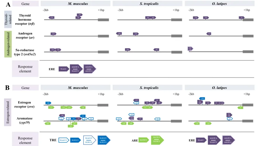
differentially regulated by THs. The presence and frequency of transcription factor binding sites in the 39
putative promoter regions of TH- and sex steroid-related genes were also examined in S. tropicalis, 40
rodent, and fish models using in silico analysis. In summary, this study provides an improved mechanistic 41
understanding of TH- and androgen-mediated actions and reveals differential transcriptional effects as a 42
function of sex in frogs. 43
1. Introduction 44
The actions of thyroid hormones (THs) are highly diverse and impact nearly every biological endocrine 45
system (Cortés et al., 2014; Duarte-Guterman et al., 2014; Mullur et al., 2014; Cooke et al., 2004). The 46
challenge remains to characterize and predict the interactions among THs and the major endocrine axes. 47
THs have been shown to cross-regulate with the hypothalamus–pituitary–gonadal axis (HPG) targeting 48
gonadotropin synthesis, steroidogenesis, and gonadal cellular differentiation in vertebrates (reviewed in: 49
Cortés et al., 2014; Duarte-Guterman et al., 2014; Flood et al., 2013; Habibi et al., 2012; Wajner et al., 50
2009; Wagner et al., 2008; Cooke et al., 2004; Maran, 2003). A large body of literature exists on the 51
molecular mechanisms underlying TH-mediated reproductive effects in gonadal tissue (Duarte-Guterman 52
et al., 2014; Flood et al., 2013; Habibi et al., 2012; Wagner et al., 2008; Cooke et al., 2004; Maran, 2003), 53
however relatively little is known with regard to sex specific effects. Transcripts of thyroid hormone-54
related machinery have been detected in testicular and ovarian tissues of numerous species (Mammals: 55
Carosa et al., 2017; Physalaemus pustulosus: Guterman et al., 2012; Silurana tropicalis: Duarte-56
Guterman and Trudeau, 2011; Scarus iseri: Johnson and Lema, 2011; Oncorhynchus mykiss: Sambroni et 57
al., 2001; Podarcis sicula: Cardone et al., 2000). TH-related transcripts have moreover been shown to 58
develop sexually-dimorphic patterns with higher mRNA levels of TH receptors (trs: trα and trβ) and 59
deiodinases (dios: dio1, dio2, and dio3) reported in testes than in ovaries of frog and fish species (S. 60
tropicalis: Duarte-Guterman and Trudeau, 2011; S. iseri: Johnson and Lema, 2011). Sex specific
61
transcriptional mechanisms in reproductive tissues may not be evident until after the completion of sexual 62
development in anamniotes once the animal has reached sexual maturity. The main goal of this study was 63
to examine the differential effects of THs as a function of gender in amphibians and to elucidate possible 64
sex specific mechanisms of crosstalk in juvenile S. tropicalis. 65
Understanding mechanisms of sex steroid-regulation of the TH axis is highly relevant to 66
amphibians due to the dependence of metamorphosis on THs. Estrogenic compounds (e.g., estradiol (E2)
67
and 17α-ethinylestradiol (EE2))have been shown to repress TH function and impede growth in aquatic
68
species (X. laevis: Sharma and Patiño, 2010; Rana pipiens: Hogan et al., 2008; R. pipiens and Rana 69
sylvatica: Hogan et al., 2006; Teleost fish: reviewed in Orozco and Valverde-R, 2005; Brown et al.,
70
2004). In contrast, androgens appear to stimulate the TH system in vertebrates. Exposure to testosterone 71
(T) and 17α-methyltestosterone has shown to elevate circulating TH levels and peripheral TH metabolism 72
in fish (reviewed in: Orozco and Valverde-R, 2005; Brown et al., 2004; Cyr and Eales, 1996; Salvelinus 73
alpinus: MacLatchy and Eales, 1988; Salmo gairdneri, Richardson: Hunt and Eales, 1979). More
74
recently, exposure to anti-androgenic compounds have been shown to alter TH-related transcription and 75
activity in developing S. tropicalis tadpoles (Langlois et al., 2011; Langlois et al., 2010b; Duarte-76
Guterman et al., 2009), which substantiates the potential for this crosstalk in amphibians. Androgen 77
response elements (AREs) have also been identified in the promoter regions of trs and dios in model fish 78
and tetrapod species (Mus musculus, S. tropicalis, and Oryzias latipes: Flood et al., 2013). Therefore, we 79
can hypothesize for direct androgenic regulation of TH-related transcription. 80
Isolating direct TH- or androgen-mediated crosstalk in vivo is difficult as this assay encompasses 81
all pathways of regulation, including compensatory feedback mechanisms by higher regulatory centres, 82
such as the hypothalamus–pituitary axis. An ex vivo assay ascertains direct and independent molecular 83
responses by eliminating factors, such as hormonal feedback loops and biotransformation of the chemical 84
by other organs (e.g., liver; Scholz et al., 2013). To characterize molecular mechanisms of TH- and 85
androgen-action as a function of sex, testicular and ovarian tissues of juvenile S. tropicalis were exposed 86
ex vivo to either triiodothyronine (T3), iopanoic acid (IOP), one co-treatment of IOP + 5α-
87
dihydrotestosterone (5α-DHT), 5α-DHT, 5β-dihydrotestosterones (5β-DHT), or T for 6 h. Gonadal TH- 88
and sex steroid-related transcript levels and sex-steroid media levels were examined in testis and ovary 89
tissue to elucidate molecular mechanisms of crosstalk with regard to the function of sex. We also 90
conducted a novel in silico promoter analysis to examine the presence and frequency of putative thyroid-, 91
androgen- and estrogen-response elements (TREs, AREs, and EREs, respectively) in S. tropicalis TH- 92
and sex steroid-related genes and made species comparisons with rodent and fish models. 93
2. Material and methods 95
2.1 Animals and exposure 96
Juvenile male and female S. tropicalis frogs were raised and housed in the Queen’s University Animal 97
Care Facility (Kingston, ON, Canada). Animals were kept in dechlorinated and aerated water (25 ± 1°C) 98
on a 12:12 h light:dark regime (light commencing at 0700 h). All aspects of animal care were performed 99
in accordance with the guidelines of the Queen’s University’s Animal Care Committee and the Canadian 100
Council on Animal Care. 101
Two ex vivo assays were performed following methods of Bissegger et al. (2014). In the first ex 102
vivo assay, we examined whether TH status affects sex steroid-related transcription and hormone
103
production in testes and ovaries of sexually mature juvenile S. tropicalis. Juvenile frogs were 104
anaesthetized by immersion in 2% of 3-aminobenzoic acid ethyl ester (MS-222; Sigma Canada Ltd., 105
Oakville, ON, Canada) and euthanized by decapitation. Four males were used per treatment. Each testis 106
was evaluated independently resulting in a total of eight whole testes per treatment. Four females were 107
used per treatment and two pieces of ovary tissue – each piece weighing between 5 to 25 mg – were 108
removed per frog. Each piece was evaluated independently resulting in a total of eight ovary pieces per 109
treatment. Once dissected – tissues were weighed and placed in separate 1.5 mL centrifuge tubes filled 110
with 500 µL of ice-cold Lebovitz (L-15 media, Sigma, Oakville, ON, Canada) containing 10 mM HEPES, 111
50 µg/mL gentamicin (Fisher Scientific, Ottawa, ON, Canada) and 2% synthetic serum replacement 112
(Sigma, Oakville, ON, Canada) at pH 7.4. Tissues were kept on ice until the exposure commenced. 113
Previous time dependent experiments (2–10 h) performed by Bissegger et al., (2014) showed that RNA 114
degradation was not evident with the time elapsed between dissection of tissues and treatment 115
incubations, and an incubation times of 6 h or less. The individual eight whole-testes or ovary-pieces were 116
then placed in eight separate designated wells in 24-well plates containing either 500 µL L-15 media 117
(control samples) or L-15 media containing T3 (50 nM; Sigma, Oakville, Ontario, CA), IOP (10 µM; TCI 118
America), or one co-treatment of IOP (10 µM, TCI America) + 5α-DHT (1 µM; Steraloids, Newport, RI, 119
USA). The individual treatments occupied a total of eight wells or two columns with the whole testes 120
assay spanning two 24-well plates (32 wells total) and ovary pieces assay spanning two 24-well plates (32 121
wells total). T3 is a potent TH and the concentration was chosen based on in vivo studies conducted with 122
S. tropicalis (Campbell and Langlois, 2017; Duarte-Guterman and Trudeau, 2011; Duarte-Guterman et
123
al., 2010). IOP is a TH antagonist that inhibits local deiodinase (dio) function. Dios are enzymes 124
responsible for the activation and deactivation of THs within individual tissues. IOP is non-specific 125
impeding all dio function, as a result the chemical locally induces both hypo- and hyperthyroid 126
conditions: (i) leading to the accumulation of THs and (ii) preventing further local synthesis of active 127
hormones. The IOP concentration was chosen based on in vivo studies conducted with S. tropicalis and X. 128
laevis (Campbell and Langlois, 2017; Fini et al., 2007). The 24-well plates were incubated for 6 h at 26°C
129
using an orbital shaker. After 6 h, the tissues and media were collected and flash frozen on dry ice. 130
Samples were stored at −80°C for subsequent gene expression and sex steroid hormone analyses. 131
In the second ex vivo assay, we investigated androgen-mediated regulation of sex steroid- and 132
TH-related transcription in isolated testis and ovary tissue of juvenile S. tropicalis. Animals were 133
euthanized and tissues were collected the same way as described above. The individual eight whole-testes 134
or ovary-pieces were transferred from the 1.5 mL centrifuge tubes filled with 500 µL of cold Lebovitz 135
into eight separate designated wells in 24-well plates containing either 500 µL of L-15 media (control 136
samples) or L-15 media containing 1 µM of T, 5α-DHT, or 5β-DHT (Steraloids, Newport, RI, USA). The 137
individual treatments occupied a total of eight wells or two columns with the whole testes assay spanning 138
two 24-well plates (32 wells total) and ovary pieces assay spanning two 24-well plates (32 wells total). 139
These concentrations were chosen based on an ex vivo study conducted with S. tropicalis (Bissegger and 140
Langlois, 2016). The 24-well plates were incubated for 6 h at 26°C using an orbital shaker. After 6 h, the 141
organs were collected and flash frozen on dry ice. Samples were stored at −80°C for subsequent gene 142
expression analysis. 143
144
2.2 Sex steroid analysis 145
Media concentrations of E2, T, and 5α-DHT were measured using commercially available enzyme-linked
146
immunosorbent assays (ELISAs; E2 and T: Cayman Chemical, Cedarlane, Burlington, ON, Canada;
5α-147
DHT: IBL America, Cedarlane, Burlington, ON, Canada). Media samples were thawed on ice and diluted 148
two-fold in the immunoassay buffer. All media samples were run in duplicate. The immunoassay 149
protocols were then followed as described by the manufacturer. The absorbance of samples were 150
measured using an Infinite® M1000 PRO plate reader (Tecan, Montreal, QC, Canada) at 405 nm for E2
151
and T, and 450 nm for 5α-DHT. The limit of detection according to the manufacturer was 15 pg/mL for 152
E2, and 6 pg/mL for both T and 5α-DHT. Note that the T and 5α-DHT levels could not be accurately
153
quantified in the co-treatment IOP + 5α-DHT because the antiserums to both T and 5α-DHT were 154
reported to cross-react with 5α-DHT by 27.4% and 100%, respectively. 155
156
2.3 In silico promoter analysis 157
To further characterize potential mechanisms of molecular crosstalk, we examined the presence and 158
frequency of TREs, AREs, and EREs in the putative promotor regions of S. tropicalis sex steroid-related 159
genes (erα and cyp19). The presence and frequency of EREs were examined in TH-related genes (trβ) and 160
androgen-related genes (ar and srd5α2). For information on tr and ar half-site motifs in these genes 161
please refer to Flood et al. (2013). All sequences used for analysis were collected from the Ensembl 162
Project (http://www.ensembl.org). Weighted matrices of tr-, ar-, and er-binding sites were obtained using 163
the PROMO matrices search engine (v.3.0.2; Farré et al., 2003) in conjunction with the TRANSFAC 164
matrices database (v.7.0). We then used the FIMO software (v.4.11.1; Grant et al., 2011) to scan for the 165
tr-, ar-, and er-motifs within the putative promoter region (−2000 to +1) of our target genes applying a
p-166
value output threshold of 0.001. The matched tr, ar, and er motif sequences were searched against the 167
core recognition motif sequence with the criterion of allowing no mismatches as a final validation step. 168
The frequency on single half-sites (TRE: TGACCT-3', TGTCCT-3'; ARE: TGTTCT-3'; ERE: 5'-169
TGACC-3'), direct repeats, and palindrome sequences were evaluated. 170
2.4 Gene expression analysis 172
Total RNA from ovary pieces and whole testes was isolated using TRIzol (Life Technologies, Burlington, 173
ON, CA) following in accordance with the manufacturer’s protocol and was purified using the TURBO 174
DNA-free™ Kit (Ambion; ThermoFisher Scientific, Ottawa, ON). The quantity of RNA was determined 175
using a NanoDrop-2000 spectrophotometer (Thermofisher, Ottawa, ON, Canada). First strand cDNA was 176
synthesized following the GoScript Reverse Transcription kit protocol with random primers (Promega, 177
Madison, WI, USA) in a Mastercycler Pro S Thermocycler (Thermo Fisher, Ottawa, ON, Canada). The 178
cDNA products were diluted 80-fold prior to qPCR amplification. 179
Primer sequences for androgen receptor (ar), aromatase (cyp19), estrogen receptor (erα), 180
deiodinases (dio1, dio2, and dio3), 5α-reductases (srd5α1, srd5α2, srd5α3), TH receptors (trα and trβ), 181
and the reference genes ornithine decarboxylase (odc) and ribosomal protein L8 (rpl8) were previously 182
designed and validated by Langlois et al. (2010b). Real-time PCR primers for dax-1 (dosage-sensitive sex 183
reversal, adrenal hypoplasia critical region, on chromosome X, gene 1) were previously designed and 184
validated by Campbell and Langlois (2017). All qPCR assays were performed using a CFX 96 Real-Time 185
System (Bio-Rad Laboratories Inc, Mississauga, ON) and GoTaq qPCR MasterMix with bryt green 186
(Promega, Madison, WI, USA). The thermocycler program included an enzyme activation step at 95°C 187
for 2 min, followed by 40 cycles at 95°C for 15 s, and 1 min at a gene-specific annealing temperature of 188
58°C, 60°C, or 62°C. After this amplification phase, there was a denaturation step of 1 min at 95°C. A 189
dissociation curve was subsequently generated to confirm the presence of a single amplicon. The 190
threshold for each gene was assessed automatically by the Bio-Rad CFX Manager Software 3.0. Pooled 191
cDNA from each treatment were serially diluted (1:4) to produce a standard curve with a starting 192
concentration of 50 ng. Each assay met the required a reaction efficiency of 100 ± 15% and an R2 ≥ 0.989. 193
For quality control purposes negative control reactions were also included (i.e., no reverse-transcriptase 194
(noRT) and no-template-controls (NTC)). The standard curve, control reactions, and samples were run in 195
duplicate. Gene expression data are presented as the fold change relative to the mean control treatment. 196
Fold change data were then normalized to the mean fold change of a reference gene. The expression of 197
reference genes can differ between some tissue types and treatments. A series of reference genes were 198
therefore profiled for ovary and testis samples (data not shown) and were only considered once the 199
absence of treatment effects were confirmed. Fold change data of testis and ovary tissue exposed to 200
androgenic compounds were normalized to the mean fold change of the reference gene odc (Fig. 2 and 201
Fig. 4). Fold change data of testis and ovary tissue exposed to TH-related compounds were then 202
normalized to the mean fold change of the reference genes odc or rpl8, respectively (Fig. 3, 5, and 6). 203
204
2.5 Statistical analysis 205
Statistical analyses were performed using Prism 6 (GraphPad Software Inc., San Diego, CA, USA) and 206
JMP (Version 12; SAS, Cary, NC, USA). Data and residuals were tested for normality and 207
homoscedasticity using the Shapiro–Wilk and Levene tests, respectively. Data were log transformed when 208
necessary to improve the fit to normality. Outlier analysis was performed using the Grubbs Test. Media 209
sex steroid data are presented as means ± 95% CL. Testis and ovary samples were analyzed as 210
independent variables. Treatments were compared to controls through one-way ANOVAs and Dunnett’s 211
post hoc analyses. Differences were accepted as significant at an alpha level of p < 0.05. 212
3. Results 213
3.1 Testis and ovary tissue modulate sex steroidlevels in response to T3 and 5α-DHT 214
Exposure to T3, IOP, or a co-treatment of IOP + 5α-DHT differentially altered levels of sex steroids (i.e., 215
E2, T, and 5α-DHT) in the media surrounding treated ovary and testis tissues (Table 1). Treatment with
216
T3 significantly increased production of 5α-DHT from testis tissue by 50%. We found that sex steroid 217
hormone levels were unaffected by IOP treatment alone; however, co-treatment with 5α-DHT 218
significantly increased E2 levels in the media surrounding testes by 50% and ovaries by 100%. Gonadal
219
tissues did not respond to TH-related compounds by modulating T levels (p > 0.05; Table 1). 220
221
3.2 Promoter analysis reveals potential for crosstalk 222
The identified putative response elements (TREs, AREs, and EREs), their relative positions to the start 223
codon, along with the core recognition motifs are shown in Fig. 1. The putative promoters of trβ, ar, and 224
srd5α2 are characterized by one to three er half-site motifs (5’-TGACC-3’) in all species. For all three
225
species, a single tr half-site motif (5’-TGACCT-3’) was detected in the putative promoter of erα. The M. 226
musculus erα promoter contained four AREs; however, EREs were not detected. In contrast, five EREs
227
were identified in the promoter region of erα in S. tropicalis, and a single er palindrome motif plus four er 228
half site motifs were identified in O. latipes. One to two AREs were found in the frog and fish erα 229
promoter. In the putative cyp19 promoter, the number of TREs decreased in a stepwise fashion in mice, 230
frogs, and fish. Five tr half site motifs and a single direct half site repeat were observed in the putative 231
promoter of cyp19 in M. musculus. A total of three tr half site motifs were identified in S. tropicalis, and 232
a single tr half site motif was detected in O. latipes. We identified two to three ar half site motifs in the 233
putative cyp19 promoter region of each species. Two er half site motifs were detected in M. musculus, a 234
single half site motif and a direct half site repeat was identified in S. tropicalis and a single palindromic 235
sequence was observed in O. latipes. 236
237
3.3 Gene expression 238
3.3.1 Androgens and T3 share analogous regulatory mechanisms of TH-related gene expression in
239
gonadal tissues
240
The relative abundances of tr and dio mRNAs were differentially modulated by testes and ovaries in 241
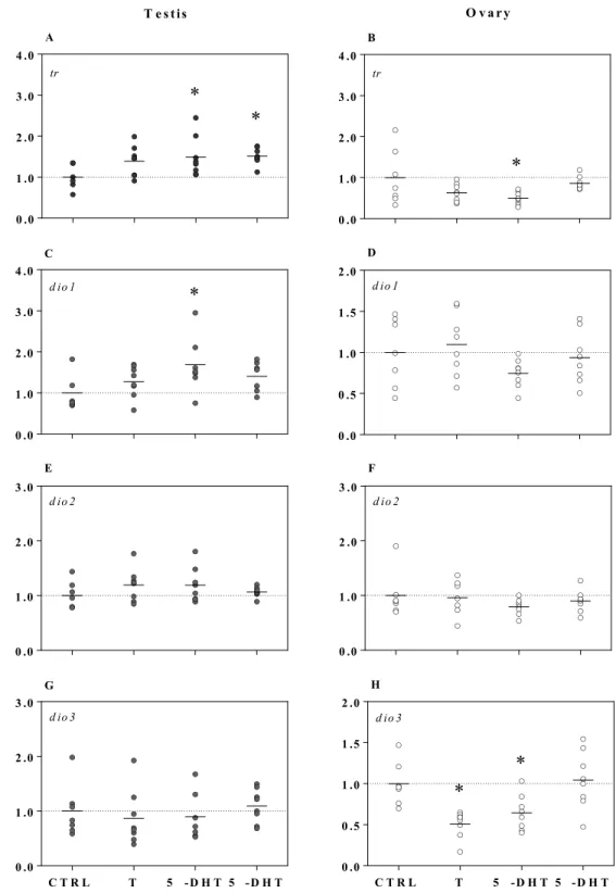
response to all three androgens (i.e., T, 5α-DHT, or 5β-DHT; Fig. 2). Expression of trβ increased on 242
average by 1.5-fold in 5α-DHT or 5β-DHT treated testes (F3, 26 = 3.8, p = 0.02); whereas, trβ transcripts
243
decreased by approximately 50% in 5α-DHT treated ovaries (F3, 26 = 5.5, p = 0.005). We found that trα
244
expression in gonadal tissues of male and female juvenile S. tropicalis was not affected by T, 5α-DHT, or 245
5β-DHT ex vivo (p > 0.05; data not shown). Exposure to 5α-DHT significantly increased dio1 expression 246
by 1.7-fold in testis tissue (F3, 22 = 3.6, p = 0.02), while dio2 and dio3 mRNA levels remained unchanged.
247
Ovary tissue did not respond to androgenic compounds by modulating dio1 or dio2 expression, but 248
exposure to T or 5α-DHT significantly decreased dio3 expression by approximately 40% (F3, 27 = 8.2, p =
249
0.001). 250
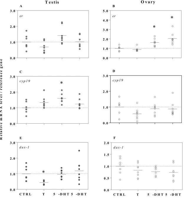
Testes and ovaries responded to T3 and IOP by differentially modulating the expression of trs 251
and dios in sex-specific manner analogous to that observed for androgens (Fig. 3). Exposure to T3 252
significantly increased trβ transcripts 2-fold in testes (F3, 26 = 20.6, p < 0.0001), in contrast to ovary tissue,
253
which did not modulate trβ expression in response to the TH exposure. A 40% decrease in trβ mRNA 254
levels was observed however in IOP-treated ovary tissue (F3, 22 = 3.3, p = 0.04). Transcriptional regulation
255
of dio3 was similar between sexes with T3 increasing dio3 expression by 3.5-fold in testes (F3, 25 = 13.2, p
256
< 0.0001) and 5-fold in ovaries (F3, 21 = 16.2, p < 0.0001). We found that TH-related gene expression in
257
ovary tissue was unaffected by the co-treatment of 5α-DHT; however, co-treatment with IOP + 5α-DHT 258
significantly increased dio1 transcripts by 1.5-fold in testes (F3, 21 = 3.3, p = 0.04). This finding
259
compliments the previously observed increase in dio1 expression in 5α-DHT treated testes. Transcripts of 260
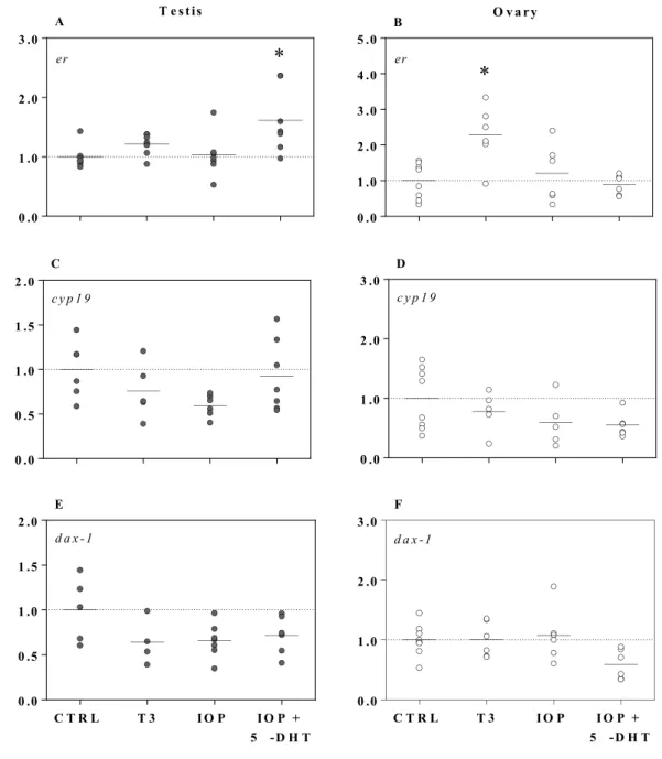
trα and dio2 were not affected by T3 or IOP treatments (p > 0.05; data not shown).
261 262
3.3.2 Androgenic compounds positively regulate estrogen-related gene expression in testis and ovary
263
tissue
Androgens differentially regulated the transcription of estrogen-related genes between sexes (Fig. 4). 265
Both 5α-DHT and 5β-DHT significantly increased erα expression in ovary tissue by 2-fold (F3, 26 = 6.3, p
266
= 0.002). A significant 1.6-fold increase in cyp19 expression was also noted in 5α-DHT-treated testes (F3,
267
25 = 3.6, p = 0.03). We observed that cyp19 transcription was however unaffected by androgenic
268
compounds in ovaries (p > 0.05). Gonadal tissues did not modulate ar, srd5α1, srd5α2, and srd5α3 269
transcription in response to different androgens (p > 0.05; data not shown). Exposure to T significantly 270
decreased dax-1 expression by 50% in testis tissue (F3, 25 = 5.20, p = 0.006); while, different androgens
271
did not modulate dax-1 transcription in ovary tissue (p > 0.05). Furthermore, we observed a sexually 272
dimorphic pattern in the gene expression of dax-1 where the mRNA levels of dax-1 in testis tissue were 273
23-fold higher than in ovary tissue (Two-tailed t-test, t11 = 11.0, p < 0.0001).
274 275
3.3.3 TH-related compounds differentially regulate sex steroid-related gene expression in testis and ovary
276
tissue
277
Gonadal tissues responded to compounds with TH-, anti-thyroid-, and androgen-related modes of action 278
by differentially modulating the relative abundance of sex steroid-related transcripts (Fig. 5 and 6). 279
Exposure to T3 significantly increased erα expression by 2.3-fold in treated ovaries (F3, 22 = 6.4, p =
280
0.003). Testis tissue did not respond to T3 or IOP by modulating erα transcription (p > 0.05); however, 281
co-treatment with IOP + 5α-DHT significantly increased erα expression by 1.6-fold in treated-testis tissue 282
relative to the control (F3, 24 = 4.3, p = 0.01), indicating an androgen mediated effect. Exposure to T3
283
significantly decreased srd5α2 expression by 47% in treated ovary tissue relative to the control (F3, 22 =
284
3.4, p = 0.04). A thyroid-mediated effect was indicated as the expression of srd5α2 did not increase in 285
ovary tissue co-treated with IOP + 5α-DHT (p > 0.05). Testis tissue did not respond to treatments by 286
modulating srd5α2 transcription (p > 0.05). Gonadal tissues did not modulate cyp19, dax-1, ar, srd5α1, or 287
srd5α3 transcription in response to TH related compounds (p > 0.05; Fig. 5 and 6).
288 289
4. Discussion 290
Androgens and THs have been shown to be extensively involved in sexual development; however, 291
relatively little is known with regard to the molecular mechanisms underlying androgen and TH crosstalk 292
as a function of sex. This study therefore investigated (i) androgenic regulation of TH-related gene 293
expression and (ii) TH-related regulation of sex-steroid gene expression in frogs using an ex vivo 294
approach to elucidate potential sex-specific mechanisms of androgenic- and TH-mediated actions in 295
reproductive tissues. 296
The transcription of several tr- and dio-isoforms within the reproductive tissues was significantly 297
altered following exposure to different androgens (T, 5α-DHT, or 5β-DHT) indicating direct crosstalk. 298
Few studies to date have reported on the molecular mechanisms underlying androgenic regulation of the 299
TH-axis between sexes, however we previously identified AREs in the putative promoter regions of trs 300
and dios in S. tropicalis using in silico analysis (Flood et al., 2013). Moreover, transcription of TH-related 301
genes has been observed within reproductive tissues and over the course of both testicular and ovarian 302
development in a wide range of vertebrate species (Duarte-Guterman et al., 2014; Flood et al., 2013; 303
Habibi et al., 2012; Wagner et al., 2008; Cooke et al., 2004; Maran, 2003). Exposure to 5α-DHT or 5β-304
DHT significantly increased trβ expression in testicular tissue ex vivo; whereas, exposure to 5α-DHT 305
significantly decreased trβ expression in ovary tissue suggesting that different androgens regulate gonadal 306
trβ expression via positive or negative mechanisms in male and female frogs, respectively. The
androgen-307
mediated increases in trβ expression in testicular tissue may be indicative of a more masculinized profile. 308
The basal endogenous TH-related gene expression exhibits a natural sexual dimorphism in S. tropicalis, 309
with testes characterized by higher trβ mRNA levels than ovaries (Duarte-Guterman and Trudeau, 2011). 310
Moreover, elevated TH-related gene expression and enzyme activity is associated with a more 311
masculinized profile (reviewed in Flood et al., 2013). Conversely, exposure to the potent androgen 5α-312
DHT further reduced trβ mRNA levels in ovary tissue, which could potentially indicate a more feminized 313
profile. We previously examined the effects of 5α-DHT on TH-related gene expression during 314
embryogenesis and observed that exposure to the androgen negatively regulated trβ expression in NF 315
stage 46 S. tropicalis larvae while also decreasing androgen-related transcription (Campbell and Langlois, 316
2017). Expression of dio1 and dio3 were also modulated in an androgen-, isoform- and sex-specific 317
manner. Exposure to 5α-DHT and IOP + 5α-DHT significantly increased dio1 expression in testes 318
compared to the control and IOP treatments, respectively. Langlois et al. (2010b) demonstrated that 319
exposure to finasteride (a srd5 inhibitor) significantly decreased dio1 expression in S. tropicalis larvae. 320
Exposure to androgenic compounds has also been shown to increase dio1 mRNA levels in hepatic tissue 321
of mice (Šošić-Jurjević et al., 2015; Miyashita et al., 1995) and fish (MacLatchy and Eales, 1988). The 322
expression of dio3 decreased following exposure to T or 5α-DHT in ovary tissue. The transcription of 323
dio3 has been proposed as biomarker for tr activation and TH levels (reviewed by Nelson and Habibi,
324
2009; Shi et al., 1996). The T3-induced response of hepatocyte dio3 gene was reduced to approximately 325
50% or 25% of the control, with inhibition of trβ expression in goldfish (Nelson and Habibi, 2008). These 326
findings suggest that TH-related gene expression can be directly regulated by different androgens and 327
highlight possible sex-specific effects of androgen-mediated actions on transcription of tr-and dio-328
isoforms in amphibians. However, the degree and direction of regulation were androgen, isoform- or sex-329
specific. 330
The androgen-mediated decreases in trβ and dio3 expression in ovarian tissue may be indicative 331
of a more feminized profile. For example, exposure to pesticides with known feminizing and TH-related 332
properties have been shown to decrease dio3 transcripts while increasing erα transcripts in various tissues 333
in fish and frogs (butachlor: Zhu et al., 2014; atrazine: Langlois et al., 2010a). In the present study, the 334
expression of erα significantly increased in 5α-DHT and 5β-DHT treated ovary tissues. We moreover 335
identified two AREs in the putative promoter regions of erα in S. tropicalis, indicating potential direct 336
androgenic regulation. Studies have also shown however that a metabolite of 5α-DHT, 5α-androstane-3β, 337
17β-diol, is weakly estrogenic in fish and rodent models (Mouriec et al., 2009; Oliveira et al., 2007). 5α-338
androstane-3β, 17β-diol can bind to either erα or erβ significantly increasing transcription of both 339
isoforms (Miller et al., 2013; Sikora et al., 2009). The increase in erα in ovary tissue was presumably 340
associated with significant increases in E2 levels of the media surrounding ovary tissue treated with IOP +
5α-DHT. Plasma levels of E2 were found to be unaffected by in vivo exposure to 5α-DHT in adult frogs
342
(X. laevis: Urbatzka et al., 2007; Coady et al., 2005), possibly as a result of hypophyseal-feedback 343
mechanisms causing the peripheral degradation and elimination of produced steroids in vivo. 344
Nevertheless, exposure to 5α-DHT has been shown to result in a rapid and consistent increase in E2
345
production in vitro from ovarian explants of P. promelas (Ornostay et al., 2016; Ornostay et al., 2013). 346
These 5α-DHT-mediated increases in E2 production are also consistent with findings in female fish 347
species exposed to the androgenic compounds 17α-methyltestosterone or 17β-trenbolone (Carassius
348
auratus of Pengze: Zheng et al., 2016; G. rarus: Gao et al., 2015; Gadus morhua: Kortner and Arukwe,
349
2009). Exposure to 5α-DHT did not modulate srd5α2 transcript levels in testes. Bissegger and Langlois 350
(2016) previously confirmed that srd5a2 expression was unaffected by exposure to T, 5α-DHT, or 5β-351
DHT ex vivo in testis tissue of frogs. However, cyp19 transcripts in- and E2 production from- whole testis
352
significantly increased following exposure to 5α-DHT. Production of E2 from testes of male S. tropicalis
353
exposed to 5α-DHT were similar to findings in male fish species exposed to 17β-trenbolone (Ankley et 354
al., 2003). Exposure to T also significantly decreased dax-1 expression by 50% in testis tissue. The 355
expression and activity of dax-1 has been shown to be essential for normal testicular development in 356
vertebrates (reviewed in: Iyer and McCabe, 2004; Lalli and Sassone-Corsi, 2003; Parker and Schimmer, 357
2002) and was identified only recently in the reproductive tissues of S. tropicalis post-metamorphosis 358
(Haselman et al., 2014). This suggests that the regulatory role of dax-1 may extend past the period of 359
sexual differentiation and later into sexual development in amphibians. Overall, research on the 360
mechanism of dax-1 in adult amphibians is limited and future studies should examine the role of dax-1 361
with regard to TH- and androgen-crosstalk in frogs. The function of these gene expression modifications 362
in the testes remains to be determined. Overall further investigation on the complex interplay between the 363
androgen and estrogen axes and the possible secondary effects on the TH-axis is warranted. 364
The present study demonstrated that exposure to T3 also increased erα expression in ovary tissue. 365
We identified one TRE half site (5’ – AGGTCA – 3’) in the putative erα promoter in S. tropicalis. Studies 366
have shown however that the tr can also bind to EREs - altering targeted gene expression as well as 367
interfering with the ability of the er to transactivate from tr-bound EREs (Vasudevan and Pfaff, 2005; 368
Vasudevan et al., 2001). Exposure to T3 also significantly decreased srd5α2 mRNA levels in ovary tissue. 369
In support of negative regulation of srd5α2 expression by T3, chronic exposure to the TH disruptor 370
potassium perchlorate was shown to significantly increase srd5α2 transcripts in vivo in hepatic (Flood and 371
Langlois, 2014) and ovary tissue (Campbell et al., 2018). Although testis tissue did not respond to T3 by 372
modulating srd5α2 expression, a significant increase in 5α-DHT levels was observed in the media 373
surrounding the T3-treated testis tissue. Exposure to methimazole, a known TH-disruptor, has been 374
shown to decrease 5α-DHT production from testis tissue (R. norvegicus: Anbalagan et al., 2010; Kala et 375
al., 2002). Morais et al. (2013) proposed that T3-modulation of steroidogenesis in Leydig cells is 376
mediated by trβ in the fish Danio rerio. We previously identified TREs in srd5α2 of M. musculus, S. 377
tropicalis, and O. latipes (Flood et al., 2013). Taken together these findings indicate that THs can regulate
378
sex-steroid related gene expression in testis and ovary tissue in amphibians, and sex-specific gene 379
expression patterns are maintained ex vivo. 380
One explanation for the differential regulation of TH-related gene expression between sexes 381
could be via DNA methylation and/or histone modification. DNA methylation has been associated with 382
transcriptional repression leading to low mRNA levels of highly methylated genes (Chen and Riggs, 383
2005). Exposure to T3 has been shown to affect histone and polymerase II modification, but does not 384
affect hyper-methylation in the promoter region of trβ in X. laevis tadpoles (Kasai et al., 2015). Moreover, 385
sexually dimorphic DNA methylation patterning has been observed in sex steroid-related genes in frogs 386
and fish (Bissegger and Langlois, 2016; Navarro-Martín et al., 2011; Contractor et al., 2004), but sex 387
differences in methylation patterns have not yet been investigated with regard to TH-related genes. Tissue 388
and age specific DNA methylation and histone modification patterns have however been thoroughly 389
studied in amphibians. During metamorphosis, different tissues in developing tadpoles (e.g., hind limbs, 390
tail tissues, etc.) have been shown to respond to exogenous T3 by differentially modulating histone 391
modifications in trs and dios (Grimaldi et al., 2013; Shi et al., 2009), indicating the potential for 392
differential epigenetic regulation of TH-related genes in reproductive tissues. In summary, epigenetic 393
regulation may play a role in differential modulation of TH- and sex steroid-related gene expression 394
between sexes. 395
This study is the first to characterize sex-specific differences in TH- and sex steroid-related gene 396
expression between testes and ovaries of sexually mature juvenile S. tropicalis following ex vivo exposure 397
to androgens (5α-DHT, 5β-DHT, or T), THs (T3), or TH-antagonists (IOP). Indeed, direct exposure to 398
different androgens led to androgen specific increases in trβ and dio1 transcripts in testes but decreases in 399
trβ and dio3 expression in ovaries, suggesting that male and female frogs can be differently affected by
400
androgenic compounds. Moreover, exposure to select androgens differentially increased estrogen-related 401
transcription (erα and cyp19) and production (E2) in ovaries and testes suggesting the activation of 402
alternate metabolic pathways yielding estrogenic metabolites. Sex steroid-related transcription (erα and 403
srd5α2) and production (5α-DHT) were differentially-regulated between sexes by T3, however
sex-404
specific gene expression patterns were maintained ex vivo. In summary, this study provides insight into 405
the molecular mechanisms underlying androgenic and TH-related actions and reveals potential 406
differential transcriptional effects as a function of sex in frogs. However, additional studies incorporating 407
in vivo and epigenetic approaches should be performed under longer-exposure conditions to firmly
408
establish these mechanisms of crosstalk. 409
Acknowledgements: 411
The authors would like to thank Dr. Sonja Bissegger, Nova Zhao, Colin Campbell, and Sarah Wallace for 412
their contributions to the project or manuscript. This work was supported by a Discovery Grant from the 413
Natural Sciences and Engineering Research Council (NSERC) of Canada (RGPIN 418576-2012) and a 414
Canada Research Chair (CRC 950-230442) to VSL, and a NSERC-PGS (PGS D2-460059-2014) to 415
DEKC. 416
References 417
Anbalagan, J., Sashi, A.M., Vengatesh, G., Stanley, J.A., Neelamohan, R., Aruldhas, M.M., 2010. 418
Mechanism underlying transient gestational-onset hypothyroidism–induced impairment of 419
posttesticular sperm maturation in adult rats. Fertil. Steril. 93, 2491–2497. 420
Ankley, G.T., Jensen, K.M., Makynen, E.A., Kahl, M.D., Korte, J.J., Hornung, M.W., Henry, T.R., 421
Denny, J.S., Leino, R.L., Wilson, V.S., Cardon, M.C., 2003. Effects of the androgenic growth 422
promoter 17β-trenbolone on fecundity and reproductive endocrinology of the fathead minnow. 423
Environ. Toxicol. Chem. 22, 1350–1360. 424
Bissegger, S., Langlois, V.S., 2016. Androgens modulate gene expression and specific DNA methylation 425
pattern of steroid 5α-reductases in the frog Silurana tropicalis. Gen. Comp. Endocrinol. 234, 426
123–132. 427
Bissegger, S., Martyniuk, C.J., Langlois, V.S., 2014. Transcriptomic profiling in Silurana tropicalis testes 428
exposed to finasteride. Gen. Comp. Endocrinol. 203, 137–145. 429
Brown, S.B., Adams, B.A., Cyr, D.G., Eales, J.G., 2004. Contaminant effects on the teleost fish thyroid. 430
Environ. Toxicol. Chem. 23, 1680–1701. 431
Campbell, D.E.K., Langlois, V.S., 2017. Expression of sf1 and dax-1 are regulated by thyroid hormones 432
and androgens during Silurana tropicalis early development. Gen. Comp. Endocrinol., 259, 34– 433
44. 434
Campbell, D.E.K., Montgomerie, R.D., Langlois, V.S. 2018. Lifecycle exposure to perchlorate 435
differentially alters morphology, biochemistry, and transcription as well as sperm motility in 436
Silurana tropicalis frogs. Environ. Pollut. 237, 196–204.
437
Cardone, A., Angelini, F., Esposito, T., Comitato, R., Varriale, B. 2000. The expression of androgen 438
receptor messenger RNA is regulated by tri-iodothyronine in lizard testes. J. Steroid Biochem. 439
Mol. Biol. 72, 133–141. 440
Carosa, E., Lenzi, A., Jannini, E.A., 2017. Thyroid hormone receptors and ligands, tissue distribution and 441
sexual behavior. Mol. Cell. Endocrinol. 457, 49–59. 442
Chen, Z., Riggs, A.D., 2005. Maintenance and regulation of DNA methylation patterns in mammals. 443
Biochem. Cell Biol. 83, 438–448. 444
Coady, K.K., Murphy, M.B., Villeneuve, D.L., Hecker, M., Jones, P.D., Carr, J.A., Solomon, K.R., 445
Smith, E.E., Van Der Kraak, G., Kendall, R.J. and Giesy, J.P., 2005. Effects of atrazine on 446
metamorphosis, growth, laryngeal and gonadal development, aromatase activity, and sex steroid 447
concentrations in Xenopus laevis. Ecotoxicol. Environ. Saf. 62, 160–173. 448
Contractor, R.G., Foran, C.M., Li, S.F., Willett, K.L., 2004. Evidence of gender- and tissue-specific 449
promoter methylation and the potential for ethinylestradiol-induced changes in Japanese medaka 450
(Oryzias latipes) estrogen receptor and aromatase genes. J. Toxicol. Environ. Health. A. 67, 1–22. 451
Cooke, P.S., Holsberger, D.R., Witorsch, R.J., Sylvester, P.W., Meredith, J.M., Treinen, K.A., Chapin, 452
R.E., 2004. Thyroid hormone, glucocorticoids, and prolactin at the nexus of physiology, 453
reproduction, and toxicology. Toxicol. Appl. Pharm. 194, 309–335. 454
Cortés, D.C.C., Langlois, V.S., Fernandino, J.I., 2014. Crossover of the hypothalamic pituitary– 455
adrenal/interrenal,–thyroid, and–gonadal axes in testicular development. Front. Endocrinol. 139, 456
1–11. 457
Cyr, D.G., Eales, J.G., 1996. Interrelationships between thyroidal and reproductive endocrine systems in 458
fish. Rev. Fish Biol. Fisher. 6, 165–200. 459
Duarte-Guterman, P., Langlois, V.S., Hodgkinson, K., Pauli, B.D., Cooke, G.M., Wade, M.G., Trudeau, 460
V.L., 2009. The aromatase inhibitor fadrozole and the 5-reductase inhibitor finasteride affect 461
gonadal differentiation and gene expression in the frog Silurana tropicalis. Sex. Dev. 3, 333–341. 462
Duarte-Guterman, P., Langlois, V.S., Pauli, B.D., Trudeau, V.L., 2010. Expression and T3 regulation of
463
thyroid hormone-and sex steroid-related genes during Silurana (Xenopus) tropicalis early
464
development. Gen. Comp. Endocrinol. 166, 428–435.
Duarte-Guterman, P., Navarro-Martín, L., Trudeau, V.L., 2014. Mechanisms of crosstalk between 466
endocrine systems: regulation of sex steroid hormone synthesis and action by thyroid hormones. 467
Gen. Comp. Endocrinol. 203, 69–85. 468
Duarte-Guterman, P., Ryan, M.J., Trudeau, V.L., 2012. Developmental expression of sex steroid-and 469
thyroid hormone-related genes and their regulation by triiodothyronine in the gonad– 470
mesonephros of a Neotropical frog, Physalaemus pustulosus. Gen. Comp. Endocrinol. 177, 195– 471
204. 472
Duarte-Guterman, P., Trudeau, V.L., 2011. Transcript profiles and triiodothyronine regulation of sex 473
steroid-and thyroid hormone-related genes in the gonad–mesonephros complex of Silurana 474
tropicalis. Mol. Cell. Endocrinol. 331, 143–149.
475
Farré, D., Roset, R., Huerta, M., Adsuara, J. E., Roselló, L., Albà, M. M., Messeguer, X., 2003. 476
Identification of patterns in biological sequences at the ALGGEN server: PROMO and 477
MALGEN. Nucleic Acids Res 31, 3651–3653. 478
Fini, J.B., Le Mével, S., Turque, N., Palmier, K., Zalko, D., Cravedi, J.P., Demeneix, B.A., 2007. An in 479
vivo multiwell-based fluorescent screen for monitoring vertebrate thyroid hormone disruption. 480
Environ. Sci. Tech. 41, 5908–5914. 481
Flood, D.E.K., Fernandino, J.I., Langlois, V.S., 2013. Thyroid hormones in male reproductive 482
development: evidence for direct crosstalk between the androgen and thyroid hormone axes. Gen. 483
Comp. Endocrinol. 192, 2–14. 484
Flood, D.E.K., Langlois, V.S., 2014. Crosstalk between the thyroid hormone and androgen axes during 485
reproductive development in Silurana tropicalis. Gen. Comp. Endocrinol. 203, 232–240. 486
Gao, J., Liu, S., Zhang, Y., Yang, Y., Yuan, C., Chen, S., Wang, Z., 2015. Effects of 17 α-487
methyltestosterone on transcriptome, gonadal histology and sex steroid hormones in rare minnow 488
Gobiocypris rarus. Comp. Biochem. Physiol. Part D Genomics Proteomics 15, 20–27.
489
Grant, C.E., Bailey, B.L., William, S.N., 2011. FIMO: Scanning for occurrences of a given motif. 490
Bioinformatics 27, 1017–1018. 491
Grimaldi, A., Buisine, N., Miller, T., Shi, Y. B., Sachs, L.M., 2013. Mechanisms of thyroid hormone 492
receptor action during development: lessons from amphibian studies. BBA-Gen. Subjects 1830, 493
3882–3892. 494
Habibi, H.R., Nelson, E.R., Allan, E.R.O., 2012. New insights into thyroid hormone function and 495
modulation of reproduction in goldfish. Gen. Comp. Endocrinol. 175, 19–26. 496
Haselman, J.T., Olmstead, A.W., Degitz, S.J., 2014. Global gene expression during early differentiation 497
of Xenopus (Silurana) tropicalis gonad tissues. Gen. Comp. Endocrinol. 214, 103–113. 498
Hogan, N.S., Duarte, P., Wade, M.G., Lean, D.R., Trudeau, V.L., 2008. Estrogenic exposure affects 499
metamorphosis and alters sex ratios in the northern leopard frog (Rana pipiens): identifying 500
critically vulnerable periods of development. Gen. Comp. Endocrinol. 156, 515–523. 501
Hogan, N.S., Lean, D.R., Trudeau, V.L., 2006. Exposures to estradiol, ethinylestradiol and octylphenol 502
affect survival and growth of Rana pipiens and Rana sylvatica tadpoles. J Toxicol. Environ. 503
Health A. 69, 1555–1569. 504
Hunt, D.W., Eales, J.G., 1979. The influence of testosterone propionate on thyroid function of immature 505
rainbow trout, Salmo gairdneri Richardson. Gen. Comp. Endocrinol. 37, 115–121. 506
Iyer, A.K., McCabe, E.R.B., 2004. Molecular mechanisms of DAX1 action. Mol. Gen. Metab. 83, 60–73. 507
Johnson, K.M., Lema, S.C., 2011. Tissue-specific thyroid hormone regulation of gene transcripts 508
encoding iodothyronine deiodinases and thyroid hormone receptors in striped parrotfish (Scarus 509
iseri). Gen. Comp. Endocrinol. 172, 505–517.
510
Kala, N., Ravisankar, B., Govindarajulu, P., Aruldhas, M.M., 2002. Impact of foetal onset 511
hypothyroidism on the epididymis of mature rats. Int. J. Androl. 25, 139–148. 512
Kasai, K., Nishiyama, N., Izumi, Y., Otsuka, S., Ishihara, A., Yamauchi, K., 2015. Exposure to 3, 3′, 5-513
triiodothyronine affects histone and RNA polymerase II modifications, but not DNA methylation 514
status, in the regulatory region of the Xenopus laevis thyroid hormone receptor βΑ gene. 515
Biochem. Biophys. Res. Commun. 467, 33–38. 516
Kortner, T.M., Arukwe, A., 2007. Effects of 17α-methyltestosterone exposure on steroidogenesis and 517
cyclin-B mRNA expression in previtellogenic oocytes of Atlantic cod (Gadus morhua). Comp 518
Biochem. Physiol. C Toxicol. Pharmacol. 146, 569–580. 519
Lalli, E., Sassone-Corsi, P., 2003. DAX-1, an unusual orphan receptor at the crossroads of steroidogenic 520
function and sexual differentiation. Mol. Endocrinol. 17, 1445–1453. 521
Langlois, V.S., Carew, A.C., Pauli, B.D., Wade, M.G., Cooke, G.M., Trudeau, V.L. 2010a. Low levels of 522
the herbicide atrazine alter sex ratios and reduce metamorphic success in Rana pipiens tadpoles 523
raised in outdoor mesocosms. Environ. Health Perspect. 118, 552–557. 524
Langlois, V.S., Duarte-Guterman, P., Ing, S., Pauli, B.D., Cooke, G.M., Trudeau, V.L., 2010b. Fadrozole 525
and finasteride exposures modulate sex steroid-and thyroid hormone-related gene expression in 526
Silurana (Xenopus) tropicalis early larval development. Gen. Comp. Endocrinol. 166, 417–427.
527
Langlois, V.S., Duarte-Guterman, P., Trudeau, V.L., 2011. Expression profiles of reproduction-and 528
thyroid hormone-related transcripts in the brains of chemically-induced intersex frogs. Sexual 529
Development, 5, 26–32. 530
Maclatchy, D.L., Eales, J.G., 1988. Short-term treatment with testosterone increases plasma 3, 5, 3′-531
triiodo-L-thyronine and hepatic L-thyroxine 5′-monodeiodinase levels in arctic charr, Salvelinus 532
alpinus. Gen. Comp. Endocrinol. 71, 10–16.
533
Maran, R.R.M., 2003. Thyroid hormones: their role in testicular steroidogenesis. Arch. Androl. 49, 375– 534
388. 535
Miller, K. K. M., Al-Rayyan, N., Ivanova, M. M., Mattingly, K. A., Ripp, S. L., Klinge, C. M., Prough, R. 536
A., 2013. DHEA metabolites activate estrogen receptors alpha and beta. Steroids 78, 15–25. 537
Miyashita, K., Murakami, M., Iriuchijima, T., Takeuchi, T., Mori, M., 1995. Regulation of rat liver type 1 538
iodothyronine deiodinase mRNA levels by testosterone. Mol. Cell. Endocrinol. 115, 161–167. 539
Morais, R.D.V.S., Nóbrega, R.H., Gómez-González, N.E., Schmidt, R., Bogerd, J., França, L.R., Schulz, 540
R.W., 2013. Thyroid hormone stimulates the proliferation of Sertoli cells and single type A 541
spermatogonia in adult zebrafish (Danio rerio) testis. Endocrinology 154, 4365–4376. 542
Mouriec, K., Gueguen, M.M., Manuel, C., Percevault, F., Thieulant, M.L., Pakdel, F., Kah, O., 2009. 543
Androgens upregulate cyp19a1b (aromatase B) gene expression in the brain of zebrafish (Danio 544
rerio) through estrogen receptors. Biol. Reprod. 80, 889–896.
545
Mullur, R., Liu, Y.Y., Brent, G.A., 2014. Thyroid hormone regulation of metabolism. Physiol. Rev. 94, 546
355–382. 547
Navarro-Martín, L., Viñas, J., Ribas, L., Díaz, N., Gutiérrez, A., Di Croce, L., Piferrer, F., 2011. DNA 548
Methylation of the Gonadal Aromatase (cyp19a) Promoter Is Involved in Temperature-Dependent 549
Sex Ratio Shifts in the European Sea Bass. PLoS Genet. 7, e1002447. 550
Nelson, E.R., Habibi, H.R., 2008. Functional significance of a truncated thyroid receptor subtype lacking 551
a hormone-binding domain in goldfish. Endocrinology, 149, 4702–4709. 552
Nelson, E.R., Habibi, H.R., 2009. Thyroid receptor subtypes: structure and function in fish. Gen. Comp. 553
Endocrinol. 161, 90–96. 554
Oliveira, A.G., Coelho, P.H., Guedes, F.D., 2007. 5-Androstane-3, 17-diol (3-diol), an estrogenic 555
metabolite of 5-dihydrotestosterone, is a potent modulator of estrogen receptor ER expression in 556
the ventral prostrate of adult rats. Steroids 72, 914–922. 557
Ornostay, A., Cowie, A.M., Hindle, M., Baker, C.J., Martyniuk, C.J., 2013. Classifying chemical mode of 558
action using gene networks and machine learning: A case study with the herbicide linuron. Comp. 559
Biochem. Physiol. Part. D. Genomics. Proteomics. 8, 263–274. 560
Ornostay, A., Marr, J., Loughery, J.R., Martyniuk, C.J., 2016. Transcriptional networks associated with 5-561
alpha-dihydrotestosterone in the fathead minnow (Pimephales promelas) ovary. Gen. Comp. 562
Endocrinol. 225, 23–32. 563
Orozco, A., Valverde-R, C., 2005. Thyroid hormone deiodination in fish. Thyroid, 15, 799–813. 564
Parker, K.L., Schimmer, B.P., 2002. Genes essential for early events in gonadal development. Ann. Med. 565
34, 171–178. 566
Sambroni, E., Gutieres, S., Cauty, C., Guiguen, Y., Breton, B., Lareyre, J.J., 2001. Type II iodothyronine 567
deiodinase is preferentially expressed in rainbow trout (Oncorhynchus mykiss) liver and gonads. 568
Mol. Reprod. Dev. 60, 338–350. 569
Scholz, S., Renner, P., Belanger, S.E., Busquet, F., Davi, R., Demeneix, B.A., Denny, J.S., Leonard, M., 570
McMaster, M.E., Villeneuve, D.L., Embry, M.R., 2013. Alternatives to in vivo tests to detect 571
endocrine disrupting chemicals (EDCs) in fish and amphibians–screening for estrogen, androgen 572
and thyroid hormone disruption. Crit. Rev. Toxicol. 43, 45–72. 573
Sikora, M.J., Cordero, K.E., Larios, J.M., Johnson, M.D., Lippman, M.E., Rae, J.M. 2009. The androgen 574
metabolite 5alpha-androstane-3beta,17beta-diol (3betaAdiol) induces breast cancer growth via 575
estrogen receptor: implications for aromatase inhibitor resistance. Breast Cancer Res. Treat. 115, 576
289–96. 577
Sharma, B., Patiño, R., 2010. Effects of cadmium, estradiol-17β and their interaction on gonadal 578
condition and metamorphosis of male and female African clawed frog, Xenopus laevis. 579
Chemosphere 79, 499–505. 580
Shi, Y.B., 2009. Dual functions of thyroid hormone receptors in vertebrate development: the roles of 581
histone-modifying cofactor complexes. Thyroid 19, 987–999. 582
Shi, Y. B., Wong, J., Puzianowska-Kuznicka, M., Stolow, M.A., 1996. Tadpole competence and tissue-583
specific temporal regulation of amphibian metamorphosis: Roles of thyroid hormone and its 584
receptors. Bioessays 18, 391–399. 585
Šošić-Jurjević, B., Filipović, B., Renko, K., Miler, M., Trifunović, S., Ajdžanovič, V., Kӧhrle, J, 586
Milošević, V., 2015. Testosterone and estradiol treatments differently affect pituitary-thyroid axis 587
and liver deiodinase 1 activity in orchidectomized middle-aged rats. Exp. Geront. 72, 85–98. 588
Urbatzka, R., Bottero, S., Mandich, A., Lutz, I., Kloas, W., 2007. Endocrine disrupters with (anti) 589
estrogenic and (anti) androgenic modes of action affecting reproductive biology of Xenopus 590
laevis: I. Effects on sex steroid levels and biomarker expression. Comp. Biochem. Physiol. C.
591
Toxicol. Pharmacol. 144, 310–318. 592
Vasudevan, N., Koibuchi, N., Chin, W.W., Pfaff, D.W., 2001. Differential crosstalk between estrogen 593
receptor (ER) α and ERβ and the thyroid hormone receptor isoforms results in flexible regulation 594
of the consensus ERE. Mol. Brain Res. 95, 9–17. 595
Vasudevan, N., Pfaff, D., 2005. Molecular mechanisms of crosstalk between thyroid hormones and 596
estrogens. Curr. Opin. Endocrinol. 12, 381–388. 597
Wagner, M.S., Wajner, S.M., Maia, A.L., 2008. The role of thyroid hormone in testicular development 598
and function. J. Endocrinol. 199, 351–365. 599
Wajner, S.M., Wagner, M.S., Maia, A.L., 2009. Clinical implications of altered thyroid status in male 600
testicular function. Arq. Bras. Endocrinol. Metabol. 53, 976–982. 601
Zheng, Y., Chen, J., Liu, Y., Gao, J., Yang, Y., Zhang, Y., Bing, X., Gao, Z., Liang, H., Wang, Z., 2016. 602
Molecular mechanism of endocrine system impairment by 17α-methyltestosterone in gynogenic 603
Pengze crucian carp offspring. Ecotox. Environ. Saf. 128, 143–152. 604
Zhu, L., Li, W., Zha, J., Wang, M., Yuan, L., Wang, Z. 2014. Butachlor causes disruption of HPG and 605
HPT axes in adult female rare minnow (Gobiocypris rarus). Chem. Biol. Interact., 221, 119–126. 606
Table 1. Total estradiol (E2), testosterone (T), and 5α-dihydrotestosterone (5α-DHT) produced by testes [pg] and ovaries [pg/g] during a 6 h ex
608
vivo incubation. Media sex steroid data are presented as means (least squares means [95% CL]. Testicular sex steroid hormone levels are
609
normalized by organ mass. Ovarian sex steroid hormone levels are reported per g tissue. Significant differences between treatments and the control 610
(*) were identified by one-way ANOVAs followed by post hoc Dunnett’s tests (p < 0.05). 611
612
Sex Sex steroid-hormones Control T3 IOP IOP + 5α-DHT
(50 nM) (10 µM) (10 µM) (1 µM) Male [pg] T (F2, 13 = 0.31, p = 0.74)b 3,605 [2588, 4622] 4,094 [2980, 5208] 3,595 [2481, 4709] NM 5α-DHT (F2, 21 = 5.79, p = 0.01) 14,186 [10991, 17381] 21,436 [18241, 24631]* 16,558 [13363, 19753] NM E2 (F3, 28 = 40.1, p < 0.0001) 227.3 [204.0, 250.6] 203.7 [180.3, 227.0] 207.8 [184.5, 231.1] 355.6 [332.3, 378.9]* Femalea [pg/g] T (F2, 21 = 1.84, p = 0.18) 14,533[5386, 23680] 22,870 [13723, 32016] 25,054 [15907, 34200] NM 5α-DHT (F2, 21 = 2.39, p = 0.12) 9,957 [7168, 12746] 13,976 [11187, 16765] 13,667 [10880, 16459] NM E2 (F3, 28 = 3.94, p = 0.02) 12,040 [6226, 17853] 16,677 [10864, 22491] 14,035 [8222, 19849] 25,814 [20000, 31627]* 613
a Female sex steroid-hormones were log-transformed to normalize residuals.
614
b For each sex per treatment n = 8, except n = 5 for levels of T in males in all treatments.
616
Figure 1. Promoter analysis of M. musculus, S. tropicalis, and O. latipes TH-related genes (trβ) and androgen-related genes (ar, srd5α2) (A), and 617
estrogen related genes (erα, and cyp19) (B). For information on tr and ar half-site motifs in thyroid- and androgen-re;ated genes please refer to 618
Flood et al. (2013). All sequences used for analysis were collected from the Ensembl Project (http://www.ensembl.org). Putative transcription 619
factor binding sites within the putative promoter (-2000 to +1) were identified using PROMO (v.3.0.2; Farré et al., 2003) and FIMO (v.4.11.1; 620
Grant et al., 2011) software. TREs are shown in blue, AREs are shown in green, and EREs are represented by purple arrows. This figure was 621
adapted from Figure 1 in Flood et al., 2013 (Ch. 1). 622 -503 nnnnn -495 -477 -1282 -1756 -953-834, -804 -1470 -1612 nnnnn -1597 -1219 -1998 nnnnn -1983 -705 -760 -524 -470 -1088 -901 -731 -1308 -1373, -1362 -1339 -1825 -1007 -827 -548 -1054 -1274 -1751 -1850 -1716 -1633 Response element TGACCT TGTCCT TGTCCT nnnnn TGTCCT TGACCT nnnnn TGTCCT TRE TGTTCT TGTTCT nnnnn TGTTCT
ARE ERE TGACC AGGTCAnnnnn
TGACCT
M. musculus S. tropicalis O. latipes
Gene +1bp -2kb AGGTCA nnnnn AGGTCA +1bp -2kb -2kb +1bp -1755 -1426 -1436, -1426 -511 -439 -904 -686 -979 -1339 -1898 -1973 -1349 -801 -1508 -1170 -695 -849 -1614 -563 Androgen receptor (ar) Estrogen receptor (erα) Aromatase (cyp19) 5α-reductase type 2 (srd5α2) Thyroid hormone receptor (trβ) -1485 -1661 -207 -45 -1301 -48 -1397 -1748 -512 T hy roi d-re la te d A ndro ge n-re la te d E st ro ge n-re la te d
M. musculus S. tropicalis O. latipes
Gene +1bp -2kb -2kb +1bp -2kb +1bp
A
B
Responseelement ERE TGACC
AGGTCA nnnnn TGACCT AGGTCA nnnnn AGGTCA
Figure 2. Expression of trs (trβ) and dios (dio1, dio2, and dio3) in testes (A, C, E, and G, respectively) and ovaries (B, D, F, and H, respectively) exposed ex vivo to testosterone (T; 1 µM),
5α-dihydrotestosterone (5α-DHT; 1 µM), and to 5β-5α-dihydrotestosterone (5β-DHT; 1 µM) for 6 h. Symbols represent individual samples (n = 6–8 per treatment). Gene expression data are normalized to odc and presented as fold changes relative to the control treatment. Significant differences between treatments and the control (*) were identified by one-way ANOVAs followed by post hoc Dunnett’s tests (p < 0.05). Note that the scales of the y-axis vary.
0 .0 1 .0 2 .0 3 .0 4 .0 tr
*
*
A 0 .0 1 .0 2 .0 3 .0 4 .0 d io 1 C*
0 .0 1 .0 2 .0 3 .0 d io 2 E C T R L T 5 - D H T 5 - D H T 0 .0 1 .0 2 .0 3 .0 d io 3 G 0 .0 1 .0 2 .0 3 .0 4 .0 tr*
B 0 .0 0 .5 1 .0 1 .5 2 .0 d io 1 D 0 .0 1 .0 2 .0 3 .0 d io 2 F C T R L T 5 - D H T 5 - D H T 0 .0 0 .5 1 .0 1 .5 2 .0 d io 3 H*
*
R e la ti v e m R N A l e v e l / r e fe r e n c e g e n e T e s t is O v a r yFigure 3. Expression of trs (trβ) and dios (dio1, dio2, and dio3) in testes (A, C, E, and G, respectively) and ovaries (B, D, F, and H, respectively) exposed ex vivo to triiodothyronine (T3; 50 nM), iopanoic acid (IOP; 10 µM), and to a co-treatment of IOP (10 µM) + 5α-dihydrotestosterone (5α-DHT; 1 µM) for 6 h. Symbols represent individual samples (n = 5–8 per treatment). Testis and ovary gene expression data are normalized to odc and rpl8, respectively and presented as fold changes relative to the control treatment. Significant differences between treatments and the control (*) were identified by one-way ANOVAs followed by post hoc Dunnett’s tests (p < 0.05). Significant differences between treatments were identified by two-tailed t tests (p < 0.05). Note that the scales of the y-axis vary.
0 .0 1 .0 2 .0 3 .0 4 .0 tr A
*
0 .0 0 .5 1 .0 1 .5 2 .0 tr B*
0 .0 1 .0 2 .0 3 .0 d io 1 C*
0 .0 1 .0 2 .0 3 .0 d io 1 D 0 .0 0 .5 1 .0 1 .5 2 .0 d io 2 E 0 .0 1 .0 2 .0 3 .0 4 .0 d io 2 F C T R L T 3 I O P I O P + 5 - D H T 0 .0 2 .0 4 .0 6 .0 8 .0 d io 3 G*
C T R L T 3 I O P I O P + 5 - D H T 0 .0 2 .0 4 .0 6 .0 8 .0 1 0 .0 d io 3 H*
R e la ti v e m R N A l e v e l / r e fe r e n c e g e n e T e s t is O v a r yFigure 4. Expression of erα, cyp19, and dax-1 in testes (A, C, and E respectively) and ovaries (B, D, and F respectively) exposed ex vivo to testosterone (T; 1 µM), 5α-dihydrotestosterone (5α-DHT; 1 µM), and to 5β-dihydrotestosterone (5β-DHT; 1 µM) for 6 h. Symbols represent individual samples (n = 6–8 per treatment). Gene expression data are normalized to odc and presented as fold changes relative to the control treatment. Significant differences between treatments and the control (*) were identified by one-way ANOVAs followed by post hoc Dunnett’s tests (p < 0.05). Note that the scales of the y-axis vary.
C T R L T 5 - D H T 5 - D H T 0 .0 1 .0 2 .0 3 .0
*
d a x - 1 E C T R L T 5 - D H T 5 - D H T 0 .0 0 .5 1 .0 1 .5 2 .0 d a x - 1 F 0 .0 1 .0 2 .0 3 .0 er A 0 .0 1 .0 2 .0 3 .0 4 .0 5 .0 er B*
*
0 .0 1 .0 2 .0 3 .0 c y p 1 9 C*
0 .0 1 .0 2 .0 3 .0 c y p 1 9 D R e la ti v e m R N A l e v e l / r e fe r e n c e g e n e T e s t is O v a r yFigure 5. Expression of erα, cyp19, and dax-1 in testes (A, C, and E respectively) and ovaries (B, D, and F respectively) exposed ex vivo to triiodothyronine (T3; 50 nM), iopanoic acid (IOP; 10 µM), and to a co-treatment of IOP (10 µM) + 5α-dihydrotestosterone (5α-DHT; 1 µM) for 6 h. Symbols represent
individual samples (n = 5–8 per treatment). Testis and ovary gene expression data are normalized to odc and rpl8, respectively and presented as fold changes relative to the control treatment. Significant
differences between treatments and the control (*) were identified by one-way ANOVAs followed by post hoc Dunnett’s tests (p < 0.05). Note that the scales of the y-axis vary.
0 .0 1 .0 2 .0 3 .0 er
*
A 0 .0 1 .0 2 .0 3 .0 4 .0 5 .0*
er B 0 .0 0 .5 1 .0 1 .5 2 .0 c y p 1 9 C 0 .0 1 .0 2 .0 3 .0 c y p 1 9 D R e la ti v e m R N A l e v e l / r e fe r e n c e g e n e C T R L T 3 I O P I O P + 5 - D H T 0 .0 1 .0 2 .0 3 .0 d a x - 1 F C T R L T 3 I O P I O P + 5 - D H T 0 .0 0 .5 1 .0 1 .5 2 .0 d a x - 1 E T e s t is O v a r yFigure 6. Expression of ar, srd5α1, srd5α2, and srd5α3 in testes (A, C, E, and G, respectively) and ovaries (B, D, F, and H, respectively) exposed ex vivo to triiodothyronine (T3; 50 nM), iopanoic acid (IOP; 10 µM), or a co-treatment of IOP (10 µM) + 5α-dihydrotestosterone (5α-DHT; 1 µM) for 6 h. Symbols represent individual samples (n = 5–8 per treatment). Testis and ovary gene expression data are normalized to odc and rpl8, respectively and presented as fold changes relative to the control treatment. Significant differences between treatments and the control (*) were identified by one-way ANOVAs followed by post hoc Dunnett’s tests (p < 0.05). Note: scales of the y-axis vary.
0 .0 1 .0 2 .0 3 .0 s r d 5 2 E 0 .0 1 .0 2 .0 3 .0
![Table 1. Total estradiol (E 2 ), testosterone (T), and 5α-dihydrotestosterone (5α-DHT) produced by testes [pg] and ovaries [pg/g] during a 6 h ex](https://thumb-eu.123doks.com/thumbv2/123doknet/2933922.78217/23.1188.61.1153.257.472/table-total-estradiol-testosterone-dihydrotestosterone-produced-testes-ovaries.webp)