HAL Id: pastel-00610998
https://pastel.archives-ouvertes.fr/pastel-00610998
Submitted on 25 Jul 2011HAL is a multi-disciplinary open access archive for the deposit and dissemination of sci-entific research documents, whether they are pub-lished or not. The documents may come from teaching and research institutions in France or abroad, or from public or private research centers.
L’archive ouverte pluridisciplinaire HAL, est destinée au dépôt et à la diffusion de documents scientifiques de niveau recherche, publiés ou non, émanant des établissements d’enseignement et de recherche français ou étrangers, des laboratoires publics ou privés.
under a high protein diet : identification of amino acids
signal and associated transduction pathways
Nattida Chotechuang
To cite this version:
Nattida Chotechuang. The role of amino acids in liver protein metabolism under a high protein diet : identification of amino acids signal and associated transduction pathways. Food and Nutrition. AgroParisTech, 2010. English. �NNT : 2010AGPT0026�. �pastel-00610998�
N° /__/__/__/__/__/__/__/__/__/__/
T H E S I S
submitted to obtain the degree of
Doctor of Philosophy
at
L’Institut des Sciences et Industries du Vivant et de l’Environnement
(AgroParisTech)
Speciality: Nutrition Science
Presented and defended in public by
Nattida CHOTECHUANG
on 22
ndMarch 2010
THE ROLE OF AMINO ACIDS IN LIVER PROTEIN METABOLISM
UNDER A HIGH PROTEIN DIET: IDENTIFICATION OF AMINO ACIDS
SIGNAL AND ASSOCIATED TRANSDUCTION PATHWAYS
Thesis director: Daniel TOMÉ
Thesis co-director: Dalila AZZOUT-MARNICHE
AgroParisTech, UMR914 Nutrition Physiology and Ingestive Behaviour, F-75005 Paris
to the jury:
Mr. Daniel TOMÉ, Professor, AGROPARISTECH President
Mr. Pierre FAFOURNOUX, DR, CNRS Reporter
Mr. Claude FOREST, DR, INSERM Reporter Mr. Bernard PORTHA, Professor, UNIVERSITE PARIS-DIDEROT Examiner Ms. Tatiana STEILER, Reseacher, DANONE Examiner Ms. Dalila AZZOUT-MARNICHE, Assitant Professor, AGROPARISTECH Examiner
Acknowledgement
I would like to express my gratitude to all those who gave me the chance to complete my PhD.
Firstly, I would like to thank Prof. Daniel TOME, my thesis director, for giving me the great opportunity to conduct research in his lab and offering me the chance to do pre-doctoral research in Japan. This PhD opened a lot of doors for my career.
I wish to thank the members of the jury, Dr. Pierre FAFOURNOUX and Dr. Claude FOREST for accepting being the reporter as well as Pr. Bernard PORTHA and Dr. Tatiana STEILER for accepting being the examiners of my thesis.
I am heartily thankful to my supervisor Dr. Dalila AZZOUT-MARNICHE, whose help, guidance and support from the start to the finish enabled me to develop an understanding of the subject, whose encouragement helped me during the period of research and later of thesis writing, and also her patience to work with a Thai who could not understand French very well.
I acknowledge the Danone for their financial support to this project and the Royal Thai Government for my scholarship and funding to study in France. I am especially thankful to the office of Educational Affairs at Royal Thai embassy in Paris for their unwavering administrative help and support.
I am particularly indebted to Catherine CHAUMONTET for her help and suggestions in the research and writing of this thesis. I want to thank all my colleagues from the PNCA, especially Gilles Fromentin, Claire Gaudichon and Cécile Bos for all their help, support, interest and valuable hints, and the internship students: Stephanie, Catherine and Wafa, for their help.
In addition, I want to thank to my office mates Anne-Sophie, Fanny, Jessica, Magda, Wahiba and Nad (post-doctoral fellow at AgroParisTech) for their great moral support and the good time that we spent together.
Last, but not least, I would like to say a big thank you to my family and friends, especially my mother, for supporting and encouraging me to pursue this degree. I am also grateful to my grandmother and my father, who encouraged my interest in science starting from an early age. Even though they are not with us, they will forever occupy a special place in my heart. Moreover, I especially want to thank to Itthikorn, Wanalee, Darin, Pete, Pimpavadee, Wanrug, Pijika, Dalad, Areeya, Karima, Patharawan, Bruno, Guillaume, and all my Thai friends for their support, great company and all the fun I had outside work.
“The important thing in science is not so much to obtain new facts as to discover new ways of thinking about them.” William Bragg, Sr.
Abstract
High Protein (HP) intake improves glucose homeostasis and reduces weight gain, body fat mass, white adipose tissue and adipocyte size in rats. The metabolic adaptation is characterized by at least a decrease in hepatic lipogenesis and an increase in hepatic amino acid (AA) conversion into glycogen. However, the role of amino acids (AAs) in the control of these metabolic adaptations has not been studied, and the transduction pathways involved in the sensing of the increase in AA supply remain unclear. Therefore, the aim of our study was to understand the effect of AAs on translation and on proteolysis, to identify the transduction pathways involved in AA signaling and the AA or the groups of AAs involved in these effects, using both in vivo and in vitro approaches. Western blot analysis was performed on protein extracts to examine the phosphorylation state of the mammalian target of rapamycin (mTOR), adenosine monophosphate-activated protein kinase (AMPK) and general control non-depressible kinase 2 (GCN2) transduction pathways which may be involved in AA sensing and in the control of translation in liver. This study demonstrated that adaptation to HP diet was characterized by the stimulation of translation, at least for the initiation step in the liver. Using primary culture of hepatocytes, we showed that this activation required both high AA levels (at least for leucine alone or a branched-chain AA mixture) and insulin, as indicated by the increase of mTOR, 4E-BP1 and S6 phosphorylation and the decrease of AMPK and GCN2 phosphorylation. Using AICAR (AMPK activator) and rapamycin (mTOR inhibitor), we demonstrated that mTOR might not be the only regulator of 4E-BP1 and S6K1 (downstream targets of mTOR) in high AA conditions and that AMPK may also play an important role in their control. Moreover, the HP diet induced the inhibition of protein breakdown in the liver and these results were concomitant with a decrease of gene expression of the major components for both autophagy and the ubiquitin-proteasome system in liver. Subsequently, ubiquitinated protein in the liver was lower and both AAs and insulin were required for the down-regulation of ubiquitination. Indeed, mTOR and AMPK were also involved in the control of the ubiquitin proteasome system in the liver in response to the increase in AA and insulin concentrations. These results suggested that the control of the catabolic and anabolic pathways of protein metabolism was regulated by the same set of signals and mediated by the same transduction signaling pathways.
Resumé
La consommation d’un régime hyper protéique (HP) améliore l’homéostasie glucidique, le gain de poids, l’adiposité, en réduisant le tissus adipeux blanc et la taille des adipocytes. Les adaptations métaboliques dues à l’augmentation de l’apport protéique sont au moins caractérisées, au niveau du foie, par la diminution de la lipogenèse et l’augmentation de la conversion des acides aminés (AA) en glycogène. Cependant, le rôle des acides aminés dans le contrôle de ces adaptations métaboliques et des voies de transduction responsables de la transmission du signal « acides aminés » n'ont pas encore été élucidés. L’objectif de notre étude a été de déterminer l’effet de l’augmentation de l’apport en acides aminés sur la traduction et la protéolyse, et d’identifier les voies de signalisation impliquées dans la détection des acides aminés ainsi que l’acide aminé ou le groupe d’acide aminés responsable de ces effets, en utilisant des approches in vivo et in vitro. Les extraits protéiques ont été analysés par western blots pour examiner l'état de phosphorylation des protéines impliquées dans les voies de signalisation qui participent à la détection des AAs et à la régulation de la traduction, à savoir les voies: « mammalian target of rapamycin » (mTOR), « adenosine monophosphate-activated protein kinase » (AMPK) et « general control non-depressible kinase 2 » (GCN2). Cette étude a montré que l'adaptation à un régime de HP est caractérisée par la stimulation de la traduction dans le foie, au moins au niveau de l'étape d'initiation. Cette activation requiert à la fois la présence de fortes concentrations en AA (au moins la leucine ou des AAs à chaîne branchée) et d'insuline, comme l’indique l'augmentation de la phosphorylation de mTOR, 4E-BP1 et S6 et la diminution de la phosphorylation de l’AMPK et GCN2. L’utilisation de l’AICAR (activateur de l’AMPK) et de la rapamycine (inhibiteur de mTOR) nous a permis de montrer qu’en présence de fortes concentrations en AA et d’insuline, mTOR n’est pas le seul régulateur de 4E-BP1 et de la S6K1 (cibles de mTOR) et que l’AMPK peut également jouer un rôle important dans la régulation de leur état de phosphorylation. En outre, l’augmentation de l’apport protéique provoque une inhibition de la dégradation des protéines dans le foie et une diminution de l'expression des gènes codant les principales protéines du système autophagie et de l’ubiquitine-protéasome. En conséquence, les protéines sont moins ubiquitinées, donc moins dégradées. Les AAs et l’insuline semblent être les principaux régulateurs de la voie de protéolyse ubiquitine-protéasome et les voies mTOR et AMPK seraient les médiateurs des effets acides aminés et de l'insuline. Ces résultats suggèrent que le contrôle des voies cataboliques et anaboliques du métabolisme des protéines sont régulées par les mêmes signaux et font intervenir les mêmes voies de signalisation.
Key words:
Translation, Protein breakdown, Ubiquitination, amino acids, Leucine, Branched chain amino acids, insulin, AMPK, mTOR, GCN2, 4E-BP1, S6K1, S6 and eIF2
Mots clés :
Traduction, Protéolyse, Ubiquitination, acide aminés, Leucine, acide aminés à chaîne branchée, insuline, AMPK, mTOR, GCN2, 4E-BP1, S6K1, S6 et eIF2 .
Table of Contents
ABSTRACT……….4 RESUME………...5 KEY WORDS……….6 MOT CLES………....6 LIST OF TABLE………..10 LIST OF FIGURES……….11 LIST OF ABBREVIATIONS………..13PUBLICATIONS AND SCIENTIFIC COMMUNICATIONS………..………..20
GENERAL INTRODUCTION………. 23
PART I: SCIENTIFIC BACKGROUND……… 25
I. Amino acid and protein metabolism………. 25
1. Amino acid metabolism……….... 25
1.1. Digestion of dietary proteins………... …25
1.2. Interorgan amino acid metabolism………. 26
2. Protein metabolism and regulation by nutritional conditions ……… 30
2.1.Protein Synthesis ……… 33
2.1.1. Initiation……… 34
2.1.2. Elongation……… 34
2.1.3. Termination……… 35
2.1.4. The regulation of the translation at the initiation step……… 36
2.2 Protein Degradation………...37
2.2.1. Lysosomal pathway………37
2.2.2. Ubiquitin-proteasome system……….. 39
2.2.3. Other cytosolic proteolytic systems……….. 41
II. Amino acids and their sensing elements……….………. 41
2. Amino Acid Sensing Pathways……….……….. 45
2.1. The mTOR Transduction Pathway……….. 45
2.1.1. The mTOR signaling pathway……….. 45
2.1.2. Effect of nutritional conditions on mTOR transduction pathway……… 48
2.1.3. The Role of mTOR in the regulation of translation……….51
2.1.4. The Role of mTOR in the regulation of translation proteolysis………. 52
2.2. The AMPK Transduction Pathway……….. 53
2.2.1. The AMPK signaling pathway………. 53
2.2.2. Effect of nutritional conditions on AMPK transduction pathway……… 55
2.2.3. The Role of AMPK in the regulation of translation……….. 55
2.2.4. The Role of AMPK in the regulation of translation proteolysis……….. 56
2.3. The GCN2 Transduction Pathway………. 57
2.3.1. The GCN2 signaling pathway……….. 57
2.3.2. Effect of nutritional condition on GCN transduction pathway………. 58
2.3.3. The role of GCN2 in the regulation of translation……….. 59
2.3.4. The role ofGCN2 in the regulation of translation proteolysis……… 60
3. Amino acid transporters and their role as nutrient sensors……….……... 61
III. Relation between protein metabolism and regulation if energy metabolism……….. 64
1. The role of mTOR in the regulation of energy metabolism..……….. 64
2. The role of AMPK in the regulation of energy metabolism………... 66
3. The role of GCN2 in the regulation of energy metabolism………69
PART II: PERSONAL WORK………. 71
I. The effect of amino acid levels on translation and the transduction signaling pathways involved in these effects in the liver……….. 72
1. Respective role of amino acids, insulin and glucose in the effect of HP diet on translation and the identification of the signaling pathways involved in these effects in rat liver……….…… 72
Publication 1: mTOR, AMPK and GCN2 coordinate the adaptation of hepatic energy metabolic pathways in response to protein intake in the rat…………..…76
2. Complementary results………. 77
2.1. Identification of the amino acid signals………. 77
2.2. Effect of AICAR and Rapamycin on the mTOR, AMPK and GCN2 transduction pathways……….78
II.Effect of high protein intake and amino acids on the proteolysis………... 80
1. Effect of high protein diet, amino acid and insulin on proteolysis and protein ubiquitination in liver……….. 80
1.1 Role of mTOR and AMPK signaling pathways in the control of ubiquitin proteasome pathway in response to the increase of amino acid concentrations and insulin……….………. 82
Publication 2: Down-regulation of the ubiquitin-proteasome proteolysis system in response to amino acids and insulin involves the AMPK and mTOR pathways in rat liver hepatocytes.………. 84
PART III: DISCUSSION AND CONCLUSION……….. 85
1. General discussion………. 85
2. General conclusion………. 92
3. Perspectives……….. 92
List of Tables
Table 1: Amino acid transport systems of mammalian cells
List of Figures
PART I: SCIENTIFIC BACKGROUND
Figure1. Overview of the catabolism of amino acids.
Figure 2. Interorgan metabolism of branched-chain amino acids (BCAA), glutamine and arginine and its role in immune function.
Figure 3. Role of Liver in the amino acid metabolism control. Figure 4. Amino group catabolism in liver.
Figure 5. The initiation step of translation.
Figure 6. The elongation and the termination step of translation.
Figure 7. Schematic model of the three forms of autophagy in mammalian cells.
Figure 8. Main characteristics of macroautophagy, microautophagy and chaperone-mediated autophagy
Figure 9. Atg conjugation systems.
Figure 10. The ubiquitinqtion mechanisms.
Figure 11. Possible mechanisms responsible for AA regulation of gene expression in cells. Figure12. mTOR, AMPK and GCN2 transduction pathways in the control of mRNA
translation and autophagy in response to nutrients, i.e. amino acids and glucose and insulin.
Figure 13. The Tor kinase exerts a negative regulatory effect on autophagy when cells are growing under nutrient-rich conditions.
Figure 14. Integration of primary (I), secondary (II), and tertiary (III) active transport (AT) mechanisms may affect transmembrane distribution of particular AAs.
Figure 15. The role of PATs.
Figure 16. Schematic diagram illustrating processes which contribute to establishment of ‘steady-state’ intracellular AA concentrations and their interactions with major intracellular nutrient signaling pathways.
PART II: PERSONAL WORK
Figure 18. Effect of Branched chain amino acids (BCAA), leucine (Leu) and insulin on mTOR, 4E-BP1 and S6 phosphorylationin a primary hepatocyte culture.
Figure 19. Effect of Branched chain amino acids (BCAA), leucine (Leu) and insulin on AMPK and GCN2 phosphorylation in a primary hepatocyte culture.
Figure 20. Effect of AICAR and Rapamycin on mTOR, 4E-BP1 and S6 phosphorylation in a primary hepatocyte culture.
Figure 21. Effect of AICAR and Rapamycin on AMPK and GCN2 phosphorylation in a primary hepatocyte culture.
PART III: DISCUSSION AND CONCLUSION
Figure 22. The stimulation of translation by amino acids and insulin through mTOR, AMPK and GCN2 transduction pathways in liver.
Figure 23. The down-regulation of protein ubiquitination by amino acids and insulin through themTOR and AMPK in liver.
List of Abbreviations
4E-BP1: Ekaryotic initiation factor 4E binding protein 1 4E-BP2: Ekaryotic initiation factor 4E binding protein 2 4E-BP3: Ekaryotic initiation factor 4E binding protein 3 5’-TOP mRNAs: 5’ terminal oligopyrimidine tract
AA: Amino acid
AAs: Amino acids
AARE: Amino acid response element-dependent transcription AT : Active transport
AAT: Amino acid transporter aa-tRNA: Amino acyl-tRNA ACC: Acetyl CoA carboxylase
AICAR: 5-ainoimidazole-4-carbomide-1--D-ribofuranoside Akt: Also called Protein kinase B
AMP: Adenosine monophosphate
AMPK: AMP–activated protein kinase
AMPK2-CA: A constitutively active from of AMPK2 ANS: Anthranilic acid
ATF: Activating transcription factor ATF2: Activating transcription factor 2 ATF3: Activating transcription factor 3 ATF4: Activating transcription factor 4 Atg: Autophagy-related gene product ATG: Autophagy-related gene Atg1: Autophagy-related gene product 1 Atg3: Autophagy-related gene product 3 Atg4: Autophagy-related gene product 4 Atg5: Autophagy-related gene product 5 Atg7: Autophagy-related gene product 7 Atg8: Autophagy-related gene product 8 Atg10: Autophagy-related gene product 10
Atg12: Autophagy-related gene product 12 Atg13: Autophagy-related gene product 13 Atg16: Autophagy-related gene product 16 ATP: Adenosine triphosphate
AVO: Adheres voraciously to TOR2 AVO1: Adheres voraciously to TOR2 1 AVO2: Adheres voraciously to TOR2 2 AVO3: Adheres voraciously to TOR2 3 BCAA: Branched chain amino acids
BCH : -aminoendobicyclo[2,2,1]heptane-2-carboxlic acid BH 4: Tetrahydrobiopterin
CaM: Ca2+/calmodulin
CaMKK2: Ca2+/calmodulin-dependent protein kinase kinase2 cAMP: cyclic AMP
CBS: Crystathionine--synthase CD98/LAT1: L-type amino acid transport 1
ChREBP: Carbohydrate response element-binding protein CNS: Central nervous system
CO: Carbon monoxide CoA: Coenzyme A
CP: Carbamoylphosphate
CPT-1: Carnitine palmitoyltransferase-1
CREB2: cAMP-response element binding protein 2 CVD: Cardiovascular disease
Cvt: Cytosol to vacuole targeting pathway DCSAM: Decarboxylated S-adenosylmethionine DNA: Deoxyribonucleic acid
E1: Ubiquitin-activating enzyme E2: Ubiquitin conjugating enzyme
E3: Ubiquitin ligase
E4: Specific multiubiquitin chain-assembly factor eEFs: Eukaryotic elongation factors
eEF1A: Eukaryotic elongation factor 1A eEF1B: Eukaryotic elongation factor 1B eEF2: Eukaryotic elongation factor 2 eEF2K: eEF2 kinase
eIFs: Eukaryotic initiation factors eIF1: Eukaryotic initiation factor 1 eIF1A: Eukaryotic initiation factor 1A eIF2: Eukaryotic initiation factor 2 eIF3: Eukaryotic initiation factor 3 eIF4A: Eukaryotic initiation factor 4A eIF4B: Eukaryotic initiation factor 4B eIF4E: Ekaryotic initiation factor 4E eIF4F: Eukaryotic initiation factor 4F eIF4G: Eukaryotic initiation factor 4G eIF5: Eukaryotic initiation factor 5 EPN: Epinephrine
ER: Endoplasmic reticulum eRFs: Eukaryotic release factors eRF1: Eukaryotic release factor 1 eRF3: Eukaryotic release factor 3 FAS: Fatty acid synthase
GL: G protein β-subunit-like protein G6Pase: Glucose 6-phosphatase GABA: -aminobutyric acid
Gap: General amino acid permease Gap1p: General amino acid permease 1p GCN2: General control nonderepressible 2 GDP: Guanosine diphosphate
GFR: Growth factor receptor GS: Glycogen synthase GSK3: Glycogen synthase kinase3 GTP: Guanosine triphosphate
GTPase: Guanosine triphosphatase Gtr1p: Yeast Ras-like small GTPase 1 Gtr2p: Yeast Ras-like small GTPase 1 HMB: β-hydroxy-β-methylbutyrate
HMG-CoA: 3-hydroxy-3-methylglutaryl-coenzyme A reductase HP: High protein
hVps15: Mammalian vascuolar protein sorting 15 hVps34: Mammalian vascuolar protein sorting 34 i.p.: intraperitoneal
ICE: Interleukin--converting enzymes. IGF: Insulin-like growth factor
IR: Intracellular receptor IRS-1: Insulin receptor substrate-1 KOG1: Kontroller of Growth-1
LCMT1: Leucine carboxyl methyltransferase 1 LKB1: Tumor suppressor LKB1 kinase
LST8: Lethal with SEC13 protein 8
LYAATs: Lysosomal amino acid transporters MCD: Malonyl-CoA decarboxylase
MeAIB: -methylaminoisobutyric acid Met-tRNAi: Initiator methionyl-transfer RNA mRNA: Messenger RNA
mSin1: Mammalian stress-activatied protein1 mTOR: Mammalian target of rapamycin mTORC1: mTOR complex 1
mTORC2: mTOR complex 2
NAD: Nicotinamide adenine dinucleotide
NADP: Nicotinamide adenine dinucleotide phosphate NAG: N-acetylglutamate
NAS: N-acetylserotonin NEPN: Norepinephrine NMDA: N-methyl-D-aspartate
NO: Nitric oxide NOS: NO synthase NP: Normal protein
ODC: Ornithine decarboxylase P-4E-BP1 4E-BP1 phosphorylation P-ACC ACC phosphorylation P-AMPK AMPK phosphorylation P-eIF2 eIF2 phosphorylation P-GCN2 GCN2 phosphorylation P-mTOR mTOR phosphorylation
P-S6 S6 phosphorylation
P5C: Pyrroline-5-carboxylate PABP: Poly (A) binding protein
PATs: Proton assisted amino transporters PB: Protein breakdown
PE: Phosphatidylethanolamine
PEPCK: Phosphoenolpyruvate carboxykinase PI3K: Class I phosphoinositide 3-kinase PIP3: Phosphatidylinositol 3,4,5-triposphate PKA: Protein kinase A
PKB: Protein kinase B
PME: Protein phosphatase methylesterase PP: Protein phosphatase
PP1: Protein Phosphatase 1 PP2A: Protein phosphatase-2A PP2C: Protein phosphatase-2C
PRAS40: Proline-rich Akt/PKB substrate 40kDa PRR5: Protein rich 5 [renal]
PS: Protein synthesis
PTG: Protein target to glycogen
Rab: Small GTP binding protein isolated from rat brain Rab5: Rab GTPase 5
Rab7: Rab GTPase 7
Rags: Small GTPases in the Ras superfamily Ras: Small GTP-binding protein Ras Rheb: Ras homolog enriched in brain RNA: Ribonucleic acid
RTKs: Receptor tyrosine kinases S6: Ribosomal protein S6
S6K1: p70 ribosomal S6 protein kinase
SEC: S. cerevisiae protein involved in secretion siRNA: Small interfering RNA
SNAT2: Sodium-coupled neutral amino acid transporter 2 SREBP-1c: Sterol regulatory element-binding protein-1c Tau-Cl: Taurine chloramine
TOR: The Ser/Thr kinase target of rapamycin TOR1: The Ser/Thr kinase target of rapamycin 1 TOR2: The Ser/Thr kinase target of rapamycin 2 TORC2: CREB activity2
tRNA: Transfer RNA
TSC: Tuberous sclerosis complex TSC1: Tuberous sclerosis complex 1 TSC2 Tuberous sclerosis complex 2 TZDs: Thiazolidinedioned
Vid: Vacuolar import and degradation pathway
ZMP: AMP analogue 5-aminoimidazole-4-carbonmide riboside
List of amino acids Ala Alanine Arg Arginine Asn Asparagine Asp Aspartic acid Cys Cysteine Gln Glutamine
Glu Glutamic acid Gly Glycine His Histidine Ile Isoleucine Leu Leucine Lys Lysine Met Methionine Phe Phenylalanine Pro Proline Ser Serine Tau Taurine Thr Threonine Trp Tryptophan Tyr Tyrosine Val Valine
Publications and Communications
Publications
Publication 1: mTOR, AMPK and GCN2 coordinate the adaptation of hepatic energy metabolic pathways in response to protein intake in the rat. (2009)
AJP Endocrinol. Met., 297: E1313–E1323.
Nattida Chotechuang, Dalila Azzout-Marniche, Cécile Bos, Catherine Chaumontet , Nicolas Gausserès, Tatiana Steiler, Claire Gaudichonand Daniel Tomé.
Publication 2: Down-regulation of the ubiquitin-proteasome proteolysis system in response to amino acids and insulin involves the AMPK and mTOR pathways in rat liverhepatocytes. Submitted to PloS ONE.
Nattida Chotechuang, Dalila Azzout-Marniche, Cécile Bos, Catherine Chaumontet,
Claire Gaudichon, and Daniel Tomé.
Scientific communications
Oral Communications
The down regulation of ubiquitin-proteasome proteolysis system in response to amino acids and insulin involves AMPK and mTOR pathways in rat liver hepatocytes.
Nattida Chotechuang, Dalila Azzout-Marniche, Cécile Bos, Catherine Chaumontet,
Claire Gaudichon, and Daniel Tomé.
Oral communication at Experimental Biology Annual Meeting, April 2010, California,
mTOR, AMPK and GCN2 coordinate the adaptation of hepatic energy metabolic pathways in response to amino acids and insulin.
Daniel Tomé, Nattida Chotechuang, Dalila Azzout-Marniche, Catherine Chaumontet, Cécile Bos, Nicolas Gausserès and Claire Gaudichon.
Oral communication at Experimental Biology Annual Meeting, April 2009, New
Orleans, USA. (Abstract published in The FASEB Journal 2009 ; 23, 228.2).
Both stimulation of mTOR and inhibition ofGCN2 and AMPK are involved in the stimulation of protein translation in response to high protein diet.
Nattida CHOTECHUANG, Dalila Azzout-Marniche, Cécile Bos, Catherine Chaumontet, Nicolas Gausserès, Sophie Vinoy, Daniel Tomé and Claire Gaudichon
Oral communication at 10th European Nutrition Conference, July 2007 in Paris,
France. (Abstract published in Annual Nutrition Metabolism 2007 ;51:62)
Poster Comunications
Both branched-chain amino acids and insulin are required for stimulation of translational control in rat primary hepatocyte culture.
Nattida Chotechuang, Dalila Azzout-Marniche1, Catherine Chaumontet, Daniel Tomé and Claire Gaudichon.
Poster communication at 19th International Congress of Nutrition, October, 2009, Bangkok, Thailand
mTOR, AMPK and GCN2 coordinate the adaptation of hepatic energy metabolic pathways in response to amino acids and insulin.
Nattida CHOTECHUANG, Dalila Azzout-Marniche, Catherine Chaumontet, Nicolas Gausserès, Tatiana STEILER, Claire Gaudichon and Daniel Tomé.
Poster communication at Biochemical Society Focused Meeting “mTOR signaling, Nutrients and Disease”, September 2008, Oxford, United Kingdom.
AMPK phosphorylation is decreased in response to amino acids and glucose in Caco-2 intestinal cells.
Nattida chotechuang, Catherine Chaumontet, Dalila Azzout-Marniche1, Claire Gaudichon1, Nicolas Gausserès, Sophie Vinoy and Daniel Tomé
Poster communication at Experimental Biology Annual Meeting, April, 2007, Wahington DC, USA. (Abstract published in The FASEB Journal 2007; 21, 289.7)
Un régime riche en protéines induit une diminution des taux de synthèse et dégradation protéiques hépatiques sans effet majeur sur les autres tissus chez le rat.
Nattida Chotechuang, Cécile Bos, Dalila Azzout-Marniche, Laure Chevalier, Catherine Luengo, Daniel Tomé, Claire Gaudichon.
Poster communication at 6èmes Journées Francophones de Nutrition, 2007, Nice,
GENERAL INTRODUCTION
General Introduction
Previous work in our lab has shown that a high-protein (HP) diet (50% energy as protein) improved glucose homeostasis and reduced weight gain, body fat mass, white adipose tissue and adipocyte size (Lacroix et al. 2004 and Blouet et al. 2006) whereas no change was observed for lean mass (Jean et al. 2001 and Pichon et al. 2006). The modulation of both energy metabolism and protein kinetics by a HP diet is mainly attributable to adaptations in hepatic protein metabolism (Blouet et al. 2006; Azzout-Marniche et al. 2007 and Chevalier et al. 2009). These adaptations were characterized by a decrease in hepatic lipogenesis (Pichon et al. 2006) and an increase in hepatic amino acid (AA) conversion into glycogen (Azzout-Marniche et al. 2007). These effects are at least due to the ability of high physiological concentrations of amino acids (AAs) to regulate gene expression of gluconeogenic enzymes (Azzout-Marniche et al. 2007). Tissue-specific differences in both protein contents and fluxes revealed an enlarged hepatic protein pool in HP rats which was associated with a strong fed-state inhibition of synthesis (-39%) (Chevalier et al. 2009).
However, even if the metabolic consequences of HP diet changes have been studied, the role of AAs in the control of these metabolic adaptations remains unclear. It was reported that AAs can control the protein phosphorylation cascades of some signaling pathways and regulate the gene expression at the level of transcription, translation and post-translational steps (Wu 2009). However, the involvement of these transduction pathways in response to the increase of AA supply in the metabolic processes remains to be determined. Moreover, AAs are important regulators of both protein synthesis and degradation but little is known about the effect of increase of dietary protein intake and thus AA supply on protein breakdown as well as the molecular mechanisms involved in the control of proteolysis.
This present study investigated the molecular mechanisms involved in the effect of a HP diet on protein metabolism in the liver. We aimed to investigate the precise effect of high AA levels on translation and proteolysis. Furthermore, we tried to determine the transduction
pathways involved in AA signaling and the AA or the groups of AAs involved in these effects. In the first part of the manuscript, we present a review of the literature. In the second part, we present the results we obtained and finally we discuss and conclude about the significance of our findings.
PART I: SCIENTIFIC BACKGROUND
PART I: SCIENTIFIC BACKGROUND
I: Amino acid and Protein Metabolism
1. Amino acid metabolism
1.1. Digestion of dietary proteins
After feeding, the dietary protein was digested in the stomach by pepsin which hydrolysed ingested protein at the peptide bonds to cleave the polypeptide chains into a mixture of smaller peptides. The digestion continues in the small intestine by trypsin, chymotrypsin, carboxypeptidase A and B and aminopeptidase. The free AAs or small peptides then enter the blood capillaries in the villi and travel to the liver (Freeman et al. 1979).
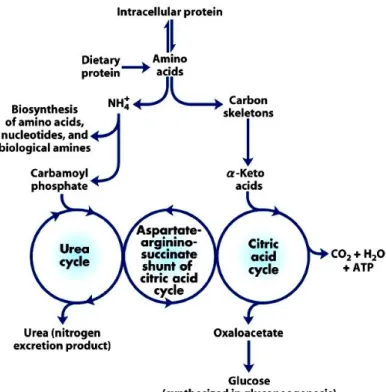
The intestine is the primary site of AAs and peptide absorption. AAs are transported into enterocytes by facilitated diffusion or specific transport systems with sodium as a carrier, that take up AAs against a concentration gradient (Stevens et al. 1984; Lerner 1987). The transport systems are specific to groups of AAs rather than to individual AAs. In addition to the AA transporters, enterocytes have an active transport system for di- and tri-peptides, independent of the one for free AAs (Adibi et al. 1976; Webb 1990). AAs arising from dietary protein digestion or from protein degradation are constantly re-synthesized or used in protein turnover. Unrequired or damaged proteins are targeted for destruction. Some AAs are used for protein synthesis in the liver (constitutive or exported plasma proteins). The branched chain amino acids (BCAA) are, along with other AAs, required for the stimulation of total liver protein synthesis (Anthony et al. 2001b). However, when protein intake increased, no change in protein synthesis rate was observed despite an increase in the tissue protein pool (Chevalier et al. 2009). The effect on protein breakdown remained unknown. Furthermore, the AA surplus may be used as metabolic fuel or converted to other compounds. The main fates of the carbon skeletons remaining from AA deamination may be their use for cellular respiration, fatty acids or ketone bodies synthesis, or gluconeogenesis
whereas the amino groups are used for the biosynthesis of AAs, nucleotides and biological amines or are converted to urea for excretion through the urea cycle (Figure 1) (Shambaugh 1977; Brosnan 2003). Ureogenesis is an important process to protect the body from potentially toxic ammonium (Shambaugh 1977; Dimski 1994; Ding et al. 1997).
1.2. Interorgan amino acid metabolism
The functions of interorgan AA traffic are to maintain the relatively constant extracellular AA concentration in which tissues are bathed and to provide AAs for protein synthesis and those used in specific functions. AAs serve as the building blocks for proteins and some of them that exceed the body’s needs for protein synthesis undergo oxidative degradation through transamination and desamination (Brosnan 2003). The liver is the main organ where many different proteins are synthesized (Brosnan 2003). For example, albumin is synthesized in the liver of a healthy adult human and 20g/day is secreted. This albumin is catabolized in the peripheral tissues, suggesting that about 20g of AAs are made available each day as a result of albumin catabolism (Maxwell et al. 1990).
The rate of AA uptake by tissues or organs depends on the activity of several transporters. In mammals, there are different AA transporters which are referred to as transport systems. Free AAs are transported across membranes through Na+-independent (facilitated transport) or Na+-dependent (secondary active transport) systems. Nomenclature of AA transport systems have letter designations based on their preferred AA substrates and the presence or absence of the requirement for sodium ion activation and co-transport (Mailliard et al. 1995), as shown in Table 1.
Table 1. AA transport systems of mammalian cells (Hyde et al. 2003)
(ai) Neutral-amino-acid transporters: sodium-dependent System Protein Gene Amino acid substrates
(one-letter code)
Notes A SNAT1 SLC38A1 Gly, Ala, Ser, Cys, Gln, Asn,
His, Met, Thr, Me-AIB, Pro, Tyr, Val
Short-chained-neutral-amino-acid transport. SNAT2 SLC38A2 Gly, Pro, Ala, Ser, Cys, Gln,
Asn, His, Met, Me-AIB
Sensitive to low pH. Ubiquitous expression. SNAT3 SLC38A4 Gly, Pro, Ala, Ser, Cys, Asn,
Met, His, Lys, Arg
SNAT3 may also function as a Na+-independent cationic amino acid transporter.
ASC ASCT1 SLC1A4 Ala, Ser, Cys
ASCT2 SLC1A5 Ala, Ser, Cys, Thr, Gln
High-affinity short-chain-amino-acid exchanger. Ubiquitous expression.
Bo ASCT2 SLC1A5 Ala, Ser, Cys, Thr, Gln, Phe, Trp, Tyr
Broad substrate specificity. Expressed on apical surface of many epithelia.
BETA GAT1 SLC6A1 GABA
GAT2 SLC6A13 GABA, betaine, Pro, -Ala transporters.
Widely expressed Cl--dependent GABA, betaine and
taurine
GAT3 SLC6A11 GABA, betaine, Tau
BGT1 SLC6A12 GABA, betaine
TAUT SLC6A6 Tau
Gly GLYT1 SLC6A9 Gly, sarcosine
GLYT2 SLC6A5 Gly, sarcosine Na+- and Cl--dependent high-affinity glycine transport. Expressed in brain and some non-neural tissues.
IMINO – – Pro Na+-dependent epithelial proline transporter, inhibited by
Me-AIB.
N SN1 SLC38A3 Gln, Asn, His Li+-tolerant transport of Gln, Asn and His. H+ antiport. SN2 SLC38A5 Gln, Asn, His, Ser, Gly Li+-intolerant variants described
Nm – – Gln, Asn, His
Nb – – Gln, Asn, His
PHE – – Phe, Met Brush-border transporter for Phe and Met
PROT PROT SLC6A7 Pro Proline transporter in central nervous system.
(aii) Neutral-amino-acid transporters: sodium-independent
System Protein Gene Amino acid substrates Notes
asc Asc1 SLC7A10 Gly, Ala, Ser, Cys, Thr Small neutral AA exchanger.
Asc2 Gly, Ala, Ser, Thr
imino PAT1/LYAAT1 SLC36A1 Pro, Gly, Ala, -Ala, GABA, Me-AIB
PAT2/LYAAT2 SLC36A2 Pro, Gly, Ala, -Ala, GABA Me-AIB
H+-coupled transport of small neutral amino acids.
Inhibited by Me-AIB.
L LAT1 SLC7A5 His, Met, Leu, Ile, Val, Phe, Tyr, Trp, Gln
LAT2 SLC7A8 Ala, Ser, Cys, Thr, Asn, Gln, His, Met, Leu, Ile, Val, Phe, Tyr, Trp
Ubiquitously expressed exchanger for large hydrophobic amino acids.
T TAT1 SLC16A10 Phe, Tyr, Trp Aromatic-amino-acid transporter. H+/monocarboxylate
transporter family – insensitive to pH, however. (bi) Anionic-amino-acid transporters: sodium-independent
System Protein Gene Amino acid substrates Notes
X-AG EAAT1 SLC1A3 Glu, Asp Widespread Glu and Asp transporter. K
+
antiport.
EAAT2 SLC1A2 Glu, Asp Substrate-dependent uncoupled anion flux.
EAAT4 SLC1A6 Glu, Asp
EAAT5 SLC1A7 Glu, Asp
(bii) Anionic-amino-acid transporters: sodium-independent
System Protein Gene Amino acid substrates Notes
x-c xCT SLC7A11 Glu, Cystine Electroneutral Glu/cystine exchanger.
– XAT2 – Glu, Asp Non-functional upon 4F2hc/rbAT heavy-chain
co-expression.
Predicted to associate with a novel glycoprotein. (ci) Cationic-amino-acid transporters: sodium-dependent
System Protein Gene Amino acid substrates Notes Bo,+ ATB(o,+) SLC6A14 Lys, Arg, Ala, Ser, Cys, Thr,
Asn, Gln, His,
Blastocysts and possibly brush-border membrane. Met, Ile, Leu, Val, Phe, Tyr, Trp Broad specificity for neutral and cationic amino acids.
Accepts BCH.
y+L y+LAT1 SLC7A7 Lys, Arg, Gln, His, Met, Leu Na+-dependent cationic/neutral-amino-acid exchanger. y+LAT2 SLC7A6 Lys, Arg, Gln, His, Met, Leu,
Ala, Cys
Electroneutral. (cii) Cationic-amino-acid transporters: sodium-independent
System Protein Gene Amino acid substrates Notes bo,+ b(o,+)AT SLC7A9 Lys, Arg, Ala, Ser, Cys, Thr,
Asn, Gln, His,
Met, Ile, Leu, Val, Phe, Tyr, Trp, Cystine
Broad-specificity cationic- and neutral-amino-acid exchanger.
y+ Cat-1 SLC7A1 Arg, Lys, His
Cat-2 SLC7A2 Arg, Lys, His
Cat-3 SLC7A3 Arg, Lys
Cat-4 SLC7A4 Unknown
Cationic-amino-acid (and Na+-dependent
neutral-amino-acid) transport. Variable degree of trans-stimulation.
The interconversion of AAs trough transamination is an important process in the transport of ammonium and to maintain acid-base balance (Wu 2009). These processes converge on the central catabolic pathways, with the removal of the -amino groups from the carbon skeleton. This was catalyzed by enzymes called aminotransferases or transaminases and then the carbon skeletons of most AAs found their way to the citric acid cycle (Metzler et al. 1982). Almost all AAs can be metabolized in the liver and it is the organ with urea cycle (Brosnan 2003).
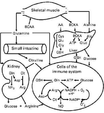
Several non-essential AAs, including glutamine, glutamate and aspartate, are oxidized by epithelial cells in the mammalian small intestine and they do not enter the portal vein (Stoll et al. 1998; Wu 1998). The small intestine uptakes the glutamine as the major fuel and nitrogenous products derived from glutamine metabolism are released into the portal vein. These include the alanine and proline which are metabolized by the liver. Moreover, the output from the small intestine also includes citrulline which is taken up and converted to
arginine in the kidney (Wu 1997; Wu and Morris 1998) (Figure 2). The kidney plays a major role in the interorgan metabolism of citrulline, arginine, glycine, and glutamine. It takes up glycine and releases serine. In addition, the kidney uptakes glutamine which is the substrate for urinary ammonia production and it contributes in this way to the maintenance of acid-base homeostasis (Brosnan 2003) (Figure 2).
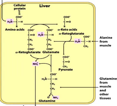
Glutamate and glutamine play critical roles in these transaminations. In the cytosol of hepatocytes, the amino groups from most AAs are transferred to -ketoglutarate to form glutamate. The glutamate serves as an amino group donor for biosynthetic pathways or excretion pathways that lead to the elimination of nitrogenous products. Glutamate is then transported from cytosol into mitochondria, where the amino group is removed to form NH4+ via oxidative deamination promoted by L-glutamate dehydrogenase (Figure 3). The -ketoglutarate formed from glutamate deamination can be used for energy production in the citric acid cycle and for glucose synthesis in the liver and kidney (Brosnan 2003). The ammonium ion is converted to urea for excretion through the urea cycle, which is distributed between the mitochondrial matrix and cytosol of hepatocytes (Shambaugh 1977).
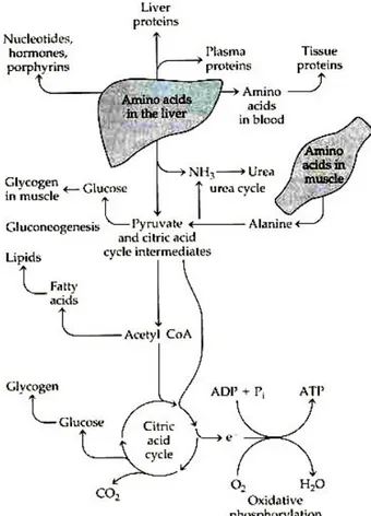
The AAs in the liver can be transaminated and degraded to other citric acid cycle intermediates and acetyl CoA or oxaloacetate which can in turn be oxidized for energy supply or converted to glucose or fat (Figure 4). However, the oxidation of AAs produces much more ATP than the liver could actually use. Therefore, it seems that the carbon skeletons of these AAs are not completely oxidized in the liver and are converted to glucose via gluconeogenesis even in the fed state (Jungas et al. 1992). During starvation, hepatic gluconeogeneis plays an important role in the production of glucose for the brain and other glucose dependent organs and the AAs from the muscle proteolysis are the major precursors for this process (Brosnan 2003). The liver also synthesizes glutathione from glutamate, glycine, and cysteine for use by extrahepatic cells (including immunocytes) and tissues (Figure 2) (Wu 2009).
Given that the bulk of the body’s protein is in the form of muscle proteins, this tissue will apparently play a critical role in the interorgan AA metabolism. The skeletal muscle is the major organ for the catabolism of BCAA and released both alanine and glutamine from BCAA
and -ketoglutarate. This alanine is taken up by the liver and converted to glucose (the glucose-alanine cycle). Thus alanine is one of the important molecules in the transport of amino groups to the liver without increasing blood ammonia concentrations (Figure 2 and 4) (Brosnan 2003; Wu 2009)
2. Protein metabolism and regulation by nutritional conditions
Protein and AA metabolism is a large, dynamic and regulated process that accomplishes a variety of physiological functions. In adult humans, some 300 g of new protein is synthesized per day for maintenance, and an equivalent amount of protein is degraded to their constituent AAs. In eukaryotes, the half-lives of proteins vary from minutes to many days (Goldberg and St John 1976; Mayer and Doherty 1986). For example, in the rat liver, proteins might turn over once every one to two days, while some regulatory enzymes have half-lives only 15 minutes. Furthermore, the more stable proteins, such as actin and myosin in skeletal muscle, might turn over once every one or two weeks. In human, hemoglobin can remain for the entire lifetime of an erythrocyte (3 months) (Lecker et al. 1999). The overall process of protein synthesis and protein degradation is referred to as protein turnover. The rates of protein turnover may vary depending on the intracellular and extracellular environmental conditions, including the availability and balance of nutrients to which cells are exposed, and the hormones and the peptide factors that bind to receptors on cell surfaces or within the cell. It has been established that alterations in dietary macronutrient intake greatly affected the balance between tissue protein synthesis and protein degradation (Darmaun 1999). Since AAs serve as the currency of protein metabolism, they are hydrolyzed from protein via proteolysis systems and serve as the building blocks for new protein synthesis. Therefore, protein cell homeostasis is maintained by a precise balance between the overall rates of synthesis and degradation (Lecker et al. 1999).
In mammals, changes in nutrient availability induce changes in the levels of hormones to adapt the metabolism. Protein synthesis requires both AAs, both as precursors, and a substantial amount of metabolic energy. Maintaining the essential AA supply is
necessary to maintain the optimal rate of protein synthesis in both the liver and skeletal muscle. Deprivation of even a single essential AA causes a decrease in the cellular protein synthesis by inhibition of the initiation phase of mRNA translation (Kimball 2002).
There is evidence that protein synthesis was stimulated in muscle and in liver by 38 and 41%, respectively when rats were fed a diet containing 20% protein whereas no change was observed in rats fed no added protein (Yoshizawa et al. 1998). Moreover, plasma insulin concentrations were the same in rats fed either diet, suggesting that feeding-induced changes in plasma insulin are not sufficient to stimulate protein synthesis. Both dietary protein and insulin may be required to stimulate translation initiation (Yoshizawa et al. 1998). However, Kimball reported that insulin alone can activate the translation at the initiation step (Kimball and Jefferson 2006a). Moreover, AAs, especially BCAA, stimulated the protein synthesis in primary hepatocytes (Dubbelhuis and Meijer 2002; Ijichi et al. 2003) whereas in the livers of rats fed a high protein diet for 2 weeks, the protein synthesis rate was decreased (Chevalier et al. 2009) A slight inhibition of synthesis rates after the high protein diet was observed in the kidney while protein synthesis rates were significantly increased in stomach and skin. These results suggested that the adaptation to high protein diet was tissue specific (Chevalier et al. 2009). Furthermore, the reduction of protein levels in diets (20.7%, 16.7% or 12.7%) decreased the protein synthesis in the pancreas, liver, kidney and muscle in piglets receiving these diets for 2 weeks (Deng et al. 2008). Muscles play a role as a protein reservoir. Skeletal muscle is also the main organ of BCAA catabolism. There is evidence that carbohydrate restricted-with high protein diet, during 7 days, stimulated muscle protein synthesis (Harber et al. 2005). In humans, increasing protein ingestion resulted in an increase in protein synthesis (up to 20%) and a decrease in protein breakdown after adaptation for 7 days to higher protein intake (Motil et al. 1981; Hoerr et al. 1993; Gibson et al. 1996; Fereday et al. 1998; Forslund et al. 1998; Harber et al. 2005). However, the acute protein intake resulted in only slight increase of protein synthesis (around 8%) and greater decrease in its breakdown (Gibson et al. 1996; Forslund et al. 1998; Cayol et al. 1997; Fereday et al. 1998). Essential AAs and BCAA (especially leucine) specifically modulate protein synthesis by activating the initiation of translation (Anthony et al. 2001a; Anthony et al. 2001b; Yoshizawa 2004; Crozier et al. 2005). In rats in vivo, infusion of the BCAA
stimulated muscle protein synthesis and essential AAs maintained this effect (Kobayashi et al. 2006).
Protein degradation is also regulated by nutrition (Kettelhut et al. 1988). High amino acid concentrations and insulin are the main inhibitors of protein degradation, whereas glucagon and low concentrations of amino acids are the principal stimulators (Gelfand and Barrett 1987; Flakoll et al. 1989; Mortimore et al. 1989; Kadowaki et al. 1992; Blommaart et al. 1997; Boirie et al. 1997; Balage et al. 2001; Kanazawa et al. 2004; Waterlow 2006; Capel et al. 2008). Under acute feeding, proteolysis is inhibited while a chronic increase in protein intake, induced proteolysis in the fed state (Price et al. 1994; Forslund et al. 1998). In the post-absorptive state, whole body protein degradation varies only very slightly (Price et al. 1994; Forslund et al. 1998). In specific tissues, only a few studies have examined the response of protein degradation to increased protein intakes (Taillandier et al. 1996; Bolster et al. 2002; Harber et al. 2005). Liver proteolysis is known to be inhibited by insulin (Duckworth et al. 1994; Hamel et al. 1997; Bennett et al. 2000; Bennett et al. 2003; Kanazawa et al. 2004) and stimulated by glucagon (Schworer and Mortimore 1979; Mortimore et al. 1989). AAs also act as a negative feed- back regulator for proteolysis in the perfused rat liver (Poso et al. 1982; Mortimore et al. 1989; Kadowaki et al. 1992; Miotto et al. 1992) and isolated hepatocytes (Mortimore and Schworer 1977; Seglen et al. 1980). In muscle, numerous publications have described the stimulation of muscle protein breakdown in response to fasting, followed by an inhibition after re-feeding with a normal diet. Muscle protein breakdown is activated in response to one or more days of starvation (Medina et al. 1991; Wing and Banville 1994; Wing et al. 1995). Several AAs also have a direct regulatory affect on proteolysis: Leu, Gln, Tyr, Phe, Pro, Met, Trp and His in the liver and Leu in the skeletal muscle (Kadowaki and Kanazawa 2003; Meijer and Dubbelhuis 2004; Oshiro et al. 2007).
2.1. Protein synthesis
Protein synthesis is one of the most complex biosynthetic processes. In eukaryotes, almost 300 macromolecules cooperate to synthesize polypeptides. These macromolecules consist of over 70 different ribosomal proteins, 20 or more enzymes to activate the AA precursors, a dozen or more auxiliary enzymes and other protein factors for the initiation, elongation and termination of polypeptides, perhaps 100 additional enzymes for the final processing of different proteins and 40 or more kinds or transfer and ribosomal RNAs.
First, the production of polypeptides follows the process of transcription, the production of messenger RNA (mRNA) from a gene’s nucleotide sequence which involves several steps. It consists of transcription initiation, elongation, RNA–processing reactions, e.g. capping and splicing, and termination. Second, the transcription is followed by the transport of mRNA to the cytosol where mRNA is decoded into protein by the translation. The mRNA transported the genetic code into the cytosol in the form of codon which is a triplet of nucleotides that codes for a specific AA. A specific first codon in the sequence of mRNA establishes an open reading frame. The reading frame is set when translation of an mRNA molecule begins and is maintained as the synthesis machinery reads sequentially from one triplet to the next. Several codons serve special functions such as the initiation codon, AUG, which signals the beginning of a polypeptide in all cells, in addition to coding for methionine residues of polypeptides. Moreover, there are the termination codons (also called stop codons or nonsense codons), UAA, UAG and UGA, which normally signal the end of polypeptide synthesis.
The three major stages of translation are: 1. initiation
2. elongation 3. termination
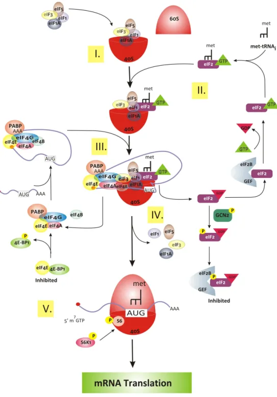
2.1.1. Initiation
(Figure 5)In eukaryotes, the initiation begins by the assembly of a complex from initiator methionyl-transfer RNA (met-tRNAi), 40S and 60S ribosomal subunits, with the aid of eukaryotic initiation factors (eIFs), into an 80S ribosome at the initiation codon of mRNA.
In the first step of translation initiation, the eukaryotic initiation factor 2 (eIF2) binds GTP and met-tRNAi, selected from the pool of tRNAs, to form the ternary complex (eIF2●GTP●met-tRNAi) and then binds to the 40S ribosomal subunit with other eIFs (eIF1, eIF1A, eIF3 and eIF5) to form the 43S preinitiation complex (Kapp and Lorsch 2004) (Figure 5). The eIF1, 1A and 3 promote the dissociation of 80S ribosomes (Kapp and Lorsch 2004). The eIF4F, including eIF4A, eIF4B, eIF4E and eIF4G, bind mRNA by a mechanism involving the initial recognition of the m7G cap at 5’-end of mRNA by eIF4E (Gingras et al. 1999). Then, mRNA binds to the 43S complex by the association of eIF3 and eIF4G (Gross et al. 2003; Prevot et al. 2003). The 43S complex scans along the mRNA in a 5’ to 3’ direction towards the initiation codon, base-paired with the anti-codon of met-tRNAi (Lopez-Lastra et al. 2005). Moreover, eIF1 interacts with the eIF1A to promote scanning of the start codon (Pestova and Kolupaeva 2002). There is another factor, the poly (A) binding protein (PABP), facilitating mRNA binding to the 43S complex. PABP interacts with eIF4G to circularise mRNA by linking the 5’ cap and poly(A) tail in a “closed loop” (Figure 5). This association stimulates mRNA binding to the 43S complex, by enhancing eIF4F binding to the capped 5’ end of mRNA (Kahvejian et al. 2005). Initiation ends when the initiation factors are released from the complex and the 60S ribosomal subunit joins to form the 80S ribosomal (initiation complex) and leave met-tRNAi in the ribosomal P site (Lopez-Lastra et al. 2005).
2.1.2. Elongation
(Figure 6)The elongation phase involves three distinct steps that are repeated many times during the formation of a polypeptide chain. The order of AAs is specified by the sequence of codons in the mRNA. Moreover, each AA is specific to its cognate tRNA to form amino acyl-tRNA (aa-acyl-tRNA) for which there is one aa-acyl-tRNA synthase per aa-acyl-tRNA pair (Guth and
Francklyn 2007). The elongation cycle requires the eukaryotic elongation factors (eEFs), including eEF1and eEF2, to catalyse this process. First, the eEF1A picks the aa-tRNA in the presence of GTP, and then the aa-tRNA●eIF1A●GTP complex enters the empty A-site on a ribosome. The anticodon of the incoming aa-tRNA needs to be matched against the mRNA codon positioned in the A-site. As the three bases in the codon can be arranged in 64 different combinations, the translational machinery must be able to select the aa-tRNA carrying the matching anticodon. When the correct three-base anticodon forms a complementary base pair with the codon on mRNA, the GTP is hydrolyzed leading to eEF1A●GDP dissociating from aa-tRNA. The resulting of eEF1A●GDP binds to eEF1B complex which facilitates exchange of GDP to GTP on eEF1A. The eEF1A●GTP now is ready to accept the next aa-tRNA. In the second step, a peptidyl transferase reaction catalysed by the ribosome itself occurs immediately after the accommodation of the correct aa-tRNA in the ribosomal A-site. The growing polypeptide in the ribosomal P-site is linked to the new AA in the A-site via a peptide bond. The reaction leaves an empty tRNA in the ribosomal P-site and the new peptidyl-tRNA in the A-site. The last step is the translocation, which promotes the ribosome’s translocation along the mRNA by the length of one codon. Translocation is catalysed by the eEF2 and subsequent GTP hydrolysis. After the translocation, the ribosome is in the position of having an empty tRNA in the E site, the peptidyl-tRNA in the P site, and the next codon of mRNA in the A site, available for interaction with a new aa-tRNA. These reaction steps are repeated until the ribosome encounters an in-frame stop-codon. At this point, the translation is terminated (Kasinath et al. 2006; Frank et al. 2007; Groppo and Richter 2009; Ling et al. 2009).
2.1.3. Termination
(Figure 6)The final step is termination which involves the release of the polypeptide chain from mRNA. The three stop codons (UAA, UAG and UGA), the eukaryotic release factors (eRFs) and one GTP are required. The eRF1 recognizes one of three stop codons and binds to the ribosome in the place of a tRNA (Kisselev et al. 2003). This event along with binding of the eRF3, facilitates eRF1 stop codon recognition and stimulates GTP hydrolysis to release the polypeptide chain (Salas-Marco and Bedwell 2004; Fan-Minogue et al. 2008).
2.1.4. Regulation of translation at the initiation step
Translation is an important regulatory step in cellular protein synthesis. It is not only a metabolic pathway, but also a signaling pathway because most regulation of protein synthesis occurs at translation. In addition, the dominant mechanism of control of global protein synthesis occurs via the phosphorylation/dephosphorylation of the translation components, primarily of initiation and elongation factors (Sonenberg and Hinnebusch 2009). It is established that AAs are important factors in the regulation of intracellular signal transduction pathways involved in the control of translation. The essential AAs have been found to regulate signaling which modulate mRNA translation through the binding of met-tRNAi to the 40S ribosomal subunit to form the 43S preinitiation complex and the binding of mRNA to the 43S preinitiation complex (Kimball and Jefferson 2005).
The first regulated step of translation at the initiation step involves the binding of met-tRNAi to the 40S ribosomal subunit to form the 43S preinitiation complex by the phosphorylation of the -subunit of eIF2. In the later step of initiation, the bound GTP of eIF2 is hydrolyzed to GDP, and the eIF2●GDP binary complex is released from the ribosome. To reform the active ternary complex, eIF2 binds to met-tRNAi, and the GDP is exchanged to GTP. This guanine nucleotide exchange reaction is catalyzed by a second initiation factor eIF2B. The mechanism for regulating eIF2B activity is through phosphorylation of the -subunit of eIF2. The phosphorylation of eIF2 converts it from a substrate into a competitive inhibitor of eIF2B, effectively sequestering eIF2B into an inactive complex. Because translation of essentially all mRNA begins with met-tRNAi, the phosphorylation eIF2 results in a decline in the synthesis of almost all proteins (Kimball 2002).
The second regulated step in translation initiation involves the binding of mRNA to 43S ribosomal subunit, a reaction mediated by the initiation factor referred to as eIF4F. The active complex of eIF4F at 5’ cap of the mRNA is regulated by the reversible interaction of eIF4E and one of its binding proteins (4E-BP1, 4E-BP2 or 4E-BP3) (Raught et al. 2000). For example, the binding of 4E-BP1 and eIF4E to eIF4G is mutually exclusive. Because the binding
of eIF4E to the 40S ribosomal subunit occurs through its interaction with eIF4G, binding of 4E-BP1 to eIF4E prevents binding of the eIF4E-mRNA complex to the 40S ribosomal subunit. The association of 4E-BP1 with eIF4E is controlled by phosphorylation of 4E-BP1. The phosphorylated 4E-BP1 does not bind to eIF4E. Thus the decrease of the association of eIF4E with eIF4G represses protein synthesis (Kimball 2002).
The latter regulated step involves the p70 ribosomal S6 protein kinase (S6K1), which phosphorylates the ribosomal protein S6 (S6), a component of the 40 ribosomal subunit. The function of S6 phosphorylation is thought to be in promoting the translation of the set of mRNAs that posses a 5’ terminal oligopyrimidine tract (5’-TOP mRNAs). The 5’ TOP encodes the translation machinery components such as ribosomal proteins and other translation factors and up–regulates their translation, thus the phosphorylation of S6 probably leads to an increase in the capacity of intracellular protein synthesis (Meyuhas 2000; Proud 2004).
2.2. Protein Degradation
The regulation of protein metabolism is essential for proper cellular function, and is a balance between synthesis and degradation. All cells possess multiple pathways for protein degradation. However, in the liver, there are at least two major degradation pathways: lysosomal system and the ubiquitin proteasome pathway.
2.2.1. Lysosomal Pathway
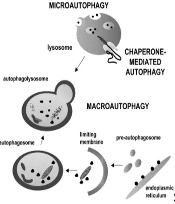
Long-lived proteins and some organelles are believed to be degraded within lysosomes (Dunn 1994; Klionsky and Ohsumi 1999). These lysosomes contain several acid-optimal proteases, including cathepsins B, H and D, and many other acid hydrolases (Lecker et al. 1999). There are at least three different pathways for lysosomal protein degradation: Cvt (cytosol to vacuole targeting pathway), Vid (vacuolar import and degradation pathway) and autophagy (Yang et al. 2005). Autophagy is a ubiquitous physiological process that occurs in all eukaryotic cells (Reggiori and Klionsky 2002). There are three primary forms of
autophagy: macroautophagy, microautophagy and chaperone-mediated autophagy (Klionsky 2005). Macroautophagy is the most prevalent form. It involves the formation of cytosolic double membrane vesicles. Initially a “C” shape double membrane structure appears in the cytoplasm, and then both ends of this membrane grow and close to form the vesicle, termed an autophagosome, that wraps the bulk of the cytoplasm and some organelles. Then, the autophagosome fuses with the lysosomal membrane, resulting in an inner vesicle (autophagic body) entering the lysosome/vacuole. The autophagic body is delivered into the lumen of the degradative compartment to degrade and carry constituent components that can be recycled (Yoshimori 2004; Klionsky 2005). Microautophagy has not been well characterized in mammalian cells. In this pathway, the lysosomal membrane itself invaginates, and then finally pinches off to form an internal vacuolar vesicle containing the materials derived from cytoplasm (Mortimore et al. 1988). Chaperone-mediated autophagy involves direct translocation of the targeted proteins (only cytosolic proteins) across the lysosomal membrane without vesicle-mediation (summarized in Figure 7) as performed in the other two processes. It is also a secondary response to starvation (Massey et al. 2004).
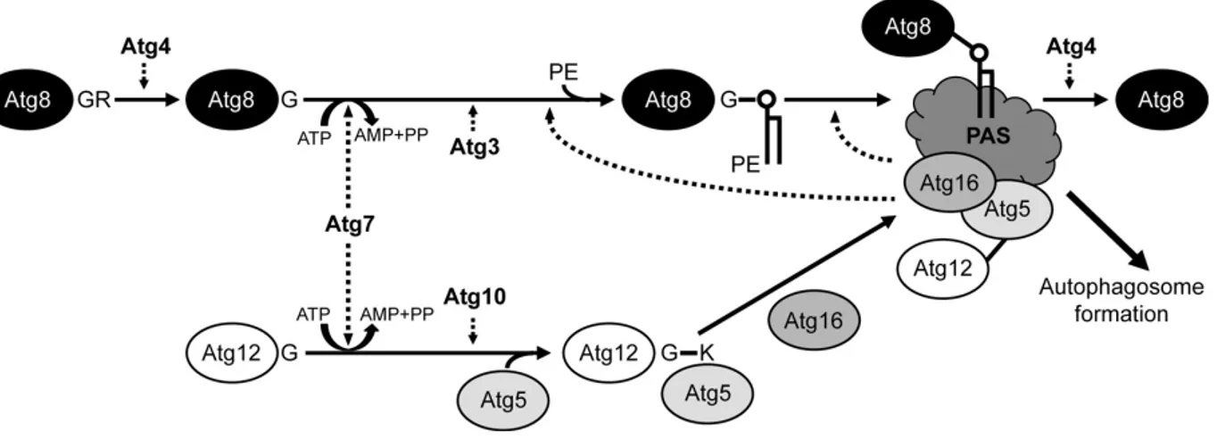
Autophagy is basically a non-specific degradation process. However, it may select to eliminate its targets in some cases, such as injured or excrescent peroxisomes, endoplasmic reticulum (ER) and mitochondria (Elmore et al. 2001). Molecular genetic studies in yeast have identified some of the components required for autophagosome formation (Klionsky et al. 2003). The autophagy-related genes and the products of these genes are named ATG and Atg, respectively. Around 31 genes involved in the autophagy has been identified (Suzuki et al. 2007), among of which 18 ATG genes, mostly conserved in the higher eukaryotes i.e. mammals (Yoshimori and Noda 2008), are essential for the autophagosome formation (Noda et al. 2009). The Atg1 is a serine/threonine protein kinase and its activity is essential for autophagy even though its substrate is not known (Yang et al. 2005). Moreover, Atg13 is hyperphosphorylated under growing conditions and dephosphorylated form can bind to Atg1 to simulated its kinase activity, which is a good candidate to induce autophagy, although no mechanism has been reported (Kamada et al. 2000). The other Atg proteins, involved in autophagosome formation are the two ubiquitin-like conjugation systems: Atg8-phosphatidylethanolamine (PE) and Atg12-Atg5. In fact one half of Atg genes are involved in these conjugation systems and these two systems are well conserved in eukaryotes. The
Atg8-PE system includes 4 Atg proteins: Atg3, Atg4, Atg7 and Atg8. The processed Atg8 is activated by Atg7, an ubiquitin-activating E1 like enzyme, and then is transferred to Atg3, an ubiquitin-activating E2-like enzyme. Finally the C-terminal glycine of Atg8 is conjugated to the amino group of PE. The Atg8-PE is deconjugated by Atg4 (Ichimura et al. 2000; Noda et al. 2009) (Figure 9). The model of Atg12-Atg5 conjugation comprised 5 proteins: Atg5, Atg7, Atg10, Atg12 and Atg16. Atg12 is activated by Atg7 as in the Atg8, and is then transferred to Atg10, an ubiquitin conjugating E2-like enzyme (Mizushima et al. 1998; Shintani et al. 1999). In the final step, the C-terminal glycine in Atg12 is conjugated to the side chain of Lysine-149 of Atg5 and the Atg12-Atg5 further form the complex with a multimeric protein, Atg16 with the Atg5 noncovalent interaction (Mizushima et al. 1999; Mizushima et al. 2003) (Figure9). It seems that this ubiquitin-like system is essential to autophagosome formation and is a constitutive process, since the formation of the Atg12-Atg5 conjugate is not dependent upon the starvation or other autophagy-inducing conditions (Mizushima et al. 1999).
2.2.2. The ubiquitin Proteasome System
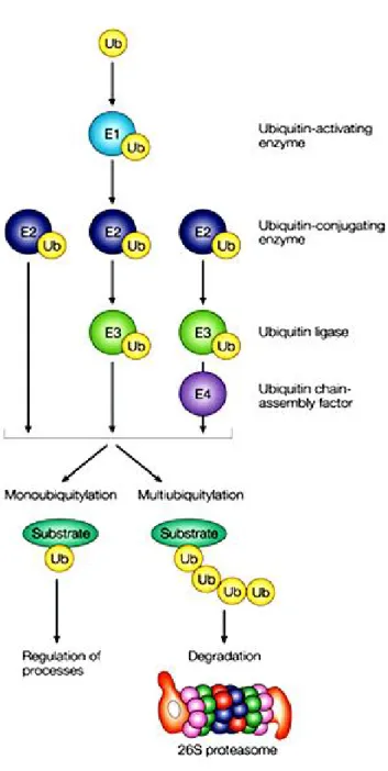
The ubiquitin proteasome pathway is the other major piece of cytosolic protein degrading machinery responsible for the breakdown of most short- and long-lived proteins in mammalian cells (Rock et al. 1994) and plays an important role in the control of the degradation of specific proteins (Kadowaki and Kanazawa 2003). This pathway includes two main steps. The first step is the covalent attachment of the polyubiquitin chain to the substrate followed by the specific recognition of this signal and degradation of the ubiquitinylated protein by the 26S proteasome (Attaix et al. 2001).
Ubiquitin is a 76-amino-acid globular protein that is highly conserved throughout eukaryotes, with only three amino-acid changes from yeast to humans (Weissman 2001). Ubiquitination is a multiple step process (Ciechanover et al. 2000; Jesenberger and Jentsch 2002). Firstly, ubiquitin is activated to a high energy thiol ester intermediate by the ubiquitin-activating enzyme (E1), then E1 transfers ubiquitin to one of the ubiquitin conjugating enzymes (E2), which also forms a thiol ester linkage between the active site