HAL Id: hal-01270688
https://hal.sorbonne-universite.fr/hal-01270688
Submitted on 8 Feb 2016
HAL is a multi-disciplinary open access
archive for the deposit and dissemination of
sci-entific research documents, whether they are
pub-lished or not. The documents may come from
teaching and research institutions in France or
abroad, or from public or private research centers.
L’archive ouverte pluridisciplinaire HAL, est
destinée au dépôt et à la diffusion de documents
scientifiques de niveau recherche, publiés ou non,
émanant des établissements d’enseignement et de
recherche français ou étrangers, des laboratoires
publics ou privés.
Physiological and Pathophysiological Insights of Nav1.4
and Nav1.5 Comparison
Gildas Loussouarn, Damien Sternberg, Sophie Nicole, Céline Marionneau,
Francoise Le Bouffant, Gilles Toumaniantz, Julien Barc, Olfat A. Malak,
Véronique Fressart, Yann Péréon, et al.
To cite this version:
Gildas Loussouarn, Damien Sternberg, Sophie Nicole, Céline Marionneau, Francoise Le Bouffant, et
al.. Physiological and Pathophysiological Insights of Nav1.4 and Nav1.5 Comparison. Frontiers in
Pharmacology, Frontiers, 2016, 6, pp.314. �10.3389/fphar.2015.00314�. �hal-01270688�
published: 14 January 2016 doi: 10.3389/fphar.2015.00314
Edited by: Mohamed Chahine, Laval University, Canada Reviewed by: Anselm Zdebik, University College London, UK Adrien Moreau, Centre de Recherche de l’Institut Universitaire en Santé, Canada *Correspondence: Gildas Loussouarn gildas.loussouarn@inserm.fr Specialty section: This article was submitted to Pharmacology of Ion Channels and Channelopathies, a section of the journal Frontiers in Pharmacology Received:05 November 2015 Accepted:21 December 2015 Published:14 January 2016 Citation: Loussouarn G, Sternberg D, Nicole S, Marionneau C, Le Bouffant F, Toumaniantz G, Barc J, Malak OA, Fressart V, Péréon Y, Baró I and Charpentier F (2016) Physiological and Pathophysiological Insights of Nav1.4 and Nav1.5 Comparison. Front. Pharmacol. 6:314. doi: 10.3389/fphar.2015.00314
Physiological and Pathophysiological
Insights of Nav1.4 and Nav1.5
Comparison
Gildas Loussouarn
1, 2, 3*, Damien Sternberg
4, 5, 6, 7, 8, 9, Sophie Nicole
4, 5, 6, 7,
Céline Marionneau
1, 2, 3, Francoise Le Bouffant
1, 2, 3, Gilles Toumaniantz
1, 2, 3, Julien Barc
1, 2, 3,
Olfat A. Malak
1, 2, 3, Véronique Fressart
9, Yann Péréon
10, 11, Isabelle Baró
1, 2, 3and
Flavien Charpentier
1, 2, 3, 121Institut National de la Santé et de la Recherche Médicale, UMR 1087, l’Institut du Thorax, Nantes, France,2Centre National
de la Recherche Scientifique, UMR 6291, Nantes, France,3Université de Nantes, Nantes, France,4Institut National de la
Santé et de la Recherche Médicale, U1127, Paris, France,5Sorbonne Universités, Université Pierre-et-Marie-Curie, UMR
S1127, Paris, France,6Centre National de la Recherche Scientifique, UMR 7225, Paris, France,7Institut du Cerveau et de la
Moelle Épinière, ICM, Paris, France,8Assistance Publique – Hôpitaux de Paris (AP-HP), Centres de Référence des
Canalopathies Musculaires et des Maladies Neuro-musculaires Paris-Est, Paris, France,9Assistance Publique – Hôpitaux de
Paris (AP-HP), Hôpital de la Pitié Salpêtrière, Service de Biochimie Métabolique, Unité de Cardiogénétique et Myogénétique, Paris, France,10Centre Hospitalier Universitaire de Nantes, Centre de Référence Maladies Neuromusculaires Nantes-Angers,
Nantes, France,11Atlantic Gene Therapies - Biotherapy Institute for Rare Diseases, Nantes, France,12Centre Hospitalier
Universitaire de Nantes, l’Institut du Thorax, Nantes, France
Mutations in Nav1.4 and Nav1.5 α-subunits have been associated with muscular
and cardiac channelopathies, respectively. Despite intense research on the structure
and function of these channels, a lot of information is still missing to delineate the
various physiological and pathophysiological processes underlying their activity at the
molecular level. Nav1.4 and Nav1.5 sequences are similar, suggesting structural and
functional homologies between the two orthologous channels. This also suggests
that any characteristics described for one channel subunit may shed light on the
properties of the counterpart channel subunit. In this review article, after a brief clinical
description of the muscular and cardiac channelopathies related to Nav1.4 and Nav1.5
mutations, respectively, we compare the knowledge accumulated in different aspects of
the expression and function of Nav1.4 and Nav1.5 α-subunits: the regulation of the two
encoding genes (SCN4A and SCN5A), the associated/regulatory proteins and at last, the
functional effect of the same missense mutations detected in Nav1.4 and Nav1.5. First, it
appears that more is known on Nav1.5 expression and accessory proteins. Because of
the high homologies of Nav1.5 binding sites and equivalent Nav1.4 sites, Nav1.5-related
results may guide future investigations on Nav1.4. Second, the analysis of the same
missense mutations in Nav1.4 and Nav1.5 revealed intriguing similarities regarding their
effects on membrane excitability and alteration in channel biophysics. We believe that
such comparison may bring new cues to the physiopathology of cardiac and muscular
diseases.
Voltage-gated sodium channels (Nav) constitute a family of 10
members in mammals, Nav1.1 to Nav1.9 and Nax, expressed in a
large variety of tissues. In excitable cells such as striated myocytes,
they initiate action potentials that, in heart as well as in skeletal
muscles, trigger, and regulate the contraction. Because of their
key role in this function, mutations impacting their activity have
tremendous consequences. This review compares the knowledge
accumulated in different aspects of the expression and function
of Nav1.4 and Nav1.5 α-subunits, and focuses on “homologous”
mutations i.e., in the same (aligned) amino acids of the skeletal
muscle Nav1.4 and of the cardiac Nav1.5 leading to a large range
of muscular and cardiac disorders also called channelopathies.
CLINICAL DESCRIPTION OF THE MAIN
Nav1.4 AND Nav1.5 RELATED
PATHOLOGIES
Clinical Description of Nav1.4 Related
Channelopathies
Nav1.4, which is encoded by the SCN4A gene, is the
pore-forming subunit of the main sodium channel present in skeletal
muscles. Nav1.4 related channelopathies that affect skeletal
muscle excitability (
Vicart et al., 2005; Jurkat-Rott et al., 2010;
Nicole and Fontaine, 2015
) are dominant diseases classified
in two opposite groups as defined by the prevalent clinical
symptoms: muscle stiffness and hypertonia (myotonia) episodes
[non dystrophic myotonias (NDM)], and muscle weakness
resulting in paralysis episodes (periodic paralyses; PP). It should
be noted that similar clinical pattern are also associated with
other channelopathies involving chloride channels (NDM) or
calcium channels (PP). Table 1 summarizes the main classes of
TABLE 1 | Main classes of Nav1.4 skeletal muscle channelopathies (Trip et al., 2009; Raja Rayan and Hanna, 2010).
Clinical manifestations Triggers Paraclinics EMG canonical
pattern First intention treatment Most frequently mutated residues References PC Stiffness followed by weakness Paradoxical myotonia Cold Myotonia Type I (repeated short effort test)
Mexiletine T1313 (ID III-IV), R1448 (DIV S4) McClatchey et al., 1992b; Ptácek et al., 1992; Hayward et al., 1996; Featherstone et al., 1998; Bouhours et al., 2004 SCM Stiffness at exertion
(most often), permanently at rest (myotonia permanens), or acetazolamide-responsive myotonia Exertion Acetazolamide Myotonia Type III (repeated short effort test)
Mexiletine G1306 (ID III-IV), G1306A/V: myotonia fluctuans G1306E: myotonia permanens
Lerche et al., 1993; Rüdel et al., 1993; Ricker et al., 1994; Hayward et al., 1996
HyperPP Short episodes (minutes) Fasting Normal or high potassium level during episodes
Some myotonia Type IV (long effort test)
Acetazolamide T704 (DIIS5), M1592 (DIV S6)
Ptácek et al., 1991; Rojas et al., 1991; Yang et al., 1994; Iaizzo et al., 1995 HypoPP Long-lasting episodes
(hours, days)
Glucide-rich meals Rest after exercise Prolonged rest
Markedly low potassium levels during episodes
No myotonia Type V (long effort test) Acetazolamide R669, R672 (DII S4) Bulman et al., 1999; Jurkat-Rott et al., 2000; Bendahhou et al., 2001; Sternberg et al., 2001
PC, Paramyotonia Congenita; SCM, Sodium channel Myotonia; Hypo, Normo, Hyper PP, Hypo, Normo, Hyper-kalemic Periodic Paralysis.
Nav1.4-related skeletal muscle channelopathies. Detailed clinical,
electromyographic (
Fournier et al., 2004, 2006
), genetic and, in
fine, pathophysiological analyses have led to distinguish several
entities among skeletal muscle sodium channelopathies.
Nav1.4-Related non Dystrophic Myotonias
Myotonia may occur at the beginning of effort and be alleviated
(myotonia, with warm-up effect), or aggravated (paradoxical
myotonia, also named paramyotonia) by continuing effort.
Those myotonic or paramyotonic symptoms are associated
with myotonic discharges analyzed with electromyographic
investigations. NDM are opposed to dystrophic myotonias
as observed in Steinert (Myotonic Dystrophy type 1, DM1)
and PROMM (PROximal Myotonic Myopathy or Myotonic
Dystrophy type 2, DM2) diseases. Among NDM, at least two
entities differ clinically and electromyographically (
Trip et al.,
2009; Raja Rayan and Hanna, 2010
).
- Paramyotonia congenita (PC) consists of cold-induced
stiffness often associated with some weakness of face and
extremities muscles, and paradoxical myotonia; it is associated
with a progressive decrease of compound muscle action
potential (CMAP) amplitude during repetitive short efforts
test at EMG (pattern I according to Fournier,
Fournier et al.,
2004
).
- Sodium channel myotonias (SCM) regroup the remaining
dominant sodium channel-related myotonias that are not
significantly cold-sensitive or paradoxical, and do not exhibit
any change of CMAP amplitude during repetitive short efforts
test at EMG (pattern III according to Fournier); this SCM
entity was initially termed “potassium-aggravated myotonia”
as potassium load triggers myotonia in some cases. This
group was further subdivided into three types: myotonia
permanens, myotonia fluctuans, and acetazolamide-responsive
myotonia. While this classification is not used in clinics, it
has some relevance: myotonia permanens designates myotonia
that is present permanently, even at rest; myotonia fluctuans
designates myotonia that appears and disappears at some
moment, with no systematic concomitance with exertion, a
peculiar circumstance being exercise-induced delayed-onset
myotonia, that occurs some time after exertion has stopped;
acetazolamide-responsive myotonia is a treatment-related
designation, that underlines the fact that some SCM are
treatable by acetazolamide.
Nav1.4-Related Periodic Paralysis
Among PP, two distinct entities are recognized (
Raja Rayan
and Hanna, 2010
): hypokalemic periodic paralysis (HypoPP)
is characterized by a marked hypokalemia concomitant with
paralysis episodes, and, on the opposite, hyperkalemic periodic
paralysis (HyperPP) is associated with a tendency to high
blood potassium levels during the paralysis episodes. From the
electromyographic point of view, both are characterized by a
marked decrease of CMAP amplitude after a 5 min-long effort
(long effort test, also referred to as McManis test).
Overlap, borderline or mixed syndromes between PP and
NDM or between their subtypes have been reported (
McClatchey
et al., 1992a; Sugiura et al., 2003; Webb and Cannon, 2008;
Yoshinaga et al., 2012
). The age at onset is usually in early to
late childhood. Neonatal symptoms are not classically reported in
the most frequent Nav1.4 channelopathies, but dominant de novo
mutations are reported in moderate to severe neonatal clinical
presentations such as severe neonatal episodic laryngospasm
(SNEL) (
Lion-Francois et al., 2010
). In a general way, respiratory
symptoms are not common in PP and NDM, however a small
number of patients are exposed to laryngeal or diaphragmatic
weakness or myotonia that may be symptomatic.
The
minimal
prevalence
of
skeletal
muscle
Nav1.4
channelopathies has been recently estimated to be 0.4:100,000
in England (
Horga et al., 2013
) and 1.4:100,000 in France.
Mutations in Nav1.4 are mostly missense or rarely in-frame
deletions or insertions, usually with a dominant effect. However
exceptional recessive homozygosity (
Arnold et al., 2015
) and
a possible recessive compound heterozygosity (
Tsujino et al.,
2003
) have been reported in congenital myasthenic syndromes.
A small number of canonical mutations account for a significant
percentage of cases (Table 1), e.g., T1313M and R1448C/H for
PC, T704M for HyperPP, V445M (
Rosenfeld et al., 1997
), V1293I
(
Koch et al., 1995
), and G1306A/V/E for SCM, mutations of
domains II and III S4 arginines (IIS4 and IIIS4) at position 669
(R>H), 672 (R>H/G/C/S), 1132 (R>Q) (
Carle et al., 2006
), 1135
(R>H) for HypoPP (
Matthews et al., 2009
). Mutations at IIS4
arginine 675 (R>Q/G/W) result in a special type of PP with both
features of HyperPP and HypoPP (
Vicart et al., 2004
). However,
beside those frequent canonical mutations, more than 70
different missense mutations at more than 55 different positions
in different domains of the protein have been reported in the
literature as causative mutations for Nav1.4 channelopathies.
The penetrance of Nav1.4 dominant mutations is variable for
each mutation: it is high for HyperPP (T704M), PC (T1313M/A
and R1448C/H) and SCM (V445M and V1293I) mutations, and
lower, with cases of gender-related non-penetrance in pedigrees,
for some other mutations such as HypoPP mutations at position
669 or 672 (
Ke et al., 2013
).
Clinical Description of Nav1.5 Related
Channelopathies
Nav1.5, which is encoded by the SCN5A gene, is the pore-forming
subunit of the main cardiac sodium channel. Nav1.5 related
channelopathies affecting cardiac excitability are dominant
diseases that, similarly to Nav1.4 in the skeletal muscles, impact
cardiac excitability through loss of function or gain of function
effects on Nav1.5 activity. Table 2 summarizes the Nav1.5 related
channelopathies that are discussed in this review, which only
considers pathologies provoked by mutations in the same, i.e.,
aligned amino acids in Nav1.4 and Nav1.5 (cf. Part Comparison
of Missense Mutations. Are there (dys-)Functional Homologies
between Nav1.4 and Nav1.5?): the Brugada syndrome (BrS),
the long QT syndrome (LQTS), and arrhythmic dilated
cardiomyopathy. The latter includes a novel form of cardiac
arrhythmia characterized by multifocal ectopic Purkinje-related
premature contractions (MEPPCs), associated or not with atrial
fibrillation and dilated cardiomyopathy. Consequently, Table 2 is
not an exhaustive list of Nav1.5 related channelopathies.
The Brugada Syndrome
The BrS is a primary electrical disorder that is characterized by a
specific ECG pattern consisting of ST-segment elevation followed
by a negative T-wave in the right precordial leads (
Brugada and
Brugada, 1992
), indicating abnormal electrical activity in the
upper part of the right ventricle (right ventricular outflow tract).
This ECG pattern is associated with an increased risk of sudden
cardiac death (SCD) resulting from polymorphic ventricular
tachyarrhythmias or ventricular fibrillation. The incidence of
BrS in the general population is currently estimated at 1:2000
(
Antzelevitch et al., 2005
). This syndrome is 8–10 times more
prevalent in males than in females and typically manifests during
adulthood, with a mean age of SCD of 41 ± 15 years (
Antzelevitch
et al., 2005
). BrS was first described as a monogenic disease,
with autosomal dominant transmission. Although more than 20
genes have been proposed as causally related to BrS, mutations in
these genes explain less than 30% of the cases (
Crotti et al., 2012;
Nielsen et al., 2013; Antzelevitch and Yan, 2015; Veerman et al.,
2015
). Around 25% of BrS patients possess a mutation in SCN5A.
So far, ≈300 mutations in SCN5A have been reported as related
to BrS (http://www.ncbi.nlm.nih.gov/clinvar). These mutations
lead to a loss of Nav1.5 function and reduce Na
+current (I
Na).
Besides BrS, loss-of-function mutations in SCN5A also cause
isolated cardiac conduction disease and sinus node dysfunction
(
Remme et al., 2008
). ECG signs of conduction defects are also
a common feature of BrS. The other genes identified so far are
coding for proteins that are involved in generating or regulating
the sodium current (
Antzelevitch and Yan, 2015
), the L-type
calcium current (
Antzelevitch et al., 2007; Burashnikov et al.,
2010; Béziau et al., 2014
) or the transient outward potassium
TABLE 2 | Nav1.5 cardiac channelopathies. Clinical
manifestations
Triggers Paraclinics ECG canonical
pattern First intention treatment References Brugada syndrome (BrS) Ventricular fibrillation or aborted sudden cardiac death, syncope, nocturnal agonal respiration, palpitations
Rest or sleep, febrile state, vagotonic conditions ST-segment elevation on right precordial leads (V1 and V2) Implantable cardioverter-defibrillator (ICD)
Brugada and Brugada, 1992; Antzelevitch et al., 2005 Type 3 Long QT syndrome (LQTS3) Polymorphic ventricular tachycardia (torsades de pointes), ventricular fibrillation, syncopes, sudden death Rest or sleep, bradycardia, hypokaliemia, drugs prolonging QT interval
Prolonged QT interval β-blockers (with or w/o mexiletine) Wang et al., 1995; Amin et al., 2013; Giudicessi and Ackerman, 2013 Arrhythmic Dilated Cardiomyopathy Systolic dysfunction, left ventricular enlargement or dilatation. Multiple arrhythmias (text)
For MEPPC: rest (exercise suppresses PVCs) For MEPPC: Quinidine Amiodarone McNair et al., 2011; Laurent et al., 2012; Mann et al., 2012; Nair et al., 2012; Beckermann et al., 2014
This list is not exhaustive, but corresponds to pathologies caused by Nav1.5 mutations that are homologous to mutations in Nav1.4 (cf. Tables 4, 5).
If BrS was first described as a monogenic autosomal dominant
disease, there is accumulating evidence suggesting that it follows
a more complex genetic model. Concerning SCN5A, segregation
studies performed in large affected pedigrees demonstrate that
mutations in this gene are characterized by a low penetrance
(47%). In some instances, a single SCN5A mutation can lead
to different cardiac arrhythmia phenotypes in the same family
or even in a single patient (
Kyndt et al., 2001; Probst et al.,
2009
). Moreover, in some pedigrees, the absence of the familial
SCN5A mutation is observed in some affected family members,
suggesting other origins for the disease (
Probst et al., 2009
).
Recently, a genome-wide association study in a large cohort of
BrS patients has provided the proof of concept that common
genetic variants outside the SCN5A gene, e.g., SCN10A and
HEY2 loci in the reported study, may have a large effect on the
development of the disease (
Bezzina et al., 2013
). Altogether,
these data suggest that the BrS most probably involves combined
contribution of different gene variants of variable impact.
The Long QT Syndrome
Congenital LQTS is defined by several criteria including a
prolongation of the QT interval corrected for heart rate, i.e.,
QTc, to values above 440 ms in males and 460 ms in females,
due to prolonged ventricular action potentials. LQTS patients
are predisposed to ventricular polymorphic tachyarrhythmias
(torsades de pointes) that may lead to syncope, seizure or SCD
(
Amin et al., 2013
). The most common form of LQTS (also
called Romano-Ward syndrome) is an autosomic dominant
disease. Its incidence in the population worldwide is about
1:2000 (
Schwartz et al., 2009
). To date, genetic defects in 15
different genes have been found in 70% of the LQTS patients
(
Amin et al., 2013; Giudicessi and Ackerman, 2013
). Similar
to BrS, the disease penetrance is most often incomplete and
highly variable, ranging from 25 to 100% (
Priori et al., 1999;
Viadero et al., 2011
). This suggests that additional genetic and
non-genetic factors may modify the clinical manifestations of a
given LQTS-causing mutation. In recent years, numerous studies
have shown that genetic variants play an important modulatory
role in establishing the disease severity (
Amin et al., 2013
).
Among non-genetic factors, hypokalemia, or treatment with
drugs inhibiting K
V11.1 (hERG) channels as side effect are well
known to favor arrhythmic events. Sex is also a well-known
modifier of QT interval duration in LQTS. Post-adolescence and
pre-menopause women have a lower repolarization reserve than
men and are therefore more prone to QT interval prolongation
and cardiac events. This is partially explained by the effects of
sex hormones on cardiac ion channel expression and function
(
Tanabe et al., 1999; Zicha et al., 2003; Bai et al., 2005; Gaborit
et al., 2010
). The most common types of LQTS are LQTS1 (30–
35% of patients;
Ackerman et al., 2011
), LQTS2 (25–40%), and
LQTS3 (5–10%), due to defects in KCNQ1 (K
V7.1 channel),
KCNH2 (K
V11.1), and SCN5A (Nav1.5) genes, respectively.
Approximately 80% of all LQTS causal mutations are found in
these three genes. Clinically, LQTS3 is characterized by unusually
increased duration of the ST segment with a late appearance
of the T wave (
Moss, 2002
). It is often more lethal, although
less frequent, than LQTS1 and LQTS2 (
Priori et al., 2003
).
Bradycardia and pauses occurring at rest or more particularly
during sleep are often at the origin of the arrhythmias, although
fatal tachycardia-induced arrhythmias have also been reported
for a third of the patients. Most of the SCN5A mutations that were
reported to be related to LQTS3 (≈200; http://www.ncbi.nlm.
nih.gov/clinvar) alter the fast inactivation process of the channel,
leading to persistent inward sodium current causing prolonged
membrane depolarizations (
Wang et al., 1995; George, 2005
).
Arrhythmic Dilated Cardiomyopathy
Dilated cardiomyopathy (DCM) is characterized by systolic
dysfunction and, in most patients, left ventricular enlargement or
dilatation. It has been associated with the mutations of more than
30 genes, including SCN5A (
McNair et al., 2011; Hershberger
et al., 2013
). Sixteen SCN5A mutations are linked to familial or
sporadic cases with DCM with various types of arrhythmias, for
example, sinus node dysfunction, conduction delay, and atrial
and/or ventricular tachy-arrhythmias (
Amin, 2014
). Among
arrhythmic DCM, the MEPPC syndrome is a recently-described
autosomal dominant form of cardiac arrhythmia (
Laurent et al.,
2012
). It is characterized by frequent premature ventricular
contractions (PVCs) originating from various ectopic foci
along the fascicular-Purkinje system occasionally associated with
dilated cardiomyopathy, non-sustained ventricular tachycardias
(NSVTs), and sudden death. A similar phenotype was first
reported in 2003 by Bezzina and collaborators in a newborn
boy and his diseased sister, both genotyped with Nav1.5 W156X
and R225W mutations (
Bezzina et al., 2003
). Both parents and
an elder sibling, each one carrier of one or the other mutation,
were asymptomatic. For the sister, arrhythmias being the cause
of the DCM is unlikely because persistent arrhythmias were
only present for a short period. Two other mutations in Nav1.5
(R222Q and R225P) have been linked to this MEPPC syndrome
in several families (
Laurent et al., 2012; Mann et al., 2012; Nair
et al., 2012; Beckermann et al., 2014
). In these families, dilated
cardiomyopathy, when present, was suggested as a consequence
of severe primary electrical dysfunctions.
Phenotypic and Genotypic Overlap
between Cardiac and Skeletal Muscle
Sodium Channelopathies?
A recently published study shows that patients carrying (or not)
SCN4A causative mutations, present with mixed phenotype (BrS
and myotonic features) (
Bissay et al., 2015
). Although SCN4A
transcripts are present in human ventricles (
Péréon et al., 2003
),
it is difficult to understand how the gain of function SCN4A
mutations can be compared to the loss of function of SCN5A
mutations classically associated with Brugada, as discussed in the
study of Bissay and collaborators. Another study on a unique
family described four patients carrying a SCN4A mutation
and presenting with PC (
Péréon et al., 2003
), two of them
having slightly prolonged QTc interval. Both PC and LQTS3
are associated with a gain of function of Nav1.4 and Nav1.5,
respectively. In this case, it is tempting to hypothesize that the
mutant Nav1.4 channels present in the heart are responsible for
the QT prolongation. Identifying more families with such overlap
phenotypes would help to confirm the potential mutual influence
of both channels on the pathogenesis of cardiac and muscular
diseases.
CHANNEL MOLECULAR BASES AND
GENE EXPRESSION
Voltage-gated sodium channels consist of an α-subunit,
constituting the pore, and accessory β-subunits controlling the
expression and activity of the pore-forming subunit. Nav1.4, the
most frequent Nav α-subunit expressed in the skeletal muscle
is a glycosylated transmembrane protein of 1836 amino acids
and has an apparent molecular weight of approximately 260 kDa
(
George et al., 1992a,b
). Nav1.5, the most frequent Nav cardiac
α
-subunit is 2015–2016 amino acid long, depending on the splice
variants, and has a similar apparent molecular weight (
Gellens
et al., 1992; Makielski et al., 2003; Balasuriya et al., 2012
).
The SCN4A gene which encodes Nav1.4 is composed of 24
exons, all containing coding sequence. No alternative splicing
events have been reported in the literature. Nav1.5 is encoded
by the SCN5A gene, composed of 28 exons, among which
exons 2–28 contain the coding sequence. Exon 1 and part of
exon 2 encode the 5
′untranslated region (UTR) while exon 28
contains the 3
′-UTR (
Wang et al., 1996
). Intron 2 of SCN4A
and intron 3 of SCN5A are AT-AC type I introns. Intron 21
of SCN4A and intron 25 of SCN5A are AT-AC type II introns
(
Wu and Krainer, 1999
). All other introns are canonical GT-AG
introns. Unlike Nav1.4, mRNA variants of Nav1.5 are detected
in the heart of mammals, resulting from alternative splicing.
In human and murine hearts, 3
′-UTRs present two different
splicing variants, generating short or long poly-adenine tails
(
Shang and Dudley, 2005
). In addition, three rare variants were
identified only in human, corresponding to alternative splicing
of exon 28A by exons 28B–28D coding for truncated and
non-functional forms of Nav1.5 (
Shang et al., 2007
). To date, only
the mechanisms of this splice site are understood. They involve
interactions with two splicing factors, the RBM25 and LUC7F3
proteins (
Gao et al., 2011; Gao and Dudley, 2013
). Four and
three splice variants, which differ from the canonical non-coding
sequence, were described for the 5
′UTR of human and mouse
SCN5A mRNAs, respectively. These transcripts originate from
the alternative splicing encompassing exons 1 (designated 1A, 1B,
1C, and 1D) and 2, and are preferentially expressed in the heart as
compared with other tissues. Also, a neonatal isoform containing
a neonatal exon 6A of 31 nucleotides has been reported. This
form presents a difference of seven amino acids in the S3–S4 loop
of domain I, in comparison with exon 6 of the adult form (
Rook
et al., 2012
). Ventricular myocardial analysis displayed abnormal
splicing of SCN5A exon 6, characterized by over-expression of
this neonatal isoform, in one patient who present DCM with
conduction system disease (
Wahbi et al., 2013
). These findings
suggest a potential implication of mis-splicing of SCN5A in the
cardiac defect observed in this patient.
Two distinct sodium currents and channels were historically
described in skeletal muscle depending upon the developmental
and innervation status of the myofiber. SkM1, the TTX-sensitive
sodium channel expressed in innervated adult myofibers,
corresponds to Nav1.4 and is the main skeletal muscle sodium
channel (
Trimmer et al., 1989; Kallen et al., 1990
). SkM2,
the TTX resistant sodium channel expressed in immature and
denervated myofibers, corresponds to Nav1.5. In rodents, SCN4A
expression increases just after birth concomitantly with the
decrease of SCN5A gene expression (
Stocksley et al., 2005
).
SCN4A expression is not sensitive to myofiber denervation by
contrast to SCN5A gene expression, which was found to be
upregulated in response to denervation (
Awad et al., 2001
).
The SCN4A promoter contains distinct positive-acting
promoter E-box and negative-acting repressor E-box that
cooperate to yield specific gene expression in differentiated
skeletal myofibers (
Kraner et al., 1998, 1999
). It is suggested
that the muscle specificity of SCN4A expression result from
the binding of two basic helix-loop-helix transcription factors
(bHLH) of the muscle-specific MyoD family, myogenin and
MRF4 for initiation and maintenance, respectively, to the
positive-acting promoter E-box located upstream the translation
initiation site. NFI would be another major regulator of SCN4A
gene expression acting in concert with bHLH factors, especially
MRF4 (
Hebert et al., 2007
). The density of Nav1.4 is around
20 times higher at the neuromuscular junction (NMJ), in part
as a result of local mRNA accumulation (
Stocksley et al.,
2005
). Although the promoter element responsible for the
transcriptional regulation of subsynaptic genes in response to
neuronal factors at the NMJ is the N-box (TTCCGG) (
Méjat
et al., 2003
), no N-box is present within the promoter of SCN4A,
suggesting the involvement of other regulatory elements.
Similarly to alternative splicing, more is known concerning
the regulation of the SCN5A promoter, compared with SCN4A.
After the identification of a first promoter region for human
SCN5A which includes multiple positive and negative
cis-acting elements extending into intron 1 (
Yang et al., 2004
),
two other promoter regions for murine SCN5A (designated
P2 and P3) containing two distinct cardiac-specific enhancer
regions were identified and functionally characterized (
Shang
and Dudley, 2005
). In human and rat, the segment immediately
upstream of the major transcription start site contains three
GC boxes that could serve as binding sites for the Sp1
transcription factor, which are homologous to the CACC boxes
recognized in promoters of muscle restricted genes, and an
E-box binding site for bHLH factors (
Yang et al., 2004
). The
human sequence also includes an additional C-rich motif which
is recognized as a major regulator of expression in myocytes.
Further, Yang and collaborators have characterized a binding
site for GATA in intron 1, which is also known as a key
regulator of gene expression in the heart. Surprisingly, variants
in SCN10A (encoding Nav1.8 of which expression is extremely
low in heart and undetectable in atrioventricular bundle) are
associated with alterations of cardiac conduction parameters
and BrS (
van den Boogaard et al., 2014
). Van den Boogaard
and collaborators have shown that the SCN10A variants act
more likely through an alteration SCN5A gene expression
level. They have demonstrated that a cis-regulatory element
located in SCN10A gene -which is immediately located next to
SCN5A- was able to interact with both SCN5A and SCN10A
promoters. Furthermore, they described, using healthy human
heart samples, a direct correlation between the SCN5A (but not
SCN10A) expression and the presence of the rs6801957
risk-associated SNP in the SCN10A intronic enhancer. Together, their
data provided a genomic mechanism explaining how a common
genetic variant at SCN10A locus influences cardiac physiology
and predispose to Brs.
ASSOCIATED/REGULATORY PROTEINS
Although expression of Nav1.5 or Nav1.4 α-subunits alone
results in the generation of functional channels in heterologous
expression systems, it is now quite clear that the regulation of
gating and/or expression of the Nav subunits substantially relies
on a variety of other accessory/regulatory proteins (
Abriel, 2010;
Rook et al., 2012
). Interestingly, the alignment of Nav1.5 and
Nav1.4 amino acid sequences could facilitate the identification of
novel associated/regulatory proteins of the counterpart channel
subunit. In addition, this direct sequence comparison has
contributed, as for Navβ1 (
Makita et al., 1996
), and will certainly
continue to contribute to localizing the structural determinants
involved in the channel regulation. In this respect, Table 3
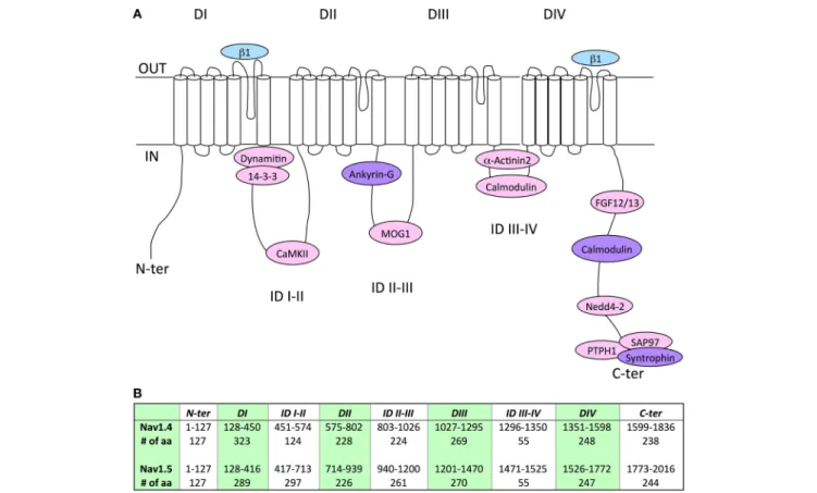
and Figure 1 recapitulate the Nav1.5 or Nav1.4 amino acid
sequences previously identified to mediate interaction with
associated/regulatory proteins, and indicates the corresponding
sequences in the other Nav α-subunit. Whereas, a number
of Nav1.5 interacting proteins, with their binding sites in the
channel subunit, have been described in the literature (see
references in Table 3), very little is known for Nav1.4 (Figure 1).
Nevertheless, it is interesting to note that the amino acid
sequence similarity obtained for some binding sites is high,
suggesting the possibility that both channel subunits share
the same associated/regulatory proteins. This is the case for
example of calmodulin, which associates with the very well
conserved (100% sequence similarity) IQ-motif on both Nav1.5
and Nav1.4 C-terminal domains (
Tan et al., 2002; Young
and Caldwell, 2005
). Most of the proteins shown to interact
with Nav1.5 on a site conserved in Nav1.4 are ubiquitously
expressed (dynamitin, 14-3-3, CaMKII, MOG-1, calmodulin,
FGF, PTPH1/PTPN3, SAP97), suggesting that an interaction with
Nav1.4 may take place in the skeletal muscle cells (
Marfatia
et al., 2001; Blair et al., 2006
). When not ubiquitously expressed,
proteins known to interact with Nav1.5 are also expressed in
skeletal muscle (α actinin2) that argue for a possible interaction
with Nav1.4 (
Foley and Young, 2014
). Conversely, weaker
sequence similarity may suggest different affinities, sites or
absence of interaction/regulation. This is the case for example
of Navβ1 for which the region within D1/S5-S6 that confers
regulation of Nav1.4 in Xenopus oocytes (
Makita et al., 1996
) is
not very well conserved in Nav1.5 (63.1% sequence similarity)
which is also regulated by Navβ1, suggesting that the structural
determinants of the interaction of Nav1.5 or Nav1.4 with
Navβ1 are different. Finally, it is striking to note the complete
absence of the PY-motif from the C-terminus of Nav1.4. This
suggests that the regulation of Nav1.4 channel internalization
and/or degradation is achieved through different mechanisms as
compared to Nav1.5 for which cell surface expression is regulated
through the ubiquitin-proteasome pathway (
van Bemmelen et al.,
2004; Rougier et al., 2005
). These mechanisms remain to be
identified.
COMPARISON OF MISSENSE MUTATIONS.
ARE THERE (DYS-) FUNCTIONAL
HOMOLOGIES BETWEEN Nav1.4 AND
Nav1.5?
Around 300 mutations in SCN5A have been identified in patients
presenting with BrS, 31% being frameshift, nonsense or
splice-site mutations, and 69% being missense or rarely in-frame
deletions/insertions (
Kapplinger et al., 2010
). When studied in
T A B L E 3 | C o mp a ri s o n o f N a v 1 .5 a n d N a v 1 .4 c h a n n e l a s s o c ia te d /r e g u la to ry p ro te in s a n d c o rr e s p o n d in g b in d in g s it e s . R e g io n N a v 1 .4 /1 .5 in te ra c ti n g p ro te in s N a v 1 .5 N a v 1 .4 % a a s e q u e n c e s imi la ri ty B in d in g s it e s M u ta ti o n s P a th o l-o g ie s R e fe re n c e s B in d in g s it e s M u ta ti o n s P a th o l-o g ie s R e fe re n c e s D I S 5 –S 6 lo o p N a vβ 1 E q u iv a le n t se q u e n c e : (2 7 8 –3 8 8 ) H K C — G K I (1 1 1 a a ) R 2 8 2 H , V 2 9 4 M , G 3 1 9 S R 2 8 2 H G 2 9 2 S K 3 1 7 N L 3 2 5 R G 3 5 1 V T 3 5 3 I D 3 5 6 N R 3 6 7 C , M 3 6 9 K R 3 6 7 H R 3 7 6 H R 3 7 6 H L 2 7 6 Q , H 2 7 8 D , R 2 8 2 C , V 3 0 0 I, L 3 1 5 P, K 3 1 7 -T 3 2 0 N , E 3 4 6 X , G 3 5 1 D R 3 6 7 C , R 3 6 7 L , M 3 6 9 K W 3 7 4 G , G 3 8 6 R , G 3 8 6 E B rS B rS B rS B rS B rS B rS B rS B rS B rS B rS B rS B rS B rS B rS B rS B rS B rS P rio ri e t a l., 2 0 0 2 It o h e t a l., 2 0 0 5 a N iim u ra e t a l., 2 0 0 4 Y ie t a l., 2 0 0 3 K e lle r e t a l., 2 0 0 5 V a tt a e t a l., 2 0 0 2 P fa h n le t a l., 2 0 0 7 M a ki ya m a e t a l., 2 0 0 5 S m its e t a l., 2 0 0 2 Ta ke h a ra e t a l., 2 0 0 4 F ru st a c ie t a l., 2 0 0 5 R o ss e n b a c ke r e t a l., 2 0 0 4 K a p p lin g e r e t a l., 2 0 1 0 K a p p lin g e r e t a l., 2 0 1 0 K a p p lin g e r e t a l., 2 0 1 0 K a p p lin g e r e t a l., 2 0 1 0 K a p p lin g e r e t a l., 2 0 1 0 (2 7 8 –4 2 2 ) Q K C –G K T (1 4 5 a a ) ( M a ki ta e t a l., 1 9 9 6 6 3 .1 % ID I-II D yn a m iti n (4 1 7 –4 4 4 ) E E Q — K K E (2 8 a a ) C h a tin e t a l., 2 0 1 4 E 4 2 8 K , H 4 4 5 D , L 4 6 1 V E 4 3 9 K E 4 4 6 K E 4 6 2 K A F B rS D C M L Q T 3 D a rb a r e t a l., 2 0 0 8 K a p p lin g e r e t a l., 2 0 1 0 M c N a ir e t a l., 2 0 1 1 Te st e r e t a l., 2 0 0 5 E q u iv a le n t se q u e n c e : (4 5 1 –4 7 8 ) A E Q –K K H (2 8 a a ) 8 5 .7 % 1 4 -3 -3 (4 1 7 –4 6 7 ) E E Q — P L A (5 1 a a ) A llo u is e t a l., 2 0 0 6 E q u iv a le n t se q u e n c e : (4 5 1 –4 8 2 ) A E Q — E A D (3 2 a a ) 6 6 .7 % C a M K II (4 1 7 –7 1 1 ) E E Q –G V K (2 9 5 a a ) A sh p o le e t a l., 2 0 1 2 E q u iv a le n t se q u e n c e : (4 5 1 –5 7 2 ) A E Q — IIH (1 2 2 a a ) 8 7 .1 % in ID fir st 3 1 a a (4 1 8 –4 4 9 :4 5 2 –4 8 4 ) 8 0 .0 % in ID la st 6 0 a a (6 5 5 –7 1 4 :5 1 7 –5 7 5 ) ID II-III A n ky rin -G (1 0 4 7 –1 0 5 5 ) V P IA V A E S D (9 a a ) M o h le r e t a l., 2 0 0 4 S 9 4 1 N R 9 7 1 C A 9 9 7 S T 1 0 6 9 M R 1 0 2 3 H L Q T 3 L Q T 3 L Q T 3 L Q T 3 B rS S c h w a rt z e t a l., 2 0 0 0 Te st e r e t a l., 2 0 0 5 A c ke rm a n e t a l., 2 0 0 1 Te st e r e t a l., 2 0 0 5 F ru st a c ie t a l., 2 0 0 5 (9 2 5 –9 3 3 ) V P IA S E E S D (9 a a ) L e m a ill e t e t a l., 2 0 0 3 S 8 0 4 N S C M F o u rn ie r e t a l., 2 0 0 6 7 7 .8 % M O G 1 (9 4 0 –1 2 0 0 ) S S F — C Y H (2 6 1 a a ) W u e t a l., 2 0 0 8 E 1 0 5 3 K , R 9 6 5 C D 1 0 5 5 G , R 9 6 5 H , A 9 9 7 T S 1 0 7 9 Y, A 1 1 1 3 V, S 1 1 4 0 T D 1 1 1 4 N A 1 1 8 0 V R 1 1 9 3 Q B rS B rS B rS L Q T 3 D C M L Q T 3 P rio ri e t a l., 2 0 0 2 K a p p lin g e r e t a l., 2 0 1 0 K a p p lin g e r e t a l., 2 0 1 0 S p la w sk ie t a l., 2 0 0 0 G e e t a l., 2 0 0 8 W a n g e t a l., 2 0 0 4 E q u iv a le n t se q u e n c e : (8 0 3 –1 0 2 6 ) S S F — C F K (2 2 4 a a ) 5 0 .2 % (C on tin ue d )
T A B L E 3 | C o n ti n u e d R e g io n N a v 1 .4 /1 .5 in te ra c ti n g p ro te in s N a v 1 .5 N a v 1 .4 % a a s e q u e n c e s imi la ri ty B in d in g s it e s M u ta ti o n s P a th o l-o g ie s R e fe re n c e s B in d in g s it e s M u ta ti o n s P a th o l-o g ie s R e fe re n c e s ID III-IV α -A c tin in -2 (1 4 7 1 –1 5 2 3 ) D N F — IF D (5 3 a a ) Z ia n e e t a l., 2 0 1 0 G 1 4 8 1 E F 1 4 8 6 L Y 1 4 9 4 N M 1 4 9 8 T L 1 5 0 1 V L Q T 3 L Q T 3 B rS L Q T 3 L Q T 3 Te st e r e t a l., 2 0 0 5 W a n g e t a l., 2 0 0 7 T ia n e t a l., 2 0 0 7 N a p o lit a n o e t a l., 2 0 0 5 S p la w sk ie t a l., 2 0 0 0 E q u iv a le n t se q u e n c e : (1 2 9 6 –1 3 4 8 ) D N F — V Y D (5 3 a a ) N 1 2 9 7 K G 1 3 0 6 E G 1 3 0 6 E G 1 3 0 6 E S N D M S C M S N E L P C G a y e t a l., 2 0 0 8 M itr o vi c e t a l., 1 9 9 5 L io n -F ra n c o is e t a l., 2 0 1 0 F le is c h h a u e r e t a l., 1 9 9 8 9 4 .5 % C a lm o d u lin (1 4 7 1 –1 5 2 3 ) D N F — IF D (5 3 a a ) P o te t e t a l., 2 0 0 9 L 1 5 0 1 V, I1 5 2 1 K G 1 5 0 2 S D Q K P 1 5 0 7 -1 5 0 9 R 1 5 1 2 W F 1 5 2 0 L B rS B rS L Q T 3 B rS D C M K a p p lin g e r e t a l., 2 0 1 0 S m its e t a l., 2 0 0 5 K e lle r e t a l., 2 0 0 3 D e sc h ê n e s e t a l., 2 0 0 0 M c N a ir e t a l., 2 0 1 1 E q u iv a le n t se q u e n c e : (1 2 9 6 –1 3 4 8 ) D N F — V Y D (5 3 a a ) G 1 3 0 6 V T 1 3 1 3 M T 1 3 1 3 A P C P C P C P la ss a rt e t a l., 1 9 9 4 F u ku d o m e e t a l., 2 0 0 3 B o u h o u rs e t a l., 2 0 0 4 9 4 .5 % D IV S 5 -S 6 lo o p N a vβ 1 E q u iv a le n t se q u e n c e : (1 7 2 0 –1 7 4 8 ) IL N — A V G (2 9 a a ) G 1 7 1 2 S B rS K a p p lin g e r e t a l., 2 0 1 0 (1 5 4 5 –1 5 7 4 ) IL N — S IG (3 0 a a ) M a ki ta e t a l., 1 9 9 6 9 0 .1 % C -t e r F G F 1 2 /1 3 (1 7 8 4 –1 8 6 4 ) E P L — L G E (8 1 a a ) L iu e t a l., 2 0 0 3 W a n g e t a l., 2 0 1 1 E 1 7 8 4 K E 1 7 8 4 K E 1 7 8 4 K S 1 7 8 7 N D 1 7 9 0 G 1 7 9 5 in sD 1 7 9 5 in sD Y 1 7 9 5 C L 1 8 2 5 P R 1 8 2 6 H Q 1 8 3 2 E , V 1 8 6 1 I D 1 8 4 0 G B rS L Q T 3 L /B L Q T 3 L Q T 3 L /B L Q T 3 L /B L Q T 3 L Q T 3 B rS L Q T 3 P rio ri e t a l., 2 0 0 2 S p la w sk ie t a l., 2 0 0 0 M a ki ta e t a l., 2 0 0 8 S p la w sk ie t a l., 2 0 0 0 A n e t a l., 1 9 9 8 B e zz in a e t a l., 1 9 9 9 va n L a n g e n e t a l., 2 0 0 3 R iv o lta e t a l., 2 0 0 1 M a ki ta e t a l., 2 0 0 2 A c ke rm a n e t a l., 2 0 0 1 K a p p lin g e r e t a l., 2 0 1 0 B e n h o rin e t a l., 1 9 9 8 E q u iv a le n t se q u e n c e : (1 6 1 0 –1 6 9 0 ) E P L — L G D (8 1 a a ) 9 5 .1 % C -t e r C a lm o d u lin (1 9 0 8 –1 9 1 9 ) IQ -mo ti f IQ R A F R R H L L Q R (1 2 a a ) Ta n e t a l., 2 0 0 2 Y o u n g a n d C a ld w e ll, 2 0 0 5 Q 1 9 0 9 R R 1 9 1 3 H L Q T 3 L Q T 3 Te st e r e t a l., 2 0 0 5 N a p o lit a n o e t a l., 2 0 0 5 (1 7 3 4 –1 7 4 5 ) IQ R A Y R R H L L Q R (1 2 a a ) Y o u n g a n d C a ld w e ll, 2 0 0 5 1 0 0 .0 % (C on tin ue d )
T A B L E 3 | C o n ti n u e d R e g io n N a v 1 .4 /1 .5 in te ra c ti n g p ro te in s N a v 1 .5 N a v 1 .4 % a a s e q u e n c e s imi la ri ty B in d in g s it e s M u ta ti o n s P a th o l-o g ie s R e fe re n c e s B in d in g s it e s M u ta ti o n s P a th o l-o g ie s R e fe re n c e s N e d d 4 -2 (1 9 7 4 -1 9 8 0 ) P Y -mo ti f P P S Y D S V (7 a a ) va n B e m m e le n e t a l., 2 0 0 4 N o h o m o lo g y N o h o m o lo g y S yn tr o p h in (2 0 1 4 –2 0 1 6 ) S IV (3 a a ) O u e t a l., 2 0 0 3 (1 8 3 4 –1 8 3 6 ) S L V (3 a a ) G e e e t a l., 1 9 9 8 1 0 0 .0 % P T P H 1 (2 0 1 4 –2 0 1 6 ) S IV (3 a a ) Je sp e rs e n e t a l., 2 0 0 6 E q u iv a le n t se q u e n c e : (1 8 3 4 –1 8 3 6 ) S LV (3 a a ) 1 0 0 .0 % S A P 9 7 (2 0 1 4 –2 0 1 6 ) S IV (3 a a ) P e tit p re z e t a l., 2 0 1 1 E q u iv a le n t se q u e n c e : (1 8 3 4 –1 8 3 6 ) S LV (3 a a ) 1 0 0 .0 % Fo r ea ch ch an ne lth e ide nti fie d bi ndi ng si te (i n b o ld ) an d th e equ iv al en t se qu en ce on th e ch an ne lc ou nte rpa rt ar e pr es en te d. Th e % am in o ac id se qu en ce si m ila riti es be tw ee n N av 1.5 (N C B IR ef er en ce S equ en ce N P _932173.1) an d N av 1.4 (N P _0 00 32 5.4 ) ch an ne ls w er e es tim ate d us in g th e fol low in g w ebs ite : http:/ /w w w .c h.e m bn et.or g/ sof tw ar e/ LA LI G N _f or m .h tm l. N av 1.4 in te ra cti on w ith an ky rin is on ly su gge ste d by ch im er ic con str uc ts , it re m ai ns to be stu di ed w ith fu ll le ngth pr ote in s ( Le m ai lle t et al ., 20 03 ). It is noti ce abl e th at N edd4-2 con se ns us bi ndi ng si te “P P S YD (E in N av 1.8) S (R in N av 1.1) ” is pr es en t in al lh um an N av ch an ne ls ex ce pt N av 1.4. S in gl e am in o ac id m uta tion s ide nti fie d in hu m an di se as e in ea ch bi ndi ng si te ar e re po rte d. D Ito D IV ,dom ai ns Ito IV ;D I(S 5-S 6) an d D IV (S 5-S 6) ,e xtr ac el lu la r con ne cti ng loops be tw ee n S 5 an d S 6 in tr am em br an e se gm en ts in dom ai ns Ia nd IV ;I D ,i ntr ac el lu la r in te rdom ai ns ;N -te r an d C -te r, N -an d C -te rm in us en ds ; B rS , B ru ga da S yn dr om e; A F, A tr ia lF ibr illa tion ; LQ T3, Ty pe 3 lon g Q T sy ndr om e; L/ B , O ve rla p of LQ T3 an d B rS ; D C M, D ila te d C ar di om yopa th y; S C M, S odi um C ha nn el My oton ia ; S N EL , S por adi c N eo na ta lEpi so di c La ry ng os pa sm ; P C ,P ar am yo to ni a C on ge ni ta ; S N D M, S ev er e ne on ata lN on -D ys tr oph ic My oton ia .
FIGURE 1 | (A) Schematic representation of the localization of the binding sites of associated/regulatory proteins identified for Nav1.4 (blue), Nav1.5 (pink), or both (purple). Nav1.4 interaction with ankyrin is only suggested by chimeric constructs, it remains to be studied with full length proteins (Lemaillet et al., 2003). DI to DIV, domains I to IV; ID, intracellular interdomains; N-ter and C-ter, N-and C-terminus ends. (B) Table presenting the amino-acid (aa) numbering and length of specific regions and domains in Nav1.4 and Nav1.5 proteins. It is noticeable that Nav1.4 IDI-II total amino-acid length is shorter than Nav1.5 IDI-II.
patch-clamp in heterologous expression systems, mutated Nav1.5
channels are showing different types of loss of function, such as a
decrease in current density, a positive shift in the activation curve,
a negative shift in the inactivation curve, or a loss of regulation
by PKA (
Tarradas et al., 2013; Zeng et al., 2013; Aiba et al., 2014
).
Mutations in SCN5A have also been found in patients presenting
with LQT syndrome (named LQTS3 when SCN5A is mutated).
As opposed to BrS, mutated channels in LQTS3 patients show
a gain a function, mainly through an increase in a persistent
Na
+current (cf. Part Clinical Description of the Main Nav1.4
and Nav1.5 Related Pathologies). As a result, BrS mutations
are associated to membrane hypo-excitability, whereas LQTS3
mutations are associated to prolonged action potential, referred
here as membrane hyper-activity.
In the skeletal muscle, a similar binary classification
is observable among the 70 mutations identified so far,
that are nearly exclusively missense or rarely in-frame
deletions/insertions. NDM are linked to membrane
hyper-excitability
, often due to defective inactivation and hence a gain
of function
of Nav1.4 channel activity (
Clarke et al., 2011
). On
the contrary, hypoPP is linked to membrane hypo-excitability,
and is often due to the apparition of an aberrant current through
the gating pore that can be a proton or a monovalent cation
current (
Sokolov et al., 2007
). This so-called “omega” current
(or gating pore current) causes paradoxical depolarization of
myofibers in low K
+, which inactivates Nav1.4 and renders
myofibers non excitable. Seemingly paradoxical, hyperPP is
associated with gain of function of Nav1.4 (as observed for
myotonia) but loss of function on skeletal muscles (paralysis). As
for myotonia, defective inactivation of Nav1.4 is often observed
and favors membrane depolarization. The paradox is resolved
if we consider that wild type Nav1.4 channels will be more
inactivated due to a slightly more depolarized membrane, thus
causing a loss of sarcolemmal excitability and myofiber paralysis
(
Cannon, 2015
). The development of myotonia or hyperPP may
depend on the degree of membrane excitability. This has been
suggested for instance when in the same family, females carrying
the M1370V mutation develop only a myotonia (PC) whereas
males are presenting with both myotonia and hyperPP (
Okuda
et al., 2001
).
Nav1.4 and Nav1.5 are similar. If we consider the aligned
region between Nav1.5 and Nav1.4, which represents 95% of
Nav1.4 sequence, 67% of the amino-acids are identical. Knowing
that, one can wonder whether mutations have been identified
at equivalent positions in both channels, and whether, in this
case, the new amino-acid is the same, such as Q270K in both
Nav1.4 and Nav1.5 or V445M in Nav1.4 and V411M in Nav1.5
(V445 is aligned with V411). It is possible to use an online
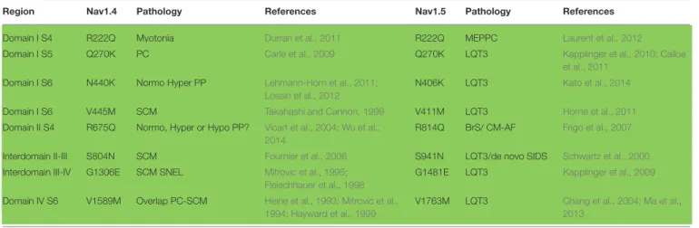
TABLE 4 | List of equivalent amino acids found to be similarly mutated in patients with cardiac (Nav1.5) or skeletal (Nav1.4) pathologies.
Region Nav1.4 Pathology References Nav1.5 Pathology References
Domain I S4 R222Q Myotonia Durran et al., 2011 R222Q MEPPC Laurent et al., 2012
Domain I S5 Q270K PC Carle et al., 2009 Q270K LQT3 Kapplinger et al., 2010; Calloe
et al., 2011
Domain I S6 N440K Normo Hyper PP Lehmann-Horn et al., 2011;
Lossin et al., 2012
N406K LQT3 Kato et al., 2014
Domain I S6 V445M SCM Takahashi and Cannon, 1999 V411M LQT3 Horne et al., 2011
Domain II S4 R675Q Normo, Hyper or Hypo PP? Vicart et al., 2004; Wu et al., 2014
R814Q BrS/ CM-AF Frigo et al., 2007
Interdomain II-III S804N SCM Fournier et al., 2006 S941N LQT3/de novo SIDS Schwartz et al., 2000
Interdomain III-IV G1306E SCM SNEL Mitrovic et al., 1995; Fleischhauer et al., 1998
G1481E LQT3 Kapplinger et al., 2009
Domain IV S6 V1589M Overlap PC-SCM Heine et al., 1993; Mitrovic et al., 1994; Hayward et al., 1999
V1763M LQT3 Chang et al., 2004; Ma et al.,
2013
Same amino acid substitutions occurring in both channels lead to consistent pathologies (in green) regarding membrane excitability. PC, Paramyotonia Congenita; MEPPC, Multifocal Ectopic Purkinje-related Premature Contraction; LQT3, Type 3 Long QT syndrome; SIDS, Sudden Infant death syndrome; Hypo, Normo, Hyper PP, Hypo, Normo, Hyper-kalemic Periodic Paralysis; SCM, Sodium channel Myotonia; BrS, Brugada Syndrome; CM, Cardiomyopathy; AF, Atrial Fibrillation; SNEL, Sporadic Neonatal Episodic Laryngospasm.
TABLE 5 | List of equivalent amino acids found to be differently mutated in patients with cardiac (for Nav1.5) or neuromuscular (for Nav1.4) pathologies.
Region Nav1.4 mutation Pathology References Nav1.5 mutation Pathology References
IS4 R222W Hypo PP Matthews et al., 2009 R222Q MEPPC Laurent et al., 2012
IS4 R225W SCM Lee et al., 2009 R225P LQT3 Beckermann et al., 2014
IS6 N440K Normo Hyper PP Lehmann-Horn et al., 2011; Lossin
et al., 2012
N406S BrS Itoh et al., 2005b
IIS4 R669H Hypo PP Struyk et al., 2000; Kuzmenkin
et al., 2002
R808P BrS Kapplinger et al., 2010
III inter S4-S5 V1149L HyperPP with myotonia Yoshinaga et al., 2015 V1323G BrS Kapplinger et al., 2010
IIIS6 V1293I SCM Koch et al., 1995; Green et al.,
1998
V1468F BrS Kapplinger et al., 2010
IVS4 R1448H PC Ptácek et al., 1992; Chahine et al.,
1994; Mohammadi et al., 2003; Holzherr et al., 2014
R1623Q LQT3 Kambouris et al., 1998;
Makita et al., 1998
IVS4 R1448C PC Ptácek et al., 1992; Chahine et al.,
1994; Featherstone et al., 1998
R1623Q LQT3 Kambouris et al., 1998;
Makita et al., 1998
IVS4 R1448P PC Featherstone et al., 1998 R1623Q LQT3 Kambouris et al., 1998;
Makita et al., 1998
IVS4 R1448S PC (mild) Bendahhou et al., 1999 R1623Q LQT3 Kambouris et al., 1998;
Makita et al., 1998
IVS4 R1451C Hypo PP Arzel-Hézode et al., 2009 R1626P LQT3 Ruan et al., 2007
IVS6 M1592V Normo Hyper PP Rojas et al., 1991; Cannon and
Strittmatter, 1993; Hayward et al., 1999; Rojas et al., 1999
M1766L LQT3 Valdivia et al., 2002; Ye
et al., 2003
Divergent amino-acid substitutions occurring in the two channels lead either to consistent (in green) or inconsistent (in red) pathologies regarding membrane excitability. Hypo, Normo, Hyper PP, Hypo, Normo, Hyper-kalemic Periodic Paralysis; MEPPC, Multifocal Ectopic Purkinje-related Premature Contraction; SCM, Sodium channel Myotonia; PC, Paramyotonia Congenita; LQT3, Type 3 Long QT syndrome; BrS, Brugada Syndrome.
compilation that has been proposed using a paralog annotation
approach in order to retrieve homologous or nearly homologous
variants in both genes (
Ware et al., 2012; Walsh et al., 2014
).
If the same mutations of homologous residues exist, do they
give rise to similar dysfunction on both channels? If yes, we
can expect that both mutations give rise to the same change in
membrane excitability. For instance, if a Nav1.5 mutation leads
to hyper-activity of cardiac cells (LQTS3), the corresponding
mutation in Nav1.4 may also give rise to a hyper-excitability
phenotype of the skeletal muscle cells such as HyperPP, PC,
or SCM. Tables 4, 5 present all the corresponding amino
acids found to be mutated
in patients with cardiac (Nav1.5)
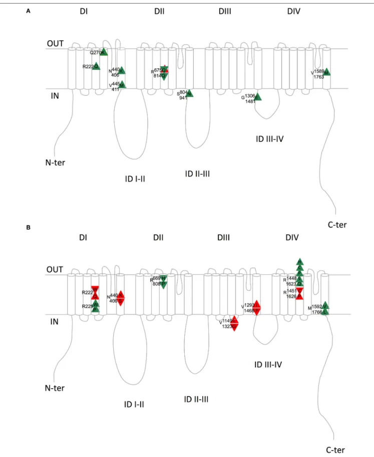
or neuromuscular (Nav1.4) pathologies. Table 4 and Figure 2A
list the mutations for which the amino acid substitutions are
the same, and Table 5 and Figure 2B those for which they are
divergent.
When looking at Table 4 and Figure 2A, it is striking to
observe that all paralog mutations give rise to clearly consistent
FIGURE 2 | Schematic representation of the equivalent Nav1.4/Nav1.5 amino acids with similar (A) or divergent (B) mutations in patients with skeletal (Nav1.4) or cardiac (Nav1.5) pathologies. Upward triangles indicate a gain of function on membrane excitability and downward triangles a loss of function on membrane excitability. Green and red triangles indicate consistent and inconsistent effects on Nav1.4 and Nav1.5 regarding membrane excitability, respectively. Upper amino acid number/letter, Nav1.4; Lower amino acid number/letter, Nav1.5. DI to DIV, domains I to IV; ID, intracellular interdomains; N-ter and C-ter, N-and C-terminus ends.
TABLE 6 | Variations of biophysical parameters compared to wild type channels for five equivalent mutations identified in Nav1.4 and Nav1.5 that have been studied extensively in patch clamp.
Subunit Mutation Pathologies Cell 1V0.5 1V0.5 fast Mutant/WT Fast Mutant/WT References
model act (mV) inac (mV) inact tau * i persistent
Nav1.4 I141V SCM HEK −10 0 77% at −10 mV ? Petitprez et al., 2008; Amarouch et al., 2014
Nav1.5 I141V ExPVC HEK −8 0 86% ? Amarouch et al., 2014; Swan et al., 2014
Nav1.4 Q270K PC HEK 1.3 12.5 168% at −25 mV 200% Carle et al., 2009
Nav1.5 Q270K LQT3 conduc CHO 5.8 9.9 260% at −25 mV 338% Calloe et al., 2011
Nav1.4 N440K Normo Hyper PP HEK 0 7.1 100% 800% Lossin et al., 2012
Nav1.5 N406K LQT3 CHO 8.6 0 217% 550% Kato et al., 2014
Nav1.4 V445M Myotonia HEK −4.1 −4.9 ? 1400% Takahashi and Cannon, 1999
Nav1.5 V411M LQT3 HEK −8.1 −7.9 75% 176% Horne et al., 2011
Nav1.4 V1589M overlap PC-SCM HEK 0 5.4 100% 362% Mitrovic et al., 1994
Nav1.5 V1763M LQT3 hiPSC-CMs 0 16.8 ? 486% Ma et al., 2013
Recording were all done at room temperature (except for Nav1.4 N440K: not indicated). *Fast inactivation tau is measured at −30 mV except when indicated. Green, consistent effect; red, inconsistent effect; SCM, Sodium Channel Myotonia; ExPVC, exercise-induced polymorphic ventricular premature complexes; PC, Paramyotonia Congenita; LQT3, Type 3 Long QT syndrome; Normo, Hyper PP, Normo, Hyper-kalemic Periodic Paralysis; Conduc, conduction disease; hiPSC-CMs, cardiomyocytes generated from human induced pluripotent cells; ?, means not determined.
functional effects, except one which is at first unclear, as detailed
below. Indeed, all Nav1.4 mutations linked to membrane
hyper-excitability (PC, SCM, and HyperPP) correspond to Nav1.5
mutations linked to membrane hyper-activity (LQTS3), except
R675Q (R814Q in Nav1.5). The comparison between Nav1.4
R675Q and Nav1.5 R814Q is not obvious because the pathology
induced by Nav1.4 R675Q mutation is difficult to classify as
Normo/Hyper PP or Hypo PP (
Vicart et al., 2004
). Indeed,
patients experienced normal as well as decreased potassium levels
concomitant to attacks. The rat ortholog of human Nav1.4 R675Q
generates an omega current activated by depolarization when
expressed in Xenopus oocytes (
Sokolov et al., 2008
) (cf. above).
The omega current represents less than 1% of the peak pore
current but it remains constant after slow inactivation of the
pore current and requires high hyperpolarizations to deactivate.
Therefore, it is suspected that this current, carried by Na
+and
K
+ions, maintained during trains of action potentials and with a
residual non-deactivated activity at resting potential could lead to
sodium accumulation and a decrease in membrane excitability. It
will be interesting to test whether the corresponding mutation in
Nav1.5 is also responsible for an omega current. Moreover, the
R675Q Nav1.4 mutation gives rise to a hyperpolarizing shift of
the inactivation curve and a slower recovery from inactivation
when expressed in HEK293 cells. (
Vicart et al., 2004; Wu et al.,
2014
). Altogether, these observations suggested us to rank it as
a hypo-excitability causing mutation, consistent with the BrS
phenotype (loss of function) induced by the homologous Nav1.5
mutation R814Q.
Table 4
and Figure 2A summarize the (dys-)functional
homology between the equivalent mutant in Nav1.4 and Nav1.5.
On the contrary, Table 5 and Figure 2B show that divergent
amino acid substitution at the equivalent position leads to some
inconsistencies (in red, 5/12). This suggests that the nature of the
amino acid substitution is determinant for the direction of the
functional net effect (loss or gain of function).
At last, we focused on five equivalent mutations that have been
studied extensively in patch-clamp in both Nav1.4 and Nav1.5.
Table 6
shows changes in each biophysical parameter for these
mutations. When looking at the direction of the functional effects
(gain or loss of function), we observe two major points. First, a
strikingly similar functional effect of the same mutations in both
channels. Second, all the gain of function mutations leading to
hyper-activity/excitability provoke an increase in the persistent
current, when measured, suggesting that this mechanism plays a
major role in the pathogenesis of Nav channelopathies.
Recently, an omega current has been observed in Nav1.5
mutant channels identified in patients presenting with
arrhythmic DCM or MEPPC (
Gosselin-Badaroudine et al.,
2012b; Moreau et al., 2015b
). This omega current, due to
mutations of arginine in the S4 of domain I, is similar to the
one observed in Nav1.4 (
Sokolov et al., 2007; Struyk et al., 2008;
Francis et al., 2011; Gosselin-Badaroudine et al., 2012a; Groome
et al., 2014
). This further strengthens the functional similarity
between Nav1.4 and Nav1.5 in pathophysiological situations. A
common feature of MEPPC, is an increase in window current
provoked by the Nav1.5 R225W, R222Q, and R225P mutations,
increasing cardiac excitability of the fascicular-Purkinje system
(
Laurent et al., 2012; Mann et al., 2012
). Another common
feature of two of these mutations: R222Q and R225W is the
presence of an omega current. This Nav1.5 omega current may
be responsible for the peculiar cardiac phenotype (
Moreau et al.,
2015a
), similar to the omega current of Nav1.4 being responsible
for the hypoPP phenotype, through sodium accumulation and a
decrease in membrane excitability (
Sokolov et al., 2008
). Indeed,
most of the SCN5A mutations linked to DCM are located
in the voltage sensor domain (VSD) as pointed by
McNair
et al. (2011)
. However, in some cases DCM may be secondary
to arrhythmias and window current increase. For instance,
preventing arrhythmias by quinidine improved the ventricular
function (ejection fraction) in patients with the Nav1.5 R222Q
mutation, via a decrease in the window sodium current (
Laurent
et al., 2012
). The use of specific inhibitor of the alpha pore and
the omega (or gating pore) current would allow to test for the
respective role of the altered gating (activation, inactivation)
and the omega current on the development of the pathology.
Noteworthy, the various localization of the Nav1.4 mutations
giving rise to omega current (in domains I, II, and III) strongly
suggests that similar mutations in Nav1.5 will be identified in
domain II and III in addition to the ones already identified in
domain I (
Moreau et al., 2015b
).
To conclude, given the sequence similarity between Nav1.4
and Nav1.5, any characteristics described for one channel subunit
may shed light on the properties of the counterpart channel
subunit, such as the presence of specific protein partners, or
the effects of a specific amino acid substitution. One can
argue that the effect of a mutation on Nav1.4 is difficult
to compare with Nav1.5 since the different molecular and
cellular environment may drastically modify the effect of the
mutation. Nevertheless, we noticed that the same mutation
lead to comparable effect regarding membrane hypo or
hyper-excitability (Table 4 and Figure 2A). This suggests that the
cellular environment is usually not able to invert the effect
of a mutation from gain to loss of function phenotypes and
reciprocally. Such comparison between Nav1.4 and Nav1.5 will
probably draw more and more interest, to address the challenge
of interpreting and understanding pathogenicity of rare SCN4A
or SCN5A variants revealed by next-generation sequencing
studies (
Arnold et al., 2015; Bergareche et al., 2015; Coll et al.,
2015
).
AUTHOR CONTRIBUTIONS
Parts were written by: Part I : DS, YP, VF (Nav1.4), FC (Nav1.5).
Part II: SN (Nav1.4), GT (Nav1.5). Part III: FL, CM. Part IV:
DS (Nav1.4), JB, OM, IB, GL (Nav1.5). DS and GL initiated the
project. IB, FC, JB, YP critically read the entire Manuscript. GL
supervised the Ms.
FUNDING
This work was supported by INSERM, CNRS, the Fondation
d’entreprise Génavie, the Fondation pour la Recherche
Médicale
(PLP20141031304),
the
Association
Française
contre les Myopathies - Téléthon (16495), the 7th European
Community Framework Programme (PIOF-GA-2011-298280,
PIRG06-GA-2009-256397,
HEALTH-F2-2009-241526),
the
ANR
(ANR-12-BSV1-0013-01),
Investissements
d’avenir
(ANR-10-IAIHU-06), and Nantes and Sorbonne universities,
UPMC-Paris 06.
REFERENCES
Abriel, H. (2010). Cardiac sodium channel Na(v)1.5 and interacting proteins: physiology and pathophysiology. J. Mol. Cell. Cardiol. 48, 2–11. doi: 10.1016/j.yjmcc.2009.08.025
Ackerman, M. J., Priori, S. G., Willems, S., Berul, C., Brugada, R., Calkins, H., et al. (2011). HRS/EHRA expert consensus statement on the state of genetic testing for the channelopathies and cardiomyopathies this document was developed as a partnership between the Heart Rhythm Society (HRS) and the European Heart Rhythm Association (EHRA). Heart Rhythm 8, 1308–1339. doi: 10.1016/j.hrthm.2011.05.020
Ackerman, M. J., Siu, B. L., Sturner, W. Q., Tester, D. J., Valdivia, C. R., Makielski, J. C., et al. (2001). Postmortem molecular analysis of SCN5A defects in sudden infant death syndrome. JAMA 286, 2264–2269. doi: 10.1001/jama.286.18.2264 Aiba, T., Farinelli, F., Kostecki, G., Hesketh, G. G., Edwards, D., Biswas, S.,
et al. (2014). A mutation causing Brugada syndrome identifies a mechanism for altered autonomic and oxidant regulation of cardiac sodium currents. Circ. Cardiovasc. Genet. 7, 249–256. doi: 10.1161/CIRCGENETICS.113. 000480
Allouis, M., Le Bouffant, F., Wilders, R., Péroz, D., Schott, J. J., Noireaud, J., et al. (2006). 14-3-3 is a regulator of the cardiac voltage-gated sodium channel Nav1.5. Circ. Res. 98, 1538–1546. doi: 10.1161/01.RES.0000229244.97497.2c Amarouch, M. Y., Kasimova, M. A., Tarek, M., and Abriel, H. (2014). Functional
interaction between S1 and S4 segments in voltage-gated sodium channels revealed by human channelopathies. Channels (Austin.) 8, 414–420. doi: 10.4161/19336950.2014.958922
Amin, A. S. (2014). SCN5A-related dilated cardiomyopathy: what do we know? Heart Rhythm 11, 1454–1455. doi: 10.1016/j.hrthm.2014.05.031
Amin, A. S., Pinto, Y. M., and Wilde, A. A. (2013). Long QT syndrome: beyond the causal mutation. J. Physiol. 591, 4125–4139. doi: 10.1113/jphysiol.2013.254920 An, R. H., Wang, X. L., Kerem, B., Benhorin, J., Medina, A., Goldmit, M., et al. (1998). Novel LQT-3 mutation affects Na+ channel activity through
interactions between alpha- and beta1-subunits. Circ. Res. 83, 141–146. doi: 10.1161/01.RES.83.2.141
Antzelevitch, C., Brugada, P., Borggrefe, M., Brugada, J., Brugada, R., Corrado, D., et al. (2005). Brugada syndrome: report of the second consensus conference. Heart Rhythm 2, 429–440. doi: 10.1016/j.hrthm.2005.01.005
Antzelevitch, C., Pollevick, G. D., Cordeiro, J. M., Casis, O., Sanguinetti, M. C., Aizawa, Y., et al. (2007). Loss-of-function mutations in the cardiac calcium channel underlie a new clinical entity characterized by ST-segment elevation, short QT intervals, and sudden cardiac death. Circulation 115, 442–449. doi: 10.1161/CIRCULATIONAHA.106.668392
Antzelevitch, C., and Yan, G. X. (2015). J-wave syndromes: brugada and early repolarization syndromes. Heart Rhythm 12, 1852–1866. doi: 10.1016/j.hrthm.2015.04.014
Arnold, W. D., Feldman, D. H., Ramirez, S., He, L., Kassar, D., Quick, A., et al. (2015). Defective fast inactivation recovery of Nav 1.4 in congenital myasthenic syndrome. Ann. Neurol. 77, 840–850. doi: 10.1002/ana.24389
Arzel-Hézode, M., McGoey, S., Sternberg, D., Vicart, S., Eymard, B., and Fontaine, B. (2009). Glucocorticoids may trigger attacks in several types of periodic paralysis. Neuromuscul. Disord. 19, 217–219. doi: 10.1016/j.nmd.2008.12.008 Ashpole, N. M., Herren, A. W., Ginsburg, K. S., Brogan, J. D., Johnson,
D. E., Cummins, T. R., et al. (2012). Ca2+/calmodulin-dependent protein kinase II (CaMKII) regulates cardiac sodium channel NaV1.5 gating by multiple phosphorylation sites. J. Biol. Chem. 287, 19856–19869. doi: 10.1074/jbc.M111.322537
Awad, S. S., Lightowlers, R. N., Young, C., Chrzanowska-Lightowlers, Z. M., Lomo, T., and Slater, C. R. (2001). Sodium channel mRNAs at the neuromuscular junction: distinct patterns of accumulation and effects of muscle activity. J. Neurosci. 21, 8456–8463.
Bai, C. X., Kurokawa, J., Tamagawa, M., Nakaya, H., and Furukawa,
T. (2005). Nontranscriptional regulation of cardiac repolarization
currents by testosterone. Circulation 112, 1701–1710. doi: