AVIS
Ce document a été numérisé par la Division de la gestion des documents et des archives de l’Université de Montréal.
L’auteur a autorisé l’Université de Montréal à reproduire et diffuser, en totalité ou en partie, par quelque moyen que ce soit et sur quelque support que ce soit, et exclusivement à des fins non lucratives d’enseignement et de recherche, des copies de ce mémoire ou de cette thèse.
L’auteur et les coauteurs le cas échéant conservent la propriété du droit d’auteur et des droits moraux qui protègent ce document. Ni la thèse ou le mémoire, ni des extraits substantiels de ce document, ne doivent être imprimés ou autrement reproduits sans l’autorisation de l’auteur.
Afin de se conformer à la Loi canadienne sur la protection des renseignements personnels, quelques formulaires secondaires, coordonnées ou signatures intégrées au texte ont pu être enlevés de ce document. Bien que cela ait pu affecter la pagination, il n’y a aucun contenu manquant.
NOTICE
This document was digitized by the Records Management & Archives Division of Université de Montréal.
The author of this thesis or dissertation has granted a nonexclusive license allowing Université de Montréal to reproduce and publish the document, in part or in whole, and in any format, solely for noncommercial educational and research purposes.
The author and co-authors if applicable retain copyright ownership and moral rights in this document. Neither the whole thesis or dissertation, nor substantial extracts from it, may be printed or otherwise reproduced without the author’s permission.
In compliance with the Canadian Privacy Act some supporting forms, contact information or signatures may have been removed from the document. While this may affect the document page count, it does not represent any loss of content from the document.
"
New high through-put assays for detecting
Transglutaminase activity
Par
Wajih Ben tahar
Département de Chimie Faculté des Arts et Sciences
Mémoire présenté à la Faculté des Études Supérieures en vue de l'obtention du grade de
Maître ès Sciences (M.Sc.) En Chimie
Avril, 2008 © wajih Ben tahar, 2008
Faculté des Études Supérieures
Ce mémoIre intitulé:
New high through-put assays for detecting
Transglutaminase activity
Présenté par:
Wajih Ben tahar
A été évalué par un jury composé des personnes suivantes:
Président -rapporteur Membre du jury Directeur de recherche Codirecteur de recherche
Mémoire accepté le :
Professeur Pelletier. Joelle N. Professeur Lortie. Robert.
Professeur Lubell, William D, Professeur Keillor. Jeffrey W.
1 wish to express my sincere gratitude to my supervisors, Prof. William D. Lubell
and Prof. Jeffrey W. Keillor for giving me the opportunity to be a member of their
group. Thanks also for their help, guidance and enthusiastic support during my
master thesis studies.
1 would like to express my sin cere gratitude to my former co-workers, Dr Claudio
Gnaccarini, Hassan Iden, Dr Gil Fridkin, and Dr Tarek Kassem for pleasant times
in the lab, helpful discussions and comments concerning chemistry, and to aIl
present and former members of the Lubell and Keillor groups for maintaining a
friendly and pleasant atmosphere.
1 wish to express my sincere gratitude to Mildred Bien Aimé for help with technical
issues, for enjoyable and interesting discussions concerning life and especially for
her spirit.
1 would like to acknowledge the assistance of Dalbir Sekhon for HPLC analyses, Dr
Alexandra Furtos and Mrs Karine Venne for mass spectral analysis, as weIl as the
Natural Sciences and Engineering Research Council of Canada (NSERC), for
financial support.
Last but not least 1 am grateful to my parents, wife and brothers for their love,
support, encouragement and patience.
Résumé
Depuis une décennie, l'industrie pharmaceutique s'intéresse de très près à la synthèse de
principes actifs constitués de peptides. Ces composés ont l'avantage d'être hautement
spécifiques, présentent de faibles problèmes toxicologiques et s'accumulent peu dans les
tissus. Cependant, il existe un réel défi pour les sociétés de mettre au point des synthèses
peu couteuses, moins polluantes et plus propres. Une synthèse peptidique par voie
enzymatique serait une alternative. La transglutaminase (TGase) enzyme a Ca2+ dépendance
qui catalyse le transfert d'un groupement glutamyle d'un donneur acyl, la glutamine à un
accepteur d'acyle, généralement une lysine, semblerait être un candidat idéal dans cette
approche. Cette enzyme catalyse la formation de la liaison ami di que entre les aminoacides
et les peptides et fonctionne dans l'eau de façon quasi-irréversible. Les mimes de TGase
sont prometteuses dans ce domaine de recherche. Dans ce type de projet, le développement
de méthodes de détection de l'activité TGase des mutants est d'importance.
Dans ce travail sera présenté le développement de deux nouvelles méthodes fluométriques
à haut débit pour la détection de l'activité de la TGase. Ces méthodes présentent de
nombreux avantages: elles sont tout d'abord très sensibles à faibles concentration en
enzyme et substrats. Ces méthodes sont reproductibles et peuvent être utilisées dans des
milieux biologiques plus complexes (lysat).
Le premier test est basé sur la mesure fluorométrique de l'activité de la TGase et a été
validé sur la transpeptidation d'un substrat donneur préalablement synthétisé contenant une
avec la TGase, le mélange est fixé à une streptavidine immobilisée et l'activité de la TGase
est mesurée par l'augmentation du signal de fluoresence.
Le second test est basé sur l'observation du phènoméne de FRET (définie comme un transfert d'énergy non radiative suite à une interaction dipole-dipole entre deux molécules
fluorescentes) suite à la ligation par la TGase d'un donneur acyle marqué avec le fluorophore Tokyo Green à une amine primaire marquée avec la 7-hydroxy-coumarine.
La synthèse des substrats, l'optimisation des tests et leurs applications dans l'étude de l'activité de la TGase et leurs possibles applications dans des criblages à haut débit sont
présentées dans ce mémoire.
Mots clés: Transglutaminase, FRET, fluorimétrie, enzyme, cinétique, synthèse sur support solide, lysat, criblage à haut débit.
Abstract
During the last decade, interest in the synthesis of peptides as drugs has grown, due to their high specificity, low toxicology problems, and minimal accumulation in tissue. Environmentally sound economical synthesis of peptides represents a challenge considering the limitation of CUITent chemistry. Enzymatic peptide synthesis may serve as an alternative for efficient peptide synthesis. Transglutaminase (TGase) is a Ca2+ dependent enzyme that catalyzes the transamidation of an acyl donor glutamine to an acyl acceptor, lysine. This enzyme represents as interesting candidate for peptide synthesis, because it already catalyzes amide bond formation between amino acids and peptides, in aqueous solution. Methods for the detection of mutant TGase activity are needed to study this enzyme for peptide synthesis.
The present study describes the development of two new high-throughput fluorometric methods for the detection of TGase activity. These methods are sensitive at low concentration of enzyme and substrate. They are also reproducible and effective in biological media expressing enzyme (lysate).
The first assay is based on the fluorometric measurement of TGase activity. Transpeptidation involves an amide donor substrate possessing an N-terminal fluorophore moiety and an acceptor amine substrate containing biotin. After treatment with TGase, the mixture is fixed to immobilized streptavidine, the resin is washed and TGase activity is monitored by the increase of fluorescence signal.
The second assay is based on the observation of the FRET (Fluorescence Resonance Energy Transfer) effect upon ligation by TGase of a donor acyl substrate labelled with the fluorophore Tokyo Green to a primary amine labelled with 7-hydroxycoumarin.
The synthesis of organic substrates and assay optimization have been perforrned to study TGase activity.
Key Words: transglutaminase, FRET, fluorometry, enzyme, kinetics, solid phase synthesis, lysate, high-through put screening, quench.
Table of contents
Acknow ledgments ... .i
Résumé ... ii
Summary ... iv
Table of Contents ... vi
List of Figures ... .ix
List of Tables ... xi
List of Schemes ... xii
List of Abbreviations ... xiii
Chapter 1: Introduction 1.1 Peptides as Drugs ... 2 1.1.1 Definition ... 2 1.1.2 Peptides as Drugs ... 2 1.1.3 Limitation ... 3 1.1.4 Proteases ... 6 1.2 Transglutaminases ... 6 1.2.1 Definition ... 6 1.2.2 Mechanism of action ... 8 1.2.3 Physiological transglutaminase ... 12 1.2.3.1 Alzheimer's disease ... 12 1.2.3.2 Huntington' s disease ... 12 1.2.3.3 Celiac disease ... 12 1.2.3.4 Cataract formation ... 13
1.2.3.5 Atherosclerosis ... 13
1.2.4 Transglutaminase structure ... 13
1.2.5 Transglutaminase binding ... 15
1.3 Aims of the Project ... , ... 16
1.5 References ... 19
Chapter 2: A fluorometric assay for the detection of transglutaminase activity. 2.1 Introduction ... 28
2.2 Materials and methods ... 29
2.2.1 Enzyme preparation ... 29
2.2.2 Preparation of crude bacteriallysate ... 29
2.3 Synthesis and kinetics for acyl donor substrate ... 30
2.4 Synthesis and kinetics for acyl acceptor substrate ... 33
2.4.1 Materials ... '" ... 34
2.5 Assay procedure using a purified enzyme ... 35
2.5.1 Methods ... 35
2.5.2 Result and discussions ... 36
2.6 Assay procedure using a crude bacteriallysate ... .41
2.6.1 Methods ... 41
2.6.2 Results and discussions ... .41
2.7 Conclusion ... 43
2.8 Experimental section ... 45
Chapter 3: a continous FRET based assay for tissue transglutaminase
3.1 Introduction ... 60
3.2 GeneraIs ... 62
3.3 Synthesis of the acyl acceptor substrate ... 64
3.4 Synthesis of the acyl donor substrate ... 64
3.5 FRET assay using purified enzyme ... 65
3.5.1 Methods ... 65
3.5.2 Results and discussions ... 65
3.5.3 Limit of detection ... '" ... 70
3.6 FRET assay using crude bacteriallysate ... 72
3.6.1 Methods ... 72
3.6.2 Results and discussions ... 73
3.7 Conclusion ... 75
3.8 Experimental section ... 76
3.8 References ... 79
Chapter 4: Concluding Statements and Future Work 4.1 Conclusion ... 81
4.2 Comparison of methods ... 82
4.3 Future work ... 83
List of Figures
Chapter 1Figure 1.1 Difference between enzymatic and non enzymatic reaction
Figure 1.2 Modified Ping-Pong mechanism ofTGase
Figure 1.3 Resemblance of the overall structure of Factor XIII and Red Sea Bream
TG2
Chapter 2
5
9
14
Figure 2.1 Model of streptavidin beads assay of detection of TGase activity 29
Figure 2.2 Comparison of assay sensitivity using acyl acceptor substrates 16a-c. 38
Figure 2.3 Assay sensitivity with acyl donor lla-c and acyl acceptor substrate 16c . 39
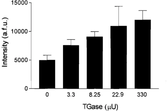
Figure 2.4 Limit of detection of the assay with respect to TGase concentration. 40
Figure 2.5 Product 17 formed by TGase ligation 42
Figure 2.6 Detection of TGase activity in crude bacterial lysate. 43
Chapter 3
Figure 3.1 FRET dependence on the overlap integrals 61
Figure 3.2 Scheme of FRET for Tokyo Green/7-Hydroxy-coumarin carboxylic acid pairs
Figure 3.4 FRET scan sensitivity with acyl donor lla-b and acyl acceptor 20 using
purified enzyme
Figure 3.5 Product forrned upon transamidation by TGase
Figure 3.6 LCMS spectrum of the enzymatic and non enzymatic reactions
Figure 3.7 Application of the FRET assay on plates
Figure 3.8 Control test for the FRET assay on plate
Figure 3.9 Limit of detection of the FRET assay
Figure 3.10 Continuous FRET assay using crude bacterial lysate
Figure 3.11 Sensitivity of the FRET assay
66 68 69 70 71 72 74 74
List of Tables
Chapter 1Table 1.1 The transglutaminase (TGase) family. Il
Chapter 2
Table 2.1 Kinetic constants for TGase with various N-terminal derivative peptides 33
Table 2.2 Kinetic constants for TGase with various acyl acceptors 33
Table 2.3 Fluorescence resulting from the coupling of lla and 16c, measured on
List of Schemes
Chapter 1Scheme 1.1 Transglutaminase catalyzed reaction
Chapter 2
Scheme 2.1 Solid support synthesis of different acyl donors.
Scheme 2.2 Synthesis of different acyl acceptors.
Chapter 3
Scheme 3.1 Synthesis of FRET donor substrate
8
32
34
Abbreviations
[a]D
optical rotationAla alanine
Asn asparagine
Asp aspartic acid
Amp ampicillin
Arg arginine
Boc tert-butoxycarbonyl
BSA bovine serum albumine
t-butyl tert-butyl bp boiling point br broad (spectral) Bz benzoyl Cbz benzyloxycarbonyl ChI chloramphenicol oC celsius Cys cysteine d doublet dd doublet of doublets D aspartic acid Da Dalton DMPDA N,N-dimethyl-I,4-phenylenediamine
DMAP DNA
DIT
DCC DIC DIEA DMF E. coli EC EDC.HCI EDTA FRET HBTU HOBt HATU Fmoc G GABA GDH Gly Glu 4-Dimethylaminopyridine deoxyribonucIeic acid dithiothreitol dicyclohexyl carbodiimide 1,3-diisopropylcarbodiimide diisopropylethylamine N,N-dimethylformamide Escherichia coli Enzyme commission l-Ethyl-3-(3-dimethyllaminopropyl)carbodiimide hydrochloride ethylenediaminetetraacetic acidfree resonance energy transfer
0-Benzotriazole-N,N,N' ,N' -tetramethyl-uronium-hexafluoro-phosphate
N-Hydroxybenzotriazole
2-(1 H-7-Azabenzotriazol-l-yl)-1, 1 ,3,3-tetramethyI uronium hexafluorophosphate methanaminium 9-fluorenylmethyI oxycarbonyl glycine y-aminobutyric acid glutamate dehydrogenase glycine glutamic acid
h HATU HOAt His HOBt HRMS IR IPTG
!Ne
LB JKI
LysKm
J.! m M mg min MHz hours O-(7-azabenzotriazole-l-yl)-1, 1,3,3-tetramethyluronium hexafluorophosphate I-hydroxy-7-azabenzotriazole histidine I-hydroxy IH-benzotriazole high-resolution mass spectrometryinfrared isopropyl-~-D-thiogalactoside incorporation Luria-Bertani coupling constant dissociation constant lysine Michaelis constant mIcro multiplet (spectral) molar milligram minutes megahertz
mL millilitre mmol millimole mp melting point MS mass speetrometry MW moleeular weight N asparagine
NEt3 triethyl amine
nm nanometers
NMR nuclear magnetie resonanee
OD
optieal densityppm part per milliom
Phe phenylalanine
Pro proline
QLPF Gln-Leu-Pro-Phe
rpm rotation per minute
s singlet
SPPS sol id-phase peptide synthesis
t triplet
Thr thryreonine
TB terrifie broth
TGase transglutaminase
Tris tris(hydroxymethyl)methane
TFA
TLC
TBTU U Àem trifluoroacetic acidthin layer chromatography
0-Benzotriazo 1-1-yl-N,N,N' ,N' -tetramethyluronium tetrafluoroborate
units
emission wavelength excitation wavelengh
CHAPTERI
1.1 Peptides
1.1.1 Introduction
A peptide is a molecule formed from the coupling of two or more a-amino acids via an amide bond resulting from the condensation of a carboxylic group of one and the amino group of the other. Peptides containing more than 50 amino acids are called proteins. Peptides play an important role in regulating many physiological processes, and can be used in various treatments for diseases such as the peptide insulin, which is used to treat diabetes.
1.1.2 Peptides as Drugs
Peptides represent promising candidates for drug discovery due to their high specificity, high affinity, low toxicity, and low accumulation in tissues. The first peptide based drug to be administered therapeutically was insulin.1 To date, insulin remains one of the most successful peptide based drugs with insulin sales of $ 7 billion (US) in 2005, and expected to reach $ 14 billion (US) by 2010.2
Solution phase synthesis was the method originally used for making peptides; however, this method presented many problems involving purification, solubility as the size of the peptide grew, reaction times, and low yields. In 1963, a new strategy for peptides synthesis, so called solid-phase peptide synthesis (SPPS) was pioneered by Bruce Merrified,3 and had a great impact on the chemistry for making peptides by overcoming the problems cited above. Five years later, the sol id phase synthesis of oxytocin, 4 and diamino oxytocin were reported.5 In fact, this method brought a 20-30 fold increase in the number of analogues
that could be synthesized compared to the solution method. The solid phase technique has become the most commonly used process for peptide synthesis, moreover, solid-phase chemistry has established itself as a useful tool in the field of organic synthesis. Bruce Merrifield was awarded the chemistry Nobel Prize in 1984 for the development of a simple and ingenious method for obtaining peptides and proteins, his original paper is the 5th most highly cited paper in the Journal of the American Chemical Society.3
The SPPS method has become commonplace for research in drug discovery.6 Over the last decade, investment of the pharmaceutical industry in peptide-based therapeutics has expanded, prompted by the advances in the understanding of the genetics of diseases and drug delivery, as weIl as developments in peptide synthesis. The global market of therapeutic peptides in 2007 was valued at around $1 billion (US), and is predicted to significantly increase over the next decade.7 In fact, interest in peptide drugs has recently intensified with the approval of the peptide-based drug Fuzeon (T-20) an anti-HIV treatment by the Roche Company. Peptides are currently being developed for therapeutic applications such as, allergy, asthma, cancer, and diabetes.
1.1.3 Limitations
To date, compared with the total number of drugs that have been delivered to market, peptide based-drugs are considered marginal. As articulated by pioneers in the field, 7 the key development of peptides as drugs is contingent on the following facts:
Enhanced synthesis of diverse peptide analog libraries to screen agoni st drug
targets.
Modification of peptides with non-natural amino acids to increase stability.
In fact, in spite of the advantages reported above, there are many issues facing this field of
research that need to be solved in order to gain an increased market for peptides as drugs.
One of the major problems in this field is the high cost of production of peptide drugs.
Roche's anti-HIV Fuzeon (T-20), the present synthetic peptide-drug paradigm, costs more
than $20 000 (US) per pers on a year which restricts its general use. As previously
mentioned, the solid phase strategy is the method of choice for peptide synthesis. This
method has two major problems:
High co st: Coupling reagents and protected ammo acids are expensive,
commonly used in excess, and rarely recycled
Pollution: Protection, deprotection and washing steps generate relatively large
quantities ofwaste per procured product.
Investment in this field is needed to resolve these two problems: high co st and pollution.
An increased peptide market demands resolution of these issues to provide affordable products in an environmentally friendly manner.
In order to find an efficient way of synthesizing peptides m low cost and with less
pollution, the pharmaceutical industry, as weIl as food companies have been investigating
other methods for large scale peptide synthesis that meet the following criteria:
Reduced cost of the synthesis. Less pollution.
Enzymes are available biocatalysts that function in aqueous conditions and more efficiently by lowering the activation energy barrier (~G") of a reaction, thus allowing the reaction to proceed much faster without affecting the equilibrium of the reaction (Figure 1.1). Industry can consider biocatalysts to be a potential strategy for efficient peptide synthesis. The proteases have been the principal targeted family of enzyme for amide bond formation between a-amino acid residues. 8
Figure1.1 Difference between enzymatic and non enzymatic reaction
non enzymatic
---
---t---Î
non enzym.tic / activation energyf ---
---r:~~~~~
1~
_____~~~rgï
__________t ________ _
enzymatic product t1.1.4 Proteases
Proteases occur naturally in aIl organisms and constitute 1-5% of the gene content. These enzymes are involved in a multitude of physiological reactions, industrial and pharmaceutical applications. 8, 9, 10 Proteases can also be used as biocatalyst for peptide bond formation via a kinetically or thermodynamically controlled approach. However, in thermodynamically controlled approach the hydrolysis of the formed peptide was favoured reducing than the yield of the reaction. Il More research was focused in favouring the
kinetically approach. 12, 13, 14 For example, subtiligase, a double mutant of subtilisin BPN', has been shown to have many uses, from the total synthesis of RNase A to the semisynthesis of a variety of other proteins via the coupling of esterified peptides onto the N-termini ofproteins in aqueous media and in high yield. 15 Moreover, asprgillus proteases were found to be efficient for peptide coupling when using the carbamoyl ester as acyl donor. 14 The two major drawbacks of proteases as peptide biocatalyst are substrate specificity and hydrolysis of the growing peptide, which respectively lower the flexibility and yields ofthis method. 16 Enzyme transglutaminase (TGase)thus has been pursued as an alternative for enzyme-catalyzed peptide coupling.
1.2 Transglutaminase
1.2.1 Definition
Transglutaminase (TGases) are a c1ass of Ca2+ -dependent enzymes that catalyze the
cross-linking of proteins through the formation of isopeptide bonds between reactive lysine and glutamine residues in proteins. An aminotransferase enzyme, TGase catalyses the transamidation of glutamine-containing proteins with a variety of other primary amine
nuc1eophiles(scheme 1.1). 17 The so formed isopeptide bonds exhibit resistance to
proteolytic degradation as weIl as physical-chemical degradation.
TGase is widely used in several different industrial products and processes. Advances in biotechnology have enabled this enzyme to be developed for processes and have made these enzymes a viable option for chemical catalysis. In particular, the microbial TGase (MTGase) derived from the Streptoverticillium mobaraense in the fermentation of sorghum straw hydrolysates has been used in the food industry for catalyzing the cross-linking of many food proteins such as caseins, soybean globulins, gluten, actin, myosins, and egg proteins, through the formation of an E-(y-glutamyl)lysine bond. Used as weIl for the incorporation of primary amines into proteins, MTGase is notable because it is a calcium independent enzyme of low molecular weight, which is smaller than of other known TGases. 18, 19, 20 Efforts continue to focus on identifying novel and alternative microbial sources for production of TGase analogs with greater substrate specificity and enhanced utility in (iso) peptide synthesis.
Scheme 1.1 Transglutaminase Catalyzed Reactions
Isopeptide bond formation
o \ . H.N
X/"y'"" ·
HN "',Il
~
GIn 0 Lys°X
H TGase "" / y N ---1.~ HN "'"Il
~
0 o y-Glu-LysPrimary amide synthesis
o \ .
X
+ H2NR 111111/"y'NHz HN "',Il
~
GIn 0 _ _T_G_as_e_---I.~ "X~."'
~
0 Amide hydrolysis°X\
111111. /"y'NH2 + HN "',.Il
~
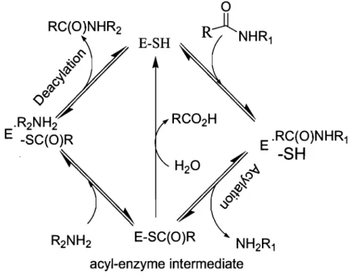
GIn 0 TGase 1,2.2 Mechanism of actionTGase follows a modified "Ping-Pong" mechanism described by Folk, J in 1969 (Figure
• Acylation step:
The acyl donor is bound at the active site of the enzyme forming the first Michaelis complex ready for the acylation step.
The catalytic cysteine thiol attacks the y-carboxamide group of the donor substrate (glutamine residue), forming the acyl-enzyme intermediate and releasing one equivalent of ammonia.22
• Deacylation step:
When the acyl-enzyme intermediate is formed, a competition occurs between hydrolysis with water forming glutamic acid (slow reaction), and the aminolysis reaction of the intermediate with lysine forming an isopeptidic bond or with a primary amine forming an amide bond.
Figure 1.2: Modified Ping-Pong Mechanism ofTGase
RC(O)NHR2
E-SH
~
RJ~
&~..:s
E-SC(O)Racyl-enzyme intermediate
E .RC(O)NHR1-SH
The Keillor laboratory has studied reactions catalyzed by the guinea pig liver TGase and demonstrated that the catalytic mechanism involves general base catalysis by characterisation of the transition state using kinetic methods. 22
The TGases family have been identified from different sources such as bacteria, invertebrate, and plants. 23 From mammalians, a total of eight different TGase enzymes have been identified (Table 1.1):
• Tissue transglutaminase (TG2):
Full length human TG2 is 687 amino acids with a predicted molecular mass of - 77 kDa. TG2 is weIl conserved, with human TG2 being 80-90% homologous to bovin, mouse and guinea pig TG2. 24 The most studied TGase, TG2 exists in the majority of tissues, transcription of TG2 is increased by retionoic acid, it plays a role in the extra-cellular matrix development, apoptosis and neural differentiation. 25 Beside having transpeptidation activity, TG2 also acts as a kinase, 26 protein disulfide isomerase, and deamidase by the deamidation of gliadin peptides thus playing an important role in the pathology of coeliac disease. 27 TG2 has also been linked to inflammation, neurodegenerative diseases,
Table 1.1: The transglutaminase (TGase) family.
Factor XIII Fibrin F13Al 732 (83) Blood clotting and Cytosol,
stabilizing wound healing extracellular
factor
Band 4.2 Erythrocyte EPB42 690 (72) Structural prote in in Membrane
membrane erythrocytes-no
prote in activity
TGase 1 Keratinocyte TGMI 814 (90) Cornified envelope Cytosol,
TGase assembly membrane
in surface epithelia
TGase 2 Tissue TGase TGM2 686 (80) Cell Cytosol,
death/ differentiation, nucleus, adhesion, matrix membrane,
assembly cell surface,
extracellular
TGase 3 Epidermal TGM3 692 (77) Cornified envelope Cytolsol
TGase assembly in surface
epithelia
TGase 4 Prostate TGM4 683 (77) Semen coagulation Unknown
TGase in rodents
TGase 5 TGaseX TGM5 719 (81) Epidermal Nuclear
differentiation matrix, cytoskeleton
TGase 6 TGase Y TGM6 Unknown Unknown Unknown
TGase 7 TGase Z TGM7 710 (80) Unknown Unknown
1.2.3 Physiological transglutaminase
TGase is implicated in different biological processes, including cell adhesion/o apoptosis/1 formation of the extra cellular matrix/2 blood coagulation.33 Deregulation of transglutaminase activity leads to different physiological disorders and diseases.34
1.2.3.1 Alzheimer's disease
The formation of the ab normal prote in structures of Alzheimer's disease have been suggested to be catalyzed by TGase because insoluble isoforms of TGase colocalized with plaques and tangles 30-50 times greater than normal brain tissue.35
1.2.3.2 Huntington's disease
Huntington's disease (HD) results from genetically programmed degeneration ofbrain cells, called neurons, in certain areas of the brain. Elevated TGase activity has been found in the affected areas. The implicated protein, Huntington, has been found to be a good substrate for the TG 2 enzyme.36
1.2.3.3 Celiac disease
Celiac disease is a digestive disease that damages the small intestine and interferes with absorption of nutrients from food. Celiac disease is precipitated by the ingestion of gliadin, a proteinaneous component of wheat gluten. The presentation of fragments of gliadin cross-linked to TG 2 results in antibodies against gliadin, TG 2 and the cross-cross-linked proteins.
1.2.3.4 Cataract formation
A cataract is an opacity that develops in the crystalline lens of the eye or in its envelope and
may cause blindness. Cataracts are distinguished by a marked elevation of intracellular
Ca2+, which may act to stimulate TGase activity either directly or indirectly suggesting
involvement of TGase in cataract formation. 37
1.2.3.5 Atherosc1erosis
Atherosc1erosis involves the build-up of a waxy deposit on the inside of blood vessels.
TGase may play a role in the formation of the atherosc1erotic plaques by catalyzing the
incorporation of lipoprotein (a) (TGase substrate) into these structures, creating insoluble
atherosc1erotic tissue. 38
1.2.4 Transglutaminase structure
To have a good understanding of the structure-function relation for enzyme activity, it is
very important to know the three-dimensional crystalline structure of the enzyme. Having
this structure could help researchers to better understand the enzyme, to design efficient
inhibitors of the enzyme, and to assess the important domains for the activity of the
enzyme.
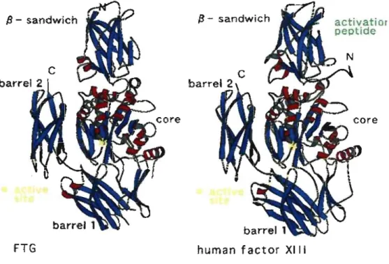
To date, the structures of four different vertebrate TGases have been resolved: the factor
XIII TGase furnished the first three-dimensional structure, 39 followed by red sea bream
In the X-ray structures, the enzyme is composed of a p-sandwich at the N-terminal containing the residues Gly-6 to Phe-134, the catalytic a/p core containing the residues Asn-l35 to Thr-461, the
P
l barrel covering residues Arg-472 to Ser-583 andP
2 barrel at the C-terminal covering residues Thr-584 to Lys-684. The active site belongs to thecatalytic a/~ core (composed of a-helices and p-sheets). Conserved among TGases, the active site is formed by three residues, a cysteine, a histidine and an aspartic acid. Red sea bream TG2 and guinea pig liver TGase share around 80% sequence homology at the active site (Figure 1.3).43
Figure 1.3: Resemblance of the Overall Structure of Factor XIII and Red Sea Bream TG2.
Il - sandwîch
FTG
13 - sandwich
human factor XIII
activatior
peptide
The red sea bream catalytic triad, composed of Cys-272, His-332 and Asp-355, is similar to the active site of the widely studied cysteine proteases. The catalytic residues Cys and His
are close enough in proximity to form a hydrogen bond. The calcium ion modifies the prote in conformation, providing the active form of the enzyme, which is a complex with the metal-ion. From this structural information, we can infer information conceming the structure and conformation of substrates.
1.2.5 Transglutaminase binding
As mentioned earlier, TGase catalyzes the cross-linking of proteins through the formation of isopeptide bonds between the side-chains of lysine and glutamine residues. TGase shows very high specificity with respect to the electrophilic substrate, and L- glutamine is usually required for enzyme recognition.44 The L-glutamine residue lies usually between the third amino acid from the N-terminal and the second amino acid from the C-terminal of the peptide substrate. 45 The presence of certain amino acids at the C-terminal as weIl as a benzyloxycarbonyl group at the N-terminal facilitate the recognition of the electrophilic substrate. For example Cbz-L-Gln-Gly is a good substrate for TGase, and was used as starting structure for the designing probes for kinetic studies of TGase. 46 Another peptide sequence, which has been demonstrated as a good substrate for TGase is PQPQLPY from the gluten protein. This sequence exhibits a high affinity for TGase (Km= 30 uM). 47, 48 Moreover, the EAQQIVM sequence, from the N-terminal of fibronectin, serves as a TGase substrate. 49
TGase tolerates a wider range of nucleophilic substrates: 46 primary alkylamines, such as monoprotected putrescine, and cadaverine. 50 The steric bulk of the nucleophilic substrate may limit the acylation step.
1.3 Aim of the Project
The general goal of this research project is to develop novel TGases that are capable of efficiently catalyzing peptide coupling for economical and environmentally sound peptide synthesis. Specifically, this project aims to develop a series of new assays for high-through put detection of TGase activity. These assays take advantage of the sensitivity of fluorescent probes and are designed to be flexible for studying a variety of TGase mutants.
Methods for monitoring TGase activity, either in its purified form or within complex solutions, are fundamental for the study of TGase and for research towards modifying its activity. The Keillor group has previously reported assays for measuring the potency of TGase, by monitoring enzyme-catalyzed release of a chromophoric anilide, 51 p-nitrophenol
22 or 7-hydroxycoumarin leaving group attached to the electrophilic substrate. 52 Although sensitive, these methods have the inherent disadvantage of requiring the measurement of a chromophoric product or leaving group. Assays that detect released ammonia from amide donor substrates 53-55 may provide better mechanistic insight into the enzyme activity than those which measure release of a1cohols from activated ester substrates. Assays detecting the formation of the acyl-enzyme intermediates have been complemented by those that measure the formation of the final product. Recently, two very sensitive methods that detect the formation of isopeptidic bonds were reported. Both are based on the increase of fluorescence of a dans yI group on the acceptor and donor substrates. In the first case, the
fluorescence increases as a result of 1t-stacking of the two dansyl moieties after ligation 56 and in the second case as a result of inclusion within the hydrophobic environment of casein. 57 These assays detect the native transamidation reaction. The nature of these assays,
however, is not compatible with the wide variation in the structural characteristics of the substrates. An assay for in situ TGase activity measurements based on protein biotinylation has also been developed. In this method human n~uroblastoma cells were preincubated with 5-(biotinamido) pentylamine, the cells were lysed and the homogenate was added to a microtiter plate. By addition of horseradish peroxidase conjugated streptavidin and
0-phenylenediamine dihydrochloride the presence of proteins into which 5-(biotinamido) pentylamine had been incorporated was detected by measuring the absorbance at 492 nm. The activity of TGase in situ was ca1culated as percentage of basal activity.58 As it relies on native protein as the acyl donor substrate, it precludes modification of the acyl donor substrate. Therefore, while all these methods are useful to characterize the wild type enzyme or for screening inhibitors, they are not suitab1e for screening TGase reactivity toward unnatural substrates. The development of two new high through-put assays for screening mutant TGases are described herein:
'" A fluorometric assay for the detection of transglutaminase activity:
This method is based on the high affinity of biotin to the streptavidin protein. The idea is to detect the increase of fluorescence featuring transpeptidation of a donor substrate containing N-terminal fluorophore moiety with an acceptor amine substrate containing biotin. After treatment with TGase, the mixture is affixed to streptavidin beads and TGase activity is monitored by the increase of the fluorescence signal. The optimization of this assay and its application in studying TGase activity will be described.
./ A continuous FRET based assay for transglutaminse activity:
Fluorescence resonance energy transfer (FRET), is an interaction of two fluorophores, in
which an excited-state donor molecule transfers energy nonradiatively to an acceptor
chromophore. A FRET -based assay for detection of ligation of a don or amine fluorophore
moiety with an acceptor su~strate containing another fluorophore has been designed. After
treatrnent with TGase, the activity is monitored by decrease in emission of the donor
fluorophore and concomitant increase of the acceptor fluororophore signal.
These assays were designed to meet two major criteria:
• High sensitivity: in order to detect activity of pure TGase and TGase in lysate.
• High throughput: in order to test a large number of mutants in a short time.
Moreover, these assays have the advantage of detecting the E-(y-glutaminyl) lysinyl bond
formed upon ligation catalyzed by the pure enzyme, and may be suitable for high-through
1.5 References
1- F. G. Banting, C. H. Best., The internaI secretion of the pancreas. J, Lab. Clin.
Med. 1922, 7, 251-256.
2- http://www.metabolic.com.aulfiles/SXH7P6WR YW IUpdateOralPeptideDelivery Platform_20Sept2007.pdf.
3- R. B. Merrifield, Solid Phase Peptide Synthesis. 1. The Synthesis of a Tetrapeptide. J Am. Chem. Soc. 1963,85,2149-2154.
4- Manning, M., Synthesis by the Merrifield method of a protected l10napeptide amide with the ami no acid sequence of oxytocin. J Am. Chem. Soc. 1968, 90,
1348-1349.
5- H. Takashima, V. Du Vigneaud, R. B. Merrifield, Synthesis of deaminooxytocin by the solid phase method. J Am. Chem. Soc. 1968, 90, 1323-1325.
6- A. Loffet., Peptides as Drugs: 1s There a Market? J. Peptide Sei. 2002,8, 1-7. 7 - http :/www.in-pharmatechnologist.com/news/ng.asp?n=5 72
90-peptide-market-finally. (See Annex page 86).
8- U. C. Banerjee , R. K. Sani, W. Azmi, R. Soni., Thermostable alkaline protease from Bacil/us brevis and its characterization as a laundry detergent additive. Pro cess Biochem. 1999,35,213-219.
9- R. Gupta,
Q.
K. Beg, P. Lorenz., Bacterial alkaline proteases: molecular approaches and industrial applications. Appl Microbiol Biotechnol. 2002, 59,10- H. Ishikawa, K. Ishimi, M. Sugiura, A. Sowa, N. Fujiwara., Kinetics and
mechanism of enzymatic hydrolysis of gelatin layers of X-ray film and release of
silver particles. J. Fermentation. Bioeng. 1993, 76, 300-305.
11- M. Lobell, M. P. Schneider., Pronase catalysed peptide syntheses. J. Chem. Soc.
Trans. 1, 1998, 319-325.
12- W. C-H, G. M. Whitesides., Enzynles in synthetic organic chemistry ( pergamOl1,
oxford). 1994,46-59.
13-D. Kumar . T. C. Bhalla., Microbial proteases in peptide synthesis: approaches
and applications. Appl Microbiol Biotechnol. 2005, 68, 726-736.
14- T. Miyazawa, M. Hiramatsu, T. Murashima, T. Yamada., Utilization of Proteases
from Aspergillus Species for Peptide Synthesis via the Kinetically Controlled
Approach. Biocatal. Biotransfor. 2003, 21, 93-100.
15- T. K. Chang, D. Y. Jackson, J. P. Bumier, J. A. Wells., Subtiligase: A tool for
semisynthesis ofproteins. Proc. Natl. Acad. Sei. USA. 1994,91,12544-12548.
16- M. Thomlann, S. Thust, Hans-Jorg, Hofmann, F. Bordusa., Protease-Catalyzed
Hydrolysis of Substrate Mimetics (Inverse Substrates): A New Approach Reveals
a New Mechanism. Biochemistry. 1999, 38, 6056-6062.
17- T. Ohtsuka, A. Sawa, R. Kawabata, N. Nio, and M. Motoki., Substrate
Specificities of Microbial Transglutaminase for Primary Amines. J. Agric. Food.
Chem. 2000,48,6230-6233.
18- M. Motoki, K. Seguro., Transglutaminase and its use for food processing, Trends
19- Z. Pietrasik, E. C. Y. Li-Chan., Binding and texturaI properties of beef gels as affected by protein, k-carrageenan and microbial transglutaminase addition. Food Res. Int. 2002, 35, 91-98.
20- P. C. Lorenzen, H. Neve, A. Mautner, E. Schlimme., Effect of enzymatic
cross-linking of milk proteins on functional properties of set-style yoghurt , Int. J. Dairy Technol. 2002, 55, 152-157.
21- J. E. Folk., Mechanism of action of guinea pig liver transglutaminase. VI. order of substrate addition. J. Biol. Chem. 1969,244,3707-3713.
22- A. Leblanc, C. Gravel, J. Labelle, J.W. Keillor., Kinetic studies of guinea pig liver transglutaminase reveal a general-base-catalyzed deacylation mechanism.
Biochemistry. 2001,40, 8335-8342.
23- M. D. Mea, D. Caparr6s-Ruiz, 1. Claparols, D. Serafini-Fracassini, J. Rigau., AtPnglp. The First Plant Transglutaminase. Plant Physiol. 2004, 135,2046-2054.
24- V. Gentile, M. Saydak, E. A. Chiocca, O. Akande, P. J. Birckbichler, K. N. Lee,
J. P. Stein, and P. J. Davies., Isolation and characterization of cDNA clones to mouse macrophage and human endothelial cell tissue transglutaminases. J. Biol. Chem. 1991, 266, 478-483.
25-R. B. Maccioni, N. W. Seeds., Transglutaminase and neuronal differentiation.
Mol. Cel!. Biochem. 1986,69, 161-168.
26- S. Mishra, L. J. Murphy., Tissue transglutaminase has intrinsic kinase activity: identification of transglutaminase 2 as an insulin-like growth factor-binding protein-3 kinase. J. Biol. Chem. 2004, 279, 23863-23868.
27- W. Sakly, V. Thomas, G Quash, S, El Alaoui., A role for tissue transglutaminase in alpha-gliadin peptide cytotoxicity. Clin. Exp. Immunol. 2006, 146, 550-558. 28- M. V. Karpuj, M. W. Becher, L. Steinman., Evidence for a role for
transglutaminase III Huntington's disease and the potential therapeutic
implications. Neurochem. Int. 2002, 40, 31-36.
29-J. S. Palumbo, K. A. Barney, E. A. Blevins, M. A. Shaw, A. Mishra, M. J. Flick,
K. W. Kombrinks, K. E. Talmage, M. Souri, A. Ichinoseand J. L. Degen., Factor XIII transglutaminase supports hematogenous tumor cell metastasis through a mechanism dependent on natural killer cell function. J. Thrombosis. Haemostasis.
2008,6,812-819.
30- Gentile, V. Thomazy, M. Piacentini, L. Fesus, P. J. A. Davies., Expression of tissue transglutaminase in balb-C 3T3 fibroblasts: effects on cellular morphology and adhesion. J. Cell. Biol. 1992, 119,463-474.
31-S. Oliverio, A. Amendola, F. Di Sano, M.G. Farrace, L. Fesus, Z. Nemes, L.
Piredda, A. Spinedi, M. Piacentini., Tissue Transglutaminase-dependent posttranslational modification of the retinoblastoma gene product III
promonocytic cells undergoing apoptosis. Mol. Cell. Biol. 1997, 17,6040-6048.
32- D. Aeschlimann, O. Kaupp, M. Paulsson, Transglutaminase-catalyzed matrix cross-linking in differentiating cartilage., identification of osteonectin as a Major glutaminyl substrate. J. Cell Biol. 1995, 129, 881-892.
33- Thung-S. Lai, Thomas F. Slaughter, Keith A. Peoples, and Charles S. Greenberg., Site-directed mutagenesis of the ca1cium-binding site of blood coagulation factor XIIIa. J Biol. Chem. 1999,274, 24953-24958.
34-S-Y. Kim, T. M. Jeitner, P. M. Steinert., Transglutaminases In disease.
Neurochem. Int. 2002,40,85-103.
35- S. Kim, Philip Grant, Jeung-Hoon Lee, Harish
e.
Pant, and Peter M. Steinert., DifferentiaI expression of multiple transglutaminases in human brain. Increased expression and cross-linking by transglutaminases 1 and 2 in alzheimer's disease.J Biol. Chem. 1999,274,30715-30721.
36- M. V. Karpuj, M. W. Becher,
L.
Steinman., Evidence for a role for transglutaminase In Huntington's disease and the potential therapeuticimplications. Neurochem. Int. 2002, 40, 31-36.
37- J. E. Folk, J. S. Finlayson., The epsilon-(gamma-glutamyl) lysine crosslink and the catalytic role oftransglutaminases. Adv. Protein Chem. 1977,31, 1-133. 38- M. A. Romanic, A. J. Arleth, R. N. Willette, E. H. Ohlstein., Factor XIII a
cross-links lipoprotein(a) with fibrinogen and is present in human atherosclerotic lesions. Circ. Res. 1998,83,264-269.
39- V.
e.
Yee,L.e.
Pedersen, 1.L.
Trong, P. D. Bishop, R.E. Stenkamp, D.e.
Teller., Three-Dimensional Structure of a Transglutaminase: Human Blood Coagulation Factor XIII. Proc. Nat. Acad. Sei. U.S.A. 1994, 91, 7296-7300.40-K. Noguchi, K. Ishikawa, K. Yokoyama, T. Ohtsuka, N. Nio, E. Suzuki Crystal Structure of Red Sea Bream Transglutaminase. J. Biol. Chem. 2001,276,
12055-12059.
41- S. Liu, R. A. Cerione, J. Clardy., Structural basis for the guanine
nucleotide-binding activity of tissue transglutaminase and its regulation of transamidation activity. Proc. Nat. Acad. Sei. US.A. 2002,99,2743-2747.
42-B. Ahvazi, K. M. Boeshans, P. M. Steinert., Crystal Structure of Transglutaminase 3 in Complex with GMP: structural basis for nucleotide specificity. J. Biol. Chem. 2004,279,26716-26725.
43- T. Ohtsuka, M. Ota, N. Nio, M. Motoki., Comparison of substrate specificities of transglutaminases using synthetic peptides as acyl donors. Biosci., Biotechnol. Biochem. 2000,64,2608-2613.
44- J. E. Folk, P. W. Cole., Transglutaminase: mechanistic features of the active site as determined by kinetic and inhibitor studies. Biochim. Biophys. Acta. 1966, 122,
244-264.
45- J. E. Folk, S. 1. Chung., Transglutaminase. Methods in Enzymology. 1985, 113, 358-375.
46- J. E. Folk., Mechanism and basis for specificity of transglutaminase-catalyzed epsilon-(gamma-glutamyl)-lysine bond formation. Adv. Enzymol. Relat. Areas Mol. Boil. 1983, 54, 1-56.
47- K. Choi, M. Siegel, J. L. Piper, L. Yuan, E. Cho, P. Stmad, B. Omary, K. M.
Rich, C. Khosla., Chemistry and biology of dihydroisoxazole derivatives:
selective inhibitors ofhuman transglutaminase 2. Chem. Biol. 2005, 12,469-475.
48- F. Hausch, T. Halttunen, M. Maki, C. Khosla., Design, synthesis, and evaluation
of gluten peptide analogs as selective inhibitors of human tissue transglutaminase.
Chem. Biol. 2003, 10, 225-231..
49- H. Sato, N. Yamada, N. Shimba, y. Takahara., Unique substrate specificities of two
adjacent glutamine residues in EAQQIVM for transglutaminase: identification
and characterization of the reaction products by electrospray ionization tandem
mass spectrometry. Anal. Biochem. 2000,281, 68-76.
50- Mary Lynn Fink, Yvonne Y. Shao, Gilbert J. Kersh., A fluorometric,
high-performance liquid chromatographic assay for transglutaminase activity. Anal.
Biochem. 1992,201, 270-276.
51-P. de Macédo, C. Marrano, J.W. Keillor., A direct continuous spectrophotometric
assay for transglutaminase activity. Anal. Biochem. 2000, 285, 16-20.
52- S. M. F. G. Gillet, J. N. Pelletier, J. W. Keillor., A direct fluorometric assay for
tissue transglutaminase. Anal. Biochem. 2005, 347, 221-226.
53- A. R. Bames, J. K. Sugden., Comparison of colourimetric methods for ammonia
determination. Pharm. Acta Helv. 1990,65,258-261.
54- S. Sheng, J. J. Kraft, S. M. Shuster., A specific quantitative colorimetric assay for
55- N. Day, J. W. Keillor., A continuous spectrophotometric linked enzyme assay for transglutaminase activity. Anal. Biochem. 1999,274, 141-144.
56- T. M. Jeitner, H. -L. Fuchsbauer, J.P. Blass, AJ.L. Cooper, A sensitive fluorometric assay for tissue transglutaminase. Anal. Biochem. 2001, 292, 198-206.
57- A. Case, J. Ni, L.-A. Yeh, R.L. Stein., Development of a mechanism-based assay for tissue transglutaminase-results of a high-throughput screen and discovery of inhibitors. Anal. Biochem. 2005,338,237-244.
58- J. Zhang, M. Lesort, R. P. Guttmann, G. V. W. Johnson., Mudulation of the in situ activity of tissue transglutaminase by calcium and GTP. J. Biol. Chem. 1998,
CHAPTER2
A fluorometric
assay
for the detection of
transglutaminase activity.
2.1 Introduction
The attachement of biotin to various chemical sites, so called biotinylation, can be used as an important laboratory technique for studying various processes, inc1uding protein localization, protein-protein interactions, DNA transcription and replication. 59, 60, 61 Biotin binds tightly to the tetrameric protein avidin (as weIl as streptavidin and neutravidin), with a dissociation constant Kd in order of 10-15 mol/L. In different biotechnological applications, biotin is added to a molecule of interest for fixation to a supported streptavidin, and subsequent measurements of activity.
Herein, we present a discontinuous assay with synthetic donor and acceptor substrates. In the as say reaction, TGase catalyzes the formation of a conjugate between a synthetic acyl-donor substrate, bearing the Tokyo Green fluorophore, 62 and an amine acceptor substrate bearing a biotin group. The resulting biotinylated fluorophore-Iabelled product is then trapped with immobilized streptavidin. Unreacted substrates are removed by washing and subsequent fluorescent measurement allows sensitive detection of activity. For ease of application, the method was adapted to use in 96-well microtiter plates. This assay is highly sensitive, allowing detection of as little as 8.25 IlU of TGase activity from a sample of purified enzyme, yet it is also sufficiently flexible to be used for detection of as little as 91 mU of TGase activity in a complex biological sample. While this assay was validated using native-like substrates, both substrates can be readily modified to allow screening for TGase reactivity toward unnatural substrates (Figure 2.1).
Figure 2.1: Model of streptavidin beads assay of detection of TGase activity. Excitation 495 nm " Emission 535 nm
~
n.., ClHNÀO
"
streptavidin"---->
2.2 Materials and methods
2.2.1 Enzyme preparation
Recombinant guinea pig liver TGase was over-expressed and purified from Escherichia
coli as previously published. 63 The purified TGase solution used in all assays had a
specific activity of 14 U/mg in 25 mM Tris-acetate (pH 7) according to the hydroxamate activity assay, 64 in which Cbz-Gln-Gly and hydroxylamine are used as acyl-donor and acyl-acceptor substrates respectively.
2.2.2 Preparation of crude bacterial lysate
First day: For expression of recombinant guinea pig liver TGase, 4 mL of Terrifie Broth (TB), 65 containing 100 !-tg/mL ampicillin (Amp) and 30 !-tg/mL chloramphenicol (ChI) was
inoculated with E. coli XLI-Blue harbouring the plasmids pDnaKJ (Chl-resistant) and pQE-32 TGase (Amp-resistant). E. coli XLI-Blue harbouring pDnaKJ only, inoculated in TB
+
ChI, was used as a control cell line not expressing TGase. All cultures were incubated for 17 h at 37 OC with shaking at 240 RPM in a C25 Incubator Shaker (New Brunswick Scientific, Edison, NJ. USA).Second day: Aliquots of 1.5 JlL of pre-culture were used to inoculate 1.5 mL of fresh
TB medium containing the appropriate antibiotics in wells of a 48-well plate (maximum volume: 6 mL/weIl) at a ratio of 1/20. The bacteria were propagated at 37 OC with shaking at 240 RPM. When the OD (600 nm) reached approximately 0.5, over-expression of TGase was induced by addition of IPTG to a final concentration of 1 mM. The plate was then incubated at 28 oC with shaking at 800 RPM for 20 hrs in a Titer Plate Shaker (Lab-Line Instruments, Inc.).
Third day: The plate was centrifuged for 30 min at 4°C and 3000 RPM (Allegra
™
6R Centrifuge). The supernatant was discarded. The bacterial cell pellets were stored at -80 oC for 10 min, thawed, resuspended in 200 JlL of CelLytic™
B cell lysis reagent (Sigma) and incubated at 28 oC for 20 min. The crude bacterial lysates thus obtained and used for subsequent tests were determined by hydroxamate test to show an activityof~18 V/mL
2.3 Synthesis and kinetics for acyl donor substrate
Recognition of the acyl donor in vivo is restricted to the y-carboxyamide of glutamine within a relatively restricted subset of sequence contexts. The tirst peptide sequence of interest was Gln-Gly. In fact, it was demonstrated that the Cbz-Gln-Gly is a substrate
for the TGase enzyme. 44 Different fluorophores (dansyl, fluoresecein, carboxy Tokyo Green) were attached at the N-terminal of Gln-Gly, and none of these peptides were recognized by the enzyme, as initially suggested by the molecular modeling study by Chica et al. 66 This was attributed to the importance of the N-terminal group for recognition by the TGase enzyme. Furthermore, the C-terminal group exhibits opposing properties, in that it is less tolerated as a recognition site by the enzyme. The alternative tetrapeptide Gln-Leu-Pro-Phe was selected as a known 67 acyl-donor substrate for TGase and conjugated to one of three fluorophores - Tokyo Green, 62 fluorescein isothiocaynate and dansyl - by way of an aminocaproate spacer. Three biotinylated amines were also explored as acyl-acceptor substrates for isopeptide bond formation with the acyl-donor substrate.
In order to verify if the identity of the fluorophore affected the extent of productive binding of the donor substrate to TGase, acyl-donor substrates 11a-e were prepared by a solid phase synthetic strategy (Scheme 2.1). Tokyo Green was chosen as a fluorophore (in compound 11a) because it is characterized by a high quantum yield (<1>
=
0.93) 62 and possesses a carboxylate that is suitably disposed to allow peptide coupling. The two additional fluorophores, dansyl chloride and fluorescein isothiocyanate, were also tested (yielding 11 band 11e, respectively) because they are commercially available and equally amenable to coupling reactions. Donor substrates l1a-e were found to have lowKM values of 28.7 ± 6.1 IlM, 13.1 ± 1.8 IlM, and 72.5 ± 6.0 IlM (Table 2.1), respectively, using the DMPDA method. 51
scheme 2.1 Solid-phase support synthesis of different acyl donors.
1) Fmoc-Phe-OH, 0
FmOCHN0o~
HO~ DICI DMF, OoC
..
Wang resin 2) DMAP, 4 h, RTU
(5)~
H 0 NN~O
. FmoCO :~
(7)"0
1) Fmoc deprotection 2) Fmoc-Gln-OH, HATU, DIEA, 6h 1) Fmoc de protection 2) Fmoc-Leu-OH, HOBt, HBtU DlEA,4h (6) 1) Fmoc deprotection 20% piperidine 1 DMF...
2) Fmoc-Pro-OH, EDC-HCI, HOBt, DIEA, DMF, 6h 1) Fmoc deprotection 2) Fmoc-amino caproic acid,EDC·HCI, HOBt, DIEA, DMF, 6h 1) Fmoc deprotection - - - -__ .. TG-AC-Gln-Leu-Pro-Phe 1) Fmoc deprotection 2) FITC,DIEA, DMF, 6h 3) TFA: DCM (1:1) FTU-AC-Gln-Leu-Pro-Phe (11b) TG HO
2) TG-OH, EDC'HCI, HOBt, DIEA, DMF, 6h
3) TFA: DCM (1:1)
1) Fmoc deprotection
2) Dansyl Chloride, DIEA, DMF, 6h 3)TFA: DCM (1:1) Dansyl-AC-Gln-Leu-Pro-Phe (11c) S Dansyl FTU ..-A NH
~I
( \ '-': N" / ' \\ 1 o ~ o HO 0 (11a)Table 2.1: Kinetic constants for TGase with various N-terminal derivative peptides substrate
Dansy-E-Ac-Gln-Leu-Pro-Phe aTG-E-Ac-Gln-Leu-Pro-Phe aIF -E-Ac-G In-Leu-Pro-Phe
72.5 ± 6.0 28.6 ± 6.1 13.1 ± 1.8 219 ± 39.6 282 ± 29.8 290 ± 14.7 a TG) 4-Carboxy-Tokyo Green, IF) isocyanate fluorescein, Ac) amino caproyl.
2.4 Synthesis and kinetics for acyl acceptor substrate
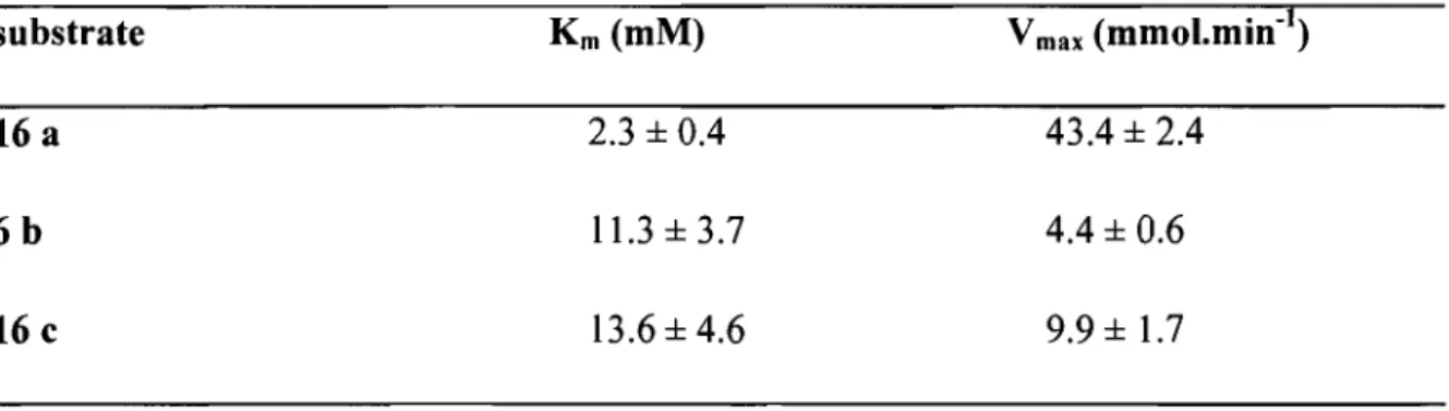
Biotinylated amines 16a-c were synthesized by coupling biotin to their corresponding diamines 13a-c (Scheme 2.2). The shortest spacer used was 1,5-diaminopentane 13a. Tris(ethylene glycol)-1,8-diamine 13b was both longer and more polar, white 2-(2-amino-ethyldisulfonyl)-ethylamine 13c was of length similar to 13a but contained a cleavable disulfide bond. Compounds 16a-c were tested as acceptor substrates using the
Cbz-L-Glu(O-p-nitrophenyl)Gly assay 23 and found to have KM values of 2.3 ± 0.4 mM,
11.3 ± 3.7 mM, and 13.6 ± 4.6 mM, respectively (Table 2.2). Thus, the different spacers tested aU provided productive binding in the same order of magnitude, validating their use in the coupling reaction.
Table 2.2: Kinetic constants for TGase with various acyl acceptor substrate 16 a 6b 16 c Km (mM) 2.3 ± 0.4 11.3±3.7 13.6 ± 4.6 V max (mmol.min-l) 43.4 ± 2.4 4.4 ± 0.6 9.9 ± 1.7
Scheme 2.2: Synthesis of different acyl acceptors
x=
(C) s-s o/S~N~X~NH
HCI HCI(1 M) ) - - \ .. IU H 2 ... ~-~..:....--HN NH1
(16): (a), (b), (c) 2.4.1 Materials .... TBTU, DIEAo
(14): (a), (b), (C)~OH
HN~NH oo
~~~X'./'NHBo<:
HN NH \ \ (15): (a), (b), (c)o
The buffer solution in aIl assays gave a final concentration of 100 mM Tris-HCl (pH 8), 0.6 mM EDTA, and 10 mM CaCh. The washing buffer solution was composed of 100 mM sodium phosphate (pH 8), 100 mM NaCl, and 0.1% SDS. Water was purified using a Millipore BioCell water purification system. Streptavidin beads (Pierce) were supplied as a suspension capable of binding 15-28 Ilg biotinimL of settled resin (a capacity corresponding to ~ 1-3 mg biotinylated BSA/mL of resin) on a support of 6% crosslinked beaded agarose.
2.5 Assay procedure using a purified enzyme
2.5.1 Methods
AIl assays were performed in triplicate. Donor substrate 11a, b, or c (10 ilL of 0.6 mM stock solution), acceptor substrate 16a, b, or c (10 ilL of 0.6 mM stock solution), 80 ilL of buffer solution, and 90 ilL of water were combined in a final volume of 190 ilL. The reaction was initiated by addition of 25 Ilg (10 Ill) of purified TGase 63 or an equivalent volume of 25 mM tris-acetate assay buffer for the blank. FoIlowing incubation for 30 min at 25 oC, 10 III of the streptavidin bead suspension was added. The mixture was incubated for 30 min at 25 oc. The tubes were centrifuged for 1 min at 1400 RPM (micro centrifuge). The supernatant was discarded. The pellets were washed by resuspension in washing buffer (200 ilL) and centrifuged as above. The pellets were washed twice more, resuspended in washing buffer (100 ilL) and transferred to a polystyrene 96-well plate. Enzyme activity was detected using a Perkin Elmer Bio Assay Reader (HTS 7000) using Aexc
=
485 nm, Aem=
535 nm for substrates 11a and11b, and Aexc = 360 nm, Aem = 535 nm for substrate 11c, by measuring the increased fluorescence signal relative to the blank.
Reduction of the disulfide-bridged acyl-acceptor substrate
Substrates 11a and 16c were incubated for 60 min at 25 oC (30 IlM 11a, 30 IlM 16c, 100 mM Tris-HCI, 10 mM CaCh, 0.6 mM EDTA, and 350 mU TGase in a total volume of 200 ilL), after which 10 ilL of streptavidin bead suspension was added. The mixture was incubated for a further 60 min. The tubes were centrifuged for 1 min at 1400 x
adding 200 ~L of wash buffer, centrifuging for 1 min at 1400 x RPM (microcentrifuge)
and discarding the supernatant. Finally the beads were suspended in 1 00 ~L of wash buffer for treatment under one of the following conditions:
1 1 00 ~L of 0.1 M DTT was added, followed by 15 min incubation, centrifugation and removal of the supernatant for fluorescence detection.
2 1 00 ~L of 0.1 M DTT was added, followed by 15 min incubation, and the entire mixture was used for fluorescence detection.
3 1 00 ~L of water was added, followed by centrifugation and removal of the supernatant for fluorescence detection.
4 1 00 ~L of water was added, and the entire mixture was used for fluorescence detection.
2.5.2 Results and discussion
In our fluorescent assay for TGase, using synthetic donor and acceptor substrates, the formation of a conjugate between a fluorescent acyl-donor substrate and an amine acceptor substrate bearing a biotin group was catalyzed by TGase. The resulting biotinylated fluorophore-Iabelled product was trapped with immobilized streptavidin.
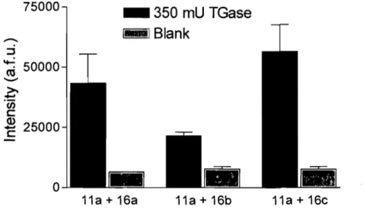
• Assay sensitivity with different acyl acceptors
To evaluate the effect of the acceptor substrate spacer on assay sensitivity, acyl donor substrate
11a
was incubated with acyl acceptor substrates 16a-c, respectively, at final concentrations of 30 JlM, in the presence or absence of TGase (Figure 2.2). Theacceptor substrates 16a-c gave different levels of fluorescence that did not correlate
with their KM values. For example, the highest and lowest fluorescence measurements were exhibited by the substrate harbouring the polyethylene glycol 16b and disulfide
16c spacer, respectively, that exhibited very similar KM values.
Acceptor substrate 16c gave the highest signal and contains a disulfide spacer, which
also offered the potential for 'reversible' biotinylation by reductive cleavage of the disulfide bond. This allowed us to verify whether quantitation of the fluorescent conjugate directly on the streptavidin beads provided a maximum signal or whether some loss of fluorescence was suffered. Thus, following enzymatic coupling of the donor and acceptor substrates Il a and 16c, trapping on the streptavidin-coated beads
and washing, reduction using D,L-dithiothreitol (DTT) and separation by centrifugation allowed measurement of the released fluorophore in the absence of the beads (Table 2.3).
The intensity of the fluorescence measured for the released fluorophore (+DTT, before or after centrifugation) was comparable to that measured directly on the beads (-DTT, before centrifugation). A low level of fluorescence was apparently released from the beads even in the absence of DTT (-DTT, after centrifugation). Nonetheless, fluorescence measurement directly on the beads was not significantly hampered and the additional steps required to release the fluorophore were not deemed to be justified. As compound 16c yielded the highest fluorescence after coupling to the donor substrate
11a (Figure 2.2), it was retained as the most suitable acceptor substrate for further
Figure 2.2: Comparison of assay sensitivity using acyl acceptor substrates 16a-c, which
differ by their spacer. Substrate lIa was the acyl donor. Results are provided for triplicates. The error bars represent the standard deviation from the mean.
75000
-
:J~
50000
--
...
>-. Ci) c:~
25000
o
_
350 mU TGase
1 iilBlank
11a + 16a 11a + 16b 11a + 16c
Table 2.3: Fluorescence resulting from the coupling of lIa and 16c, measured on
streptavidin beads or following release.
Before centrifugation
Supematant after centrifugation
a Àex
=
485nm,
Àem=
535nm
Fluorescence (a.f.uf +DTT -DTT 6.3 X 10⁴ 4.2 X 10⁴ 4.6 X 10⁴ 1.0 X 10⁴• Assay sensitivity with different acyl donors
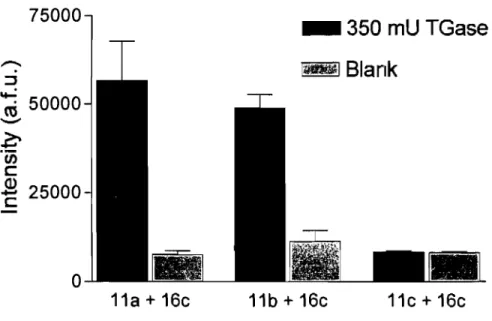
To evaluate the effect of the fluorophore on assay sensitivity, acyl-acceptor substrate 16c was incubated with the acyl-donor substrates 11a-c and purified TGase. Substrate lle was shown to be an insufficiently intense fluorophore and was not retained for further testing. The substrates lIa and lib showed significant enhancement of signal compared to the control, but the fluorescence of the blank for lib was significantly higher than that of 11a. Among these acyl donor substrates, 11a gave the best results and was therefore chosen for subsquent tests (Figure 2.3).
Figure 2.3: Assay sensitivity with acyl donor lla-c and acyl acceptor substrate 16c . Results are provided for triplicates. The error bars represent the standard deviation from the mean. 75000