Viral Integration Sites in Human Papilloma
Virus-33-Immortalized Cervical Keratinocyte Cell Lines
Christine Gilles a,b, *, Jacques Piette a, Dominique Ploton c, Martine Doco-Fenzy d, and Jean-Michel Foidart ba Laboratory of Fundamental Virology, University of Liège, C.H.U. Sart-Tilman, Liège, Belgium b Laboratory of General Biology, University of Liège, C.H.U. Sart-Tilman, Liège, Belgium c INSERM U.314, C.H.U., Maison Blanche, Jay, Reims, France.
d Laboratory of Cytogenetic, Maison Blanche, Jay, Reims, France.
ABSTRACT
The viral organization of HPV-33 was determined by Southern blotting in 2 HPV-33-immortalized cervical cell lines (CK11 and CK12) and compared to our previous results obtained on 10 other already characterized HPV-33-immortalized cell lines (CK1 to CK10). As observed in CK1 to CK10, the viral DNA was found integrated in the cellular genome of CK11 and CK12. However, in CK11 and CK12, the integrated viral genome was deleted and mostly limited to the URR and the E6-E7 ORFs, stressing the importance of those sequences in the
immortalization process. Furthermore, CKll and CK12 showed a unique and identical integration site, as observed in CK1 to CK10, which also harbored HPV-33 integrated at a unique and identical site (which was however different from the one evidenced in CK11 and CK12). Indeed, in situ hybridizations on chromosomes allowed the precise localization of the viral DNA on chromosome 13q33-34 in CK1 to CK10 whereas it was mapped to chromosome 9p13 in CK11 and CK12. We discuss the possibility that integration of HPV-33 at those two particular sites has conferred some growth advantages to the cells and could have thus played a crucial role in the immortalization.
INTRODUCTION
About 20 human papillomavirus (HPV) types have been implicated in the etiology of genital cancers [1]. Some of them, such as HPV-6 or -11, are called nononcogenic HPVs because of their preferential association with benign lesions. Others, such as HPV-16, -18, and -33 are closely related to malignant lesions and have therefore been termed oncogenic HPVs [2]. Oncogenic HPVs display the ability to immortalize many cell types in vitro, including human keratinocytes. The immortalizing functions of these HPVs have been mapped to the E6-E7 open reading frame (ORF) as they are necessary and sufficient to immortalize primary keratinocytes. The oncogenic potential of these genes might partly rely on their ability to bind p53 and pl05Rb respectively, thus interfering with the control of cell proliferation mediated by these two cellular genes [3, 4]. In most cases and in contrast with nononcogenic HPVs, oncogenic HPVs are found integrated into the chromosomes of carcinoma cells or in vitro HPV-transfected cells. Integration of HPV-16 or -18 often leads to the disruption of the viral E2 ORF [5-7], which encodes a transcriptional repressor of the viral early promoter, driving the transcription of the E6 and E7 genes. This phenomenon might be related to the observation that the E6-E7 ORFs are often retained and expressed in cancer cells, supporting the idea that these genes are implicated in cellular transformation [5-9]. In contrast with these data, the contribution of the integration site within the chromosomes of the host cells to the tumor phenotype remains unclear. Even though it is believed that HPV integration happens initially at random in the cellular genome, some data have shown that most cancer cells harboring HPV-16 or HPV-18 have integrated the virus at chromosomal loci containing fragile sites and oncogenes [10, 11].
An in vitro model of HPV-33-transfected cervical keratinocytes has recently been developed in our laboratory [12, 13]. Like HPV-16 or -18, HPV-33 is an oncogenic HPV [14] but its ability to integrate within the cellular genome is not well documented. Ten of these HPV-33-immortalized cell lines (CK1 to CK10) were previously shown (by Southern analyses) to harbor HPV-33 integrated at a unique and identical site within their genome, emphasizing the importance of the integration site in the immortalization of these lines [12].
In the present paper, we studied the organization and transcription of 2 newly established HPV-33-immortalized cell lines (CK11 and CK12) and compare these characteristics with those observed in CK1 to CK10. In situ hybridizations on chromosomes were carried out to localize precisely the HPV-33 integration sites in CK1 to CK10 and in CK11 and CK12.
MATERIAL AND METHODS Cell Lines
Twelve HPV-33-immortalized cervical keratinocyte cell lines were used (CK1 to CK12). The establishment of 10 of these lines (CK1 to CK10) as well as the viral organization and transcription within these lines were previously described [12]. Briefly, primary cervical keratinocytes were transfected by electroporation with the linearized HPV-33 DNA. Transfected cells were pooled until about passage 10, passage at which untransfected cells died, leading to the emergence of distinguishable small islands of surviving cells. These islands were isolated and expanded in culture, raising different cell populations. The transfected cell populations underwent several growth crises before the emergence of stable cell lines. The two other cell lines (CK11 and CK12) were obtained by a similar isolation procedure but were established from another HPV-33 transfection performed on a distinct culture of normal cervical keratinocytes. The transfected lines were cultivated as described previously [12].
Southern and Northern Analyses
DNA extracted from the different cell lines was digested by the appropriate restriction enzymes and run on a 1% agarose gel in TEA. After denaturation and neutralization, DNA was transferred from gels to positively-charged nylon membranes (Zetaprobe membranes, Biorad, Richmond, CA, USA) in an alkaline buffer by capillary blotting.
Total RNA was isolated from a 4 × 10⁶ cell pellet and was transferred by a vacuum method onto a positively-charged nylon membrane. Southern and Northern hybridizations were carried out according to the
manufacturer's instructions (Amersham, Buckinghamshire, U.K.). 32P-labeled probes (> 5 × 10⁸ cpm/mg) consisted of either the total HPV-33 genome or the E6-E7 region (from nucleotides 97 to 785 isolated from the total genome following a RsaI digestion).
In Situ Hybridization on Chromosomes
The viral integration sites were determined by fluorescence in situ hybridization (FISH) on chromosomes combined with R-banding procedure. Briefly, BrdU incorporated cells (cultivated 6 hours with 10 µg/ml BrdU) were spread on glass slides. Before hybridization, slides were treated with Hoechst 33258 (0.1 mg/ml in 2 × SSC) for 20 min and UV-irradiated for 30 min. Slides were incubated with RNAse (100 µg/ml), denaturated, and then hybridized at 37°C for 18 hours. Hybridization was carried out using a biotinylated probe in 50% (v/v) formamide-2 × SSC-10% (w/v) dextran sulfate. Washings were performed in 50% (v/v) formamide-2 × SSc at 45°C. Immunodetection of the biotinylated probe was carried out using a FITC-conjugated avidine amplification system. The slides were successively incubated with a fluorescein-conjugated avidin, with an anti-avidin biotinylated goat antibody and again with a fluorescein-conjugated avidin. Slides were then observed under a fluorescence microscope or under a confocal microscope (Biorad MRC 600). For each cell line analyzed, 50 metaphases were observed.
The biotinylated probes (either a total HPV-33 probe or a probe covering the E6-E7 region of HPV-33) were made by nick translation in presence of biotinylated 16-D-UTP (Boehringer, Mannheim, Germany) and biotinylated 14-D-ATP (Gibco, Paisley, U.K.).
RESULTS
HPV-33 Organization in CK11 and CK12 Cell Lines
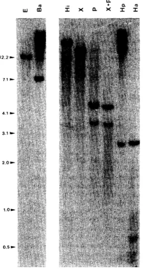
To study the organization of HPV-33 in CK11 and CK12 and compare it with our previous observation in CK1 to CK10, Southern blotting analyses were performed on genomic DNA extracted from CK11 and CK12 and digested with a set of restriction enzymes. Fig. 1 shows the Southern patterns obtained for DNA extracted from CK11 and hybridized with a total HPV-33 probe. The same analyses performed on DNA extracted from CK12 gave similar results (data not shown). The expected restriction map of HPV-33 for these enzymes is given in Fig. 2a. EcoRI and BamHI have no restriction site within the HPV-33 genome and, therefore, the observation of a single band representing the viral sequences integrated within the cellular DNA was expected. However, the observation that BamHI digestion gave rise to a band smaller than the size of HPV-33 (7.9 kb) strongly suggested that a rather large deletion in the viral genome probably occurred during the integration process. The unique restriction fragment detected after HindIII or XbaI digestion, each of which have two restriction sites within the HPV-33 genome, confirmed this hypothesis suggesting that the four restriction sites were lost within the integrated viral sequences. Also, no differences were observed between the PstI and the double PstI-XbaI restriction patterns. This observation demonstrated that the XbaI site was lost and that only a single PstI site (which normally cleaves HPV-33 at four sites) was retained, giving two bands corresponding to two junctional fragments between HPV-33 and the cellular genome. For HpaII digestion, an expected band probably
A very weak band of about 0.4 kb could also represent an internal expected fragment of 387 bp. Another very weak band of 0.8 kb as well as a high—molecular-weight band (visible within the smear of undigested DNA) could correspond to the junctional fragments between viral and cellular DNA. In the same way, the HaeIII pattern showed a band of about 2.7 kb, which could be the expected internal fragment of 2694 bp. Also, a set of bands of low-molecular-weight was detected, corresponding either to small expected fragments (615 bp, 299 bp) or to junctional fragments if one considers that the virus was integrated close to HaeIII cellular sites. However, the absence of other expected fragments following HpaII or HaeIII digestions also supported the idea that an important viral region was deleted following or during integration. Therefore, we can conclude from our restriction analyses, that the minimal viral region integrated within the cellular genome of CK11 and CK12 cell lines extended from the HaeIII site at nucleotide 5907 to the HaeIII site at nucleotide 1497, implicating that the conserved PstI site was located at nucleotide 6745. As shown in Fig. 2b, our results led to the conclusion that in these two cell lines (CK11 and CK12), the integrated viral sequences encompassed the entire URR as well as the E6-E7 ORFs but also a part of L1 (3' border) and El (5' border). These observations contrasted with the results previously obtained for CK1 to CK10 cell lines where the viral genome was only slightly altered [12] but where, anyhow, the URR and the E6-E7 ORFs were also retained.
Figure 1 Southern hybridizations of total genomic DNA extracted from CK11, digested by a set of the following
restriction enzymes and hybridized with a total HPV-33 probe. E: EcoRI, Ba: BamHI, Hi: HindIII, X: XbaI, P: PstI, X+P: double digestion XbaI + PstI, Hp: HpaII, Ha: HaeΠI.
Figure 2 (a) Restriction map of HPV-33 for the restriction enzymes indicated. The virus is interrupted within the
E2 ORF at a BgIII site. The length of the main expected fragments are indicated in bp (b) Postulated organization of HPV-33 integrated within CK11 and CK12 cell lines.
Northern Analyses
Northern blotting analyses were performed on mRNA extracted from CK11 and CK12 and hybridized with a total HPV-33 probe (Fig. 3a) or an E6-E7 probe (Fig. 3b). These analyses clearly showed the presence of several viral mRNAs that could be similarly detected with the total HPV-33 probe or with the E6-E7 probe. This pattern of transcription was different from the one observed for CK1 to CK10 in which the pattern of mRNAs detected with the total HPV-33 probe was more complicated than the one detected with the E6-E7 probe, as previously reported [12].
Figure 3 Northern analyses of total RNA extracted from CK11 and hybridized with (a) total genomic HPV-33
probe and (b) E6/ E7 probe.
Evolution of the Integration Pattern
To analyze the evolution of the organization of HPV-33 in parallel with the progression towards the
immortalized phenotype, Southern analyses were performed on DNA extracted from CK11 at different passage levels (P29, P50) (Fig. 4). Earlier passages (P9 and P22) corresponding to the cell population from which the stable CK11 line was established, were also studied. As shown in Fig. 4, the hybridization pattern observed at passage 9 clearly showed the internal expected fragments of 3741, 1693, 1173, and 318 bp (the 120 bp fragment
could be too small to be detected). The presence of other weaker bands could correspond to various junctional fragments suggesting that the viral DNA, at that early passage level, was integrated at several distinct sites in the cellular genome. These bands could also represent fragments obtained from various episomal forms of the viral DNA. It is important to notice that the hybridization pattern clearly evolved towards the one observed in the stable CK11 line in parallel to passage level, suggesting that cells possessing the integration pattern observed in CK11 were selected during cultivation.
Figure 4 Evolution of the integration pattern according to the passage level. Southern analyses were performed
on DNA extracted from the initial transfected cell population at early passage levels (P9, P22) or from CK11 (P29, P50) once established in culture and digested with XbaI and PstI.
In Situ Hybridizations on Chromosomes
The precise viral integration sites within our two sets of cell lines was determined by fluorescence in situ hybridizations (FISH) on chromosomes combined with R-banding procedure. Fifty metaphases were observed in each cell line.
For CK1 to CK10 cell lines, the results, obtained with the whole HPV-33 as a probe, clearly localized HPV-33 at a single site, on locus 13q33-34 in more than 90% of the hybridized metaphases (Fig. 5a). For CK11 and CK12, which have integrated only a part of the viral genome, the hybridizations were carried out using an E6-E7 probe to optimize the detection. We could therefore map the integration site to 9p13 in 82% of the hybridized
metaphases in CK11 and CK12 (Fig. 5c). Analyses by confocal microscopy greatly improved the precision of the localization as compared with classical fluorescence microscopy (Fig. 5b,d).
Figure 5 Localization of HPV-33 integration sites by FISH on R-banded chromosomes analyzed by classical
fluorescence microscopy (bar = 9 µm) (a,c) and confocal microscopy (b,d). The integration sites in CK1 and CK11 are shown as representative of the site of integration in CK1 to CK10 and CK11 and CK12 cell lines respectively.
DISCUSSION
Our previous [12] and present results have shown that 12 HPV-33-transfected cervical cell lines harbor the viral DNA integrated within the cellular genome. These data are in agreement with many reports describing HPV-16 or -18 integration in cancer cells of the cervix [5, 6, 15-17]. Moreover, in contrast with nononcogenic HPVs, which are mostly in an episomal state, HPV-16 or -18 are preferentially found integrated in high grade lesions [18-23]. These findings emphasize the importance of the integration event in the cellular transformation. Also, as previously shown in CK1 to CK10, our Southern analyses clearly revealed that HPV-33 is integrated at a single and identical site (which is however different from the integration site in CK1 to CK10). The observation (in CK11) that the viral organization evolved in parallel with the passage level has led us to state the following hypothesis. HPV-33 integration would have occurred as an early event, at several sites in the cellular genome (as revealed by the complex hybridization pattern observed in CK11 at low passage levels). Cells possessing some growth advantages would have then been selected as they were passaged in vitro. The simplification of the integration pattern reflects this phenomenon of selection. It is thus likely that all the cell lines evolved from a common set of cells that had integrated the viral DNA at a particular site, and were progressively selected during in vitro cultivation. It could thus be hypothesized that the integration at those two particular sites (in CK1 to CK10 on one hand and in CK11 and CK12 on the other hand) might have played an important role in the immortalization process by conferring to the cells some growth advantages. This was further strengthened by our previous observation [12] that cell populations that have integrated the viral DNA at different sites could not be stably established in culture. This interpretation of our results is in agreement with many data obtained by Southern blotting describing a single integration site of HPV-16 or -18 in cervical cancer cell lines or in in vitro transfected lines [5, 7, 15, 16, 24-28]. Moreover, some studies reporting HPV integration at a single site in high grade lesions emphasize the monoclonal origin of cervical cancers and support our results [19].
Viral integration may lead to the reorganization of both viral and cellular genes. Our previous results have shown that the viral DNA integrated in CK1 to CK10 was only slightly altered and could generate several viral mRNAs. These results contrast with our present findings showing that, in CK11 and CK12, an important deletion
occurred in the viral DNA. Indeed, it appeared that the URR and the E6-E7 ORFs were the main conserved regions in these two lines. This finding was further documented by Northern analyses, which showed the same transcription pattern using a complete HPV-33 probe or an E6-E7 restricted probe. Therefore, it is clear that the selection phenomenon has favored cells that have conserved the E6-E7 genes and that express viral mRNAs for this region. This indirectly suggests that these two genes have played an important role in the immortalization process. This is in agreement with numerous data reporting that the E6-E7 region is invariably conserved and expressed in cervical carcinoma cells or in cells immortalized in vitro by HPV-16 or -18 [5-9]. This is also consistent with the fact that E6-E7 of oncogenic HPVs were shown to possess some transforming functions and that the cells expressing these genes display some growth advantages [3, 4]. Even though the data concerning HPV-33 are limited, Kitasato [29] has shown that the E6 and E7 of that HPV possess some transforming functions in NIH3T3 cells, strengthening our results.
Despite the fact that the cells that had conserved the E6-E7 region were selected, it is clear that the importance of the integration event also rely on the cellular site implicated. Indeed, our in situ hybridizations on chromosomes confirmed our Southern analyses by evidencing a unique and identical site of integration in CK1 to CK10 on one hand and in CK11 and CK12 on the other hand. This strongly suggests that integration at those particular sites might have conferred some growth advantages to the cells and might have thus been crucial in their
immortalization and their selection. In agreement with our results, in situ hybridization analyses on chromosomes have described HPV-16 or -18 integrated at a single site within cervical carcinomas [30] or cervical carcinoma cells such as SW756 [31], SiHa [32] or C4-I [33] cell lines, even though HeLa and Caski cells were shown to harbor HPV-18 and HPV-16 respectively at several distinct sites [32, 34, 35]. From those localization studies, it appeared that HPV integration sites were frequently mapped in regions containing oncogenes or fragile sites [10, 11]. The integration might therefore alter cellular genes (such as protooncogenes) that could then be implicated in the transformation process. Indeed, two chromosomal loci, 8q24 and 12q13-15, have been most frequently involved in HPV integration. Furthermore, studies reporting HPV-16 or -18
integration on 8q24 have also described an overexpression of the c-myc oncogene, which is located at the same locus [25]. The locus 9p13, on which HPV-33 was mapped within CK11 and CK12, is actually not very well documented. However, in agreement with the concept expressed above, HPV-33 integration on the locus 13q33-34 could have altered some cellular genes. Indeed, a fragile site as well as several genes such as RAP-2, a member of the ras oncogene family, and ERCC5, a DNA excision-repair enzyme the disregulation of which could lead to the accumulation of mutations, were also localized in that region [36, 37]. The detection of HPV-16 integrated at a similar locus in SiHa cells (13q21-31) also reinforces our observation.
It could thus be concluded that HPV-33 integration has contributed to the transformed phenotype of our cell lines. Even though the E6-E7 region was invariably conserved in our lines, suggesting the implication of these genes in the immortalization process, it is clear that the integration at particular cellular sites might also have contributed to the transformation of our lines. Because HPV-16 and -18 integration has been shown to occur at sites involving genes possessing oncogenic potential, it is likely that HPV-33 integration could also implicate chromosomal loci harboring such genes. Nevertheless, additional data are necessary to address the precise mechanisms by which integration at 13q33-34 or 9p13 could confer growth advantages to cervical keratinocytes.
ACKNOWLEDGEMENT
This work was supported by grants from: the "Association sportive contre le Cancer," the "Centre Anticancéreux près l'Université de Liège," FNRS grant number 3.9003.92, the "Communauté Française": ARC number 90/94-139, the "Fonds de Recherche Facultaire-Université de Liège." C.G. is supported by a FNRS-Télévie grant 1992 number 7.4555.92 and J.P. is Research Director from the National Fund for Scientific Research (Brussels, Belgium).
REFERENCES
1. DeVilliers EM. Heterogeneity of the human papillomavirus group (1989): J Virol 63:4898-4903.
2. zurHausen H, Schneider A (1987): The role of papillomaviruses in human anogenital cancers, In: The Papovaviridae, Salzman N. and Howley RM, eds. Plenum Press, New York, pp 245-263.
3. Münger K, Scheffner M, Huibregste JM, Howley PM. Interactions of HPV E6 and E7 oncoproteins with tumor suppressor gene products (1992): Cancer Surveys 12:197-217.
5. El Awady M, Kaplan J, O'Brien S, Burk R (1987): Molecular analysis of integrated papillomavirus 16 sequences in the cervical cancer cell line SiHa. Virology 159:389-398.
6. Choo KB, Pan CC, Han SH (1987): Integration of human papillomavirus type 16 into DNA of cervical carcinoma: Preferential deletion of the E2 gene and invariable retention of the long control region and the E6/E7 open reading frames. Virology 161:259-261.
7. Schwarz E, Freese UK, Gissmann L, Mayer W, Roggenbuck B, Stemlau A, zurHausen H (1985): Structure and transcription of human papillomavirus sequences in cervical carcinoma cells. Nature (London) 314:111-114.
8. Schwarz E, Schneider-Gadicke A, Roggenbuck B, Mayer W, Gissman L, zurHausen H (1986): Expression of human papillomavirus DNA in cervical carcinoma cell lines. Banbury Rep 21:281-290.
9. Romanczuk H, Howley PM (1992): Disruption of either El or the E2 regulatory gene of human papillomavirus type 16 increases viral immortalization capacity. Proc Natl Acad Sci USA 89:3159-3163.
10. Popescu NC, DiPaolo JA (1989): Preferential sites for viral integration on mammalian genome. Cancer Genet Cytogenet 42:157-171. 11. Lazo PA, Galleg MA, Ballester S, Feduchi E (1992): Genetic alterations by human papillomaviruses in oncogenesis. FEBS letters 300:109-113.
12. Gilles C, Piette J, Rombout S, Laurent C, Foidart JM (1993): Immortalization of human cervical keratinocytes by human papillomavirus type 33. Int J Cancer 53:872-879.
13. Gilles C, Piette J, Peter W, Fusenig NE, Foidart JM (1994): Differentiation ability and oncogenic potential of 33- and HPV-33+ras-transfected keratinocytes. Int J Cancer 58:847-854.
14. Beaudenon S, Kremsdorf D, Croissant O, Jablonska S, Wain-Hobson S, Orth G (1989): A novel type of human papillomavirus associated with genital neoplasia. Nature 321:246-249.
15. Durst M, Dzarlieva-Petrusevka RT, Boukamp P, Fusenig NE, Gissmann L (1987b): Molecular and cytogenetic analyses of immortalized human primary keratinocytes obtained after transfection with human papillomavirus type 16 DNA. Oncogene 1:251-256.
16. Pecoraro G, Morgan D, Defendi V (1989): Differential effects of human papillomavirus type 6, 16, and 18 DNAs on immortalization and transformation of human cervical epithelial cells. Proc Natl Acad Sci USA 86:563-567.
17. Woodworth CD, Doniger J, DiPaolo JA (1989): Immortalization of human foreskin keratinocytes by various human papillomavirus DNAs corresponds to their association with cervical carcinoma. J Virol 63:159-164.
18. Durst M, Kleinheinz A, Hotz M, Gissmann L (1985): The physical state of human papillomavirus type 16 DNA in benign and malignant genital tumors. J Gen Virol 66:1515-1522.
19. Lehn H, Krieg P, Sauer G (1985): Papillomavirus genomes in human cervical tumors: Analysis of their transcriptional activity. Proc Natl Acad Sci USA 82:5540-5544.
20. Pater MM, Pater A (1985): Human papillomavirus type 16 and 18 sequences in carcinoma cell lines of the cervix. Virology 145:313-318.
21. DiLuca D, Pilotti S, Stefanon B, Rotola A, Monini P, Tognon M, DePalo G, Rilke F, Cassai E (1986): Human papillomavirus type 16 DNA in genital tumors: A pathological and molecular analysis. J Gen Virol 67:583-589.
22. Durst M, Schwarz E, Gissmann L (1986): Integration and persistence of human papillomavirus DNA in genital tumors. Banbury Rep 21:273-280.
23. Lehn H, Villa LL, Marziona F, Hilgarth M, Hillemans HG, Sauer G (1988): Physical state and biological activity of human papillomavirus genomes in precancerous lesions of the female genital tract. J Gen Virol 69:187-193.
24. Matsukura T, Kanda T, Furuno A, Yoshikawa H, Kawana T, Yoshiike K (1986): Cloning of monomeric human papillomavirus type 16 DNA integrated within cell DNA from a cervical carcinoma. J Virol 58:979-982.
25. Durst M, Croge CM, Gissmann L, Schwarz E, Huebner K (1987a): Papillomavirus sequences integrate near cellular oncogenes in some cervical carcinomas. Proc Natl Acad Sci USA 84:1070-1074.
26. Shirasawa H, Tomita Y, Sekiya S, Takamizawa H, Simizu B (1987): Integration and transcription of human papillomavirus type 16 and 18 sequences in cell lines derived from cervical carcinomas. J Gen Virol 68:583-591.
27. Pirisi L, Creek KE, Doniger J, DiPaolo JA (1988): Continuous cell lines with altered growth and differentiation properties originate after transfection of human keratinocytes with human papillomavirus type 16 DNA. Carcinogenesis 9:1573— 1579.
28. Wagatsuma M, Hashimoto K, Matsukura T (1990): Analysis of integrated human papillomavirus type 16 DNA in cervical cancers: Amplification of viral sequences together with cellular flanking sequences. J Virol 64:813-821.
29. Kitasato H, Hillova J, Lenormand M, Hill M (1991): Tumorigenicity of the E6 and E6-E7 gene constructions derived from human papillomavirus type 33. Anticancer Res 11: 1165-1172.
30. Couturier J, Sastre-Garau X, Schneider-Manoury S, Labib A, Orth G (1991): Integration of papillomavirus DNA near myc genes in genital carcinomas and consequence on protooncogene expression. J Virol 65:4534-4538.
31. Popescu NC, Amsbaugh SC, DePaolo JA. (1987a): Human papillomavirus type 18 DNA is integrated at a single site in cervical carcinoma cell line SW756. J Virol 51:1682-1685.
32. Mincheva A, Gissmann L, zurHausen H (1987): Chromosomal integration sites of human papillomavirus DNA in three cervical cancer cell lines mapped by in situ hybridization. Med Microbiol Immunol 176:245-256.
33. Cannizaro LA, Durst M, Mendez MJ, Hecht BK, Hecht F (1988): Regional chromosome localization of human papillomavirus integration sites near fragile sites, oncogenes, and cancer chromosome breakpoints. Cancer Genet Cytogenet 33:93-98.
34. Ambros PF, Karlic HI (1987): Chromosomal insertion of human papillomavirus sequences in HeLa cells detected by non isotopic in situ hybridization and reflexion contrast microscopy. Hum Genet 77:251-254.
35. Popescu NC, DiPaolo JA, Amsbaugh SC (1987): Integration sites of human papillomavirus type 18 DNA sequences on HeLa cells chromosomes. Cytogenet Cell Genet 44:58-62.
36. Takahashi E, Shiomi N, Shiomi T (1988): Precise localization of the excision-repair gene, ERCC5, to human chromosome 13q32.3-q33.1 by direct fluorescence in situ hybridization. Jpn J Cancer Res 83:1117-1119.
37. Rousseau-Merck MF, Pizon V, Tavitian A, Berger R (1990): Chromosome mapping of the human RAS-related RAP1A, RAP1B, RAP2 genes to chromosomes 1p12-p13, 12q14, and 13q34, respectively. Cytogenet Cell Genet 53:2-4.