O R I G I N A L A R T I C L E
Sex Differences in White Matter Microstructure in
the Human Brain Predominantly Re
flect Differences
in Sex Hormone Exposure
J. van Hemmen
1,2,5
, I. M. J. Saris
6
, P. T. Cohen-Kettenis
2
, D. J. Veltman
3,5
,
P. J. W. Pouwels
4,5
and J. Bakker
1,2,7
1
Netherlands Institute for Neuroscience, Amsterdam, The Netherlands,
2Department of Medical Psychology,
3Department of Psychiatry,
4Department of Physics and Medical Technology and,
5Neuroscience Campus
Amsterdam, VU University Medical Center, Amsterdam, The Netherlands,
6Department of Psychiatry,
GGZ inGeest, Amsterdam, The Netherlands and
7GIGA Neurosciences, University of Liège, Liège, Belgium
Address correspondence to Judy van Hemmen, VU University Medical Center Amsterdam, Department of Medical Psychology, HB 1H11, PO Box 7057, 1007 MB Amsterdam, The Netherlands. Email: j.vanhemmen@vumc.nl, judy_van_hemmen@hotmail.comAbstract
Sex differences have been described regarding several aspects of human brain morphology; however, the exact biological mechanisms underlying these differences remain unclear in humans. Women with the complete androgen insensitivity syndrome (CAIS), who lack androgen action in the presence of a 46,XY karyotype, offer the unique opportunity to study isolated effects of sex hormones and sex chromosomes on human neural sexual differentiation. In the present study, we used diffusion tensor imaging to investigate white matter (WM) microstructure in 46,XY women with CAIS (n = 20), 46,XY comparison men (n = 30), and 46,XX comparison women (n = 30). Widespread sex differences in fractional anisotropy (FA), with higher FA in comparison men than in comparison women, were observed. Women with CAIS showed female-typical FA throughout extended WM regions, predominantly due to female-typical radial diffusivity. Thesefindings indicate a predominant role of sex hormones in the sexual differentiation of WM microstructure, although sex chromosome genes and/or masculinizing androgen effects not mediated by the androgen receptor might also play a role.
Key words: androgens, CAIS, DTI, sexual differentiation, white matter
Introduction
Sex differences have been described regarding several aspects of human brain morphology, although the specific biological mechanisms driving neural sexual differentiation remain to be determined. Numerous neuroimaging studies have focused on macro- and mesoanatomical sex differences, such as in overall or regional gray and white matter (WM) volume derived from structural magnetic resonance imaging (MRI) scans (for meta-analysis seeRuigrok et al. 2014). More recently, diffusion tensor imaging (DTI), an MRI technique that measures characteristics of water molecule diffusion (Basser et al. 1994), has been widely
used to study sex differences in WM microstructure. Quantitative measures that can be derived from the diffusion tensor include fractional anisotropy (FA), axial (AD), and radial diffusivity (RD) (Pierpaoli and Basser 1996). FA provides information about the degree of diffusion anisotropy. A low FA value reflects isotropic diffusion, that is, equal diffusion in all directions (e.g., in cerebro-spinalfluid). A high degree of anisotropy is found in WM fiber bundles, in which water diffusion is restricted in the direction perpendicular to the axon, and relatively unrestricted along the axon. AD, which equals the largest eigenvalue of the diffusion tensor, and RD, the mean of the two smaller eigenvalues, are
© The Author 2016. Published by Oxford University Press. All rights reserved. For Permissions, please e-mail: journals.permissions@oup.com
Cerebral Cortex, 2016, 1–8
doi: 10.1093/cercor/bhw156 Original Article
1
at University of Liege on May 30, 2016
http://cercor.oxfordjournals.org/
interpreted as the diffusivity parallel and perpendicular to the WM tract, respectively, and are typically assessed to further characterize FA.
There is evidence for sex differences in WM microstructure as assessed by diffusivity measures. The majority of whole-brain DTI studies have found higher FA in men in major WM regions (Chou et al. 2011;Inano et al. 2011;Menzler et al. 2011;Rametti et al. 2011;Den Braber et al. 2013;Kanaan et al. 2014;Schoonheim et al. 2014;Takao et al. 2014). While sex effects in other diffusivity measures are less frequently studied, and results are less consist-ent compared with FA, a study in a large sample of 547 men and 310 women, showed that a higher FA in men is generally associated with a higher AD and a lower RD than in women (Inano et al. 2011).
At present, the exact factors and mechanisms underlying the sexual differentiation of WM microstructure are unknown. The classical theory of the sexual differentiation of the brain empha-sizes a pivotal role for steroid hormones (Phoenix et al. 1959). Ex-posure to high levels of androgens during the pre- and/or early postnatal period of neural development is thought to have a per-manent, that is, organizing, effect on the brain. Human brain structure, specifically gray matter morphometry, has for instance been associated with fetal testosterone levels in boys at age 8–11 years in some, but not all, sexually differentiated brain regions (Lombardo et al. 2012), and with androgens (with unknown tim-ing) in a study in boys with early androgen excess due to familial male precocious puberty (Mueller et al. 2011). In several species androgens masculinize and defeminize the brain after being aromatized to estrogens (reviewed inBaum 1979), whereas stud-ies in human primates and rare clinical conditions in humans suggest that androgens act directly, that is, by activation of the androgen receptor (AR), in the development of the human brain (Cohen-Bendahan et al. 2005;Wallen 2005;Baum 2006;Hamann et al. 2014;van Hemmen et al. 2016). The absence of androgens during early development would induce female-typical neural development, although in mice a role for estrogens in feminizing the brain has recently been suggested (Brock et al. 2011). High sex hormone levels during adolescence and adulthood are thought to have activational, that is, transient, effects on sexually differ-entiated neural characteristics, while more recently puberty has been proposed as a second sensitive period for organizational sex hormone effects on brain structure (Sisk and Zehr 2005;Schulz et al. 2009).
Even though sex hormones have received most attention from researchers, animal studies have shown that sex chromo-some composition might also directly influence neural and behavioral sexual differentiation (e.g.,McCarthy and Arnold 2011). It is, however, difficult to disentangle potential direct sex chromosomal effects from sex hormone actions, since sex chromosomes determine gonadal development and are therefore intrinsically related to gonadal hormone levels. While these factors can be manipulated and separately studied in animal models, we depend on naturally occurring disorders/ differences of sex development (DSDs) when studying humans. Studies of human brain structure have been performed in men with Klinefelter syndrome (KS), which is characterized by a Y- and two or more X-chromosomes, and women with Turner syndrome (TS), who have one X-chromosome and lack (all or part of ) the second sex chromosome. These sex chromosome an-euploidies usually result in a sex hormone deficiency; reduced androgen levels in KS and reduced estrogen levels in TS. Neuro-anatomical differences between these groups and comparison men and women include increased or decreased regional volumes (for reviews, seeSavic 2012;Hong and Reiss 2014) as
well as aberrant WM microstructure, including lower FA through-out major WM tracts in TS (Holzapfel et al. 2006;Yamagata et al. 2012;Villalon-Reina et al. 2013, but seeMolko et al. 2004), and a lower FA within the bilateral anterior cingulate and the left internal capsule and arcuate bundle in KS (DeLisi et al. 2005). The exact implications of thesefindings for the relative influence of sex hormones and sex chromosome genes on the sexual differentiation of human brain structure are, however, unclear, be-cause it is difficult to establish whether these findings represent direct genetic factors related to sex chromosome gene dosage, sex hormone effects, or a combination of these factors.
The complete androgen insensitivity syndrome (CAIS) is a DSD that provides a unique opportunity to assess the relative role and importance of sex hormones and sex chromosomes in human neural sexual differentiation. This rare condition is characterized by a 46,XY karyotype and normally functioning abdominal testes in combination with a complete androgen resistance. The under-lying cause is a mutation in the X-linked AR gene, resulting in a nonfunctional AR (Oakes et al. 2008;Hughes et al. 2012). Despite testosterone concentrations within or above the male range (Melo et al. 2003;Doehnert et al. 2015), CAIS results in a female phenotype and nearly all women with CAIS are androphilic (sexu-ally attracted to men), have a female gender identity, and show female-typical gender role behavior (Masica et al. 1971;Wisniewski et al. 2000;Hines et al. 2003). Two functional MRI (fMRI) studies in women with CAIS revealed a female-typical activation pattern in response to sexual images (Hamann et al. 2014) and during mental rotation (van Hemmen et al. 2016). This indicates that sex differences in regional brain function related to these tasks are most likely not directly driven by genetic sex, but rather re-flect differences in sex hormone exposure.
Measures of brain structure have not yet been studied in women with CAIS. Therefore, in the present study, we acquired DTI scans in 46,XY women with CAIS, as well as 46,XY compari-son men and 46,XX comparicompari-son women, to determine the relative role of sex hormones versus sex chromosomes in the sexual differentiation of WM characteristics.
Materials and Methods
Participants
From a total of 85 participants, the DTI data from 5 participants had to be excluded from the present study because of anatomical abnormalities (3 comparison women), excessive head movement (1 comparison man) and technical failure (1 women with CAIS). Thefinal sample consisted of 20 women with CAIS (46,XY), 30 comparison men (46, XY), and 30 comparison women (46, XX). Groups were matched for age and level of education. Participants reported no history of a serious medical, neurological or psychi-atric disease, or MRI contraindications. All participants had a right-hand preference for writing and accordingly, with 1 excep-tion (score + 3, i.e., ambidexter), had scores indicating“extreme right-handedness” (+8 to +10) as determined by the Dutch Handedness inventory (van Strien 1992). All participants were heterosexual, that is, women with CAIS were androphilic, and had a sex-typical gender identity and gender role behavior (see
van Hemmen et al. 2016for further details regarding gender-related psychological functioning questionnaires). In this con-text,“sex-typical” refers to gender-of-rearing, which is male in comparison men and female in comparison women and women with CAIS.
The diagnosis CAIS was based on clinical characteristics in all women. In addition, mutation analyses of the AR gene were
at University of Liege on May 30, 2016
http://cercor.oxfordjournals.org/
performed using genomic DNA, showing a mutation of the AR gene confirmative of the clinical diagnosis in 13 women, an un-classified variant of the AR gene mutation in 6 women and an in-conclusive result in 1 participant. All women with CAIS were gonadectomized and therefore, with one exception, used hor-mone replacement (estrogens n = 15, combined estrogens and progestins n = 4) to compensate for the lack of gonadal sex hor-mone production. Comparison women were not using hormonal contraceptives. Venous blood samples were obtained in all parti-cipants to assess circulating levels of estradiol and total (TT) and free testosterone (FT) (seevan Hemmen et al. 2016for further de-tails on hormone assessment).
Women with CAIS were recruited from the databases of the VU University Medical Center Amsterdam and the Erasmus Uni-versity Medical Center—Sophia Children’s Hospital Rotterdam and from the support group DSDNederland. Comparison men and women were recruited usingflyers and advertisements in a local newspaper. The study was approved by the Medical Ethics Committee of the VU University Medical Center Amsterdam (ap-plication number NL32740.029.10) and the Erasmus University Medical Center (application number MEC-2010-350). All partici-pants gave their written informed consent according to the Dec-laration of Helsinki.
MRI Data Acquisition
MRI data were acquired at 3T (Signa HDxt, General Electric, Milwaukee, WI, USA). To reduce head motion, foam padding was used in the 8-channel head coil. For DTI, single-shot echo-planar imaging (EPI) was used to acquire 5 nondiffusion weighted (b0) volumes and 30 volumes with noncollinear diffusion gradients (b 1000 s/mm2, repetition time [TR] 13 s, echo time [TE] 86 ms,
field of view [FoV] 256 mm, matrix size 128 × 128, 45 axial slices, 2 × 2 × 2.4 mm3voxels). T
1-weighted anatomical images were
ac-quired using a 3D fast spoiled gradient echo sequence (TR 7.8 ms, TE 3.0 ms, inversion time [TI] 450 ms, 1 mm isotropic resolution).
Data Analysis
Sample Characteristics
Statistical analyses of sample characteristics were performed with IBM SPSS Statistics for Windows, Version 20.0 (IBM Corp., Armonk, NY, USA). For between-group analyses, one-way analyses of vari-ance (ANOVA) with post hoc pairwise tests or, when the assump-tions for the use of parametric tests were violated, Kruskal–Wallis and post hoc Mann–Whitney U-tests were used. For all analyses, a P < 0.05 was considered significant and all post hoc test statistics were Bonferroni-corrected for multiple comparisons.
MRI data
DTI data were preprocessed using FSL 5.04 (Functional Magnetic Resonance Imaging of the Brain Software Library;http://fsl.fmrib. ox.ac.uk/fsl/, last accessed 15 May, 2015,Smith et al. 2004). After motion and eddy current correction and reorientation of gradient vectors, the tensor model wasfitted. Three volumes revealing motion-related artifacts were deleted from the raw data in one participant, after which the previous steps were repeated. Further processing and voxel-wise analysis of FA, AD, and RD data were performed with tract-based spatial statistics (TBSS;Smith et al. 2006). TBSS projects all subjects’ FA data onto a mean FA tract skel-eton, thresholded at 0.2, before applying voxel-wise cross-subject statistics. The registration and projection parameters used for the FA data were also applied to the AD and RD images.
The skeletonized FA, AD, and RD images were used for whole-brain between-group analyses. First, data from comparison men and women were compared in order to verify sex differences. Subsequently, whole-brain comparisons between these groups and women with CAIS were conducted. A non-parametric per-mutation method was used (FSL’s randomize;Winkler et al. 2014) with 5000 permutations for each between-group compari-son, using threshold-free cluster enhancement (TFCE;Smith and Nichols 2009) and a family-wise error (FWE) corrected P < 0.05 threshold. In addition, between-group analyses of mean FA, AD, and RD, extracted from the regions of the skeleton showing sex differences in FA, were performed to further characterize the overall differences in FA and the contribution of AD and RD to these effects. Although groups were successfully matched for age, the age range was large (18.1–52.7 years). Therefore, age was added as a covariate to all analyses to ensure that any observed differences between the groups were independent of age-related effects.
Total white matter volume (TWMV) was calculated from T1 images after brain extraction using BET (implemented in FSL) with image bias and residual neck voxel reduction (-B) and a 0.1 fractional intensity threshold, and segmentation using the voxel-based morphometry toolbox (VBM8;http://dbm.neuro.uni-jena. de/vbm8/, last accessed 15 May, 2015) for statistical parametric mapping (SPM8; Wellcome Trust Center for Neuroimaging, Insti-tute of Neurology at UCL, UK) with a medium bias regularization.
Results
Sample Characteristics
Sample characteristics are summarized in Table1. The three groups did not differ in age and level of education. Serum
Table 1 Sample characteristics
Comparison men Comparison women CAIS F/χ2
P-value
n 30 30 20
Age 31.7 (9.5) 32.0 (9.7) 31.0 (11.0) 0.26 0.878
Level of education (C) 5.5 (1.8) 5.8 (1.5) 5.8 (1.6) 0.52 0.770
Level of education (E) 6.3 (1.9) 6.3 (1.7) 6.0 (1.6) 0.69 0.708
TWMV (mL) 567.8 (70.0) 490.6 (55.3) 541.5 (47.3) 12.88* <0.001
Estradiol ( pmol/L) 82.3 (18.2) 391.8 (305.5) 243.3 (151.6) 37.74 <0.001
Total testosterone (nmol/L) 13.3 (6.0) 0.9 (3.0) 0.3 (0.1) 68.72 <0.001
Free testosterone (pmol/L) 297.6 (128.4) 14.7 (5.2) 3.7 (1.7) 69.08 <0.001
Mean and (SD) per group. Scores for the level of education range from 1 ( primary school) to 8 (university degree). Bold P-values represent a significant (P < 0.05) main effect of group.
C, current level at the day of testing; E, expected level once the participant hasfinished the current educational trajectory (if applicable); TWMV, total WM volume. *F-Test statistic.
at University of Liege on May 30, 2016
http://cercor.oxfordjournals.org/
hormone levels obtained at the day of testing showed significant between-group differences. Estradiol levels in comparison women and women with CAIS were not significantly different, whereas both groups of women had higher estradiol levels than men (P < 0.001). TT and FT levels were highest in comparison men, followed by comparison women, and women with CAIS having the lowest TT and FT levels (all P < 0.001). TWMV showed a significant effect of group, with larger TWMV in men and women with CAIS than comparison women (P < 0.001 and 0.012, respectively).
Diffusion Tensor Imaging (TBSS)
Fractional Anisotropy
Whole-brain between-group TBSS analyses revealed widespread sex differences in FA (Fig.1). Comparison men showed higher FA than comparison women in a single cluster covering a large part of the skeleton, including major WM tracts, subcortical regions such as the bilateral thalamus and basal ganglia, and the brain-stem. Comparison women did not show any regions with higher FA than comparison men. Similar differences in FA were found between comparison men and women with CAIS (Fig.1); com-parison men had higher FA in a large part of the skeleton, where-as the reverse contrwhere-ast did not show any significant differences. No differences in FA were observed between comparison women and women with CAIS.
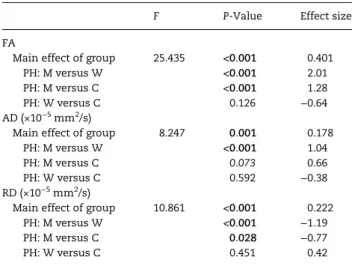
Similar between-group effects, with large effect sizes for the differences between comparison men and both comparison women and women with CAIS, were observed for the mean FA values extracted from the FA sex difference cluster (Fig.2and Table2).
Axial, Radial Diffusivity
Whole-brain TBSS analyses of AD and RD also revealed between-group differences (Fig.1). Comparison men showed higher AD va-lues than comparison women throughout extensive parts of the skeleton, without differences in the reverse contrast. Women with CAIS showed differences in AD relative to both comparison groups: lower AD than comparison men in a left hemisphere cluster, including ( parts of ) the internal and external capsule, sa-gittal stratum, superior longitudinal fasciculus, and posterior thalamic radiation, and higher AD than comparison women in a right hemisphere cluster, comprising ( parts of ) the internal and external capsule, corona radiata, superior longitudinal and fronto-occipital fasciculus, corpus callosum (splenium), cerebral peduncle, and posterior thalamic radiation.
A sex difference in RD, with higher RD values in comparison women than in men, was observed in a cluster covering ( parts of ) the left internal and external capsule, corona radiata, cerebral peduncle, uncinate and superior fronto-occipital fas-ciculus, pallidum, and amygdala. Women with CAIS showed higher RD values than comparison men in a small cluster of 25 voxels located within the right anterior corona radiata. Women with CAIS also showed higher RD than comparison women in a cluster covering part of the right superior longitu-dinal fasciculus.
Mean AD and RD (Fig.2and Table2), extracted from the FA sex difference region, showed sex differences with large effect sizes. In comparison men, a higher AD and lower RD relative to comparison women was found. Compared with women with CAIS, a tendency towards higher AD and a statistically sig-nificantly lower RD, with a large effect size, was found in com-parison men. Comcom-parison women did not differ from women with CAIS.
Discussion
The present study investigated the relative role of sex hormones versus sex chromosome genes on the sexual differentiation of human WM microstructure. This question was addressed by comparing DTI data from a unique group, that is, 46,XY women with CAIS, to 46,XY comparison men and 46,XX comparison women. Anisotropyfindings suggest a more important role for sex hormones, most likely masculinizing/defeminizing andro-gen and/or feminizing estroandro-gen effects, than for andro-genetic effects related to sex chromosome genes in the sexual differentiation of WM microstructure. Furthermore, ourfindings regarding the underlying DTI metrics showed that these sex hormone influ-ences are most pronounced in RD, while AD, to some extent, might also reflect other factors directly related to sex chromo-some composition and/or masculinizing androgen actions that are not mediated by the AR.
Widespread sex differences in FA were found, showing higher FA in comparison men than women, in line with the majority of findings in DTI studies using TBSS to investigate sex differences (e.g.,Inano et al. 2011). Similar to comparison women, women with CAIS had lower FA than comparison men throughout exten-sive regions of the skeleton, showing great overlap with sex dif-ference regions. Furthermore, their FA values did not differ from those of comparison women. Thesefindings were also sub-stantiated by the mean FA values in the region demonstrating a sex difference in FA.
In addition to FA, we analyzed the underlying DTI metrics AD and RD, to assess their contribution to the differences observed in FA. Based on the whole-brain voxel-wise analyses of AD and RD, higher FA in men appeared to be predominantly associated with higher AD. However, the mean AD and RD in areas with FA sex differences showed that the sex difference in FA was due to both higher AD and lower RD in men than in women, with equal-ly large effect sizes. Although results from previous studies on sex effects in these DTI measures are less consistent than in FA, the current results are generally in accordance with a study in a large sample of 857 subjects (Inano et al. 2011).
In the whole-brain voxel-wise comparisons to women with CAIS, higher AD contributed significantly to higher FA in men. This contribution of AD was, however, observed in a considerable smaller area than in the comparison groups, only partially cover-ing the areas where FA differences were found. In addition, a dif-ference relative to comparison women was found, with higher AD in women with CAIS in part of the right hemisphere skeleton. In accordance with these voxel-wise results, the mean AD in the FA sex difference region showed a tendency, but did not reach statistical significance, toward a female-typical AD in women with CAIS. While a higher RD in women with CAIS relative to both comparison groups was apparent in relatively small regions within the skeleton, the mean RD in the part of the skeleton showing FA sex differences was female-typical in women with CAIS, that is, higher than in comparison men. Within the FA sex difference region, both the sex difference in mean RD, and the female-typical mean RD in women with CAIS, appears to re-sult from a widespread, subtle effect, which is only detected in the mean RD calculated from all individual voxel values and, apart from some small clusters, is not detected in the whole-brain voxel-wise analysis.
Taken together, the female-typical FA in women with CAIS is in agreement with previousfindings in neural activity patterns in response to visual sexual stimuli and mental rotation perform-ance (Hamann et al. 2014;van Hemmen et al. 2016). The present findings thus argue against a dominant role for sex chromosome
at University of Liege on May 30, 2016
http://cercor.oxfordjournals.org/
Figure 1. Whole-brain between-group differences (P < 0.05 FWE-corrected) in FA, AD, and RD. Clusters of significant voxels are projected on the mean FA skeleton (green), using the“TBSS fill” procedure in FSL, and the MNI152 T1-weighted standard brain (FSL). Slice labels refer to MNI z-coordinates. MvsW, comparison men versus comparison women; MvsC, comparison men versus women with CAIS; WvsC, comparison women versus women with CAIS. For comparison XvsY: X > Y effects are displayed in red–yellow, X < Y effects are displayed in blue–green. Left is left.
at University of Liege on May 30, 2016
http://cercor.oxfordjournals.org/
genes and rather indicate that the sexual differentiation of human WM microstructure is primarily associated with differ-ences in sex hormone exposure. Due to the complete androgen resistance in CAIS, it is likely that these sex hormone effects reflect masculinizing and/or defeminizing androgen actions through direct activation of the AR. An important role for andro-gens acting on the AR would indeed be consistent withfindings from animal studies, showing the presence of ARs in cortical axons, dendrites, and glial cells (Sarkey et al. 2008). Furthermore, an association between testosterone and WM development in human adolescents was modulated by functional AR gene var-iants (CAG repeat-length polymorphisms) (Perrin et al. 2008). However, since all women with CAIS, with 1 exception, were on estrogen replacement therapy and estrogen receptors in women with CAIS function normally, the female-typical FA values in women with CAIS might also result from estrogenic actions. Therefore, sex differences in WM microstructure might also reflect a feminizing influence of estrogens. At present,
studies investigating an association between circulating estradiol levels and FA have not shown consistent results (Herting et al. 2011;Peper et al. 2013;Kranz et al. 2014).
The female-typical FA in women with CAIS was predominant-ly due to a female-typical RD, although a contribution of AD, at least in some of the WM tracts with a sex difference in FA , cannot be ruled out. Consequently, these results indicate that the proposed sex hormone effects are most pronounced in WM microstructural characteristics related to RD. The absence of a compelling female-typical AD in women with CAIS was unex-pected based on previous neuroimaging studies in these women (Hamann et al. 2014;van Hemmen et al. 2016), although a recent behavioral study on spatial learning in women with CAIS also did not observe an overall female-typical pattern (Mueller et al. 2016); women with CAIS did not differ significantly from comparison men and women on any of the variables measuring spatial learning, and effect size analyses revealed a more female-like pattern in women with CAIS on some, but not all variables. Regarding the sexual differentiation of the microstructural WM characteristics associated with AD, the present findings are suggestive of a contribution of other factors than the previously proposed sex hormone effects. These factors may include direct effects of sex chromosome genes, or other masculinizing/ defeminizing androgen actions that are not mediated by the AR. It is important to note that AD in women with CAIS differed from comparison women in only a small part of the skeleton, while the effect size of the summarizing mean AD within the FA sex difference region was in the female direction. Combined with the predominant female-typical RD and FA, this indicates that the contribution of these other factors is less pronounced and less extensive than the hypothesized masculinizing/ defeminizing androgen or feminizing estrogen effects.
An interestingfinding in light of the female-typical FA in women with CAIS is that their TWMV is more similar to that in comparison men. A potential explanation is that overall WM vol-ume is susceptible to direct sex chromosomal effects or androgen actions that are not mediated by the AR. Nonetheless, the present findings in WM microstructure are not likely to result from differences in TWMV for 2 reasons. First, susceptibility to partial volume effects (PVEs), known to affect the accuracy of DTI mea-sures, is influenced by volume size. Smaller volumes, or thinner fiber bundles, are more affected by PVEs, which might result in artificially lower FA values in women. However, the TBSS ap-proach greatly reduces PVEs and, as argued by Smith et al. Figure 2. Mean corrected for age (±SEM) FA, AD, and RD within FA sex difference regions. M, comparison men; W, comparison women; CAIS, women with CAIS.
Table 2 Between-group statistics and effect sizes for mean FA, AD, and RD within FA sex difference regions
F P-Value Effect size
FA
Main effect of group 25.435 <0.001 0.401
PH: M versus W <0.001 2.01
PH: M versus C <0.001 1.28
PH: W versus C 0.126 −0.64
AD (×10−5mm2/s)
Main effect of group 8.247 0.001 0.178
PH: M versus W <0.001 1.04
PH: M versus C 0.073 0.66
PH: W versus C 0.592 −0.38
RD (×10−5mm2/s)
Main effect of group 10.861 <0.001 0.222
PH: M versus W <0.001 −1.19
PH: M versus C 0.028 −0.77
PH: W versus C 0.451 0.42
Bold and italic P-values represent a significant (P < 0.05) effect and a trend (P < 0.1), respectively. For post hoc (PH) tests a Bonferroni-correction for multiple comparisons was applied. Effect sizes are expressed in partialη2for the main
effect of group and Cohen’s d for pairwise comparisons. M, comparison men; W, comparison women; C, women with CAIS.
at University of Liege on May 30, 2016
http://cercor.oxfordjournals.org/
(2006), FA values in largefiber tracts, that are wider than the voxel size, are unaffected by PVEs. Second, the overall female-typical findings in women with CAIS are in the opposite direction as would be expected when TWMV drives the between-group differences.
The tracts showing sex differences in WM microstructure con-nect extensive parts of the cerebral cortex and subcortical brain regions. The widespread differences in FA were found throughout major WM tracts, including projectionfibers such as the internal capsule and the corona radiata, containing axons going to and from the cerebral cortex, associationfibers such as the external capsule, connecting different cortical regions within a hemi-sphere, and parts of the corpus callosum, which is responsible for interhemispheric connections. Thesefindings might reflect general differences in structural connectivity of the male and fe-male brain, which does not show a clear association with regional gray matter sex differences, which have generally been found to be limited to certain brain regions (for meta-analysis seeRuigrok et al. 2014). Based on the present study it is, therefore, difficult to draw conclusions about the exact functional implications of thesefindings with regard to specific behavioral and cognitive sex differences.
It is also difficult to interpret the current results with respect to the underlying physiological characteristics. FA is a sensitive, but rather nonspecific measure, since differences in FA could re-flect several physiological differences. Even though AD and RD provide more specific information about FA, the exact physio-logical correlates of these measures are still under investigation (Jones et al. 2013). The predominant contribution of RD to the fe-male-typical FA in women with CAIS in sexually differentiated WM regions might, for instance, reflect sex hormone effects on axonal density, diameter or the degree of myelination.
A limitation of the present study is that no inferences can be made about the exact timing of the proposed sex hormone ef-fects. Since women with CAIS are insensitive to androgens throughout life, the currentfindings might reflect organizational and/or activational sex hormone effects during the pre-/neonatal phase, adolescence and/or adulthood. Longitudinal studies in women with CAIS, starting before adolescence, could provide in-formation regarding the timing of sex hormone effects on the sexual differentiation of human WM microstructure.
In conclusion, this DTI study in women with CAIS presents unique insights into the biological mechanism underlying the sex-ual differentiation of human WM microstructure. Ourfindings are supportive of an important role for sex hormones, most likely mas-culinizing and/or defeminizing androgen effects through the AR, feminizing estrogen effects, or a combination of both. Furthermore, currentfindings argue against substantial genetic effects related to differences in sex chromosome composition, or androgenic effects after aromatization to estrogens, acting on the estrogen receptor, although a more subtle contribution of these factors should not be ruled out for some aspects of WM microstructure.
Funding
This work was supported by the Netherlands Organization for Scientific Research (NWO-VICI grant number 453-08-003 to J.B.). J.B. is a senior research associate of the Fonds National de la Re-cherche Scientifique.
Notes
The authors are very grateful to Dr A.B. Dessens for her contribu-tion to subject recruitment, Ms W. Menks and Ms J. Boel for their
help with data acquisitions, the support group DSDNederland, and all subjects who participated in this study. Conflict of Interest: None declared.
References
Basser PJ, Mattiello J, LeBihan D. 1994. MR diffusion tensor spec-troscopy and imaging. Biophys J. 66:259–267.
Baum M. 1979. Differentiation of coital behavior in mammals: a comparative analysis. Neurosci Biobehav Rev. 3:265–284. Baum MJ. 2006. Mammalian animal models of psychosexual
dif-ferentiation: when is“translation” to the human situation possible? Horm Behav. 50:579–588.
Brock O, Baum MJ, Bakker J. 2011. The development of female sex-ual behavior requires prepubertal estradiol. J Neurosci. 31:5574–5578.
Chou K-H, Cheng Y, Chen I-Y, Lin C-P, Chu W-C. 2011. Sex-linked white matter microstructure of the social and analytic brain. NeuroImage. 54:725–733.
Cohen-Bendahan CCC, Van De Beek C, Berenbaum SA. 2005. Prenatal sex hormone effects on child and adult sex-typed behavior: methods and findings. Neurosci Biobehav Rev. 29:353–384.
DeLisi LE, Maurizio AM, Svetina C, Ardekani B, Szulc K, Nierenberg J, Leonard J, Harvey PD. 2005. Klinefelter’s syndrome (XXY) as a genetic model for psychotic disorders. Am J Med Genet B Neuropsychiatr Genet. 135B:15–23. Den Braber A, Van’t Ent D, Stoffers D, Linkenkaer-Hansen K,
Boomsma DI, De Geus EJC. 2013. Sex differences in gray and white matter structure in age-matched unrelated males and females and opposite-sex siblings. Int J Psychol Res. 6:7–21. Doehnert U, Bertelloni S, Werner R, Dati E, Hiort O. 2015.
Charac-teristic features of reproductive hormone profiles in late adolescent and adult females with complete androgen insensitivity syndrome. Sex Dev. 9:69–74.
Hamann S, Stevens J, Vick JH, Bryk K, Quigley CA, Berenbaum SA, Wallen K. 2014. Brain responses to sexual images in 46,XY women with complete androgen insensitivity syndrome are female-typical. Horm Behav. 66:724–730.
Herting MM, Maxwell EC, Irvine C, Nagel BJ. 2011. The impact of sex, puberty, and hormones on white matter microstructure in adolescents. Cereb Cortex. 22:1979–1992.
Hines M, Ahmed SF, Hughes IA. 2003. Psychological outcomes and gender-related development in complete androgen insensitivity syndrome. Arch Sex Behav. 32:93–101.
Holzapfel M, Barnea-Goraly N, Eckert MA, Kesler SR, Reiss AL. 2006. Selective alterations of white matter associated with visuospatial and sensorimotor dysfunction in Turner syn-drome. J Neurosci. 26:7007–7013.
Hong DS, Reiss AL. 2014. Cognitive and neurological aspects of sex chromosome aneuploidies. Lancet Neurol. 13:306–318. Hughes IA, Davies JD, Bunch TI, Pasterski V, Mastroyannopoulou K,
MacDougall J. 2012. Androgen insensitivity syndrome. Lancet. 380:1419–1428.
Inano S, Takao H, Hayashi N, Abe O, Ohtomo K. 2011. Effects of age and gender on white matter integrity. AJNR Am J Neuroradiol. 32:2103–2109.
Jones DK, Knösche TR, Turner R. 2013. White matter integrity, fiber count, and other fallacies: the do’s and don’ts of diffu-sion MRI. NeuroImage. 73:239–254.
Kanaan RA, Chaddock C, Allin M, Picchioni MM, Daly E, Shergill SS, McGuire PK. 2014. Gender influence on white mat-ter microstructure: a tract-based spatial statistics analysis. PLoS One. 9:e91109.
at University of Liege on May 30, 2016
http://cercor.oxfordjournals.org/
Kranz GS, Hahn A, Kaufmann U, Küblböck M, Hummer A, Ganger S, Seiger R, Winkler D, Swaab DF, Windischberger C, et al. 2014. White matter microstructure in transsexuals and controls investigated by diffusion tensor imaging. J Neurosci. 34:15466–15475.
Lombardo MV, Ashwin E, Auyeung B, Chakrabarti B, Taylor K, Hackett G, Bullmore ET, Baron-Cohen S. 2012. Fetal testoster-one influences sexually dimorphic gray matter in the human brain. J Neurosci. 32:674–680.
Masica DN, Money J, Ehrhardt AA. 1971. Fetal feminization and female gender identity in the testicular feminizing syndrome of androgen insensitivity. Arch Sex Behav. 1:131–142. McCarthy MM, Arnold AP. 2011. Reframing sexual differentiation
of the brain. Nat Neurosci. 14:677–683.
Melo KFS, Mendonca BB, Billerbeck AEC, Costa EMF, Inácio M, Silva FAQ, Leal AMO, Latronico AC, Arnhold IJP. 2003. Clinical, hormonal, behavioral, and genetic characteristics of andro-gen insensitivity syndrome in a Brazilian cohort:five novel mutations in the androgen receptor gene. J Clin Endocrinol Metab. 88:3241–3250.
Menzler K, Belke M, Wehrmann E, Krakow K, Lengler U, Jansen A, Hamer HM, Oertel WH, Rosenow F, Knake S. 2011. Men and women are different: diffusion tensor imaging reveals sexual dimorphism in the microstructure of the thalamus, corpus callosum and cingulum. NeuroImage. 54:2557–2562. Molko N, Cachia A, Riviere D, Mangin JF, Bruandet M, LeBihan D,
Cohen L, Dehaene S. 2004. Brain anatomy in Turner syn-drome: evidence for impaired social and spatial-numerical networks. Cereb Cortex. 14:840–850.
Mueller SC, Merke DP, Leschek EW, Fromm S, VanRyzin C, Ernst M. 2011. Increased medial temporal lobe and striatal grey-matter volume in a rare disorder of androgen excess: a voxel-based morphometry (VBM) study. Int J Neuropsychopharmacol. 14:445–457.
Mueller SC, Verwilst T, Van Branteghem A, T’Sjoen G, Cools M. 2016. The contribution of the androgen receptor (AR) in human spatial learning and memory: a study in women with complete androgen insensitivity syndrome (CAIS). Horm Behav. 78:121–126.
Oakes MB, Eyvazzadeh AD, Quint E, Smith YR. 2008. Complete an-drogen insensitivity syndrome—a review. J Pediatr Adolesc Gynecol. 21:305–310.
Peper JS, Mandl RCW, Braams BR, de Water E, Heijboer AC, Koolschijn PCMP, Crone EA. 2013. Delay discounting and fron-tostriatalfiber tracts: a combined DTI and MTR study on im-pulsive choices in healthy young adults. Cereb Cortex. 23:1695–1702.
Perrin JS, Hervé P-Y, Leonard G, Perron M, Pike GB, Pitiot A, Richer L, Veillette S, Pausova Z, Paus T. 2008. Growth of white matter in the adolescent brain: role of testosterone and androgen receptor. J Neurosci. 28:9519–9524.
Phoenix CH, Goy RW, Gerall AA, Young WC. 1959. Organizing ac-tion of prenatally administered testosterone propionate on the tissues mediating mating behavior in the female guinea pig. Endocrinology. 65:369–382.
Pierpaoli C, Basser PJ. 1996. Toward a quantitative assessment of diffusion anisotropy. Magn Reson Med. 36:893–906.
Rametti G, Carrillo B, Gómez-Gil E, Junque C, Segovia S, Gomez Á, Guillamon A. 2011. White matter microstructure in female to male transsexuals before cross-sex hormonal treatment. A diffusion tensor imaging study. J Psychiatr Res. 45:199–204.
Ruigrok ANV, Salimi-Khorshidi G, Lai M-C, Baron-Cohen S, Lombardo MV, Tait RJ, Suckling J. 2014. A meta-analysis of sex differences in human brain structure. Neurosci Biobehav Rev. 39:34–50.
Sarkey S, Azcoitia I, Garcia-Segura LM, Garcia-Ovejero D, DonCarlos LL. 2008. Classical androgen receptors in non-classical sites in the brain. Horm Behav. 53:753–764. Savic I. 2012. Advances in research on the neurological and
neuropsychiatric phenotype of Klinefelter syndrome. Curr Opin Neurol. 25:138–143.
Schoonheim MM, Vigeveno RM, Rueda Lopes FC, Pouwels PJW, Polman CH, Barkhof F, Geurts JJG. 2014. Sex-specific extent and severity of white matter damage in multiple sclerosis: im-plications for cognitive decline. Hum Brain Mapp. 35:2348–2358. Schulz KM, Molenda-Figueira HA, Sisk CL. 2009. Back to the fu-ture: the organizational-activational hypothesis adapted to puberty and adolescence. Horm Behav. 55:597–604.
Sisk CL, Zehr JL. 2005. Pubertal hormones organize the adolescent brain and behavior. Front Neuroendocrinol. 26:163–174. Smith SM, Jenkinson M, Johansen-Berg H, Rueckert D, Nichols TE,
Mackay CE, Watkins KE, Ciccarelli O, Cader MZ, Matthews PM, et al. 2006. Tract-based spatial statistics: voxelwise analysis of multi-subject diffusion data. NeuroImage. 31:1487–1505. Smith SM, Jenkinson M, Woolrich MW, Beckmann CF, Behrens TE,
Johansen-Berg H, Bannister PR, De Luca M, Drobnjak I, Flitney DE, et al. 2004. Advances in functional and structural MR image analysis and implementation as FSL. NeuroImage. 23(Suppl 1):S208–S219.
Smith SM, Nichols TE. 2009. Threshold-free cluster enhancement: addressing problems of smoothing, threshold dependence and localisation in cluster inference. NeuroImage. 44:83–98. Takao H, Hayashi N, Ohtomo K. 2014. Sex dimorphism in the
white matter: fractional anisotropy and brain size. J Magn Reson Imaging. 39:917–923.
van Hemmen J, Veltman DJ, Hoekzema E, Cohen-Kettenis PT, Dessens AB, Bakker J. 2016. Neural activation during mental rotation in complete androgen insensitivity syndrome: the in-fluence of sex hormones and sex chromosomes. Cereb Cortex. 26:1036–1045.
van Strien JW. 1992. Classificatie van links- en rechtshandige proefpersonen [Classification of left-handed and right-handed test subjects]. Ned Tijdschr Psychol. 47:88–92. Villalon-Reina J, Jahanshad N, Beaton E, Toga AW, Thompson PM,
Simon TJ. 2013. White matter microstructural abnormalities in girls with chromosome 22q11.2 deletion syndrome, Fragile X or Turner syndrome as evidenced by diffusion tensor im-aging. NeuroImage. 81:441–454.
Wallen K. 2005. Hormonal influences on sexually differentiated behavior in nonhuman primates. Front Neuroendocrinol. 26:7–26.
Winkler AM, Ridgway GR, Webster MA, Smith SM, Nichols TE. 2014. Permutation inference for the general linear model. NeuroImage. 92:381–397.
Wisniewski AB, Migeon CJ, Meyer-Bahlburg HF, Gearhart JP, Berkovitz GD, Brown TR, Money J. 2000. Complete androgen insensitivity syndrome: long-term medical, surgical, and psy-chosexual outcome. J Clin Endocrinol Metab. 85:2664–2669. Yamagata B, Barnea-Goraly N, Marzelli MJ, Park Y, Hong DS,
Mimura M, Reiss AL. 2012. White matter aberrations in pre-pubertal estrogen-naive girls with monosomic Turner syn-drome. Cereb Cortex. 22:2761–2768.
at University of Liege on May 30, 2016
http://cercor.oxfordjournals.org/