The catalytic, glycosyl transferase and acyl transferase modules of the cell
wall peptidoglycan-polymerizing penicillin-binding protein 1b of
Escherichia coli
Mohammed Terra k,1Tushar K. Ghosh,1 Jean van Heijenoort,2 Jozef Van Beeumen,3 Maxime Lampilas,4Jozsef Aszodi,4 Juan A. Ayala,5 Jean-Marie Ghuysen1and Martine Nguyen-Distèche1
1Centre d'Ingénierie des Protéines, Université de Liège, Institut de Chimie, B6, B-4000 Sart Tilman (Liège), Belgium. 2Biochimie moléculaire et cellulaire, Université Paris-Sud, CNRS-URA 1131, Bâtiment 432, F-91405 Orsay Cedex, France. 3
Laboratorium voor Eiwitbiochemie en Eiwitengineering, Rijksuniversiteit-Gent, K. L. Ledeganckstraat, 35, B-9000 Gent, Belgium. 4Hoechst Marion Roussel, 102, Route de Noisy, F-93235 Romainville Cedex, France.
5Centro de Biologia Molecular, Universidad Autonoma, Canto Blanco, E-28049 Madrid, Spain.
Summary
The penicillin-binding protein (PBP) 1 b of Escherichia coli catalyses the assembly of lipid-transported N-acetyl glucosaminyl-β-1,4-N-acetylmuramoylL-alanyl-γ-D-glutamyl-(L)-meso-diaminopimelyl-(L)-D-alanyl-D-alanine disaccharide pentapeptide units into polymeric peptidoglycan. These units are phosphodiester linked, at C1 of muramic acid, to a C55 undecaprenyl carrier. PBP1b has been purified in the form of His tag (M46-N844) PBP1bγ. This derivative provides the host cell in which it is produced with a functional wall peptidoglycan. His tag (M46-N844) PBP1bγ possesses an amino-terminal hydrophobic segment, which serves as transmembrane spanner of the native PBP. This segment is linked, via an 100-amino-acid insert, to a D198-G435 glycosyl transferase module that possesses the five motifs characteristic of the PBPs of class A. In in vitro assays, the glycosyl transferase of the PBP catalyses the synthesis of linear glycan chains from the lipid carrier with an efficiency of 39000 M-1 s-1. Glu-233, of motif 1, is central to the catalysed reaction. It is proposed that the Glu-233 γ-COOH donates its proton to the oxygen atom of the scissile phosphoester bond of the lipid carrier, leading to the formation of an oxocarbonium cation, which then undergoes attack by the 4-OH group of a nucleophile N-acetylglucosamine. Asp-234 of motif 1 or Glu-290 of motif 3 could be involved in the
stabilization of the oxocarbonium cation and the activation of the 4-OH group of the N-acetylglucosamine. In turn, Tyr-310 of motif 4 is an important component of the amino acid sequence-folding information. The glycosyl transferase module of PBP1b, the lysozymes and the lytic transglycosylase Slt70 have much the same catalytic machinery. They might be members of the same superfamily. The glycosyl transferase module is linked, via a short junction site, to the amino end of a Q447-N844 acyl transferase module, which possesses the catalytic centre-defining motifs of the penicilloyl serine transferases superfamily. In in vitro assays with the lipid precursor and in the presence of penicillin at concentrations sufficient to derivatize the active-site serine 510 of the acyl transferase, the rate of glycan chain synthesis is unmodified, showing that the functioning of the glycosyl transferase is acyl transferase independent. In the absence of penicillin, the products of the Ser-510-assisted double-proton shuttle are glycan strands substituted by cross-linked tetrapeptide-pentapeptide and tetrapeptide-tetrapeptide dimers and uncross-linked pentapeptide and tetrapeptide monomers. The acyl transferase of the PBP also catalyses aminolysis and hydrolysis of properly structured thiolesters, but it lacks activity onD-alanyl-D-alanine-terminated peptides. This substrate specificity suggests that carbonyl donor activity requires the attachment of the pentapeptides to the glycan chains made by the glycosyl transferase, and it implies that one and the same PBP molecule catalyses transglycosylation and peptide cross-linking in a
sequential manner. Attempts to produce truncated forms of the PBP lead to the conclusion that the multimodular polypep-tide chain behaves as an integrated folding entity during PBP1b biogenesis.
Introduction
The bacterial cell wall peptidoglycan is a net-like polymer in which glycan strands made of alternating β-1,4-linked N-acetylglucosamine and N-acetylmuramic acid residues are cross-β-1,4-linked by peptides. In Escherichia coli, the carboxyl groups of the N-acetylmuramic acid residues are amide-linked to L-alanyl-γ-D-glutamyl-(L)-meso-diaminopimelyl-(L)-D-alanine peptide units, and the cross-linkages between glycan strands are, mainly, direct
D-alanyl-(D)-meso-diaminopimelic acid peptide bonds (van Heijenoort, 1996). The immediate biosynthetic precursor of the peptidoglycan is lipid II. A disaccharide pentapeptide (L-alanyl-γ-D-glutamyl-(L)-meso-diaminopimelyl-(L)-D-alanyl-D-alanine) is linked to a C55H89 undecaprenyl carrier via a pyrophosphate bridge involving C1 of N-acetylmuramic acid. From this precursor, glycosyl transferases catalyse glycan chain elongation, and acyl transferases catalyse peptide cross-linking between glycan chains. This latter reaction proceeds via the formation of a serine-ester-linked peptidyl (~L-alanyl-γ-D-glutamyl-(L)-meso-diaminopimelyl-D-alanyl) enzyme intermediate. Penicillin is a suicide substrate. The reaction produces a stable and inactive serine-ester-linked penicilloyl enzyme, and the inactivated enzymes behave as penicillin-binding proteins, in short PBPs (Frère et al., 1992).
The 850-amino-acid residue PBP1 a and the 844-amino-acid residue PBP1 b of E. coli each combine in one single polypeptide chain, the required enzymatic activities for peptidoglycan assembly from lipid II (Broome-Smith et al., 1985). Essentially, a transmembrane spanner is linked to the amino end of a non-penicillin-binding glycosyl transferase module that is linked to the amino end of a penicillin-binding acyl transferase module, and the polypeptide chain folds on the exterior of the plasma membrane (Edelman et al., 1987). PBP1 b and PBP1 a have been characterized biochemically as bifunctional glycosyl transferase-acyl transferase enzymes in in vitro assays (Ishino et al., 1980; Suzuki et al., 1980; Nakagawa et al., 1984; Mottl et al., 1995). They are regarded as the prototypes of the multimodular class A PBPs (Ghuysen, 1991; Goffin and Ghuysen, 1998). Further studies have been carried out to substantiate the structure-function relationships of E. coli PBP1b. The results are reported below.
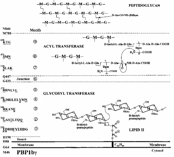
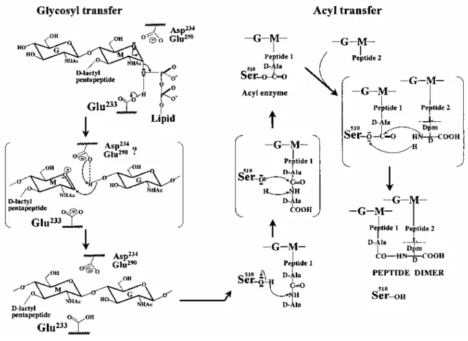
Fig. 1. Modular design of E. coli PBP1b and transfer reactions involved in the conversion of the C55H89
lipid-transported disaccharide pentapeptide units into polymeric peptidoglycan. The motifs characteristic of the glycosyl transferase and acyl transferase modules are identified. The amino acid residues strictly conserved in the class A PBPs are underlined. *Amino acid residues modified by site-directed mutagenesis. G,
Results
The multimodular design of PBP1b is shown in Fig. 1 (Goffin and Ghuysen, 1998). The D198-G435 glycosyl transferase module has the amino acid sequence signature of the class A PBPs with five conserved motifs. Glu-233 and Asp-234 are in motif 1, Glu-290 is in motif 3, and Tyr-310 is in motif 4. The associated Q447-M780 acyl transferase module carries the three catalytic centre-defining motifs characteristic of the penicilloyl serine transferases superfamily: Ser-510LeuAlaLys (where Ser-510 is the active-site serine residue), Ser-572MetAsn and Lys-698ThrGly. A conserved junction site links the two modules. PBP1b has structural peculiarities. It contains a D88-N185 insert located between the carboxy end of the transmembrane spanner and motif 1 of the glycosyl transferase module. This insert provides the protein with an additional membrane association site (Wang et al., 1996). PBP1b has a carboxy-terminal extension. Residues 762-780 are essential, but residues 781-844 are dispensable (Kato et al., 1984; Lefèvre et al., 1997). PBP1b has cytosolic tails of varying lengths.
M1-N844 PBP1bα and M46-M1-N844 PBP1bγ result from the presence of two alternative translation initiation codons in
the encoding gene (Kato et al., 1984). PBP1 bβ arises by proteolysis of PBP1bα (Henderson et al., 1994). The bifunctional, glycosyl transferase-acyl transferase, His tag (M46-N844) PBP1b (pDML924)
pDML924 had the information for the overproduction of the (M46-N844) PBP1bγ whose cytosolic tail was His tag-labelled via a thrombin cleavage site. The gene was under the control of the T7 promoter-/ac operator and it was IPTG inducible.
Overproduction and purification. E. coli BL21 (DE3)/ pDML924 was grown at 37°C in Luria-Bertani medium containing 50 µg kanamycin ml-1. When the culture reached an optical density (OD) of 0.6 at 600 nm, 1 mM IPTG was added, and the culture was maintained at 37°C for 2 h. The yield was 5 mg PBP l-1 of culture. The cells were disrupted and fractionated, the membrane-bound PBP was solubilized in 25 mM Tris-HCl, pH 7.5, 1 M NaCl, 1% 3-[(3-cholamidopropyl)dimethylammonio]1-propane sulphonate (CHAPS), and the PBP was purified by chromatography on a Ni2+-nitrilotriacetic acid (NTA)-agarose column (Nicholas et al., 1993). His tag (M46-N844) PBP1bγ was about 95% pure (Fig. 2). The final yield was about 1.4mgl-1 culture. The PBP bound [3H]-benzylpenicillin in a 1:1 molar ratio. It was less stable than the membrane-bound native PBP1b or the membrane-bound His tag (M46-N844) PBPIbγ (half-lives at 60°C: 8min versus 45 min). It was stored at -20°C in 25 mM Tris-HCI, pH 7.5, 0.7% CHAPS, 0.5 M NaCl (buffer A) without detectable alterations, although aggregation may occur to some extent without loss of activity.
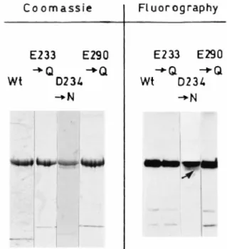
Fig. 2. SDS-PAGE analysis of the purified, His tag (M46-N844) PBP1bγ (Wt) and Glu-233→Gln, Asp-234→Asn
and Glu-290→Gln PBP mutants. Coomassie blue staining (3 µg of protein). Fluorography after labelling with 100 µM [3H]-benzylpenicillin (0.5 µg of protein).
Peptidoglycan polymerization. Purified His tag (M46-N844) PBP1bγ (15 nM) and [14C]meso-diaminopimelic acid-labelled lipid II (0.8-4 µM) were incubated in 50 mM Tris-HCI, pH7.5, containing 0.046% CHAPS, 33 mM NaCl, 10 mM MgCI2, 0.5% polyethyleneglycol 300 mono-1-decylether (decyl PEG), 12.5% 1-octanol and 25% dimethyl sulphoxide (DMSO) for 15-30min at 30°C (Hara and Suzuki, 1984; Mottl et al, 1995). The reaction mixtures were submitted to paper chromatography in isobutyric acid-1 M ammonia (5:3), under which conditions the polymerized peptidoglycan remained immobile on the chromatograms. On the basis of initial rate measurements of radioactive peptidoglycan synthesis and Michaelis-Menten equation, the reaction proceeded with a Kmvalue of 1.8 µM, a kcat value of 70 x 10-3s-1 and, therefore, a kcat/Kmefficiency of about 39000M-1s-1. The efficiency was 60-fold larger than that of systems containing a mixture of methanol/deoxycholate instead of the mixture 1-octanol/DMSO/decyl PEG (van Heijenoort et al., 1992). Moenomycin, used at a concentration of 14 nM and an antibiotic-PBP molar ratio of 3:1, reduced the amount of polymerized material by 50%.
Complementation assays in vivo. The in vivo functionality of His tag (M46-N844) PBP1b7 was tested by complementation assays carried out in E. coli EJ801. E. coli EJ801 is defective in PBP1b, and it possesses a thermosensitive (42°C) PBP1a (Hara and Suzuki, 1984). Consequently, the strain grows as rod-shaped cells at 30°C, but it fails to grow at 42°C. E. coli EJ801/pDML924 was grown at 30°C in Luria-Bertani medium containing 30 µg kanamycin ml-1. When the culture reached an OD value of 0.3 at 600 nm, one volume was diluted into two volumes of fresh medium, and cultures were maintained for 4 h at 30°C and at 42°C
respectively. The E. coli EJ801 transformant grew at 30°C and at 42°C (Fig. 3), indicating that His tag (M46-N844) PBP1bγ was produced in sufficient amounts to achieve complementation in an E. coli strain that lacked the T7 RNA polymerase, and that the presence of the His tag label at the amino end of the polypeptide chain had no adverse effects.
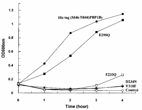
Fig. 3. Complementation activity of plasmids (pDML924) encoding the non-mutated His tag (M46-N844)
PBP1bγ and the Glu-233→Gln, Asp-234→Asn, Glu-290→Gln and Tyr-310→Phe protein mutants. E. coli EJ801, used as host, was grown in Luria-Bertani medium at 42°C.
The glycosyl transferase module
His tag (M46-N844) PBP1bγ was penicilloylated with benzylpenicillin, resulting in the complete inactivation of the acyl transferase module. The penicilloylated PBP and the untreated PBP had the same peptidoglycan-polymerizing activity in the in vitro assay, indicating that the determined Kmand kcatvalues ( 1.8 µM and 70 x 10-3s-1) applied to the synthesis of linear glycan chains and not to the ensuing synthesis of glycan chain cross-linkages.
Roles of conserved amino acid residues. The His tag E233→Q; D234→N; E290→Q; Y310→F (M46-N844) PBP1bγ mutants (pDML924A, B, C, D). By analogy with glycosyl transferases of known three-dimensional structure (Thunnissen et al., 1995), it was predicted that Glu-233 and Asp-234 of motif 1, Glu-290 of motif 3 and Y310 of motif 4 could be important elements of the catalytic centre of the glycosyl transferase module of PBP1b (see Discussion). To test this hypothesis, pDML924A, B, C and D had the information for the IPTG-inducible overproduction of His tag (M46-N844) PBP1bγ mutants in which Glu-233 was changed into Gln, Asp-234 was changed into Asn, Glu-290 was changed into Gin, and Tyr-310 was changed into Phe.
The Glu-233→Gln, Asp-234→Asn and Glu-290→Gln PBP mutants were produced, bound to the plasma membrane, by the respective E. coli BL21 (DE3) transformants. They were purified by chromatography of the CHAPS/NaCl membrane extracts on Ni2+-NTA agarose, as described for the non-mutated PBP. The PBP mutants were indistinguishable from the non-mutated PBP with respect to their levels of production by the E. coli transformants and their sensitivities to β-lactam antibiotics (see below). Marked differences were observed with respect to their capacities for catalysing glycan chain elongation from lipid II in in vitro assays, and the abilities of the corresponding plasmids to achieve complementation in E. coli EJ801 at 42°C. The Glu-233→Gln and Glu-290→Gln PBP mutants were as stable as the non-mutated PBP. In contrast, the Asp-234→Asn PBP mutant was susceptible to proteolysis, and the purified PBP did not migrate as a sharp band on SDS-PAGE (arrow in Fig. 2).
The Glu-233→Gln PBP mutant had negligible glycan chain elongation activity, 0.2% of that of the non-mutated PBP, and the values of the kinetic parameters could not be estimated. As expected, pDML924A, which encoded the inactive PBP mutant, lacked complementation activity in E. coli EJ801 at 42°C (Fig. 3). The Asp-234→Asn and Glu-290→Gln mutants had a much decreased catalytic efficacy (kcat/Km: 5600 M-1 s-1 and 3600 M-1 s-1 respectively, versus 39000M-1 s-1 for the non-mutated PBP). The Asp-234→Asn mutation affected the kcat value only ( 10 x 10-3s-1 versus 70 x 10-3s-1), and the Glu-290→Gln mutation affected both the Kmvalue ( 0.4 µM versus 2 µM) and the kcat value (1.5 x 10⁻³s-1). Somewhat surprisingly, pDML924C, which encoded the Glu-290→Gln PBP mutant, had complementation activity, whereas pDML924B, which encoded the Asp-234→Asn PBP mutant, lacked complementation activity (Fig. 3).
Attempts to produce the Tyr-310→Phe PBP mutant in E. coli BL21 (DE3)/pDML924D failed. The mutated protein was very unstable and susceptible to proteolysis. It could not be isolated. Consistently, pDML924D, which encoded the Tyr-310→Phe protein mutant, lacked complementation activity (Fig. 3).
The glycosyl transferase module as independent entity. His tag (M46-Q423) and His tag (M46-D478)
polypeptides. (pDML927, 926). Monofunctional glycosyl transferases, endowed with (moenomycin-insensitive) glycan chain elongation activity on lipid II, have been identified in several bacterial species (Spratt et al., 1996). The E. coli PBP1b polypeptide terminating at Glu-423, i.e. 12 amino acid residues upstream from the
intermodule junction site, performs glycosyl transferase in in vitro assays (Nakagawa et al., 1984). Moenomycin protects against trypsin proteolysis of the hybrid resulting from the fusion of the Ser-37-Lys-263 polypeptide of the class A Streptococcus pneumoniae PBP1a, i.e. 32 amino acid residues from the intermediate junction site, to the glutathione S-transferase (Di Guilmi et al., 1998). E. coli PBP1b mutants in the 470 region, i.e. downstream from the intermediate junction site, had folding problems, and this region was predicted to be located in a β-sheet (Lefèvre et al., 1997).
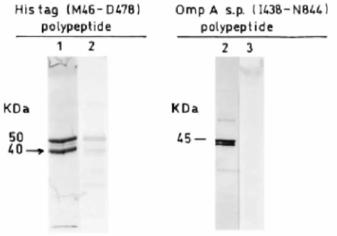
Attempts were made to produce acyl transferase-less glycosyl transferase modules of PBP1bγ (terminating at Glu-423 and Asp-478 respectively). E. coli BL21(DE3)/ pDML927 and E. coli BL21 (DE3)/pDML926 had the information for the synthesis of the truncated His tag (M46-Q423) and His tag (M46-D478) polypeptides respectively. The transformants were grown in Luria-Bertani medium at 37°C and induced with 1 mM IPTG for 2h. Ni2+-NTA agarose chromatography of the CHAPS-NaCl membrane extracts yielded several polypeptides that reacted with the anti-PBP1b antibodies (Fig. 4). Their molecular masses ranged from 44 kDa to 50 kDa, as expected for the full-size His tag (M46-Q423) and His tag (M46-D478) polypeptides, respectively, to about 30 kDa. As derived from microsequencing data, the main 40 kDa degradation product of the His tag (M46-D478) polypeptide (isolated by SDS-PAGE; arrow in Fig. 4) started with the sequence GlySerx(His)6and terminated with the sequence GlnLeuVal-418, suggesting that, essentially, proteolysis proceeded from the free carboxy end. His tag (M46-Q423) polypeptide was especially susceptible to degradation. The use of a cocktail of protease inhibitors had little protective effect. The polypeptides underwent spontaneous precipitation during the purification steps and storage at 4°C and -20°C.
The preparations obtained after chromatography of the CHAPS-NaCl membrane extracts on Ni2+ agarose columns exhibited glycosyl transferase activities on lipid II, and the reaction was completely inhibited by
moenomycin used at a 6 x 10⁻⁸M concentration and at an antibiotic-protein molar ratio of about 3:1. However, the enzymatic activities were low. The specific activities of the His tag (M46-Q423) and His tag (M46-D478) preparations were 13% and 23% that of the full-size PBP (all other conditions being identical and using average molecular masses of 40000 and 47000 respectively).
Fig. 4. SDS-PAGE analysis of the His tag (M46-D478) polypeptide (1, 8 µg of proteins as obtained after Ni2+
-NTA agarose chromatography; 2, cell extract from 100 μl of culture at OD = 0.8) and the OmpA signal peptide transported I438-N844 polypeptide (30 µg of proteins from the periplasmic fraction of the producing
transformant). 1, Coomassie blue staining; 2, immunoblotting; 3, fluorography of the [3
H]-benzylpenicillin-labelled preparation. The lactamase present in the periplasmic fraction was inhibited by 50 µM
β-iodopenicillanate. The peptide indicated by an arrow has been sequenced.
The acyl transferase module
To check that the acyl transferase module was able to cross-link the linear pentapeptide-substituted glycan strands made by the glycosyl transferase module, purified His tag (M46-N844) PBP1bγ and radioactive lipid II were incubated as described above. Then, the N-acetylmuramyl-N-acetylglucosamine bonds of the polymerized material were hydrolysed with lysozyme and the Chalaropsis muramidase (in order to conduct the cleavage of the glycan chains into disaccharides to completion), and the radioactive compounds were isolated and estimated (Glauner et al., 1988). The lipid precursor used in these experiments was a mixture of lipid-linked disaccharide units substituted by pentapeptide [L-alanyl-γ-D-glutamyl-(D)-meso-diaminopimelyl-(L)-D-alanyl-D-alanine] and tetrapeptide [L-alanyl-γ-D-glutamyl-(D)-meso-diaminopimelyl-(L)-D-alanine] monomers. The acyl transferase of PBP1b hydrolysed and aminolysed theD-alanyl-D-alanine peptide bonds using water and theD-amino group of meso-diaminopimelic acid of pentapeptide and tetrapeptide monomers as acceptors. As a result of the PBP-catalysed reaction, the glycan chains of the polymeric peptidoglycan consisted of disaccharide pentapeptide monomers (46% versus 70% in the lipid precursor), disaccharide tetrapeptide monomers (27% versus 23%), cross-linked bisdisaccharide tetrapeptide-pentapeptide dimers (2%) and cross-linked bisdisaccharide
tetrapeptide-tetrapeptide dimers (8%). Peptide oligomers larger than peptide dimers were not identified. Both transpeptidation and hydrolysis were inhibited by benzylpenicillin.
Acyl transfer reactions on β-lactam antibiotics. β-Lactam compounds inactivated His tag (M46-N844) PBP1bγ
as stable acyl (penicilloyl, cephalosporoyl) derivatives (Table 1). As derived from the values of the first-order rate constant of breakdown, the half-lives of the acyl PBP derivatives, at 37°C, ranged from 23 min for cefuroxine to 550 min for cefoperazone. As derived from the values of the second-order rate constants of acylation, the cephalosporins were more potent inactivators than the penicillins. The acylation rate values, at 37°C, varied from 100 M-1s-1for carbenicillin to 2600 M-1s-1 for cefotaxime. These values were two- to fourfold larger than those measured at 30°C on the native, membrane-bound PBP1b (Curtis et al., 1979). Irrespective of the PBP forms, the antibiotics fell roughly in the same order of decreasing acylating activity. The mutations Glu-233→Gln, Asp-234→Asn and Glu-290→Gln, which affected the non-penicillin-binding glycosyl transferase module (see above), did not significantly affect the values of the rate constants of acylation of the
penicillin-binding acyl transferase module by cefotaxime, cephaloridine and benzylpenicillin.
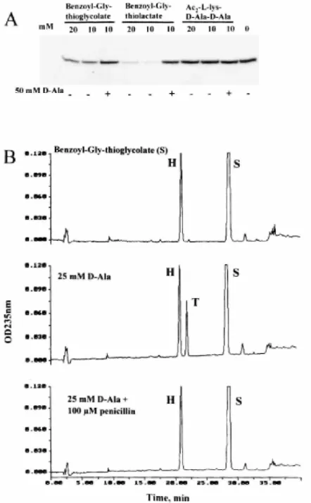
Fig. 5. Acyl transfer activities of His tag (M46-N844) PBP1bγ. A. SDS-PAGE analysis. Effects of preincubation
of the PBP with bisacetylL-lysyl-D-alanyl-D-alanine, benzoylglycylthiolactate and benzoylglycylthiolglycolate on subsequent binding of [3H]-benzylpenicillin to the PBP in the absence and in the presence ofD-alanine. For conditions, see text. B. HPLC analysis. PBP-catalysed hydrolysis and aminolysis of benzoylglycylthiolglycolate (S) in the absence and in the presence of benzylpenicillin. H, benzoylglycine; T, benzoylglycyl-D-alanine. For more details, see text.
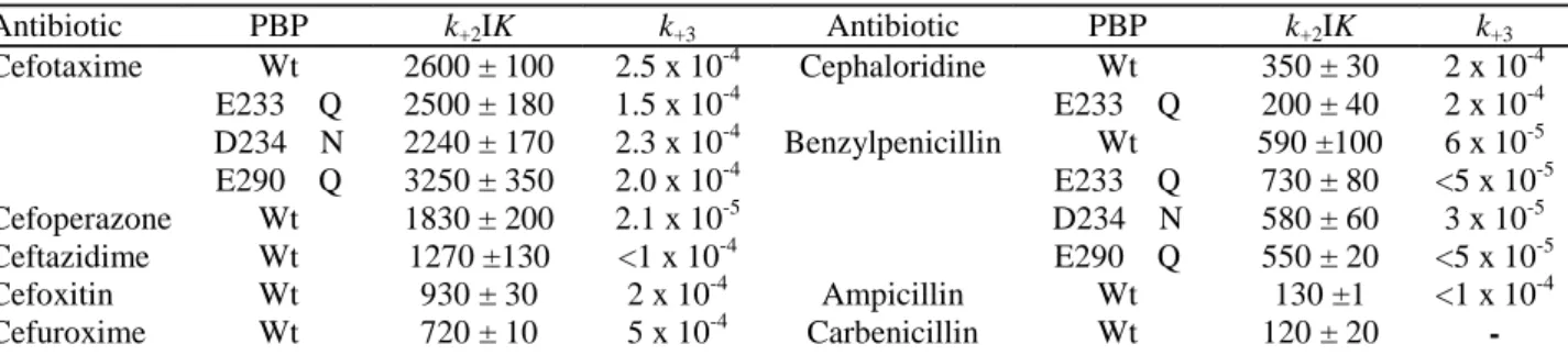
Table 1. Interaction between β-lactam antibiotics and non-mutated (Wt) and mutated His tag (M46-N884) PBP1bγ. Antibiotic PBP k+2IK k+3 Antibiotic PBP k+2IK k+3 Cefotaxime Wt 2600 ± 100 2.5 x 10⁻⁴ Cephaloridine Wt 350 ± 30 2 x 10⁻⁴ E233→Q 2500 ± 180 1.5 x 10⁻⁴ E233→Q 200 ± 40 2 x 10⁻⁴ D234→N 2240 ± 170 2.3 x 10⁻⁴ Benzylpenicillin Wt 590 ±100 6 x 10⁻⁵ E290→Q 3250 ± 350 2.0 x 10⁻⁴ E233→Q 730 ± 80 <5 x 10⁻⁵ Cefoperazone Wt 1830 ± 200 2.1 x 10⁻⁵ D234→N 580 ± 60 3 x 10⁻⁵ Ceftazidime Wt 1270 ±130 <1 x 10⁻⁴ E290→Q 550 ± 20 <5 x 10⁻⁵ Cefoxitin Wt 930 ± 30 2 x 10⁻⁴ Ampicillin Wt 130 ±1 <1 x 10⁻⁴ Cefuroxime Wt 720 ± 10 5 x 10⁻⁴ Carbenicillin Wt 120 ± 20
-The k+2/K values at 30°C for the interaction with the native membrane-bound PBP1b (Curtis et al., 1979) were 785 M-1s-1(cefotaxime), 510
M-1s-1(cefoperazone), 130 M-1s-1 (cefoxitin), 320 M-1s-1 (cefuroxime), 210 M-1s-1 (cephaloridin), 110 M-1s-1 (ampicillin) and 100 M-1s-1
(carbenicillin). Second-order rate constants k+2/K (M-1s-1) of acylation and first-order rate constants k+3 of deacylation, at 37°C.
Acyl transfer reactions on thiolester and peptide carbonyl donors
Preincubation of the thiolester benzoylglycylthiolactate (10-20 mM) with His tag (M46-N844) PBP1bγ (2 µM) for 10 min at 37°C (in 20 mM sodium phosphate, pH 7.5, 0.7% CHAPS, 0.4 M NaCl, 5mM MgCI2, 0.2 mM dithiothreitol) prevented binding of benzylpenicillin to the PBP upon subsequent incubation for 1 min at 37°C
with the β-lactam used at a 25 µM concentration (Fig. 5A). Benzoylglycylthiolglycolate also had competing
activity, but to a lesser extent (Fig. 5A). Penicillin binding occurred when preincubation of the thiolester with the PBP was made in the presence of 50 mMD-alanine, all the other conditions being identical (Fig. 5A). These observations were indicative of a competition between the thiolester and penicillin as carbonyl donors and between water and the amino group ofD-alanine as nucleophile acceptors of the transfer reaction.
The PBP (9 µM) was incubated with benzoylglycylthiol-glycolate (1 mM) in the absence and in the presence of D-alanine (25 mM) for 80 min at 37°C, and the reaction products were separated by high-performance liquid chromatography (HPLC). The results of the analyses (Fig. 5B) were that, in the absence ofD-alanine, the thiolester was hydrolysed (reaction product benzoylglycine). The yield of the reaction could not be quantified because of the spontaneous breakdown undergone by the carbonyl donor during the incubation. In the presence ofD-alanine, the thiolester was aminolysed (reaction product benzoylglycyl-D-alanine; yield 7%). The two reactions were inhibited by 1 x 10⁻⁴M benzylpenicillin.
In contrast to thiolesters, the tripeptide Nα,NЄ bisacetylL-lysyl-D-alanyl-D-alanine did not compete with penicillin under conditions in which the benzoylglycylthiolactate prevented binding of the β-lactam to the PBP. Moreover, this tripeptide (5 mM), the pentapeptide L-alanyl-γ-D-glutamyl-(L)-meso-diaminopimelyl-(L)-D-alanyl-D-alanine (2.2 mM) and the N-acetylmuramoyl pentapeptide (2.2 mM) lacked detectable carbonyl donor activity in the absence and in the presence ofD-alanine (50 mM), meso-diaminopimelic acid (10 mM) and the tetrapeptide L-alanyl-γ-D-glutamyl (amide)-(L)-meso-diaminopimelyl-(L)-D-alanine (2.8 mM), used as potential amino acceptors.
The acyl transferase module as independent entity. OmpA signal peptide-transported (Q270-N844) and (I438-N844) polypeptides (pDML9234 and 9232). Gln-270 occurs within motif 2 of the glycosyl transferase module, and lle-438 occurs within the intermodule junction site of PBP1b (Fig. 1). Plasmids pDML9234 and pDML9232 encoded the OmpA signal peptide fused to the amino end of the polypeptides Q270-N844 (pDML9234) and I438-N844 (pDML9232) respectively. The genes were under the control of the Ipp promoter, lac promoter operator. E. coli HB101/pDML9234 and E. coli HB101/pDML9232 each produced the desired polypeptides in the periplasm. The polypeptides had the expected molecular masses, and they reacted with the anti-PBP1b antibodies, but they failed to bind [3H]-benzylpenicillin, and they were susceptible to proteolysis (Fig. 4). The D88-N185 insert. PelB (E144-N844) polypeptide (pDML922)
The Asp-88-Asn-185 insert occurs downstream from the transmembrane spanner and upstream from motif 1 of the glycosyl transferase module (Fig. 1). This insert possesses a membrane association site (Wang et al., 1996). To check the role that it may play in the stability and/or the catalytic activities of PBP1b, pDML922 encoded a
truncated PBP1bγ polypeptide in which the Met-46-Gly-143 sequence was replaced by the pullunase signal
amount of a membrane-bound polypeptide that had the expected molecular mass and reacted with the anti-PBP1b antibodies.
The question of whether the produced polypeptide remained membrane associated via the sequence 144-185 of the insert or via a non-cleaved PelB signal peptide was not examined. But, to check that the bulk of the
polypeptide chain was exposed on the outer face of the plasma membrane, E. coli transformants were grown and 1 µM IPTG induced for 1 h at 20°C, or they were grown and 1 mM IPTG induced for 30 min at 30°C. The cells were converted into spheroplasts, and the intact spheroplasts were treated with 0.5 mg proteinase K ml-1 at 20°C for 15 min. As a control, spheroplasts were lysed with 1% Triton X-100 and then treated with proteinase K. The amounts of polypeptide left intact were estimated by SDS-PAGE and Western blotting.
The results (not shown) were that the PelB (Glu-144-Asn-844) polypeptide was essentially resistant to
proteolysis in intact spheroplasts from cells grown at 30°C. In contrast, it was completely degraded by proteinase K in intact spheroplasts prepared from cells grown at 20°C, showing that, during cell growth at this temperature, the PelB polypeptide was transported across the plasma membrane. However, the membranes isolated from the transformants grown and IPTG induced at 20°C failed to bind [3H]-benzylpenicillin and lacked glycosyl transferase activity on lipid II. E. coli BL21 (DE3)/pDML922 lacked complementation activity.
Discussion
The D198-G435 glycosyl transferase module of PBP1b catalyses the polymerization of the lipid-transported N-acetylmuramoyl(pentapeptide)-N-acetylglucosamine disaccharide units into linear glycan chains. Carbon C1 of the lipid-phosphodiester-linked N-acetylmuramic acid undergoes attack by the acceptor nucleophile OH at C4 of N-acetylglucosamine (Fig. 1). The reaction is an SN2 displacement leading to an inversion of configuration at C1, from the α-configuration in the precursor to the β-configuration in the glycosylated nucleophile acceptor. Elongation of the glycan chain occurs either by transfer of the lipid-linked growing chain to C4 of N-acetylglucosamine of a lipid II precursor molecule as observed in Bacillus licheniformis (Ward and Perkins, 1973) or by transfer of a lipid-linked disaccharide to C4 of N-acetylglucosamine at the non-reducing end of the growing chain. In E. coli, a nascent peptidoglycan was not detected (Goodel et al., 1983; van Heijenoort, 1996), implying that its binding to pre-existing peptidoglycan is concomitant with its synthesis. Linked to the glycosyl transferase module, the Q447-N844 acyl serine transferase module catalyses peptide cross-linking between glycan strands. The carbonyl carbon atom of theD-alanyl-D-alanine peptide bond of a pentapeptide unit undergoes attack by the acceptor nucleophile NH2 at theD-centre of meso-diaminopimelic acid of another peptide (Fig. 1). In E. coli, the glycan chains of the completed peptidoglycan are substituted, essentially by peptide monomers and cross-linked peptide dimers. Peptide oligomers larger than dimers are also detected (Höltje, 1998).
The glycosyl transferase
The hen egg white lysozyme (HEWL), the goose-type lysozyme of the Australian black swan (SEWL), the lysozyme of phage T4 (T4L) and the lytic transglycosylase Slt70 of E. coli are of known three-dimensional structures (Thunnissen et al., 1995). These four enzymes are involved in the breakdown and mobilization of the disaccharide (peptide) units of the completed peptidoglycan. They each catalyse cleavage of
β-1,4-N-acetylmuramyl-N-acetylglucosamine bonds, and they bring about the dissolution of the polymer. Slt70 is multimodular. Residues 1-448 form a ring-shaped structure with a large central hole. On top of one side of the ring lies the catalytic domain (residues 449-618), which itself divides into two lobes with the catalytic centre located in the groove. The lysozymes and the catalytic domain of Slt70 lack similarity in the amino acid sequences, but they have much the same catalytic machinery, and the overall packing of the secondary structure elements of the catalytic centres is similar. They can help to shed light on the catalytic mechanism of the glycosyl transferase of PBP1b.
Three features deserves attention, (i) A glutamic acid, namely Glu-35 in HEWL, Glu-73 in SEWL, Glu-11 in T4L and Glu-478 in Slt70 is central to the catalysed reactions (Fig. 6). The catalytic Glu γ-COOH donates its proton to the oxygen atom of the peptidoglycan scissile glycosidic bond, resulting in the formation of an oxocarbonium cation. Then, the lysozymes catalyse attack of carbon C1 of the oxocarbonium by a water molecule, thus completing the hydrolysis of the glycosidic bond with retention of configuration at C1. In contrast, Slt70 catalyses attack of the intermediate by the C6 hydroxyl group of N-acetylmuramic acid, thus resulting in the formation of a 1,6-anhydro-N-acetylmuramic acid, (ii) In HEWL and T4L, the scissile glycosidic bond straddles subsites D and E, and the carboxyl groups of the pair 35-Asp-52 in HEWL and the pair Glu-11-Asp-20 in T4L are disposed on either site, suggesting that the βCOO- groups of Asp-52 in HEWL and Asp-20
in T4L each stabilize the oxocarbonium intermediate (Fig. 7). Amidation of Glu-35 of HEWL abolishes the enzymatic activity, and amidation of the associated Asp-52 decreases the activity by 95% (Malcolm et al., 1989). SEWL and Slt70, however, have no aspartates occurring in a position similar to Asp-52 in HEWL or Asp-20 in T4L. (iii) The pairs Glu-35-Trp-108 in HEWL, Glu-73-Tyr-147 in SEWL, Glu-11-Phe-104 in T4L and Glu-478-Tyr-552 in Slt70 occupy equivalent positions in the corresponding catalytic centres, and the spanning distances between the two amino acid residues are well conserved, comprising 72, 73, 92 and 73 intervening amino acid residues respectively (Fig. 7).
Amidation of the Glu-233 of motif 1 of the glycosyl transferase module of His tag (M46-N844) PBP1b7 has no detectable effects on the folding of the polypeptide chain in terms of stability, proteolytic susceptibility and penicillin-binding capacity of the protein. But it abolishes almost completely the in vitro glycan chain polymerization activity and, in parallel to this, a plasmid encoding the Glu-233→Gln protein mutant has no complementation activity in E. coli EJ801. Remarkably, the intervening distance between Glu-233 and Tyr-310 comprises 76 amino acid residues so that, in this respect, the pair Glu-233-Tyr-310 in PBP1b is similar to the pairs Glu-35-Trp-108 in HEWL, Glu-73-Tyr-147 in SEWL, Glu-11 -Phe-104 in T4L and Glu-478-Tyr-552 in Slt70. One may note that the glutamic residue of motif 1 and the tyrosine residue of motif 4 are strictly conserved in all class A PBPs (Goffin and Ghuysen, 1998).
Amidation of Asp-234 of motif 1 and amidation of Glu-290 of motif 3 of the glycosyl transferase module of His tag (M46-N844) PBP1bγ have no detectable effects on the folding of the polypeptide chain in terms of penicillin binding. Amidation of Glu-290 decreases both the Kmand the kcat values, resulting in a sevenfold decreased catalytic efficiency. Binding effects may compensate for the altered catalytic participation of the modified amino acid residue, so that the residual activity of the Glu-290→Gln protein mutant is sufficient to allow the E. coli EJ801 transformant to synthesize the wall peptidoglycan in a cell cycle-dependent fashion. In turn, amidation of Asp-234 selectively decreases the kcat value, resulting in a 10-fold decreased catalytic efficiency. In addition, it causes an increased susceptibility of the protein to proteolytic degradation. The Asp-234→Asn protein mutant may lack functionality in vivo because it is susceptible to proteolysis.
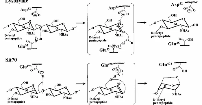
Fig. 6. Glutamic acid-assisted glycosyl transfer reactions catalysed by hen egg white lysozyme and E. coli lytic
transglycosylase Slt70. G, N-acetylglucosamine; M, N-acetylmuramic acid. The transition states shown in parentheses represent attack of the oxocarbonium cation intermediate by a water molecule (lysozyme) and the C6 hydroxyl group of N-acetylmuramic acid (Slt70).
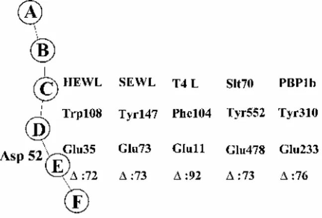
Fig. 7. Schematic illustration of the catalytic centre of hen egg white lysozyme (HEWL) and the glycosyl moieties
of a hexasaccharide (A-F) in the cleft region. Trp-108 in HEWL, Tyr-147 in swan egg white lysozyme (SEWL), Phe-104 in phage T4 lysozyme (T4L) and Tyr-552 in the lytic transglycosylase Slt70 occur at equivalent positions with respect to the corresponding catalytic glutamic acid in the crystal structures. Δ, spanning
distances in terms of intervening amino acid residues between the catalytic glutamic acid and the corresponding hydrophobic amino acid residue. Glu-233 and Tyr-310 of PBP1b are shown for comparison.
From the foregoing, it follows that Glu-233 is a key element of the glycosyl transferase catalytic centre of PBP1 b (Fig. 8). In all probability, it is involved in proton donation to the oxygen atom of the scissile phosphoester bond, resulting in the formation of an oxocarbonium intermediate. The second step of the reaction is the subject of more speculation. Asp-234 or Glu-290 could be responsible for the activation of the 4-OH of the nucleophile N-acetylglucosamine and the substitution on the β-face of the N-acetylmuramic acid. They might also play a role in the stabilization of the oxocarbonium as Asp-52 in HEWL and Asp-20 in T4L. The pair Glu-233-Tyr-310 suggests similarities in the design of the catalytic centre to those of the lysozymes and lytic transglycosylase Slt70. Substitution of a phenylalanine residue for Tyr-310 in PBP1b gives rise to a protein mutant that is very rapidly degraded during biogenesis, and the polypeptide produced cannot be isolated. Trp-108 in HEWL, which is the equivalent of Tyr-310 in PBP1b, has not been mutated, but the mutation of Val-109, immediately
downstream from Trp-108, into Pro abolishes the lysozyme activity (Hadfield et al., 1994). The acyl transferase
The Q447-M780 acyl transferase module of PBP1 b, which is linked to the D198-G435 glycosyl transferase module, possesses the characteristic motifs of the penicilloyl serine transferases superfamily. On this basis, motifs 7, 8 and 9 form the boundary of the catalytic centre, and Ser-510 of motif 7 is central to catalytic activity (Fig. 8). Cross-linking between peptide-substituted glycan strands implies that the Michaelis complex, which is formed upon binding of aD-alanyl-D-alanine-terminated pentapeptide, adopts a conformation that allows the proton of Ser-510γOH to be abstracted, the activated oxygen to attack the carbonyl carbon of the scissile peptide bond and the abstracted proton to be back-donated to the adjacent nitrogen atom. Then, the Ser-510-ester-linked peptidyl enzyme adopts a conformation that allows the proton of the NH2group at theD-centre of meso-diaminopimelic acid of another peptide to be abstracted, the activated nitrogen to attack the carbonyl carbon atom of the ester bond and the abstracted proton to be back-donated to the Oγ atom of the serine residue.
In in vitro assays using lipid II as substrate, the acyl transferase module of PBP1b catalyses the conversion of the pentapeptide units borne by the glycan chains into cross-linked peptide dimers and uncross-linked tetrapeptides. Under these unnatural conditions, the reaction proceeds at a water interface, and water also functions as acceptor of the transfer reaction. It is possible that, in the in vivo situation, the acyl transferase works as a strict
transpeptidase and that the uncross-linked tetrapeptides found in the completed peptidoglycan are the reaction products of monofunctional DD-carboxypeptidases/PBPs.
Thiolesters act as carbonyl donor analogues of the pentapeptide units. Under certain conditions, they behave as binding competitors of penicillin. In the absence of penicillin, thiolester binding leads to an acyl enzyme derivative whose acyl moiety is transferred to water and an amino acceptor. One may note that PBP1b is a weak thiolesterase when compared with the class A Mycobacterium leprae PBP1* (Lepage et al., 1997) and
In contrast to the thiolesters,D-alanyl-D-alanine-terminated peptides and amino compounds used as exogenous analogues of the natural peptide-substituted glycan chains lack substrate activities. In all appearances, the acyl transferase catalytic centre of PBP1b is designed to use, as carbonyl donors and amino acceptors, the peptide substituents borne by the glycan chains synthesized by the associated glycosyl transferase module. Conversely, derivatization of Ser-510 of the acyl transferase by penicillin in the form of a stable and inactive serine-ester-linked penicilloyl derivative does not alter the rate of glycan chain elongation, showing that the activity of the glycosyl transferase is acyl transferase independent.
Fig. 8. Functioning of the catalytic, Glu-233 glycosyl transferase and Ser-510 acyl transferase modules of the
wall peptidoglycan-polymerizing PBP1b of E. coli. The transition states shown in parentheses represent: (1) attack of the oxocarbonium cation intermediate by the 4-OH of N-acetylglucosamine; (2) attack of the carboxy-terminalD-alanyl-D-alanine bond of a pentapeptide by the Ser-510γOH, leading to the formation of an acyl (peptidyl) enzyme; and (3) attack of the ester bond of the peptidyl enzyme by the amino group at theD-centre of meso-diaminopimelic acid of another peptide, leading to cross-linked peptide dimer. Dpm, meso-diaminopimelic acid.
The full-size polypeptide
The Gln-270-Asn-844 and IIe-430-Asn-844 polypeptides of PBP1b have been produced in the periplasm of E. coli as attempts to obtain a catalytically active acyl transferase module disconnected from the glycosyl transferase module. The assays have failed. However, one may note that an active acyl transferase module in terms of penicillin binding has been obtained by tryptic digestion of the folded S. pneumoniae PBP1a (Di Guilmi et al., 1998).
The His tag (Met-46-Asn-423) and His tag (Met-46-Asp-478) polypeptides have been produced bound to the plasma membrane of the host cell. Essentially, these polypeptides comprise the glycosyl transferase module disconnected from the acyl transferase module. They adopt a conformation endowed with the desired activity,
but they are unstable and susceptible to proteolysis.
The membrane-bound, PelB-transported (Glu-144-Asn-844) polypeptide has been produced with the bulk of the polypeptide chain exposed on the outer face of the plasma membrane. The transported polypeptide lacks glycosyl transferase activity on lipid II, and it does not bind penicillin, indicating that the Leu-87-Asn-185 insert contains an essential amino acid sequence folding information. All these converging observations lead to the conclusion that PBP1b folds as a fully integrated polypeptide hybrid.
In contrast to E. coli PBP1b, the E. coli PBP1a has no insert downstream from the membrane anchor, and it contains two inserts that are located downstream from the intermodule junction site and between motifs 8 and 9 of the acyl transferase module respectively (Goffin and Ghuysen, 1998). PBP1a also catalyses peptidoglycan polymerization from lipid II. Loss of either PBP1a or PBP1 b is tolerated, showing that the two PBPs can substitute for each other, but loss of the two PBPs is fatal (Suzuki et al., 1978; Kato et al., 1985). PBP1a and PBP1b may prescribe distinct, subtle traits in peptidoglycan assembly that are difficult to detect. The occurrence of adducts at specific places along the corresponding polypeptide chains may be of importance in this respect. PBP1b mutants at positions 99-103 within the insert adopt a correct folding in terms of penicillin binding, but they have a reduced complementation activity in vivo (Lefèvre et al., 1997), suggesting that this adduct, which is involved in polypeptide folding, may also have other functions.
Experimental procedures
Bacterial strains, plasmids and oligonucleotides
E. coli EJ801 [ponA 1104(ts), ponB 1085, dacA 1191, dacB 12, lac, strA, tonA, metB, proA, tsx] was a gift from H. Hara (Hara and Suzuki, 1984). pHK2414 was the source of the PBP1b-encoding ponB (Kato et al., 1984), and pMJR160 was the source of laclq(Stark, 1987). The oligonucleotides were from Amersham Pharmacia Biotech Benelux.
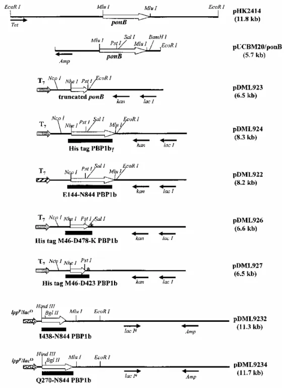
Recombinant plasmids (Fig. 9)
plN OmpA Hind/laclq. The /ac/-containing Kpnl-Hpal segment of plN OmpA Hind (Rentier-Delrue et al., 1988) was replaced by the corresponding /ac/-containing segment of pMJR160. giving rise to plN OmpA Hind/laclq. pDML924. The ponB-containing Mlul segment of pHK2414 was inserted into pUCBM20. A Nhel site was introduced at the 5' end of ponB by polymerase chain reaction (PCR). The 5' end of the sense primer cgccaagctagcatgccgcg-caaaggtaagggc-3') possessed a Nhel site, and the 5' end of the antisense primer (5'-ccggaattcctccctgcagctcctgacg-cacc-3') possessed an EcoRI site. These two primers were used to amplify from pUCBM20/ponB a 1160bp DNA segment that encoded the M46-Q423 sequence of PBP1b. The Nhel-EcoRl PCR segment was inserted between the corresponding sites of pET28a(+) (Novagen), giving rise to pDML923. The nucleotide sequence was analysed to detect any error. Then, the Pstl-EcoRI DNA segment carrying the end of ponB was introduced into pDML923, giving rise to pDML924. The His tag (M46-N844) PBP-encoding gene was under the control of the T7 promoter, lac operator.
pDML922. The truncated ponB encoding the E144-N844 sequence of PBP1b was inserted into pET22b/kan (Basu et al., 1996) downstream from the segment encoding the pullulanase signal peptide, using the same strategy as above. A Ncol site was added at the 5' end of the truncated ponB by PCR. The sense primer was 5'-cgccgcccatggaatt-taccgtgcaggccaac-3', and the antisense primer was that described above. The resulting plasmid was called pDML922. The gene was under the control of the T7 promoter, lac operator.
pDML926. A TAA codon was introduced into pET28a(+) downstream from Sall using the Quick Change Mutagenesis kit (Stratagene), the nucleotide 5'-cgagctccgtcgacaagtaa gcggccgcactcgag-3' and its reverse. The Ncol-Xhol 142 bp segment was sequenced and exchanged with the corresponding wild-type segment of pET28a(+), giving rise to pET28/Hind-. The Ncol-Sall 1400 bp segment encoding His tag (M46-D478) PBP1b was excised from pDML924 and inserted between the corresponding sites of pET28/Hind-, giving rise to pDML926. Because of the construction, the encoded M46-D478 PBP1b had an additional lysine at the carboxy end.
pDML927. A stop codon was mutagenized downstream from Pstl in pDML923 using the oligonucleotide 5'-cgtc aggagctgcagtaaggaattcgagc-3' and its reverse (Quick Change Mutagenesis kit). After sequencing the modified region, the Pstl-TAA-Clal 1488 bp segment was exchanged with the corresponding unmodified segment of
pDML923, giving rise to the His tag (M46-Q423) PBP1b-encoding pDML927.
Fig. 9. Maps of pHK2414, pUCBM20/ponB, pDML923, pDML924, pDML922, pDML926, pDML927,
pDML9232 and pDML9234.
pDML924A, pDML924B, pDML924C and pDML924D. The 1300 bp NcoI-PstI segment encoding His tag (M46-Q423) PBP1 b was excised from pDML924 and inserted into pUCBM20. The resulting pDML920C served as template for site-directed mutagenesis (Quick Change Mutagenesis) using the oligonucleotides
gctggcgacacaggaccgtcatttttacgag-3' and aat-gacggtcctgtgtcgccagcaaagtatc-3' (mutation E233→Q), gctggcgacagaaaaccgtcatttttacg-3' and cgtaaaaatgacggttttct gtcgccagc-3' (mutation D234→N), gcgtaaagcgaaccagcttaca tggcgctg-3' and cgccatgtaagctggttcgctttacgccag-3' (mutation E290→Q) and
5'-attcttgagctgttcatgaacgaggtgtat-3' and its reverse (mutation Y310→F). The mutated genes were selected on the basis of the appearance of an AvaW site (E233→Q), the disappearance of a HindIII site (E290→Q) and by DNA sequencing (D234→N and Y310→F). The 840bp NheI-SacII DNA segments carrying the desired mutations were completely sequenced and exchanged with the unmodified NheI-SacII segment of pDML924. Plasmids pDML924A, B, C and D encoded His tag (E233-Q) PBP1bγ, His tag (D234-N) PBP1bγ, His tag (E290-Q) PBP1bγ and His tag (Y310-F) PBP1bγ respectively.
pDML9232. The BglII-EcoRI 4874bp segment encoding the I438-N844 sequence of PBP1b was excised from pHK2414 and inserted between HindIII and EcoRI in plN OmpA Hind/laclq. The insertion was made via a DNA duplex encoding the L433 to K437 sequence of PBP1b and possessing protruding HindIII and BglII sites. The resulting plasmid pDML9232 contained the desired gene under the control of the Ipp promoter, lac promoter-operator.
pDML9234. pIN OmpA Hind/lac/q was digested with HindIII and EcoRI, and the HindIII ends were filled up with the Klenow polymerase. The PvuII-EcoRI 4355 bp segment encoding the Q270-N844 sequence of PBP1b was excised from pHK2414 and inserted into the modified plN OmpA Hind/laclq. The resulting plasmid, pDML9234, contained the desired gene under the control of the Ipp promoter, lac promoter-operator. Production, purification and analysis of His tag (M46-N844) PBP1b and derivatives
E. coli BL21 (DE3)/pET and E. coli HB101/plN OmpA Hind transformants were grown at 37°C in Luria-Bertani (LB) medium containing 50 µg kanamycin ml-1 and 50 µg ampicillin ml-1 respectively. When the optical density (OD) reached a value of 0.6-600 nm, the cultures were supplemented with 1 mM IPTG and maintained for an additional 2h. The produced proteins were isolated using the same procedure as that described below for His tag (M46-N844) PBP1b. The cells from 151 of culture (75 mg of PBP) were suspended in 750 ml of 25 mM Tris-HCI, pH7.5, containing 1 mM phenylmethylsulphonyl fluoride (PMSF), disrupted at 4°C in a cell disintegrator (Constant System), and the membranes were collected by centrifugation at 40 000 g at 4°C. The PBP was purified as described by Nicholas et al. (1993). Essentially, the membrane-bound PBP was solubilized at least partially (50-60%) by incubating the membranes (5 mg protein ml-1) in 25 mM Tris-HCI, pH7.5, 1 M NaCl, 1% CHAPS for 1 h at room temperature. The PBP (37.5 mg) was precipitated with 33% (NH4)2SO4 (2h at 4°C), the pellet was solubilized in 25 mM Tris-HCI, pH7.5, 0.5 M NaCl, 0.7% CHAPS (buffer A), and the solution was loaded on a Ni++-NTA agarose column equilibrated with the same buffer. The column was washed with buffer A, except that it contained 1 M NaCl (10 volumes), then with buffer A containing 40 mM imidazole (10 volumes), and finally with buffer A containing 150 mM imidazole. The PBP was eluted under these latter conditions. The fractions were pooled, concentrated, dialysed against buffer A and stored at -20°C.
The proteins were estimated using the method of Lowry et al. (1951). Immunological detection was carried out with polyclonal antibodies directed against PBP1b according to the Bio-Rad protocol. The antigen-antibody complexes were detected with alkaline phosphatase coupled to the anti-rabbit IgG. Proteins were submitted to SDS-PAGE and electro-blotted on Immobilon membranes (Millipore). N-terminal and C-terminal sequence analyses were carried out, respectively, on a 477 pulsed liquid sequencer and a Procise 494 sequencer (both from Perkin-Elmer, Applied Biosystems Division).
Penicillin binding
Non-radioactive β-lactam antibiotics were gifts from pharmaceutical companies. [3H]-benzylpenicillin (5 µCi nmol-1) and [14C]-benzylpenicillin (54 nCi nmol-1) were from Amersham International. The interaction between
PBP (E) and β-lactam compounds (I) was analysed on the basis of the three-step reaction:
K is the dissociation constant of the Henri-Michaelis complex E∙l; k2/K (M -1
s-1) is the second-order rate constant of acyl enzyme (El*) formation; and k3(s-1) is the first-order rate constant of enzyme deacylation. P is the product.
Determination of k3. PBP samples (5.5 µM) and β-lactams were incubated in 20 µl of buffer A (see above) at
37°C for 15 min, and then supplemented with 1 mg BSA ml-1 and an equal volume of precooled acetone. After 1 h at -20°C, the samples were centrifuged at 20000 g for 20 min at -10°C, and the pellets were solubilized in 173 µl of buffer A. The rate of breakdown of the [3H]-benzylpenicilloyl PBP was estimated by SDS-PAGE
fluorography and densitometry of the fluorograms (Fraipont et al., 1994). For non-radioactive antibiotics, the rate of active PBP recovery was estimated by labelling the samples with [3H]-benzylpenicillin (10-4M) for 2 min at 37°C.
Determination of k2/K. PBP samples (0.26 µM) were incubated with various concentrations of β-lactams in
buffer A. At increasing time intervals, samples (8 µl) were incubated with 1 × 10⁻⁴ M [3H]-benzylpenicillin for 2 min at 37°C, and the rate of formation of the [3H]-benzylpenicilloyl PBP was estimated as described above. The k2/K-value was also estimated by competition between [3H]-benzylpenicillin and non-labelled β-lactam antibiotic as described by Frère et al. (1992).
Lipid II and peptidoglycan polymerization
The [14C]-meso-diaminopimelic acid (Dpm)-labelled lipid II intermediate
[N-acetylglucosaminyl-N-acetylmuramoyl(L-Ala-γ-D-Glu-(L)-meso-Dpm-(L)-D-Ala-D-Ala)-pyrophosphate-undecaprenol] was prepared essentially as described previously (van Heijenoort et al., 1992), except that the membranes of a MurG-overproducing strain were used, resulting in a 1.7-fold increased yield of lipid II
Glycosyl transfer reaction (glycan chain synthesis). Typical assays were performed under the following conditions: [14C]-lipid II (1.5-2.5 µM; 0.126 µCi nmol-1) and His tag (M46-N844) PBP1bγ (15nM) were incubated in 50 mM Tris-HCl, pH7.5, 0.046% CHAPS, 33 mM NaCl, 10 mM MgCl2, 0.5% decyl PEG, 12.5% 1 -octanol, 25% DMSO for 30 min at 30°C. The reaction products were separated by an overnight chromatography on Whatman no. 1 filter paper in isobutyric acid-1 M ammonia (5:3) (Hara and Suzuki, 1984). The radioactive compounds were detected and analysed with a Phosphor Imager Scanner. The radioactive spots were also counted as described previously (van Heijenoort et al., 1992).
Acyl transfer reaction (peptide cross-linkage synthesis). His tag (M46-N844) PBP1bγ samples (110 nM in buffer A) as such or previously inactivated by benzylpenicillin (0.28 mM for 5 min at 30°C) were incubated with [14 C]-lipid II (1.7 µM for 1 h at 30°C). The polymerized material (83% versus 72% in the absence of benzylpenicillin) was isolated by paper chromatography (see above) and then incubated with lysozyme (0.25 mg ml-1) for 20 h. The polymerized material recovered in the supernatant (50%) was treated for 20 h with the Chalaropsis muramidase (20 µg ml-11) and analysed by HPLC as described by Glauner et al. (1988).
Acyl transfers on thiolester and peptide carbonyl donors
Benzoylglycylthiolactate, benzoylglycylthiolglycolate, Ac2L-Lys-D-Ala-D-Ala, L-Ala-γ-D-Glu-(L)-mesoDpm-(L)-D-Ala-D-Ala andL-Ala-γ-D-Glu(NH2)-(L)-mesoDpm-(L)-D-Ala were described previously (Nguyen-Distèche et al., 1974; Adam et al., 1991; Granier et al., 1994). Unlabelled and [14C]-mesoDpm-labelled UDP-MurNAcL-Ala-γ-D-Glu-(L)-mesoDpm-(L)-D-Ala-D-Ala were gifts from M. Guinand.
His tag (M46-N844) PBP1bγ samples (20-50 µg) and thiolester substrates (0.2 mM) were incubated at 37°C in 500 µl of 25 mM sodium phosphate buffer, pH7, 0.5 M NaCl, 0.7% CHAPS (buffer B). The reaction was monitored by measuring the decrease in A250 (Δє = 2200 M-1 cm-1) with a UVIKON 860 spectrophotometer coupled to a microcomputer via an RJ322 interface (Jamin et al., 1991). The carbonyl donor benzoyl-Gly-thiolglycolate (D), the hydrolysed product (H) and the aminolysed product benzoyl-Gly-D-Ala (T) obtained with D-Ala as acceptor of the transfer reaction were separated by HPLC on an ET250/8/4 Nucleosil-5 C18 column (Macherey Nagel). The flow rate was 1 ml min-1, and the retention times for D, H and T were 28.58, 20.94 and 22.06 min respectively. Solvent A was 0.05% trifluoroacetic acid, and solvent B was 0.035% trifluoroacetic acid in acetonitrile (gradient 0-30%).
His tag [M46-N844] PBP1bγ samples (5-10 µg) were incubated at 37°C for 3-20 h withD-Ala-D-Ala-terminated peptides (2-5 mM) in the absence or in the presence of various amino acceptors (2-10 mM) in buffer B.
Enzymatic activity was probed by estimating the releasedD-alanine using theD-amino acid oxidase test (Granier et al., 1994).
Acknowledgements
We thank Dr M. Guinand for the gracious gift of substrates, J. Wierenga and Dr W. Keck for their contribution to the production of the acyl transferase module as an independent entity. This work was supported in part by the Belgian programme on Interuniversity Poles of Attraction initiated by the Belgian State, Prime Minister's Office, Services fédéraux des affaires scientifiques, techniques et culturelles (PAI no. P4/03), the Fonds de la Recherche
Fondamentale Collective (Contract no. 2.4534.95) and the French Centre National de la Recherche Scientifique (EP1088). Some of the work described in this paper is part of a dissertation presented by M.T. in partial fulfilment of a PhD degree at the University of Liège.
References
Adam, M., Damblon, C., Jamin, M., Zorzi, W., Dusart, V., Galleni, M., et al. (1991) Acyltransferase activities of the high-molecular-mass essential penicillin-binding proteins. Biochem J 279: 601 -604.
Basu, J., Mahapatra, S., Kundu, M., Mukhopadhyay, S., Nguyen-Distèche, M., Dubois, P., et al. (1996) Identification and overexpression in Escherichia coli of a Mycobacterium leprae gene, pon1, encoding a high-molecular-mass class A penicillin-binding protein, PBP1. J Bacteriol 178: 1707-1711.
Broome-Smith, J.K., Edelman, A., Yousif, S., and Spratt,
B.G. (1985) The nucleotide sequences of the ponA and ponB genes encoding penicillin-binding proteins 1A and 1B of Escherichia coli K12. Eur J Biochem 147: 437-446.
Curtis, N.A.C., Orr, D., Ross, G.W., and Boulton, M.G. (1979) Affinities of penicillins and cephalosporins for the penicillin-binding proteins of Escherichia coli K12 and their antibacterial activity. Antimicrob Agents Chemother 16: 533-539.
Di Guilmi, A.M., Mouz, N., Andrieu, J.P., Hoskins, J., Jaskunas, S.R., Gagnon, J., et al. (1998) Identification, purification, and
characterization of transpeptidase and glycosyltransferase domains of Streptococcus pneumoniae penicillin-binding protein 1a. J Bacteriol 180: 5652-5659.
Edelman, A., Bowler, L., Broome-Smith, J.K., and Spratt, B.G. (1987) Use of a β-lactamase fusion vector to investigate the organization of penicillin-binding protein 1B in the cytoplasmic membrane of Escherichia coli. Mol Microbiol 1: 101-106.
Fraipont, C., Adam, M., Nguyen-Distèche, M., Keck, W., Van Beeumen, J., Ayala, J.A., et al. (1994) Engineering and overexpression of periplasmic forms of the penicillin-binding protein 3 of Escherichia coli. Biochem J 298: 189-195.
Frère, J.M., Nguyen-Distèche, M., Coyette, J., and Joris, B. (1992) Mode of action: interaction with the penicillin-binding proteins. In The Chemistry of β-Lactams. Page, M.I. (ed.). London: Blackie Academic and Professional, pp. 148-197.
Ghuysen, J.M. (1991) Serine β-lactamases and penicillin-binding proteins. Annu Rev Microbiol 45: 37-67.
Glauner, B., Höltje, J.V., and Schwarz, U. (1988) The composition of the murein of Escherichia coli. J Biol Chem 263: 10088-10095. Goffin, C, and Ghuysen, J.M. (1998) Multimodular penicillin-binding proteins: an enigmatic family of orthologs and paralogs. Microbiol Mol Biol Rev 62: 1079-1093.
Goodell, E.W., Markiewicz, Z., and Schwarz, U. (1983) Absence of oligomeric murein intermediates in Escherichia coli. J Bacteriol 156: 130-135.
Granier, B., Jamin, M., Adam, M., Galleni, M., Lakaye, B., Zorzi, W., et al. (1994) Serine-typeD-Ala-D-Ala peptidases and penicillin-binding proteins. Methods Enzymol 244: 249-266.
Hadfield, A.T., Harvey, D.J., Archer, D.B., MacKenzie, D.A., Jeenes, D.J., Radford, S.E., et al. (1994) Crystal structure of the mutant D52S hen egg white lysozyme with an oligosaccharide product. J Mol Biol 243: 856-872.
Hara, H., and Suzuki, H. (1984) A novel glycan polymerase that synthesizes uncross-linked peptidoglycan in Escherichia coli. FEBS Lett 168: 155-160.
van Heijenoort, J. (1996) Murein synthesis. In Escherichia coli and Salmonella. Neidhardt, F.C. (ed.).Washington, DC: American Society for Microbiology Press, pp. 1025-1034.
van Heijenoort, Y., Gomez, M., Derrien, M., Ayala, J., and van Heijenoort, J. (1992) Membrane intermediates in the peptidoglycan metabolism of Escherichia coli: possible roles of PBP1b and PBP3. J Bacteriol 174: 3549-3557.
Henderson, T.A., Dombrosky, P.M., and Young, K.D. (1994) Artifactual processing of penicillin-binding proteins 7 and 1b by the OmpT protease of Escherichia coli. J Bacteriol 176: 256-259.
Höltje, J.V. (1998) Growth of the stress-bearing and shape-maintaining murein sacculus of Escherichia coli. Microbiol Mol Biol Rev 62: 181-203.
Ishino, F., Mitsui, K., Tamaki, S., and Matsuhashi, M. (1980) Dual enzyme activities of cell wall peptidoglycan synthesis, peptidoglycan transglycosylase and penicillin-sensitive transpeptidase, in purified preparations of Escherichia coli penicillin-binding protein 1A. Biochem Biophys Res Commun 97: 287-293.
Jamin, M., Adam, M., Damblon, C., Christiaens, L., and Frère, J.M. (1991) Accumulation of acyl-enzyme in DD-peptidase-catalysed reactions with analogues of peptide substrates. Biochem J 280: 499-506.
Kato, J., Suzuki, H., and Hirota, Y. (1984) Overlapping of the coding regions for α and γ components of penicillin-binding protein 1 b in Escherichia coli. Mol Gen Genet 196: 449-457.
Kato, J., Suzuki, H., and Hirota, Y. (1985) Dispensability of either penicillin-binding protein 1a or 1b involved in the essential process for cell elongation in Escherichia coli. Mol Gen Genet 200: 272-277.
Lefèvre, F., Rémy, M.H., and Masson, J.M. (1997) Topographical and functional investigation of Escherichia coli penicillin-binding protein 1b by alanine stretch scanning mutagenesis. J Bacteriol 179: 4761-4767.
Lepage, S., Dubois, P., Ghosh, T.K., Joris, B., Mahapatra, S., Kundu, M., et al. (1997) Dual multimodular class A penicillin-binding proteins in Mycobacterium leprae. J Bacteriol 179: 4627-4630.
Lowry, O.H., Rosebrough, N.J., Farr, A.L., and Randall, R.J. (1951) Protein measurement with the folin phenol reagent. J Biol Chem 193: 265-275.
Malcolm, B.A., Rosenberg, S., Corey, M.J., Allen, J.S., de Baetselier, A., and Kirsch, J.F. (1989) Site-directed mutagenesis of the catalytic residues Asp-52 and Glu-35 of chicken egg white lysozyme. Proc Natl Acad Sci USA 86: 133-137.
Mottl, H., Anderluzzi, D., Kraft, A., and Höltje, J.V. (1995) Towards the enzymology of the transglycosylase reaction: studies on the transglycosylase activity of the penicillin-binding protein 1A of Escherichia coli. In Abstracts of the Symposium on The Envelope In Bacterial Physiology and Antibiotic Action, Garda, Italy, p. 70.
Nakagawa, J., Tamaki, S., Tomioka, S., and Matsuhashi, M. (1984) Functional biosynthesis of cell wall peptidoglycan by polymorphic bifunctional polypeptides. J Biol Chem 259: 13937-13946.
Nguyen-Distèche, M., Ghuysen, J.M., Pollock, J.J., Reynolds, P., Perkins, H.R., Coyette, J., et al. (1974) Enzymes involved in wall peptide crosslinking in Escherichia coli K12 strain 44. Eur J Biochem 41: 447-455.
Nicholas, R.A., Lamson, D.R., and Schultz, D.E. (1993) Penicillin-binding protein 1B from Escherichia coli contains a membrane association site in addition to its transmemγ brane anchor. J Biol Chem 268: 5632-5641.
Rentier-Delrue, F., Swennen, D., and Martial, J. (1988) plN-\W-ompA secretion vectors: modification of the ompA signal peptide sequence for easier insert cloning. Nucleic Acids Res 16: 8726.
Spratt, B.G., Zhou, J., Taylor, M., and Merrick, M.J. (1996) Monofunctional biosynthetic peptidoglycan transglycosy-lases. Mol Microbiol 19: 639-647.
Stark, M.J.R. (1987) Multicopy expression vectors carrying the lac repressor gene for regulated high-level expression of genes in Escherichia coli. Gene 51: 255-267.
Suzuki, H., Nishimura, Y., and Hirota, Y. (1978) On the process of cellular division in Escherichia coli: a series of mutants of E. coli altered in the penicillin-binding proteins. Proc Natl Acad Sci USA 75: 664-668.
Suzuki, H., van Heijenoort, Y., Tamura, T., Mizoguchi, J., Hirota, Y., and van Heijenoort, J. (1980) In vitro peptidoglycan polymerization catalysed by penicillin binding protein 1b of Escherichia coli K12. FEBS Lett 110: 245-249.
Thunnissen, A.M.W.H., Isaacs, N.W., and Dijkstra, W. (1995) The catalytic domain of a bacterial lytic transglycosylase defines a novel class of lysozymes. Proteins: Struct Funct Genet 22: 245-258.
Wang, C.C., Schultz, D.E., and Nicholas, R.A. (1996) Localization of a putative second membrane association site in penicillin-binding 1B of Escherichia coli. Biochem J 316: 149-156.