Université de Montréal
Role of Receptor and Non-Receptor Protein Tyrosine
Kinases in Vasoactive Peptide-Induced Signaling
par
George Vardatsikos
Programme de Sciences Biomédicales Faculté de Médecine
Thèse présentée à la Faculté de Médecine
en vue de l’obtention du grade de Philosophiæ Doctor (Ph.D.) en Sciences Biomédicales
option Générale
Janvier, 2012
Université de Montréal
Faculté des études supérieures et postdoctorales
Cette thèse intitulée:
Role of Receptor and Non-Receptor Protein Tyrosine Kinases in Vasoactive Peptide-Induced Signaling
Présenté par : George Vardatsikos
a été évalué par un jury composé des personnes suivantes :
Dr. Eugenio Rasio, président-rapporteur Dr. Ashok Srivastava, directeur de recherche Dr. Suhayla Mukaddam-Daher, membre du jury
Dr. Ghassan Bkaily, examinateur externe Dr. Jean St-Louis, représentant du doyen de la FESP
Résumé
L'endothéline-1 (ET-1) et l'angiotensine II (Ang II) jouent un rôle important dans le maintien de la pression artérielle et l'homéostasie vasculaire. Une activité accrue de ces peptides vasoactifs est présumée contribuer au développement de pathologies vasculaires, telles que l'hypertension, l'athérosclérose, l'hypertrophie et la resténose. Ceci est causé par une activation excessive de plusieurs voies de signalisation hypertrophiques et prolifératives, qui incluent des membres de la famille des Mitogen Activated Protein Kinases (MAPK), ainsi que la famille phosphatidylinositol 3-kinase (PI3-K) / protéine kinase B (PKB). Bien que l'activation de ces voies de signalisation soit bien élucidée, les éléments en amont responsables de l'activation des MAPK et de la PKB, induite par l'ET-1 et Ang II, demeurent mal compris. Durant les dernières années, le concept de la transactivation de récepteurs et/ou non-récepteurs protéines tyrosine kinases (PTK) dans le déclenchement des événements de signalisation induits par les peptides vasoactifs a gagné beaucoup de reconnaissance. Nous avons récemment démontré que la PTK Insulin-like Growth Factor type-1 Receptor (IGF-1R) joue un rôle dans la transduction des signaux induits par l’H2O2, menant à la phosphorylation de la PKB. Étant donné que les peptides vasoactifs génèrent des espèces réactives d'oxygène, telles que l’H2O2 lors de leur signalisation, nous avons examiné le rôle de d’IGF-1R dans la phosphorylation de la PKB et les réponses hypertrophiques dans les cellules muscle lisse vasculaires (CMLV) induites par l'ET-1 et Ang II. AG-1024, un inhibiteur spécifique de l'IGF-1R, a atténué la phosphorylation de la PKB induite à la fois par l'ET-1 et Ang II. Le traitement des CMLVs avec l’ET-1 et Ang II a également induit une phosphorylation des résidus tyrosine dans les sites d'autophosphorylation d'IGF-1R, celle-ci a été bloquée par l’AG-1024. En outre, l’ET-1 et l’Ang II on tous les deux provoqué la phosphorylation de c-Src, une PTK non-récepteur, bloqué par PP-2, inhibiteur spécifique de la famille Src. La PP-2 a également inhibé la phosphorylation de PKB et d’IGF-1R induite par l’ET-1 et l’Ang II. De plus, la synthèse de protéines ainsi que d’ADN, marqueurs de la prolifération cellulaire et de l’hypertrophie, ont également été atténuée par l’AG-1024 et le PP-2
Bien que ce travail démontre le rôle de c-Src dans la phosphorylation PKB induite par l'ET-1 et Ang II, son rôle dans l’activation des MAPK induit par l’ET-1 dans les CMLVs reste controversé. Par conséquent, nous avons examiné l'implication de c-Src dans l’activation de ERK 1/2, JNK et p38MAPK, par l'ET-1 et Ang II, ainsi que leur capacité à régulariser l’expression du facteur de transcription Early growth transcription factor-1 « Egr-factor-1 ». ET-factor-1 et Ang II ont induit la phosphorylation de ERK factor-1/2, JNK et p38MAPK, et ont amplifié l'expression d'Egr-1 dans les CMLVs. Cette augmentation de la phosphorylation des MAPK a été diminuée par la PP-2, qui a aussi atténué l’expression d’Egr-1 induite par l’ET-1 et l’Ang II. Une preuve supplémentaire du rôle de c-Src dans ce processus a été obtenue en utilisant des fibroblastes embryonnaires de souris déficientes en c-Src (Src -/- MEF). L’expression d’Egr-1, ainsi que l’activation des trois MAPKs par l’ET-1 ont été atténuées dans les cellules Src -/- par rapport au MEF exprimant des taux normaux Src. En résumé, ces données suggèrent que l'IGF-1R et c-Src PTK jouent un rôle essentiel dans la régulation de la phosphorylation de PKB et des MAPK dans l’expression d’Egr-1, ainsi que dans les réponses hypertrophiques et prolifératives induites par l'ET-1 et Ang II dans les CMLVs.
Mots-clés : Endotheline-1, Angiotensin II, PKB, MAPK, IGF-1R, c-Src, VSMC, Egr-1, prolifération, hypertrophie
Abstract
Endothelin-1 (ET-1) and angiotensin II (Ang II) play important roles in maintaining blood pressure and vascular homeostasis, and a heightened activity of these vasoactive peptides is thought to contribute to the development of vascular pathologies, such as hypertension, atherosclerosis, hypertrophy and restenosis. This is caused by an excessive activation of several growth and proliferative signaling pathways, which include members of the mitogen-activated protein kinase (MAPK) family, as well as the phosphatidylinositol 3-kinase (PI3-K)/protein kinase B (PKB) pathway. While the activation of these signaling pathways is well elucidated, the upstream elements responsible for ET-1 and Ang II-induced MAPK and PI3-K/PKB activation remain poorly understood. During the last several years, the concept of transactivation of receptor and/or non-receptor protein tyrosine kinases (PTK) in triggering vasoactive peptide-induced signaling events has gained much recognition. We have recently demonstrated that insulin-like growth factor-1 receptor (IGF-1R) plays a role in tranducing the effect of H2O2, leading to PKB phosphorylation.
Since vasoactive peptides elicit their responses through generation of reactive oxygen species, including H2O2, we investigated whether IGF-1R transactivation plays a similar
role in ET-1 and Ang II-induced PKB phosphorylation and hypertrophic responses in VSMC. AG-1024, a specific inhibitor of IGF-1R, attenuated both ET-1 and Ang II-induced PKB phosphorylation in a dose-dependent manner. ET-1 and Ang II treatment also induced the phosphorylation of tyrosine residues in the autophosphorylation sites of IGF-1R, which was blocked by AG-1024. In addition, both ET-1 and Ang II evoked tyrosine phosphorylation of c-Src, a non-receptor PTK, and pharmacological inhibition of c-Src PTK activity by PP-2, a specific inhibitor of Src-family tyrosine kinase, significantly reduced PKB phosphorylation as well as tyrosine phosphorylation of IGF-1R induced by the two vasoactive peptides. Furthermore, protein and DNA synthesis, markers of cell growth and proliferation, enhanced by ET-1 and Ang II were also attenuated by AG-1024 and PP-2.
While this work demonstrates the role of c-Src in ET-1 and Ang II-induced PKB phosphorylation, its role in ET-1-induced MAPK signaling and regulation of transcription factors, such as early growth response factor-1 (Egr-1), which was recently shown to be expressed in atherosclerotic plaque, remains controversial in VSMC. Therefore, we have also investigated the involvement of c-Src in ET-1 and Ang II-induced ERK 1/2, JNK and p38mapk activation, as well as Egr-1 regulation. ET-1 and Ang II-induced the phosphorylation of ERK 1/2, JNK and p38mapk, and enhanced the expression of Egr-1 in aortic VSMC. This increased phosphorylation was decreased by PP-2. Further proof for the role of c-Src in this process was obtained by using mouse embryonic fibroblasts (MEF) deficient in c-Src (Src -/- MEF). ET-1-induced Egr-1 expression, as well as MAPK activation, were found to be downregulated in Src -/- MEF, as compared to MEF expressing normal Src levels. In summary, these data demonstrate that IGF-1R and c-Src PTK play a critical role in mediating both PKB and MAPK phosphorylation and Egr-1 expression, as well as hypertrophic and proliferative responses induced by ET-1 and Ang II in VSMC.
Keywords: Endothelin-1, Angiotensin II, PKB, MAPK, IGF-1R, c-Src, VSMC, Egr-1, proliferation, hypertrophy
Table of Contents
Résumé... i Abstract ... iii Table of Contents ... v List of Tables ... ix List of Abbreviations ... xi Acknowledgements ... xvi Chapter 1 ... 1 Introduction... 11.1 Obesity, Diabetes and Hypertension: The Modern Epidemic... 1
1.1.1 Insulin resistance and the metabolic syndrome... 2
1.2 The metabolic Syndrome, Hypertension and Cardiovascular Disease ... 5
1.3 Hypertension ... 5
1.3.1 Definition of hypertension ... 6
Table 1: Categories of hypertension ... 1
Table 2: Lifestyle Modifications to Manage Hypertension ... 1
1.3.2 Varying forms of hypertension ... 11
Cardiovascular Diseases ... 13
1.4 Endothelins... 14
1.4.1 Structure, Regulation and Biosynthesis of ET-1... 15
1.4.2 ET-1 Receptors ... 19
1.4.3 Biological actions of ET-1 ... 22
1.5 Role of ET-1 in cardiovascular disease... 31
1.5.1 Role of ET-1 in essential and experimental hypertension ... 31
1.5.2 Role of ET-1 in atherosclerosis and heart failure... 33
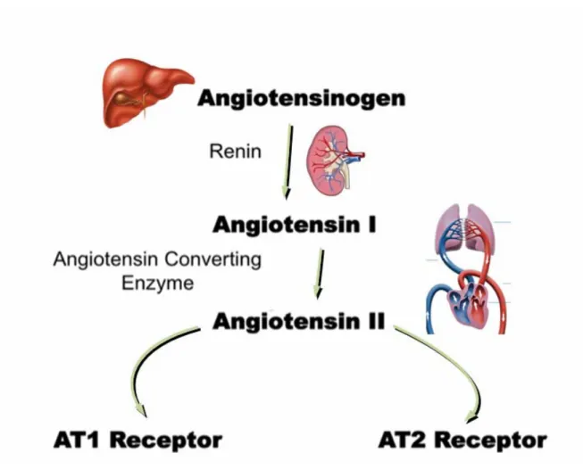
1.6 Angiotensin II and the Renin-Angiotensin-system (RAS) ... 36
1.6.1 Structure, Regulation and Biosynthesis of Angiotensin II through the RAS... 36
1.6.3 Biological actions of Ang II... 42
1.7 Role of Ang II in Cardiovascular Disease... 48
1.7.1 Role of Ang II in hypertension ... 48
1.7.2 Role of Ang II in atherosclerosis and heart failure ... 49
1.8 ET-1 and Ang II-induced signalling events in VSMC... 51
1.8.1 ET-1 and Ang II-induced activation of the phosphatidylinositol 3-kinase (PI3-K)/protein kinase B (PKB) pathway ... 53
1.8.2 ET-1 and Ang II-induced activation of the Mitogen-Activated Protein Kinase (MAPK) pathway... 56
1.8.3 ET-1 and Ang II-induced transactivation of receptor and non-receptor protein tyrosine kinases (R-/NR-PTK)... 59
1.9 Objectives of the present study ... 69
Chapter 2 ... 71
Role of insulin-like growth factor 1 receptor and c-Src in endothelin-1- and angiotensin II-induced PKB phosphorylation, and hypertrophic and proliferative responses, in vascular smooth muscle cells. ... 71
2.1 Abstract ... 73
2.2 Introduction... 74
2.3 Materials and Methods... 75
2.3.1 Materials... 75
2.3.2 Methods... 76
2.4 Results... 78
2.4.1 Attenuation of ET-1 and Ang II-induced PKB phosphorylation by AG-1024 in A-10 VSMCs... 78
2.4.2 ET-1 and Ang II induce tyrosine phosphorylation of IGF-1R... 78
2.4.3 Attenuation of ET-1 and Ang II-induced PKB phosphorylation by PP-2 in A-10 VSMCs... 79
2.4.5 Attenuation of ET-1 and Ang II-induced tyrosine phosphorylation of IGF-1R β
subunit by PP-2. ... 79
2.4.6 Lack of Involvement of IGF-1R in ET-1/Ang II-induced c-Src phosphorylation. ... 80
2.4.7 Attenuation of ET-1 and Ang II-induced protein and DNA synthesis by AG-1024 and PP-2. ... 80 2.5 Discussion ... 81 2.6 Acknowledgements... 83 2.7 Figure legends ... 84 2.8 References... 88 Chapter 3 ... 103
Requirement of c-Src, a Non-Receptor Tyrosine Kinase in both Endothelin-1 and Angiotensin II-induced ERK 1/2, JNK and p38 MAPK, as well as Egr-1 activation in Vascular Smooth Muscle cells... 103
3.1 Abstract ... 105
3.2 Introduction... 106
3.3 Materials and Methods... 108
3.3.1 Materials... 108
3.3.2Methods... 108
3.4 Results... 111
3.4.1 Inhibition of c-Src PTK attenuates ET-1 and Ang II-induced phosphorylation of ERK 1/2 in A10 VSMC. ... 111
3.4.2 ET-1 and Ang II-induced phosphorylation of JNK/SAPK and p38 MAPK is attenuated by c-Src PTK inhibition in A10 VSMC. ... 111
3.4.3 Inhibition of c-Src PTK attenuates ET-1-induced early growth response factor-1 (Egr-1) transcription factor expression in A10 VSMC. ... 112
3.4.4 Effect of c-Src knockdown in ET-1 and Ang-II-induced ERK 1/2, JNK and p38 MAPK phosphorylation and Egr-1 expression ... 113
3.6 Acknowledgments... 116
3.7 Figure legends ... 117
3.8 Reference List ... 120
3.9 Figures... 126
3.10 Supplementary Figures ... 133
3.11 Supplementary Figure Legends ... 137
Chapter 4 ... 139
General Discussion ... 139
Chapter 5 ... 150
Conclusion ... 150
List of Tables
Table 1: Categories of hypertension ... 1 Table 2: Lifestyle Modifications to Manage Hypertension ... 1
List of Figures
Figure 1: Structure of the Endothelins. ... 1 Figure 2: Factors affecting regulation of ET-1 synthesis and its subsequent ET receptor
mediated actions on vascular smooth muscle cells... 1 Figure 3: Biological actions of ET-1 on various systems leading to multiple
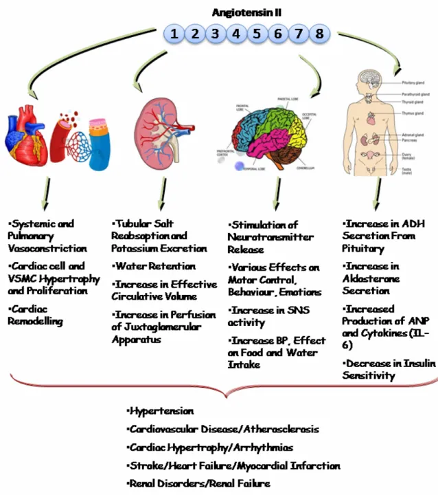
pathophysiological states.. ... 1 Figure 4: Events leading to the dysfunction of endothelial cells and VSMCs leading to the development of atherosclerosis. ... 1 Figure 5: Synthesis of Angiotensin II (Ang II) by the renin-angiotensin system (RAS)... 1 Figure 6: Structure of Angiotensins.. ... 1 Figure 7: Biological actions of Ang II on various systems leading to multiple
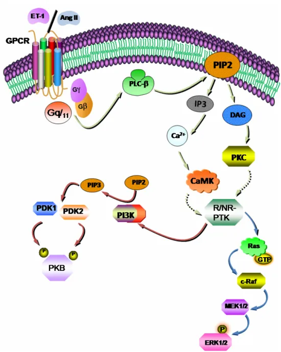
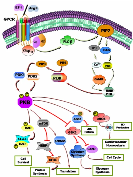
pathophysiological states. A. ... 1 Figure 8: Vasoactive peptide-induced activation of the phosphoinositide cascade through
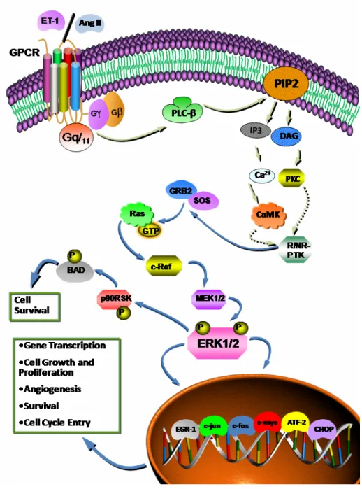
GPCR activation in VSMC ... 1 Figure 9: Vasoactive peptide-induced activation of the PI3-K/PKB cascade in VSMC. . ... 1 Figure 10: Vasoactive peptide-induced activation of the MAPK pathway in VSMC. . ... 1 Figure 11: Key steps in the production of reactive oxygen species (ROS) by vasoactive
peptides ... 1 Figure 12: Schematic model summarizing the mechanism by which ET-1 and Ang II induce downstream activation and modulation of PKB, ERK1/2, JNK, p38mapk and Egr-1, leading to subsequent hypertrophic and proliferative events in VSM. ... 1
List of Abbreviations
2K1C Two kidney 1 clip rodent model of renal hypertension ANF Atrial natriuretic factor
Ang II Angiotensin II
ANP Atrial natriuretic peptide AP-1 Activator protein 1
ApoE Apolipoprotein E
AT1R Angiotensin II type 1 receptor
AT2R Angiotensin II type 2 receptor
BAD Bcl-2 associated death promoter
Bcl-2 B-cell lymphoma 2
bFGF Basic fibroblast growth factor BNP Brain natriuretic peptide
BQ 123 Cyclo(D-Asp-Pro-D-Val-Leu-D-Trp, Na), a potent ETA receptor
antagonist
BQ 788 N-cis-2,6-Dimethylpiperidinocarbonyl-L-γ-MeLeu-D-Trp(MeOCO)-D-Nle-OH, Na, a potent ETB receptor antagonist
Ca2+ Calcium
cGMP Cyclic guanosine monophosphate CHF Chronic heart failure
COX-2 Cyclooxygenase-2
CSF Colony-stimulating factors
CVD Cardiovascular disease
DAG Diacyglycerol
DOCA Deoxycorticosterone acetate ECE Endothelin converting enzyme EGF Epidermal growth factor
EGF-R Epidermal growth factor receptor Egr-1 Early growth response factor 1
eNOS Endothelial nitric oxide synthase ERK Extracellular signal-regulated kinase
ET Endothelin
ETA Endothelin receptor type A
ETB Endothelin receptor type B
FGF Fibroblast growth factor G protein Guanosine nucleotide binding protein
GDP Guanosine diphosphate
GPCR Guanosine nucleotide binding protein-coupled receptor Grb2 Growth factor receptor-bound protein 2
GSK-3 Glycogen synthase kinase 3
GSH Glutathione
GST Glutathione-s-transferase
GTP Guanosine triphosphate H2O2 Hydrogen peroxide
HDL High density lipoprotein-cholesterol HIF Hypoxia inducible factor
IGF-1 insulin-like growth factor 1
IGF-1R insulin-like growth factor type 1 receptor
IL interleukin
iNOS inducible nitric oxide synthase
IR insulin receptor
IRS insulin receptor substrate IP3 inositol triphosphate
JNK c-Jun N-terminal kinase
kDa kiloDalton
MAPK mitogen activated protein kinase MEF2 myocyte enhancer factor 2
MEK mitogen extracellular regulated kinase
MI myocardial ischemia
MMP matrix metalloproteinase
mTOR mammalian target of rapamycin
NADPH oxidase nicotamide adenine dinucleotide phosphate oxidase
NF-1 nuclear factor 1
NF-κB nuclear factor kappa-light-chain-enhancer of activated B cells
NO nitric oxide
NR-PTK Non-receptor protein tyrosine kinase (O2˙-) superoxide anion
(OH˙) hydrogen radicals
(ONOO-) peroxynitrite p70s6k p70 ribosomal S6 kinase
p90rsk p90 ribosomal kinase
PDGF platelet derived growth factor
PDGF-R platelet derived growth factor receptor PDK phosphoinositide-dependent kinase
PH pleckstrin homology
PI3-K phosphatidylinositol 3-kinase
PI phosphatidylinositol
PIP phosphatidylinositol 3 monophosphate PIP2 phosphatidylinositol 4, 5 bisphosphate
PIP3 phosphatidylinositol 3, 4, 5 triphosphate
PLC β phosphoinositide-specific phospholipase C β
PKB protein kinase B
PKC protein kinase C
PTK protein tyrosine kinase PTPase Protein tyrosine phosphatase PTP-1B Protein tyrosine phosphatase 1B PYK-2 proline-rich tyrosine kinase R-PTK receptor protein tyrosine kinase ROS reactive oxygen species SAPK stress-activated protein kinase
SH2 src homology 2
Shc src homology collagen
SHR spontaneously hypertensive rat
SHP-2 SH2 domain-containing tyrosine phosphatase-2
SOD Superoxide dismutase
SOS son of seven less
STAT signal transducer and activator of transcription TNF-α tumour necrosis factor-α
TGF-β transforming growth factor-β VSMC vascular smooth muscle cell
To my Mom and Dad, who have worked tirelessly for 30 years to help get me where I am today.
Acknowledgements
First and foremost, I would like to thank my supervisor, Dr. Ashok Srivastava, for your immeasurable support, constant encouragement, endless patience, timely advice and most of all, your friendship, both in and out of the lab. The most humble person I have ever met, you truly are a remarkable mentor, having given me an amazing work environment, as well as plenty of room and time to learn not only science, but how to be a good researcher. I will forever be indebted to you for your teachings. Thank you, Ashok. Being your student has truly been an honour.
My sincere thanks go to Drs. Marc Prentki and Vincent Poitout for your constant encouragement and support, as well as your helpful suggestions and genuine interest in my research project and presentations during journal clubs, data sessions, conferences and retreats. Truly amazing scientist and people, their doors have always been open to me, and they have always gone out of their way to help me when I needed it. I cannot thank you enough for the support and encouragement you have both provided me!
I must also take this opportunity to thank my thesis jury members, who have been burdened with the painstaking task of evaluating this thesis. Your comments and corrections will truly improve this work, and I thank you for investing so much time and effort to accomplish this task. A special thank you goes to Dr. Eugenio Rasio, who has been a constant inspiration and continues to be, who has supported me and my work, and has helped guide me through the internal workings of the CRCHUM during my tenure as student representative on the comité pédagogique and the comité de direction du CRCHUM.
Over the past seven years, I have had the chance to work with truly remarkable labmates. I would like to thank all of them for helping achieve my goal. My thanks go out to Georgia Kapakos, Estelle Simo, Viktoria Youreva and Marilyne Blain, for helping me make all those acrylamide gels, lyse thousands of petri dishes full of cells, quantify insane amounts of protein, and for helping me with all those western blots! You girls came in early in the morning for me, stayed late at night for me and spent weekends at the lab for
me. This work is as much yours as it is mine. Thank you doesn’t even begin to express my gratitude for all your help and support. I would also like to thank Dr. Ali Bouallegue, with whom I also worked for part of this project, for his continuous help, and for teaching me that one should not let a couple of bad results get the best of you. I must also thank Dr. Mohamad Z. Mehdi, who was not only my labmate, but my mentor when I began my graduate studies. He not only taught me the technical aspects of the science we were doing, but also helped me understand and to appreciate scientific research. Thank you my brother, for your help, guidance, constant support and above all, your friendship.
A special mention must also be given to all of my labmates and technical staff at the MDRC, especially Dr. Melkam Kebede, whose constant encouragement and aide have helped me not only in completing this project, but also to create an exciting work atmosphere, full of ideas, friendship and comradery.
I would also like to take this opportunity to thank the Montreal Diabetes Research Center, the Faculté de Médecine and the Faculté d’Études supérieures et post-doctorales de l’Université de Montréal, the Programme de Sciences Biomédicales, as well as the Centre de recherche du Centre hospitalier de l’Université de Montréal for the many studentships and awards they have granted me over the years.
Last but not least, I would like to thank my friends and family for their constant love and support. To Greg, Leo, Marco, Johnny 15, Giancarlo, Ariana, Vivienne and Laura, all of whom I am very proud of for all their accomplishments; thanks guys, for being there and always supporting the cause. To my family, words cannot express my love and appreciation to you; my Mom and Dad, my brothers Billy and Kosta and my sister Effie, all three of whom I am especially proud of, all four of my beautiful grandparents who have taught me that hard work and perseverance always wins out, and even Moulo the dog, for always keeping me company, especially during the writing of this thesis, probably hoping I would give him a puppy snack! Your unfaltering support and help in whatever endeavors I undertake amazes even me at times, but I guess that’s what family is for. I am deeply and forever indebted to you for your love and encouragement.
Chapter 1
Introduction
1.1 Obesity, Diabetes and Hypertension: The Modern Epidemic
The insulin resistance syndrome, more commonly known as the metabolic syndrome, has been described as a cluster of multiple risk factors leading to the development of type 2 diabetes mellitus (T2DM) and cardiovascular disease (CVD). Among these risk factors are obesity, irregular blood lipids (dyslipidemia), insulin resistance and hypertension 1. These risk factors, beginning with insulin resistance, are caused by both genetic (where certain individuals or populations may be more likely to develop the metabolic syndrome due to their unique genetic background) and environmental/lifestyle factors, such as diet and exercise. Globally, the incidence of metabolic syndrome is rising at an alarming rate 2, yet the true prevalence of the disease is unknown, partly due to the lack of an accepted definition to define the metabolic syndrome. To understand insulin resistance and the metabolic syndrome, and how this dysregulation of hormonal signaling affects various aspects of normal physiology and pathophysiological states, it is important to define insulin, and to understand its functions in normal physiology.
Insulin is the primary hormone involved in blood glucose control. In response to increasing blood glucose levels, pancreatic β-cells secrete insulin, which stimulates glucose uptake in muscle and fat tissues via stimulation of glucose transporter 4 (GLUT4) from intracellular sites to the plasma membrane 3 and triggers inhibition of gluconeogenesis and glucose release by the liver by stimulating glycogen synthesis and inhibiting glycogenolysis and gluconeogenesis, respectively. In addition, insulin is an anabolic hormone, which promotes lipid synthesis and suppresses lipid degradation. These actions are all mediated by an intricate signaling cascade initiated by the binding of insulin to its receptor. An impaired insulin signaling and action results in a drastic decrease in glucose transport, glycogen synthesis, and lipid generation, leading to dysregulation of the physiological effects of insulin, which normally leads to insulin resistance, and T2DM 4. When added to other environmental and genetic factors, this impaired signaling leads to symptoms of the metabolic syndrome, which can turn in to T2DM and CVD.
1.1.1 Insulin resistance and the metabolic syndrome
Simply put, insulin resistance is a condition in which the body produces insulin, but cannot use it properly. In a more scientific fashion, it is a condition in which insulin, exogenous or endogenous, produces an abnormally low biological response with respect to a decrease in blood glucose levels 5. Insulin resistance is the earliest detectable metabolic trait of pre-diabetes, and possibly the most important risk factor in the development of Type 2 diabetes mellitus (T2DM). In the pre-diabetic state, or insulin resistance, serum insulin concentrations are elevated to compensate for insulin resistance in peripheral tissues, and to maintain a normal glucose balance. This is a compensatory mechanism, known as hyperinsulinemia.
Hyperinsulinemia exists to maintain normal glucose levels, or normoglycemia, during the prediabetic/insulin resistant state. Even though normoglycemia may be maintained in some, it has been shown that other patients will have increased fasting glucose or impaired glucose tolerance (IGT) after a 2 hour oral glucose challenge, of 140 to 199 mg/dL 6. Hyperinsulinemia is a stress on the pancreas, and may, at some point, not be able to handle this increased work load, and as a result its insulin secretory response may decrease. A decrease in insulin levels may cause a rise in blood glucose, leading to diabetes.
Today, it is almost impossible to speak of insulin resistance without talking about the metabolic syndrome. In fact, after having done an in depth review of the literature on the subject, I have come to realize that if not for the clinical definition of insulin resistance, the terms metabolic syndrome and insulin resistance have come to be almost interchangeable. Essentially, the metabolic syndrome is a constellation of abnormalities that is associated with increased risk for the development of T2DM and atherosclerotic vascular disease, such as heart disease and stroke 2.
As mentioned earlier, insulin levels may decrease after prolonged insulin resistance, yet in most of these insulin resistant individuals, large amounts of insulin continue to be secreted to overcome this defect in insulin action, thereby maintaining normal or near-normal glucose tolerance. While this compensatory hyperinsulinemia prevents the
development of hyperglycemia, these individuals run a risk of suffering from a certain degree of IGT, high plasma triglyceride (TG) and low high-density lipoprotein cholesterol (HDL-C), or “good cholesterol” levels, obesity, and most importantly, essential hypertension 7, 8. These seem to constitute the cluster of risk factors leading to T2DM and CVD.
The first guide lines for the diagnosis of metabolic syndrome were presented in 1998 by the World Health Organization (WHO) 9. This definition was later modified by the European Group for the Study of Insulin Resistance (EGIR) to focus more on glucose intolerance and insulin resistance 10. In 2001, the National Cholesterol Education Programme Adult Treatment Panel III (ATPIII) again revised the metabolic syndrome diagnosis criteria. It is based more on plasma glucose levels and dyslipidemia, and omits insulin resistance as a criteria 11. More recently, the American Heart Association (AHA) released its own criteria, which mirror the ATPIII, with the exception of stricter fasting glucose criteria 1. The most recent definition of the metabolic syndrome criteria comes from the International Diabetes Federation, which names abdominal obesity and insulin resistance as the most important factors leading to metabolic syndrome 12. Despite these
more recent definitions, the most widely used definition and criteria to diagnose the metabolic syndrome remain those of the ATP III, and as such, we will concentrate on those criteria here. The ATPIII identifies 5 components of the metabolic syndrome that relate to CVD: abdominal obesity (waist circumference), atherogenic dyslipidemia, high density lipoprotein (HDL) cholesterol levels, glucose intolerance, and high blood pressure 11.
Visceral obesity is the form of obesity most strongly linked with insulin resistance
and with the presence of related metabolic abnormalities of the metabolic syndrome. Clinically, it presents increased waist circumference. However, the mechanisms underlying the association between abdominal obesity and the metabolic syndrome are not fully understood. It has been assumed that abdominal adipose tissue releases an excess of fatty acids and cytokines that contribute to insulin resistance. For example, visceral adipose tissue secretes adipokines like adiponectin and inflammatorycytokines such as interleukin-6 (IL-interleukin-6) and tumor necrosis factor (TNF)-α, which contribute to the insulin resistant,
proinflammatory, and hypertensive states 13. In fact, visceral adiposity and insulin resistance are connected by TNF-α at the molecular level. Studies have shown that in adipocytes, TNF-α causes serine phosphorylation of IR and IRS-1, impairing insulin signaling. This leads to increased lipolysis, as well as decreased adiponectin levels, and an increase in circulating free fatty acids (FFA) 14, 15
Dyslipidemia and HDL levels present in routine lipoprotein analysis by high
triglyceride levels, exceeding 200mg/dL and reduced concentrations of high density lipoproteins (HDL) cholesterol, usually below 40mg/dL 5. In addition, it is characterized by other lipoprotein abnormalities, such as elevated apolipoprotein B, small low density lipoprotein (LDL) particles, caused by enriched triglycerides, and small HDL particles 5. The combination of these factors increases the atherogenicity of LDL, even though LDL levels are usually not increased. Lipoprotein metabolism is regulated by genetic and environmental (diet, exercise or lack thereof) factors and both can aggravate dyslipidemia.
Glucose intolerance presents as impaired fasting glucose (levels of 110 to 125
mg/dL) or IGT (levels of 140 to 199 mg/dL) after a 2 hour oral glucose challenge, of 1.75 Kg of glucose per Kg of body weight for children and 75 g of glucose for adults 6. These
glucose levels are intermediates between normal values and overt diabetes. When glucose intolerance evolves into diabetes-level hyperglycemia due to the failure of the hyperinsulinemia mechanism described earlier, high glucose levels constitute a major, independent risk factor for CVD.
Blood pressure has been shown to be elevated in overweight/obese patients with
insulin resistance or glucose intolerance. In fact, 50-60% of patients suffering from essential hypertension are insulin resistant to some degree 16. Blood pressure regulation is complex and affected by dietary factors, physical activity and renal/adrenal organ function.
1.2 The metabolic Syndrome, Hypertension and Cardiovascular
Disease
Obesity, diabetes, and the metabolic syndrome have clearly been shown to cause endothelial dysfunction 17. Endothelial dysfunction is commonly characterised by reduced nitric oxide (NO) dependent activity, which leads to hypertension, coronary heart disease, and accelerated atherosclerosis 18. Glucotoxicity (deleterious effects of hyperglycemia) and lipotoxicity (negative effects of increased plasma FFA and low HDL), as well as increased pro-inflammatory signaling are all adverse effects of the metabolic syndrome, which worsen insulin resistance, and contribute to endothelial dysfunction, hypertension and CVD.
1.3 Hypertension
Blood pressure is defined as the force that blood exerts on the vessel walls. Hypertension (HT), or high blood pressure, is a chronic medical condition characterized mainly by elevated arterial blood pressure 19. The worldwide prevalence of hypertension is estimated to be at 1 billion people, and contributes to approximately 7.1 million deaths per year worldwide 20. Persistent HT is a major risk factor contributing to pathophysiological events such as stroke, heart attack, heart failure and chronic renal failure 21. In the United
States, it is estimated that approximately 50 million people (almost 1 in 4 adults) suffer from hypertension 19, with proportions varying according to race, age, geographic location, gender and economic status. To add to these factors, not all people suffering from HT suffer from the same type of HT, as multiple types of HT exist. Furthermore, as blood pressure is not static, and varies from one minute to another in the day due to multiple external stimuli, it is difficult to define HT, in order to establish accepted treatment guidelines. Here, I will define blood pressure and discuss several of the proposed definitions of HT, as well as several different types of HT, and the differences between them.
1.3.1 Definition of hypertension
Blood pressure is calculated by measurements of systolic pressure (SBP) over diastolic blood pressure (DBP), measured in millimeters of mercury (mmHg). Systolic pressure is the peak pressure in the arteries, which occurs near the end of the cardiac cycle, during ventricular contraction. Diastolic pressure is the lowest pressure in the arteries, occurring at the start of the cardiac cycle, when the ventricles are filling with blood. It can be said that “normal” or healthy adult blood pressure, when taken at a resting state (normally how blood pressure is taken) is 115mmHg systolic over 75mmHg diastolic, read as 115/75 mmHg 22. As mentioned earlier, blood pressure is not static, as it undergoes natural variations from one heart beat to another, in response to simple stimuli, such as movements, nutrients or metabolites in the circulation, exercise, disease and stress. High blood pressure (or hypertension (HT)) can be clinically diagnosed by either a SBP above 140mmHg or a DBP above 90 mmHg, measured at least twice, on at least 2 subsequent visits to have blood pressure taken 19. Although these criteria help diagnose HT so that a patient can receive the proper treatment, recent studies show that a great deal of damage may have already occurred to the cardiovascular system by the time or before a patient’s blood pressure reaches 140/90 mmHg. In fact, the Framingham Heart Study has shown that for every increase of 20/10 mmHg, from a base blood pressure of 120/80 mmHg, the risk of development of cardiovascular disease doubles 23. Further to this data which highlights the increased risk of cardiovascular complications associated with blood pressure levels that used to be considered normal, the Joint National Committee on prevention, detection, evaluation and treatment of high blood pressure (JNC) convened to re-evaluate the definition of hypertension 22. In this new definition, blood pressure is separated into 4 different categories (Table 1):
Normal: This category is composed of individuals with an SBP under 120 mmHg and a DBP under 80 mmHg.
• Prehypertension: This category is composed of individuals with an SBP between 120-139 mmHg and a DBP between 80-89 mmHg.
• Stage 1 hypertension: This category is composed of individuals with an SBP between 140-159 mmHg and a DBP between 90-99 mmHg.
• Stage 2 hypertension: This category is composed of individuals with an SBP over 160 mmHg and a DBP over 100 mmHg.
Longutidinal studies, such as the Framingham Heart Study, have shown the long-term effects of prolonged hypertension. Based on such studies and others, the new JNC definitions have made it easier to diagnose hypertension and the increased blood pressure conditions which precede HT. By adding a category to include “pre-hypertensive” patients (Table 1), individuals in whom early intervention could reduce blood pressure could easily be identified. These patients would be counseled to adopt lifestyle changes, which may not only lower blood pressure, but help prevent the progression of HT with age, and may prevent HT altogether 22. It is important to note however, that the prehypertension category does not imply that an individual suffers from HT. This category was created to identify patients at risk of developing HT, such as individuals suffering from obesity, diabetes and kidney disease. While there is no pharmacological therapy suggested to treat the increase in blood pressure in the prehypertenvise group, patients in both stage 1 and stage 2 hypertension groups must receive pharmacological therapy, in addition to lifestyle changes, to decrease HT, and its associated risks, such as angina, myocardial infarction, stroke or chronic kidney disease 24. Lifestyle changes include weight reduction through decreased caloric intake and exercise (Table 2), the adoption of an exercise/physical activity plan consisting of moderate exercise (at least 30 minute moderate walk 3 to 4 times per week), a reduction in dietary sodium intake (Table 2), as well as limiting the consumption of alcohol to no more than 2 drinks per day for men and 1 drink per day for women 24. Adoption of lifestyle changes alone have been shown to reduce SBP by 2-20 mmHg 25-28, prevent/delay the incidence hypertension and increase the efficiency of antihypertensive pharmacological therapy 29. When the adoption of lifestyle changes is not sufficient, pharmacological
therapies must be included in treatment plans. These pharmacological therapies include diuretics, angiotensin converting enzyme (ACE) inhibitors, angiotensin II (Ang II) antagonists, and α- and β-blockers. Unfortunately, not all people suffering from HT suffer
from the same type of HT, as multiple forms of HT exist, and as such, treatments must be personalized.
Ta
bl
e 1: Cat
egor
ies of h
ypert
en
sion
Chobanian AV et al. for JNC 7, Hypertension
Chobanian AV et al. for JNC 7, Hypertension 2003, 42(6):1206-52.
Ta
bl
e 2: Lifest
yl
e Mod
ificat
ions t
o Manag
e Hypert
ension
1.3.2 Varying forms of hypertension
As mentioned above, multiple forms of HT exist, and specific classifications have been defined to identify them. HT is classified under 2 main categories: essential, or idiopathic HT, and secondary HT.
Essential hypertension (EHT) is diagnosed when no clear medical cause can be identified to explain the increased blood pressure 30. This is the most common type of hypertension, with approximately 95% of all HT being diagnosed at EHT 19, 30. It is unassociated with secondary causes, such as renovascular disease, renal failure, pheochromocytoma, aldosteronism or monogenic HT 19. Multiple pathophysiological factors have been suggested to contribute to the development of essential HT, among them, obesity, high sodium intake, high alcohol intake, and inadequate potassium and calcium intake 31-37. It is for this reason that HT has been termed a heterogeneous disorder, as different patients have different causal factors leading to HT 19. Insulin resistance (IR) and/or glucose intolerance has also been shown to play an important role in the development of EHT. Blood pressure has been shown to be elevated in overweight/obese patients with IR. In fact, 50-60% of patients suffering from EHT are insulin resistant to some degree 16. Blood pressure regulation is complex and affected by dietary factors, physical activity and renal/adrenal organ function.
To complicate matters further, the presence of obesity, IR and EHT are hallmarks of the metabolic syndrome, a constellation of abnormalities that is associated with an increased risk of development of type 2 diabetes mellitus (T2DM) and atherosclerotic vascular disease, such as heart disease and stroke. Obesity, diabetes, and the metabolic syndrome have clearly been shown to cause endothelial dysfunction 17, commonly characterized by reduced nitric oxide production, leading to HT, coronary heart disease and accelerated atherosclerosis 18.
Secondary hypertension (SHT), unlike EHT, is caused by an identifiable secondary cause, usually a pre-existing medical condition, such as congestive heart failure,
liver damage, kidney damage, hormonal irregularities, certain types of cancers and different types of medications 38. It is less frequent as well, affecting only about 5% of people with HT 38. Here, I will briefly describe SHT associated to 1) chronic kidney disease and 2) gestational hypertension, which are two of the more common forms of SHT encountered.
Chronic kidney (renal) disease, also known as renal parenchymal disease, is the most common cause of SHT. HT may be an early sign of renal disease and may be present even before a decline in renal function, yet it is mostly caused by renal artery stenosis. Renal artery stenosis causes the kidney attached to the constricted arteries to become ischemic, leading to an increased activation of the renin-angiotensin system (RAS) 39. An increased production of Ang II leads to augmented aldosterone secretion, causing sodium and water retention, leading to hypertension 39, 40. Renal artery stenosis may be unilateral, affecting the artery going only to one kidney, or bilateral, affecting the arteries going to both kidneys. The latter is the most common cause of renal failure. In the case of unilateral stenosis, volume overload of the affected kidney is avoided, as the contralateral kidney responds to the rise in blood pressure through increased excretion of sodium through the urine, or natriuresis 41. In the case of bilateral stenosis, or in patients with only
one functional kidney, there is no compensatory diuresis, thus inhibiting sodium excretion. Open surgical revascularization was at one point the only treatment available for renal artery stenosis. In fact, this method is still the primary treatment used in patients where the stenosis extends into segmental arteries, or involves multiple small arteries or the early branching primary renal artery 41. Balloon angioplasty and especially renal artery stent insertion have also become popular “surgical” treatments, especially with the development of stents coated with different types of medications 42. Pharmacological treatment options include angiotensin-converting enzyme inhibitors, angiotensin receptor blockers and calcium channel blockers, which are recommended for the treatment of unilateral renal artery stenosis-induced hypertension only. However, caution must be exercised with the use of these medications, and their administration must be carefully monitored by constant laboratory testing, as they may cause acute renal failure 41, 43. These treatments are, for the most part, very successful in terms of restoring blood pressure to
normal levels, as well as preventing further renal, endocrine or cardiac complications, such as atherosclerosis, cardiovascular disease, heart failure and stroke. Unlike EHT, adoption of healthy lifestyle changes will not cure HT due to renal artery stenosis, but will certainly cause no harm!
Gestational hypertension (GHT) is another type of secondary hypertension. Gestational HT is defined as the onset of HT more than 20 weeks into the pregnancy 44. To be considered GHT, blood pressure must return to normal within 12 weeks after the birth. Therefore, the diagnosis of GHT is often made in retrospect 45. GHT is the most common medical complication of pregnancy 44. Treatments for GHT are very limited, due to the fact that most antihypertensive medications have been found to be teratogenic to some degree.
It is also a temporary diagnosis, which may include the eventual development of preeclampsia 46. Up to 50% of women diagnosed with GHT develop preeclampsia 47, which is defined as the development of hypertension and proteinuria after 20 weeks of gestation, even into the first week postpartum 48. Preeclampsia is a condition endangering both the mother and the fetus, and is responsible for up to 20% of maternal mortality, as well as the majority of morbidities, prenatal deaths, preterm births and fetus growth restrictions 48. Treatment options for GHT and preeclampsia are limited, as many pharmacological treatments may pose severe risks to the fetus 45. The most common
antihypertensive used in GHT is the sympathetic nervous system inhibitor α-methyldopa, which has shown few or no lasting side effects to the mother or foetus 45, yet in depth studies on methyldopa, as well as many other classes of antihypertensives, are lacking. For the most part, treatment of GHT includes bed rest and limiting movement, and if the pregnancy is close to term, labour is induced. If the development of preeclampsia occurs, the most common treatment is for labour to be induced, especially if gestation is close to or at 32 weeks 45, 48.
Cardiovascular Diseases
According to the World Health Organization (WHO), more people die of cardiovascular diseases (CVD) yearly than any other cause or pathological condition, with
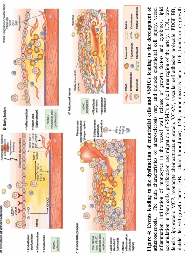
approximately 17.3 million deaths due to CVDs in 2008, a number which is predicted to rise to 23.6 million by the year 2030 49. Alterations in vascular smooth muscle cell (VSMC) growth, migration, proliferation and plasticity is believed to contribute to abnormal vascular functions associated with or leading to CVDs, such as hypertension, atherosclerosis, and stenosis after angioplasty 50-52. Under normal physiological conditions, vasoactive peptides, such as angiotensin II (Ang II) and endothelin-1 (ET-1), normalize blood pressure through the regulation of salt and/or water homeostasis, sympathetic nervous system modulation, as well as VSMC contraction and relaxation 53-56. Increased levels of both ET-1 and Ang II, present in certain pathophysiological states, such as essential hypertension, obesity, or advanced stages of diabetes, have been suggested to contribute to the pathogenesis of CVDs, by activating signaling events intimately linked to migration and proliferation of VSMC 53, 57-59.
1.4 Endothelins
Originally identified in 1988 by Yanagisawa et al. from porcine aortic endothelial cell cultures 60, endothelin (ET) is the most potent vasoconstrictor peptide known. Today, ET has been characterized in almost all organs and physiological systems, and remains one of the most important regulators of blood pressure, sodium and water homeostasis in the body 61. ET also exhibits important mitogenic and inotropic properties, can stimulate the renin-angiotensin-aldosterone system (RAAS) as well as the sympathetic nervous system (SNS) 54-56. The general role of ET is to increase vascular tone and blood pressure in
response to potential hypotensive states. However, through its mitogenic properties, ET is thought to play an important role in vascular remodeling associated with hypertension, which contributes to the development of various CVDs 62, by increasing cell proliferation, hypertrophy and cell migration, through the activation of several signal transduction pathways linked to these events in the cardiovascular system 63, 64.
1.4.1 Structure, Regulation and Biosynthesis of ET-1
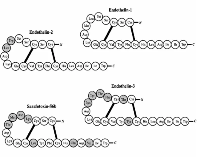
Three members of the mammalian ET gene family exist and have been characterized: ET-1, ET-2 and ET-3 61. All three ET peptides are 21 amino acids long connected by two interchain disulfide bonds (Cys1-Cys15 and Cys3-Cys11) at the N-terminal end, with a cluster of three polar charged side chains on amino acid residues 8-10 and a hydrophobic C-terminus (residues 16-21) containing the aromatic indole side chain at Trp21, essential for its loop configuration and its bioactivity 61 (Figure 1). The ET-2 peptide shares a 90% sequence homology with ET-1, varies from it by only two amino acids (Trp6-Leu7), while ET-3 shares 71 % sequence homology with ET-1 and ET-2, and varies by six amino acids (Thr2, Phe4-Thr5-Tyr6-Lys7 and Tyr14). ET-1 is encoded by a gene on chromosome 6 in humans (chromosome 13 in mouse), while ET-2 and ET-3 are encoded by independent genes located on chromosomes 1 and 20, respectively 65. The sequences for all ET family members are preserved in mammals, and are related to the sarafotoxin snake venom 66 (Figure 1). Among the three ET peptides, ET-1 is the most important isopeptide in the vasculature.
Multiple stimuli, including hypoxia, shear stress, lipoproteins, hormones and growth factors, free radicals and endotoxins, can induce and/or increase ET-1 generation, while others, such as nitric oxide (NO), natriuretic peptides, nitrovasodilators, heparin and prostaglandins, all of which increase intracellular cyclic guanosine monophosphate (cGMP) levels, inhibit ET-1 production 67. Production of ET-1 is regulated by the preproET-1 gene, at the transcriptional level 68. Binding sites for multiple regulatory elements, such activating protein 1 (AP-1), CAAT-binding nuclear factor 1 (NF-1), nuclear factor of activated T-cells (NFAT)-binding domains and GATA binding protein 2 are located in the 5’ promoter region of the preproET-1 gene, which mediate ET-1 mRNA induction by several factors, including Ang II and transforming growth factor-β (TGF-β) 69. The adenine-uracil-rich motifs present in the non-translated 3’ region mediate selective destabilization of preproET-1 mRNA, contributing to the relatively short biological half-life of ET-1 (15-20 minutes) 65.
Figure 1: Structure of the Endothelins. Amino acid sequences of the three members of the endothelin family and of the structurally related snake venom toxin sarafotoxin S6b. All three ET peptides are 21 amino acids long connected by two interchain disulfide bonds (Cys1-Cys15 and Cys3-Cys11) at the N-terminal end ET-1 (top right) is a 21 amino acid cyclic peptide with two disulphide bridges joining the cysteine residues at positions 1-15 and 3-11. Grey circles indicate where amino acids differ from those of endothelin-1. (Haynes WG, J. Hypertens 1998, 16(8):1081-98)
Compared to all other cell types, ET-1 is primarily produced in vascular endothelial cells, which express high levels of ET-1 mRNA, preproET-1 mRNA and its converting enzyme
65. ET-1 has also been shown to be produced by the heart, kidney, the posterior pituitary
gland and the CNS 67. ET-1 is also expressed in VSMC, yet its production in this cell type is 100 fold less than in endothelial cells 70. While ET-2 is secreted by heart, kidney and endothelial cells (albeit in limited quantities), ET-3 does not seem to be present in endothelial cells, and is secreted mainly by the CNS, gastro-intestinal (GI) and endocrine systems 71, 72.
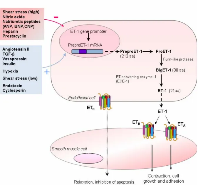
The formation of the mature ET-1 peptide is preceded by multiple steps, including the cleavage of precursor peptides. Human ET-1 mRNA encodes a 212 amino acid peptide named preproET-1 61. The signal sequence is then cleaved by a signal peptidase to form proET-1, which is further cleaved by dibasic-pair-specific endopeptidases, including furin and PC7, to from bigET-1, a 38 amino acid peptide whose vasoconstrictor efficacy is two orders of magnitude less than that of the mature ET-1 peptide 73. BigET-1 is then cleaved between Trp21 and Val22 by one of several endothelin converting enzymes (ECE) to form the mature 21 amino acid ET-1 peptide (Figure 2).
Three isoforms (ECE-1, ECE-2 and ECE-3) of ECE have been identified, all belonging to a family of zinc peptidases and related to neutral endopeptidase-24.11 and Kell protein, but not to angiotensin converting enzyme (ACE). ECE-1 is the isoform predominantly found in endothelial cells, and has the greatest affinity for BigET-1, but has been found to proteolyze other peptides as well. Four isoforms of ECE1 exist (ECE1a, -1b, -1c and -1d) and all are derived from alternative splicing of the same gene 74, 75. ECE-1a is primarily expressed in intracellular vesicles and on the cell surface of ET-1 “producer” cells, such as endothelial cells, while ECE-1b is located mostly near the trans-Golgi network, in the endosomal compartment of “responder” cells, such as VSMC. ECE-1b is then transported to the plasma membrane where it is responsible for the cleavage of extracellular BigET-1 61, 76. ECE-1c and ECE-1d are both located on the extracellular face of the plasma membrane 73.
ECE-2 hydrolyzes ET-1, and has a 60% homology with ECE-1. It also has four isoforms, which vary at the amino acid terminus level, possibly responsible for their different intracellular localization 77. The optimal functional pH of ECE-2 is 5.5, and it has virtually no activity at pH 7.0, indicating that ECE-2 is involved in intracellular processes, particularly at the level of the trans-Golgi network 61, 77. ECE-1 and ECE-2 are relatively selective for big ET-1, having much less activity in cleaving big ET-2 and big ET-3, yet ECE-1 and ECE-2 knockout mice still display significant levels of 1, indicating that ET-1 formation may also be regulated by other proteases and/or enzymes, yet their physiological relevance is not clear 78. While ECE-3 has been identified, studies have demonstrated that it has a preferential activity for ET-3 76.
In healthy subjects, circulating concentrations of ET-1 in venous plasma are in the range 0.1-10 pmol/l 79. While these concentrations are lower than the concentrations able to induce vascular constriction in vivo and in vitro, ET-1 concentrations at the interface between VSMC and endothelial cells are many times higher 61, 70. This is supported by
studies demonstrating that ET-1 is secreted by cultured endothelial cells into the basolateral (towards VSMC) compartment and not apically 80. In a similar fashion, renal tubule cells secrete ET-1 towards the interstitium, and only minimally into the urine 61. As such, rather than acting as a circulating endocrine peptide, ET-1 acts primarily as a paracrine factor, affecting local cell and tissue metabolism. Consequently, ET-1 levels in circulation do not reflect its full physiological impact. This phenomenon has led to the use of venous plasma BigET-1 and inactive ET-1 C-terminal fragment concentrations as markers for endothelial ET-1 synthesis, as they better reflect the quantities of ET-1 generated, as opposed to measuring ET-1 generation. This is due to the efficient clearance mechanism of ET-1, owing to its short half life (approx. one minute, although its pressor effects are maintained for up to one hour) and the unusual binding characteristics of the ET receptors for their ligand, which is almost irreversible 81, 82.
Circulating ET-1 is eliminated mainly by the kidney, liver and lungs, the latter of which is responsible for more than 50% of the elimination of ET-1 in humans 81. ET is degraded mainly by endopeptidases, such as neutral endopeptidase (NEP), found in the
proximal tubules of the kidney, and cathepsin G, generated in vascular endothelial and pulmonary epithelial cells 83. ET-1-binding to its cell-surface receptors and subsequent lysosomal internalization and degradation are also important mechanisms in ET-1 clearance. This theory stems from studies showing that pulmonary clearance of labelled ET-1 can be blocked by pre-treatment of the cells with a large dose of unlabelled ET-1, suggesting that ET-1 clearance is receptor-dependent 84. Investigations have also shown an increase in plasma ET-1 levels following a pharmacological blockade of the ETB receptor 85, yet in most studies the pharmacological blockade of the ET
A receptor had little impact on
plasma ET-1 levels 61. However, studies using specific disease models in which there is thought to be some degree of ETB dysfunction, such as the ETB-deficient rat, or the
DOCA-salt hypertensive rat model, pharmacological blockade of the ETA receptor led to an
increase in circulating ET-1 levels 84, 86. As such, it cannot be concluded that the ETA
receptor does not play a role in ET clearance. Moreover, plasma ET levels are increased within 15 minutes of ET receptor blockade, without any effect on C-terminal fragment and Big ET-1 concentrations, confirming that the increase in ET levels is not entirely due to peptide synthesis, but is mediated by ET-1 receptor displacement 87, 88.
1.4.2 ET-1 Receptors
All mammalian ET receptors are coded from two separate genes 61. ETA and ETB
are the two main ET-1 receptor subtypes through which ET-1 exerts its biological effects in a paracrine/autocrine fashion. Both of these receptors have been cloned in humans 89, 90, and belong to the rhodopsin class A of seven transmembrane guanine nucleotide-binding protein (G protein)-coupled receptors (GPCRs), which signal through activation of G proteins. GPCRs have an approximate 400 amino acid sequence with an N-terminal extracellular region and C-terminal intracellular region, and contain seven 22-27 hydrophobic amino acid transmembrane domains.
The ETA receptor contains 427 amino acids, and its gene is located on chromosome
4. ETA has the strongest affinity for ET-1, yet binds ET-2 to a lesser or equal extent, and
recently reported the existence of splice variants of the ETA receptor in the rat anterior
pituitary gland, of which one variant was found to have reduced efficacy in stimulating adenylyl cyclise activity and mobilizing intracellular calcium (Ca2+) 92. Nevertheless, it remains to be seen if these splice variants exist in other tissues and have a significant physiological contribution.
The ETB receptor, whose gene is located on chromosome 13, contains 442 amino
acids, and has the capacity to bind all ET peptides with equal affinity 89, 93. Several splice variants of the ETB receptor have been identified. One in particular contains an additional
10 amino acids, is found only in humans, and appears not to present any differences in cellular signalling events 94. A second ETB receptor variant was found to have important
differences in the cytoplasm domain and the 3’-untranslated domain, yet this splice variant is thought to function primarily as a clearance receptor, as it has shown little to no cellular signalling activity 95. An ETB receptor splice variant has also been discovered in the rat
brain and possibly other tissues, yet its functional characteristics have yet to be identified 96. ETB receptors are found predominantly in endothelial and renal tubule cells, yet are
expressed in smaller quantities in VSMC, cardiomyocytes, hepatocytes, osteoblasts, neurons, epithelial cells and fibroblasts 61, 90.
ET receptors couple to the Gi, Gq, Gs and Gα12/13 members of the G protein family,
regulating various signalling cascades, including adenylyl cyclases, nitric oxide synthase (NOS), serine/threonine kinases, tyrosine kinases, cyclooxygenases, amongst others 61, 69. More often than not, ETA and ETB receptors have opposite actions, depending on their
localization, even in the same organ, yet many exceptions exist. For example, in the vasculature, both ETA and ETB receptors located on VSMC induce ET-1-induced
vasoconstriction, cell adhesion and cell growth (Figure 2). In opposition, ETB receptor
binding of ET-1 in endothelial cells induces vasorelaxation, through the release of nitric oxide (NO) and prostacyclin on to VSMC 97, as well as contributing to the prevention of endothelial cell apoptosis, inhibition of ECE-1 expression and increase in ET-1 clearance 98 (Figure 2). Many pharmacological ET receptor agonists and antagonists have been
Figure 2: Factors affecting regulation of ET-1 synthesis and its subsequent ET receptor mediated actions on vascular smooth muscle cells. The generation and secretion of ET-1 is regulated by multiple factors, including hypoxia, shear stress and various growth factors and peptides. The formation of the mature ET-1 peptide is preceded by multiple steps. In humans, ET-1 mRNA encodes a 212 amino acid peptide named preproET-1, which is cleaved by a signal peptidase to form proET-1. ProET-1 is again cleaved by dibasic-pair-specific endopeptidases, including furin and PC7, to from bigET-1, a 38 amino acid peptide. BigET-1 is then cleaved between Trp21 and Val22 by one of several endothelin converting enzymes (ECE) to form the mature 21 amino acid ET-1 peptide. In the vasculature, binding of ET-1 to ETA and ETB receptors on VSMCs induces vasoconstriction, cell adhesion and cell growth. In
opposition, binding of ET-1 to ETB receptor on endothelial cells induces vasorelaxation, through the release of nitric
oxide (NO) and prostacyclin on to VSMCs. ANP, atrial natriuretic peptide; BNP, brain natriuretic peptide; CNP, C-type natriuretic peptide; TGF-β, transforming growth factor β.(Adapted from Remuzzi, G. et al., Nat.Rev.Drug. Disc 2002, 1(12):986-1001)
developed and used to better identify and understand the role of ET receptors in physiological states. For example, BQ123 and BQ788 are highly selective ETA and ETB
receptor antagonists, respectively, while sarafotoxin 6c is a powerful and highly selective ETB receptor agonist 99. Orally active ET receptor antagonists have also been developed
and used clinically, such as Bosentan, an ETA/B receptor antagonist, as well as Sitaxsentan, a
selective ETA receptor inhibitor 99. Use of such compounds has also helped to identify the
phenomenon of ETA and ETB receptor heterodimerization, possibly affecting receptor
functionality, since ETA/ETB heterodimers were shown to have delayed ET receptor
internalization and a prolonged increase in intracellular Ca2+ in response to ET-1 100, 101.
1.4.3 Biological actions of ET-1
1.4.3.1 ET-1 in the vasculature and heart
Hickey et al. were the first to demonstrate that media taken from cultured endothelial cells produced constriction in classic muscle bath preparations 102. Yanagisawa
et al. later isolated and purified the peptide and gave it the name endothelin, stemming from its endothelial cell origin 60 and demonstrated that a bolus injection of ET caused a transient hypotension immediately followed by a prolonged increase in blood pressure 60. Later studies indicated that these effects are due to ETB and ETA receptor, respectively. However,
further detailed studies have shown that ETB receptor is also implicated in vasoconstriction
and induction of hypertensive effects of ET. In VSMC, ETA and ETB activation by ET
leads to the vasoconstrictor response of ET (Figure 2). Both receptor types were shown to be present on the plasma membrane, cytosol, nuclear envelope membrane and the nucleplasm of human VSMC 103.
On the other hand, ETB activation by ET in the endothelium leads to the transient
vasodilatation response described earlier, usually followed by vasoconstriction (Figure 2). Studies have attributed this vasodilation to the generation of NO via the activation of endothelial NOS (eNOS). Activation of eNOS is thought to occur through ET-1-induced protein kinase B (PKB) phosphorylation and activation in endothelial cells 104. The
subsequent ETB receptor-dependent vasoconstriction mediated by ET-1 varies, however,
from one vascular bed to another 61. Veins seem to have a more potent ETB
receptor-dependent vasoconstriction action than arteries, yet the functional implication of these findings remains to be determined 105. Results from several investigations have suggested that NO release and vasodilatation are mediated by ETB1 receptor isotype, while the
subsequent vasoconstriction requires ETB2 receptor isotype 106. It is interesting to note that
hypotension does not occur when ET concentrations rise slowly, as opposed to a bolus dose
107. As mentioned earlier, ET response varies from one vascular bed to another. Renal and
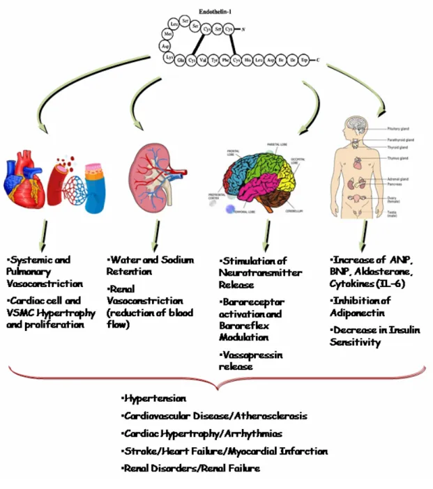
coronary vascular beds are the most sentitive to the systemic vasoconstrictor effects of ET-1, however, hindquarter skeletal muscle beds display only a minimal ET-1-induced constriction 108-110. Mesenteric vascular beds, like their coronary counter parts, have a potent ET-1-induced vasoconstrictor response 110 (Figure 3). The concentration and localisation of the different ET receptors in these vascular beds may be the cause of this varying degree of ET-1-induced response. In VSMC, ET-1 is known to also act as a mitogenic factor, as long-term treatment of VSMC resulted in an increased growth and proliferation of this cell type, which was shown to be mediated through ET-1-induced ETA
receptors activation 111, 112 (Figure 3). As well as being a mitogenic peptide in VSMC, ET-1
also displays co-mitogenic properties, potentiating the effects of such growth factors as epidermal growth factor (EGF), platelet derived growth factor (PDGF) and basic fibroblast growth factor (bFGF) 112.
In the heart, the regulation of cardiac function regulated by the ET system has been fairly well studied, particularly the role of ET in cardiac pathophysiological conditions, such as cardiac hypertrophy, ischemia/reperfusion injury, arrhythmias and congestive heart failure 61 (Figure 3). In terms of production, there is some controversy as to whether or not cardiomyocytes produce ET-1. Several studies have reported ET-1 mRNA, mature peptide and/or production in rat and chick neonatal cardiomyocytes, as well as adult porcine cardiomyocytes 113-116. In contrast, others have demonstrated the absence of ET-1 mRNA in cardiomyocytes from adult pig and rat hearts 117, 118. This has led to the speculation that
neonatal or embryonic hearts may produce larger amounts of ET-1 than healthy adult, which may produce small amounts 61.
While it may only be produced in small amounts in the heart, ET-1 secreted from VSMC and/or endothelial cells has an impact on cardiomyocyte function 119. In general, ET has positive inotropic effects on the heart, which is to say an increase in cardiac contractility, yet this varies from species to species, ET dose and duration of exposure, sympathetic nerve activity, underlying cardiac pathology and other contributing factors (reviewed in 120). Several studies have shown an increase in cardiomyocyte contractility by ET-1 in several species, including mouse, rat, dog and human, yet other studies have detected little or no ET-1-enhanced cardiomyocyte contractility, possibly due to the aforementioned factors 121-125. Despite the fact that the inotropic effects of exogenous ET-1 were examined in these studies, they did not look at the inotropic effect of endogenous ET-1. In studies where the ET-1 gene was knocked out specifically in mouse cardiomyocytes, the mice displayed an increase in cardiac cell apoptosis associated with the development of a dilated cardiomyopathy as of seven months of age. Until then, no difference in left ventricular function was observed. These studies suggest that while ET-1 production by cardiomyocytes in required for their survival, it may not be crucial for a normal heart function for a large part of the animals’ life 126. In a similar fashion, mice in which the ET
A
receptor gene was deleted, specifically in cardiomyocytes, displayed normal baseline and Ang II-induced cardiac contractility, suggesting that ET-1-induced cardiomyocyte contractility is not of physiological relevance 127. These conclusions are based on the hypothesis that the ETA receptor is responsible for the inotropic effects of ET (reviewed in 61, 128). However, diffusion of BQ123, a selective pharmacological inhibitor of ET
A receptor
subtype, into left coronary artery of patients presenting with atypical chest pain, reduced contractility. These results confirm that ET exerts a positive inotropic effect via ETA
receptor 129. Cardiac contractility in response to endogenous ET was also examined in vitro, in studies examining the response of cardiac muscle to stretch test. An increase in both rapid and slow cardiac muscle shortening and/or developed force, due to the Frank-Starling mechanism and slow force response, respectively, was observed after myocardial stretch,
with the study concluding that ET may be involved in slow force response 130. This was confirmed by studies showing that ETA receptor antagonism inhibited slow force response
in cat cardiac papillary muscle after mechanical stretch 131, yet had no effect on the Frank-Starling response in intact hearts of normal rats 132, substantiating the notion that cardiomyocyte-derived endogenous ET is important to cardiac adjustments, and plays a physiological role 61.
1.4.3.2 ET-1 in the nervous system
ET is known to play an important role in the development of the neural crest and of enteric neurons, as well as of peripheral sympathetic ganglia which modulate systemic hemodynamics 133, 134, where both ET-1 and ET-3 production has been demonstrated in neurons cultured from superior cervical ganglion (SCG) 135, as well as in the dorsal root ganglia 136. These results demonstrate a clear presence of ET in neurons within the sympathetic ganglia. Spontaneously hypertensive rats (SHR) have been shown to have increased SCG ET-1 levels, as compared to Wistar Kyoto rats (WKY), yet it is not clear as to whether or not this is a significant contributing factor to the increased hypertensive state of SHR 137.
ET mRNA and mature peptides are also present in regions of the hypothalamus and brain stem known to regulate brain function, such as the paraventricular nuclei, dorsal motor nucleus of the vagus nerve, medulla oblongata, choroid plexus and rostral brain regions 136, 138. Intracerebroventricular (ICV) administration of ET-1 or ET-3 caused an increase in blood pressure and a decrease in heart rate in conscious rats, effects which were reversed by paraventricular nuclei produced NO, suggesting that the hypertensive effect of centrally administered ET may involve baroreflex activity, yet direct ET-induced baroreflex modulation is not clearly demonstrated 139, 140. ET-1 ICV injection was also shown to produce a pressor response in SHR and WKY 141. Furthermore, ETA receptor blockade
decreased blood pressure in SHR, but not in WKY, suggesting that endogenous ET-1 from the central nervous system (CNS) causes a tonic hypertensive effect through the ETA